Article

Comparative Analysis and Phylogeny of the Complete Chloroplast Genomes of Nine Cynanchum (Apocynaceae) Species

Key Laboratory of Ecological Protection of Agro-Pastoral Ecotones in the Yellow River Basin, National Ethnic Affairs Commission of the People’s Republic of China, College of Biological Science & Engineering, North Minzu University, Yinchuan 750021, China
*
Author to whom correspondence should be addressed.
Genes 2024, 15(7), 884; https://doi.org/10.3390/genes15070884
Submission received: 9 June 2024 / Revised: 27 June 2024 / Accepted: 4 July 2024 / Published: 5 July 2024
(This article belongs to the Section Genes & Environments)

Abstract

:
Cynanchum belongs to the Apocynaceae family and is a morphologically diverse genus that includes around 200 shrub or perennial herb species. Despite the utilization of CPGs, few molecular phylogenetic studies have endeavored to elucidate infrafamilial relationships within Cynanchum through extensive taxon sampling. In this research, we constructed a phylogeny and estimated divergence time based on the chloroplast genomes (CPGs) of nine Cynanchum species. We sequenced and annotated nine chloroplast (CP) genomes in this study. The comparative analysis of these genomes from these Cynanchum species revealed a typical quadripartite structure, with a total sequence length ranging from 158,283 to 161,241 base pairs (bp). The CP genome (CPG) was highly conserved and moderately differentiated. Through annotation, we identified a total of 129–132 genes. Analysis of the boundaries of inverted repeat (IR) regions showed consistent positioning: the rps19 gene was located in the IRb region, varying from 46 to 50 bp. IRb/SSC junctions were located between the trnN and ndhF genes. We did not detect major expansions or contractions in the IR region or rearrangements or insertions in the CPGs of the nine Cynanchum species. The results of SSR analysis revealed a variation in the number of SSRs, ranging from 112 to 150. In five types of SSRs, the largest number was mononucleotide repeats, and the smallest number was hexanucleotide repeats. The number of long repeats in the cp genomes of nine Cynanchum species was from 35 to 80. In nine species of Cynanchum, the GC3s values ranged from 26.80% to 27.00%, indicating a strong bias towards A/U-ending codons. Comparative analyses revealed four hotspot regions in the CPG, ndhA-ndhH, trnI-GAU-rrn16, psbI-trnS-GCU, and rps7-ndhB, which could potentially serve as molecular markers. In addition, phylogenetic tree construction based on the CPG indicated that the nine Cynanchum species formed a monophyletic group. Molecular dating suggested that Cynanchum diverged from its sister genus approximately 18.87 million years ago (Mya) and species diversification within the Cynanchum species primarily occurred during the recent Miocene epoch. The divergence time estimation presented in this study will facilitate future research on Cynanchum, aid in species differentiation, and facilitate diverse investigations into this economically and ecologically important genus.

1. Introduction

Cynanchum belongs to the Apocynaceae family and is a morphologically diverse genus that includes around 200 shrub or perennial herb species [1,2]. Cynanchum species are primarily found in eastern Africa; the Mediterranean region; and the tropical, subtropical, and temperate regions of Europe and Asia [3]. These plants can be utilized for medicinal purposes, food, as forage resources, and for managing wasteland and barren land [4,5]. Some species with climbing vines and tuberous roots have been traditionally used in Korean and Chinese medicine [4,5,6,7,8,9]. Various studies have shown that extracts from these plants can help prevent and treat conditions like rheumatoid arthritis, lumbago, abdominal pain, and traumatic injuries [8,10,11]. Previous research has mainly focused on the growth, development, medicinal properties, and breeding of Cynanchum [12,13,14,15]. Despite its economic and ecological importance, there is still much to learn about Cynanchum phylogeny, interspecific relationships, and evolutionary history [16,17]. While many studies have identified and examined phylogenetic relationships within the Cynanchum genus using DNA barcode fragments [18,19], there is a lack of research on chloroplast genome (CPG) phylogeny and lineage diversification.
Chloroplasts play a crucial role in photosynthesis and carbon fixation in plant cells. Advances in sequencing technologies have made whole-chloroplast genome sequencing more accessible [20,21,22]. The CPG is characterized by two large inverted repeat regions (IRa and IRb) separated by two single-copy regions, known as the large single-copy region (LSC) and the small single-copy region (SSC) [23]. Compared to DNA fragments, the plant CPG has a relatively small molecular size (107~218 kb) and moderate rates of nucleotide substitution, making it a valuable tool for species identification, phylogeny, and genetic diversity studies [24]. CPGs have recently been utilized for comparative and phylogenetic analyses, proving beneficial for species identification, genetic diversity assessment, nucleotide diversity evaluation, resolving phylogenetic relationships, and understanding evolutionary history. For instance, comparative analyses of CPGs have successfully revealed the phylogenetic relationships within Brassicaceae [25,26], Salicaceae [27], and the genus Yulania [28].
Despite the utilization of CPGs, few molecular phylogenetic studies have endeavored to elucidate infrafamilial relationships within Cynanchum through extensive taxon sampling. Previous studies mostly relied on single or limited molecular loci or faced constraints in sampling, resulting in an unclear understanding of the phylogenetic relationships and divergence timescales of the genus [29,30]. In this research, we conducted sequencing and alignment of the chloroplast genomes of nine Cynanchum species. Our primary goals were to (a) establish a phylogeny based on the CPGs of nine species from the Apocynaceae family; (b) estimate the divergence of the Cynanchum clade; and (c) investigate structural alterations in the PCGs of the sampled Cynanchum species.

2. Results

2.1. Genome Size and Features

After quality control and pre-processing, a minimum of 6 Gb of whole-genome sequencing data were obtained for each of the nine species included in this study (Table 1). These clean reads were used to assemble complete chloroplast genomes using a reference-guided approach. All the newly assembled CP genomes displayed a typical quadripartite structure, with two IR regions separating the LSC and SSC regions (Figure 1).
For each of the nine Cynanchum species, the CP genome size ranged from 158,283 bp (Cynanchum. acutum subsp. sibiricum) to 161,241 bp (C. wilfordii) (Table 1). All the CPGs had a typical quadripartite circular structure (Figure 1) consisting of an LSC and an SSC region separated by a pair of IR regions (Figure 1 and Table 1). The LSC region’s length varied from 89,424 bp (C. acutum subsp. sibiricum) to 92,051 bp (C. rostellatum), and the lengths of the SSC and IR regions ranged from 18,281 bp (C. thesioides) to 20,943 bp (C. wallichii) and 23,834 bp (C. rostellatum) to 24,658 bp (C. wilfordii), respectively (Table 1). The GC content of the entire plasmid sequence and the LSC, SSC, and IR regions was similar across all Cynanchum CPGs. Specifically, the GC content of the entire CPGs sequence was 37.8–38.1%, while the GC content of the IR regions was 42.69–44.55%, which was higher than that of the LSC and SSC regions (35.53–36.24% and 30.2–32.57%, respectively; Figure S1 and Table S1). Additionally, the number of annotated genes in each CPG ranged from 129 (C. acutum subsp. sibiricum) to 133 (C. wallichii) and included 37 tRNA and 8 rRNA genes.

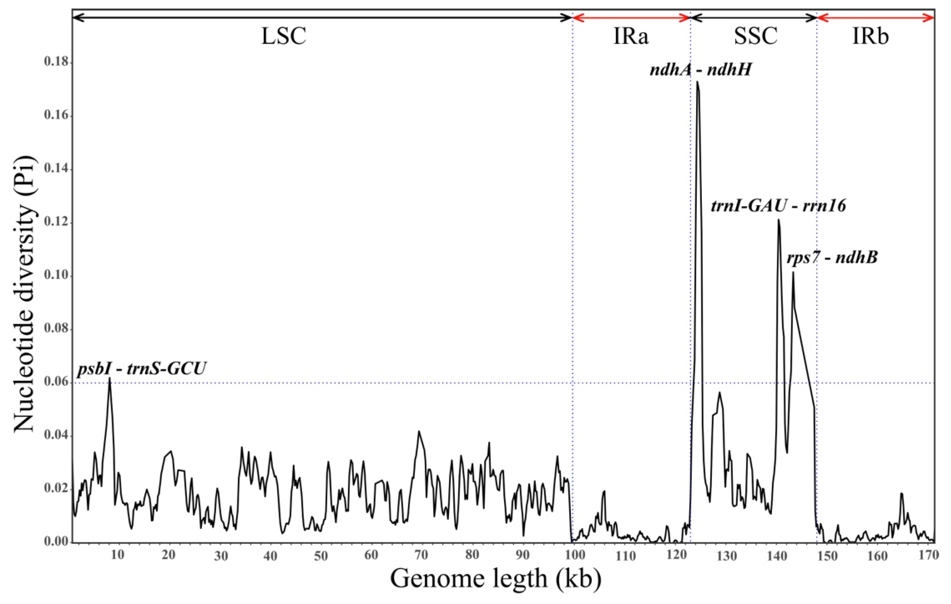
2.2. Comparative Genomics and Divergence Hotspots

Using V. mongolicum as a reference, the CPGs of the nine Cynanchum species were visually compared with those obtained using the mVISTA online database. The results showed that the cp genomes of the nine Cynanchum species were conserved. Moreover, there were differences in the LSC and SSC regions compared with the IR region, and the same was true for the non-coding and coding regions (Figure 2). The regions with greater variation were located mainly in rps16-trnQ-UUG-psbK, atpH-atpI, rps2-rpoC2, rpl20-psbB, rpl16-rps3, ndhG-ndhI, and ndhF-rpl32 (Figure 2). The greatest variation among coding regions was observed in accD, ycf2, and ycf1. No major genomic rearrangements or insertions were detected among the nine CPGs (Figure 2). DNA molecular markers are usually highly variable regions of sequences that can be used for the differentiation of relationships between species. Therefore, to further understand the DNA polymorphisms (Pi), mutation hotspot regions in the cp genomes of the nine Cynanchum plants were screened using DnaSP (Figure 3). Pi analysis revealed that the pi values ranged from 0 to 0.18, and the cp genome was relatively structurally conserved, small, and highly variable among the species. A total of six mutation hotspot regions (Pi > 0.06) were detected, and they can be used as potential molecular markers. Among these, ndhA-ndhH, trnI-GAU-rrn16, and rps7-ndhB were located in the SSC region; psbI-trnS-GCU was located in the LSC region (Figure 3). None of the mutation hotspots were located in the IR region.

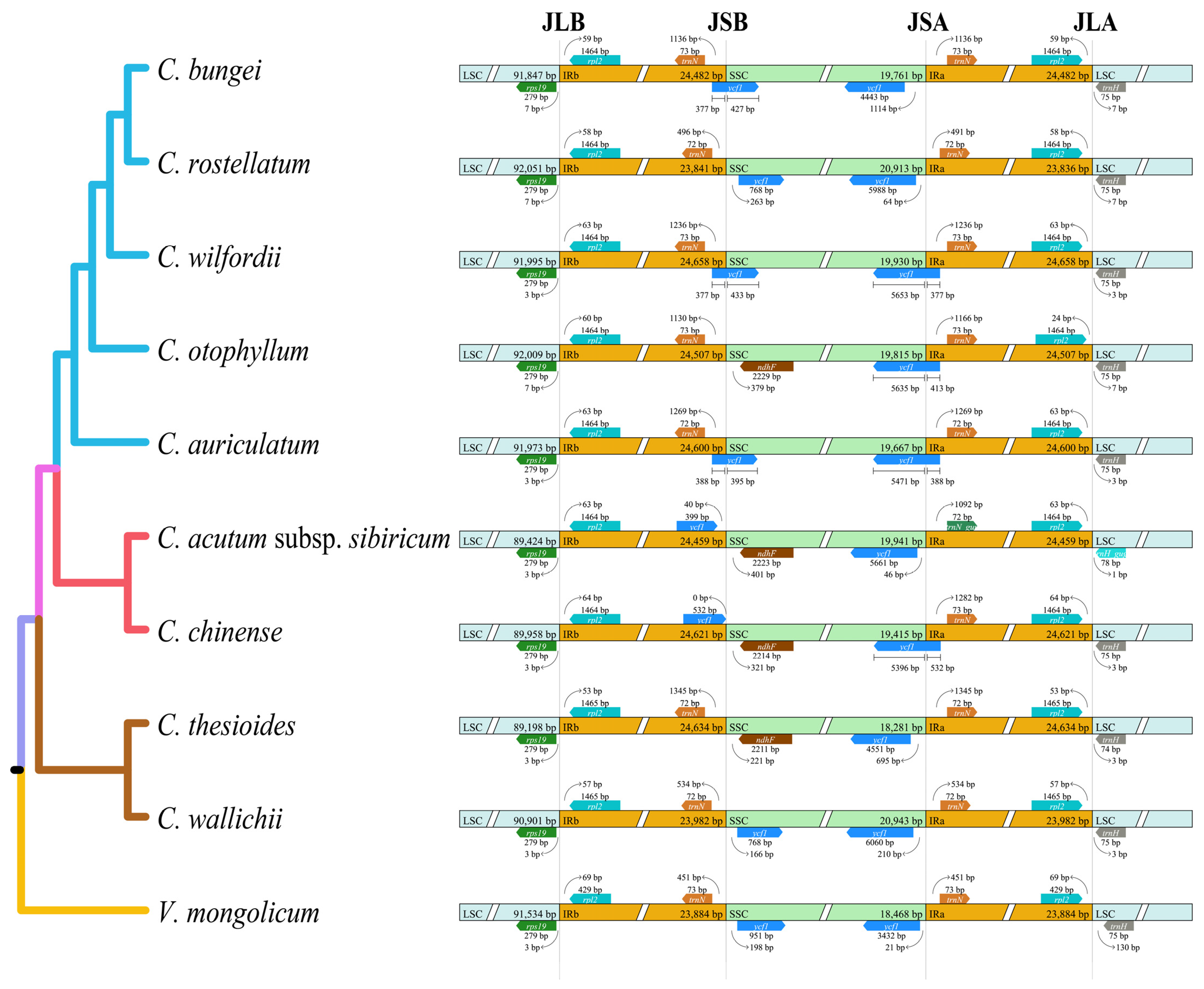
2.3. Boundaries between IR and SC Regions

We analyzed the dynamics of the IR boundaries of the cp genomes of nine Cynanchum species. The results showed that Cynanchum chloroplast genomes were relatively conserved; however, some structural variations were still identified, especially at the boundaries between the IR and SSC regions (Figure 4), with the length of the rps19 gene located in the IRb region, varying from 46 to 50 bp. At both ends of the SSC region, IRb/SSC junctions were located between the trnN and ndhF genes. However, the ycf1 genes of C. bungei, C. rostellatum, C. acutum subsp. sibiricum, C. thesioides, and C. wallichii were located in the SSC region, and the ycf1 genes of the other four species were located in the LSC-IRb boundary. SSC/IRa was found in the ycf1 gene, with the length of the ycf1 gene located in the IRa region varying from 377 to 532 bp. Here, trnH was shown to be the first gene in the LSC region at the junction between IRa and LSC (i.e., IRa/LSC). At the other end of the LSC region, the IRb/LSC junctions were located between the rps19 and rpl2 genes in all Cynanchum species.

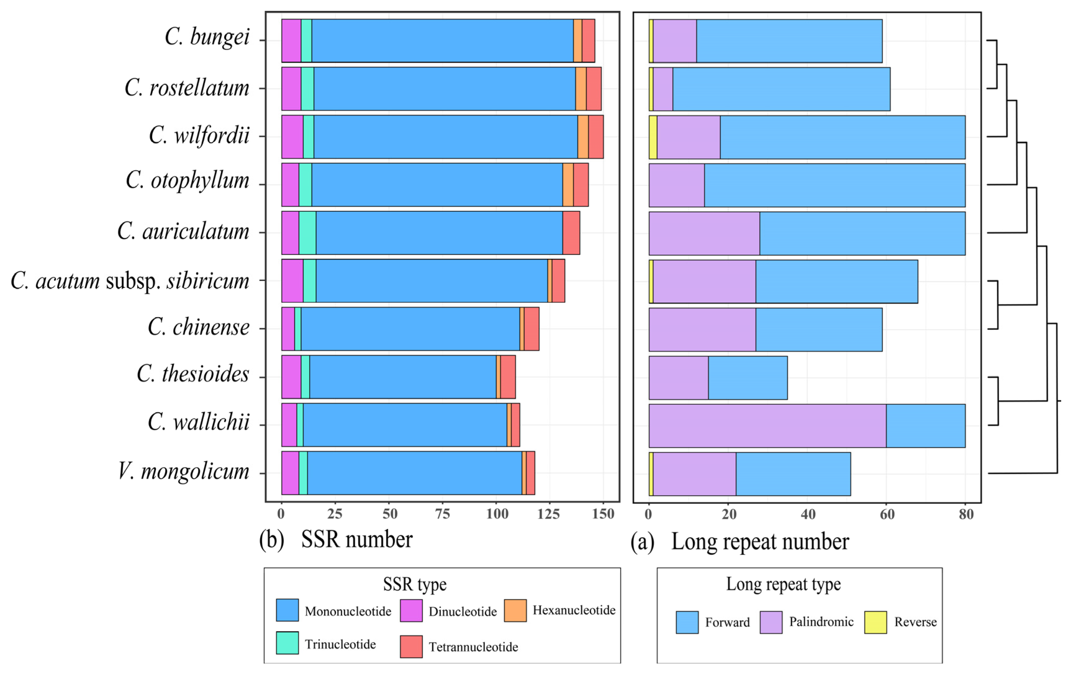
2.4. Repeat Identification

The MISA v. 1.0 software was utilized to detect simple sequence repeats (SSRs) in nine cp genomes of Cynanchum (Figure 5a; Table S1). The results of SSR analysis revealed a variation in the number of SSRs, ranging from 112 to 150. These SSRs were predominantly located in the LSC and SSC regions of the gene spacer among the five types of SSRs, and the largest number was mononucleotide repeats, accounting for 82.3%, followed by dinucleotide and tetranucleotide repeats, accounting for 6.3% and 4.9%, respectively. The smallest number was hexanucleotide repeats, accounting for only 2.6%. We examined the number and distribution of long repeats in the cp genomes of nine Cynanchum species, which ranged from 35 to 80, with an average of 67 repeats, mainly in the IR and LSC regions (Figure 6; Table S2). Five Cynanchum species contained only forward and palindromic repeats, namely C. otophyllum, C. auriculatum, C. chinense, C. thesioides, and C. wallichii.

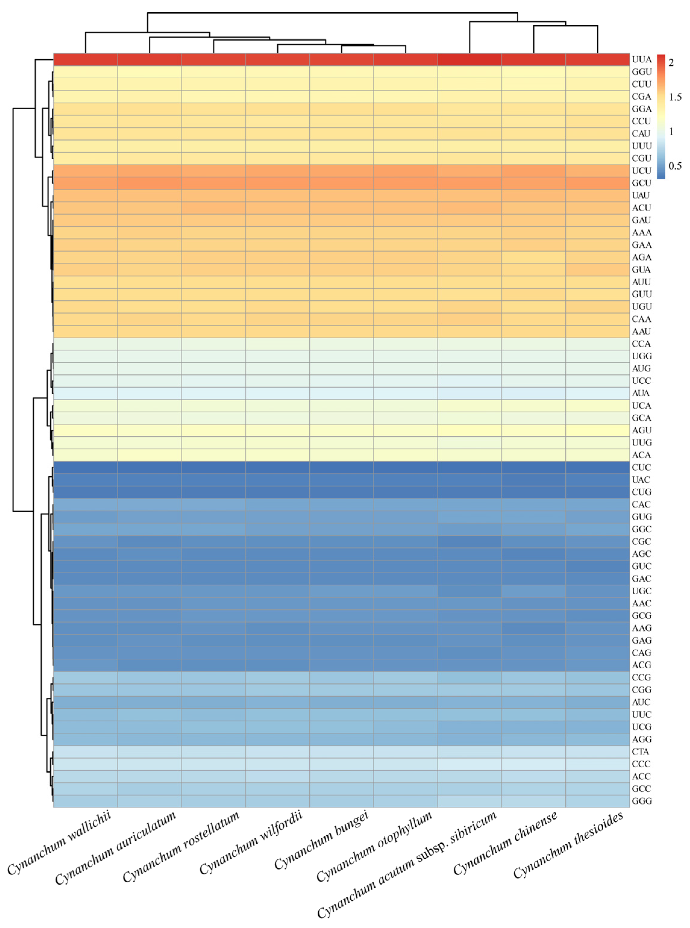
2.5. Codon Usage Analysis

The amino acid frequency, the number and bias of codon usage, and RSCU were investigated among the nine Cynanchum species cp genomes. The results indicated that 21 different amino acids were encoded in the cp genome, and a comprehensive set of 64 codons were deduced. Among these codons, 32 were frequently utilized in various Cynanchum species (Tables S3–S11). A total of 30 codons displayed RSCU values > 1, 29 of which had A or U terminal nucleotides. The rest of the 34 codons had RSCU values ≤ 1, where 31 of these ended in G or C nucleotides. Additionally, the codon UUA had the highest frequency, followed by GCU, while CUC was the least common (Figure 6). Leucine and aspartic acid had the highest and lowest number of codons, respectively. Moreover, unlike other amino acids, which were encoded by at least two synonymous codons, methionine and tryptophan were encoded by only one respective codon. The GC content of synonymous third codon positions (GC3s) is correlated with codon bias to evaluate codon usage patterns. In nine species of Cynanchum, the GC3s values ranged from 26.80% to 27.00%, indicating a strong bias towards A/U-ending codons. In addition, both the frequency of optical codons and the codon adaptation index were less than 0.4, and the effective proportion of codons ranged from 49.80% to 50.05% (Table S12). The codon usage of cp genomes in C. wallichii, C. auriculatum, and C. thesioides was relatively similar (Figure 6), suggesting that a minor bias existed in codon use across the nine Cynanchum species.

2.6. Phylogenetic Analyses and Divergence Time Estimation

To infer the phylogenetic relationships of the 36 Apocynaceae species, we included 3 Gentianaceae species whose PCGs are publicly available in the GenBank database. These species (Comastoma falcatum, Gentianella pygmaea, and Gentiana algida) were used as the outgroup for phylogenetic analyses. The final concatenated dataset included 60 plastid genes and 43,362 sites, after trimming poorly aligned regions and gaps with missing genes (Table S13). In the phylogenetic trees, maximum likelihood (ML) analyses supported similar relationships to those indicated by maximum parsimony and Bayesian analyses (BI). The ML and BI analyses of each dataset contained nine internal clades with high bootstrap support (BS = 100%) and posterior probability (PP = 1) values (Figure 7 and Figures S2–S5). There were nine species of Cynanchum and two species of Vincetoxicum and Parsonsia goniostemon gathered into one clade, in which Parsonsia goniostemon resolved (BS; PP = 100%, 1) as a sister to Cynanchum plus Vincetoxicum. Further, in the CDS phylogeny, Cynanchum species were clustered on one large branch, confirming that the independence of this genus is highly supported (BS; PP = 100%, 1) (Figure 7 and Figures S2–S5).
We estimated the divergence timescales of the Cynanchum according to the time constrations of the gene tree constructed based on 60 plastid genes. The split between Cynanchum and its sister group was estimated to have occurred 23.85 Mya. The crown ages of all subclades in the genus Cynanchum were dated mainly within the Miocene, suggesting that numerous species of this genus originally diversified in the recent past (18.87 Mya) (Figure 7).

3. Discussion

Recent studies have reported that the CPG of seed plants range in size from 107 kb in Pinaceae to 170 kb in Geraniaceae, with the IR region typically spanning 20–30 kb [31,32]. In the present study, a comparative analysis of the CPGs indicated that those of nine Cynanchum species were ranging from 158,283 bp (C. acutum subsp. sibiricum) to 161,241 bp (C. wilfordii), at the larger end of the spectrum for seed plant organelle genomes. All Cynanchum species comprising the LSC region (89,424–92,05 bp), SSC region (18,281–20,943 bp), and two identical IR regions (23,834–24,658 bp) were similar to other vascular plants. The GC content of the entire plastid sequence and the LSC, SSC, and IR regions was similar across all Cynanchum CPGs. In agreement with numerous studies on angiosperms, the IR regions exhibited the highest GC content [26]. The conversion between sequences and higher GC content may contribute to the greater conservation of IR regions [27,28]. In angiosperms, the IR region is relatively conserved in sequence and structure. The narrowing and widening of its edges are not only important factors for length variation, but also the main cause of the emergence of pseudogenes [33,34]. While cp genes have evolved slowly and are relatively conserved in terms of sequence and structure, boundary contraction and expansion in the IR regions are common phenomena. In our study, we analyzed nine CPGs within the highly conserved Cynanchum species and noticed that no major expansions or contractions occurred in the IR regions.
Highly variable regions offer valuable phylogenetic information. For example, variable regions aid in species kinship identification and gene pool construction [25,26,35,36,37]. A good DNA barcode must be a short, representative DNA fragment with high variability and amenability to amplification [38]. In this study, both the sequence and structure of Cynanchum CPGs were highly conserved. mVISTA analysis revealed that most of the variation in nucleotide sequences occurred in non-coding regions, consistent with previous reports, suggesting this variation as a common feature of angiosperms [39,40,41]. In the Cynanchum, several highly variable regions, such as accD, ycf2, and ycf1, are recognized as potential DNA barcoding sites [42,43]. A comparable number of SSRs and long repeats were identified in nine Cynanchum species. However, the types of SSRs and long repeats differed among the species. These repeats were predominantly found in the intergenic spacer (IGS) of the large single-copy (LSC) region. Mononucleotide repeats were the most common types of SSRs, while forward and palindromic repeats were the predominant types of long repeats (Figure 5; Tables S1 and S2). In addition, our nucleotide diversity (Pi) analysis led to the identification of five highly variable regions with Pi values greater than 0.06, including three SSC regions (ndhA-ndhH, trnI-GAU-rrn16 and rps7-ndhB) and one LSC region (psbI-trnS-GCU). None of the mutation hotspots were located in the IR region. In conclusion, these mutation hotspot regions play an important role in the identification and characterization of Cynanchum plant species.
In nature, plants are often influenced by environment. Some genes may be subject to positive selection in response to environmental changes. In protein synthesis, codons represent the rule of a certain amino acid [44]. In this study, we found over 90% (RSCU ≥ 1) of Cynanchum codons terminated in A/U, and the GC3s value ranged from 26.80% to 27.00%. Cynanchum exhibited a high coding efficiency and a strong preference for A/U termination codons, possibly owing to the overall high AT content in the cp genome. This trend was also evident in other angiosperms [45]. Our investigation of Cynanchum genomes revealed that non-coding regions displayed more substantial variation than coding regions. Four highly variable regions in non-coding regions identified in this study effectively distinguished the most common Cynanchum species. These findings aligned with earlier studies of Cynanchum [36]. Non-coding regions evolve rapidly and contain valuable variations for genus phylogenetic analysis [37]. Therefore, the significance of non-coding regions in the cp genome for identifying Cynanchum species should be underscored.
The CPGs are central to molecular biology research and have become a prominent focus. In particular, species identification, phylogenetic relationships, and the reconstruction of evolutionary history via whole-genome sequencing have become important tools because of improvements in sequencing technology and low costs. Phylogenetic trees constructed based on a single or a few gene sequences often yield inconsistent or even conflicting topologies due to variations in evolutionary rates and horizontal shifts between genes. This complication affects the determination of accurate evolutionary relationships among species [46,47]. In this study, we constructed a phylogenetic tree using the BI and ML methods. The CPGs of nine Cynanchum species converged into branches with high support (Figures S4 and S5). Notably, Parsonsia goniostemon resolved (BS; PP = 100%, 1) as a sister to Cynanchum plus Vincetoxicum. The phylogenetic result was consistent with previous findings in Apocynaceae species [17,18,19,30], indicating that high-resolution CPG sequences offer valuable resources for extensive research on the genetic phylogeny and species identification of Cynanchum spp. Furthermore, further studies on Cynanchum are warranted, particularly to validate the relationships of this genus with other sister genera, such as Vincetoxicum. Expanding the number of cp genomes of Cynanchum and Apocynaceae will yield deeper insights into the evolution of this ecologically and economically important phytogroup.
To calibrate the divergence and origin of Cynanchum, we used 60 highly conserved and stable alignment plastid genes (Figure 7). We also used fossil date and time constrations to estimate the diversification. While the estimated ages should be used with caution, our findings indicate that the Cynanchum and Vincetoxicum diverged from the sister genus around 32.67 Mya, and the two Cynanchum and Vincetoxicum diverged 23.85 and 18.87 Mya, suggesting relatively late clade diversifications. Specifically, most species diversification within the subclades of Cynanchum, as estimated from these plastid genes, appeared to have occurred in the recent past, mostly after 20 Mya. The divergence timescales estimated here for the major subclades will serve as a basic timescale for diverse studies on this economically and ecologically important genus.

4. Conclusions

We analyzed the complete CPGs of nine Cynanchum species and found that all exhibited a quadripartite structure, typical of most angiosperms. The CPG arrangement was highly conserved, with great sequence variation observed in the SSC region compared with that in the IR region. We did not detect major expansions or contractions in the IR region, nor did we find any rearrangements or insertions in the CPGs of the nine Cynanchum species. We identified highly variable regions within Cynanchum that are likely to be useful for species delimitation. Mononucleotide repeats were the most common types of SSRs, while forward and palindromic repeats were the predominant types of long repeats. Cynanchum exhibited a high coding efficiency and a strong preference for A/U termination codons. Phylogenetic tree construction based on the CPGs showed that all nine Cynanchum species formed a monophyletic group, divided into two typical subbranches and three minor branches. Molecular dating suggested that Cynanchum diverged from its sister genus around 23.85 Mya, with species diversification of the Cynanchum species of China occurring mainly within the recent Pliocene epoch. Overall, our findings and the estimated divergence times will facilitate future studies on Cynanchum, assist in species differentiation, and facilitate diverse studies of this economically and ecologically significant genus.

5. Materials and Methods

5.1. Taxon Sampling, DNA Extraction, and PCG Sequencing

A total of 37 PCGs representing Apocynaceae and related families were included in this study (Table S14). Thirty-four PCGs from Apocynaceae were selected, including nine Cynanchum species found in China. Further, three additional PCGs from related families in Gentianaceae were chosen as outgroups for phylogenetic analysis. Among these 37 PCGs, 3 complete PCGs were newly sequenced, and the others were obtained from GenBank (Table S14). The leaves used in this study were collected from natural populations in China. The plant materials and specimens were deposited in the Herbarium of North Minzu University (NMU; Yinchuan, China). For each species, we extracted total DNA from dried leaves and preserved them in silica gel using the CTAB protocol [48]. Paired-end libraries with an insert size of 500 base pairs (bp) were constructed by Illumina (Qingdao, China) following sequencing with a HiSeq × Ten System (Jizhi, Qingdao, China).

5.2. Chloroplast Genome and Annotation

At least two gigabases (Gb) of 2 × 150 bp short read data were generated for each sample. Reads with quality scores of less than 7 and with more than 10% ambiguous nucleotides were filtered. The remaining reads were assembled using NOVOPlasty version 2.7.2 [49] software. The contigs were aligned into sequence in Geneious version 9.1.8 [50] software using the V. mongolicum PCG as a reference. The PCGs were annotated using Plann version 1.1 [51]. Protein-coding genes were extracted using customized Python scripts. The alignment of chloroplast genes across all species was performed using PRANK version 130410 [52] software. Poorly aligned regions were trimmed using Gblocks version 0.91b [53] with the option “−t = c,” selected to set the type of sequence to codons. Genes that were absent in at least one species were excluded, and the aligned sequences were combined into a super matrix. Additionally, circular maps of the CP genomes were created using OGDRAW version 1.2 [54], and all annotated CP genomes were submitted to GenBank [55].

5.3. Comparative Genomics and Structural Analyses

The structural variation and identification of arrangement events across Cynanchum was conducted for the nine CPGs of Cynanchum. The results of the comparative analysis of the CPGs were visualized with the mVISTA program [56], and the annotated CPG of V. mongolicum was used as the reference in the LAGAN mode [57]. The junction sites of the four structural regions (IRA, LSC, SSC, and IRB) and adjacent genes in nine Cynanchum PCGs were visualized and compared using IRscope v0.1.R [58] software to obtain a macroscopic view of the CP genome structure. Following sequence alignment, nucleotide diversity (Pi) analysis of the CP genome was performed using DnaSP version 6.0 [59].

5.4. Repeat Sequence and Codon Preference Analyses

The Geneious 9.1.8 software [50] was employed to conduct a GC content analysis. Furthermore, the REPuter program (https://bibiserv.cebitec.uni-bielefeld.de/reputer accessed on 6 July 2016) was utilized to recognize dispersed repeat sequences, including forward (F), complementary (C), palindromic (P), and reverse (R) [60], with the setting of >30 bp, ≥90% sequence identity, and a Hamming distance at 3. Simple sequence repeats (SSRs) in the cp genomes were analyzed on the MISA-web (http://pgrc.ipk-gatersleben.de/misa/ accessed on 5 August 2016) [61], and those with different repeat units were regarded as hexanucleotides, pentanucleotides, tetranucleotides, trinucleotides, dinucleotides, or mononucleotides. Additionally, the amino acid usage frequency and relative synonymous codon usage (RSCU) were identified via the CodonW 1.4.2 software [62]. Lastly, TBtools, a software that integrates various biological data handling tools [63], generated a heatmap of the RSCU values.

5.5. Phylogenetic Inference and Divergence Time Estimation

We generated two datasets for phylogenetic analysis: a protein-coding region (CDS) set and a whole PCG (WP) set. Protein-coding genes (PCGs) were extracted from the GenBank formatted file containing 37 PCGs using customized Perl scripts that removed the start and end codons. After excluding possible pseudogenes, 60 PCGs were retained in all species. Each PCG was aligned using PRANK version130410 based on the translated amino acid sequences. Genes that had been lost in at least one species were discarded, and then, the remaining aligned sequences were concatenated into a super matrix. Independent phylogenetic analyses were performed for each dataset (CDS and WP) using the maximum likelihood (ML) and Bayesian inference (BI) methodologies. We used RAxML version 8.1.24 [64] to conduct ML analyses with a general time reversible model with a gamma distribution (GTR + Γ). The best-scoring ML tree was obtained using the rapid hill-climbing algorithm (i.e., the option “-f d”) with 1000 bootstrap replicates. The optimal model (GTR + I + G) was identified using jModeltest 2.1.10 software, and BI analysis was conducted using MrBayes version 3.2.6 [65]. Additionally, FigTree version 1.4.2 [66] was used to visualize phylogeny.
We estimated divergence times from the PCG dataset using an approximate likelihood method, as implemented in MCMCtree in PAML version 4 [67] software, with independent relaxed-clock and birth–death sampling [68] strategies. Fossil dates were used as calibration points to reduce bias for more accurate age estimates [69]. In order to estimation the divergence time of Cynanchum, we used one fossil and two other time constrations to calibrate the phylogeny: (1) the root of the phylogeny occurred during the upper Cretaceous (~89 Mya, 95% HPD = 78–102 Mya) [70]. According to the ancient seed fossil of Carissa manghas [70], 47.02–50.78 Mya was the age range assigned to the split between Carissa manghas and its sister group [71]. (3) The split between Apocynum and Beaumontia was assigned an age range of 10.29–29.59 Mya as previously estimated [72]. The best-fit GTR + Γ model was selected, and the prior of the substitution rate (rgene) was modeled by a Γ distribution as Γ (2, 200, 1). We set parameters for the birth–death process with species sampling and σ2 values of (1, 1, 0.1) and G (1, 10, 1), respectively. We executed the MCMC runs for 2000 generations as burn-in and then sampled every 750 generations until 20,000 samples were obtained. We compared two MCMC runs for convergence using random seeds and obtained similar results.

Supplementary Materials

The following supporting information can be downloaded at https://www.mdpi.com/article/10.3390/genes15070884/s1: Table S1. Statistics of SSR analysis results from nine Cynanchum species. Table S2. Statistics of long repeats from nine Cynanchum species. Table S3. Codons in cp genome of Cynanchum auriculatum. Table S4. Codons in cp genome of Cynanchum bungei. Table S5. Codons in cp genome of Cynanchum chinense. Table S6. Codons in cp genome of Cynanchum otophyllum. Table S7. Codons in cp genome of Cynanchum rostellatum. Table S8. Codons in cp genome of Cynanchum acutum subsp. sibiricum. Table S9. Codons in cp genome of Cynanchum thesioides. Table S10. Codons in cp genome of Cynanchum wallichii. Table S11. Codons in cp genome of Cynanchum wilfordii. Table S12. GC content at different positions of CDS sequence codon. Table S13. Comparison of plastid protein-coding genes. Table S14. Species and data description of plastomes used in this study. Figure S1. Changes in plastid GC content of nine Cynanchum species. This graph shows the total GC content (red bar) and the third codon position GC content (green bar) of each species. Figure S2. Phylogenetic tree obtained using the maximum likelihood (ML) methods for the Cynanchum species based on 60 PCGs. Figure S3. Phylogenetic tree obtained using the Bayesian inference (BI) methods for the Cynanchum species based on 60 PCGs. Figure S4. Phylogenetic tree obtained using the maximum likelihood (ML) methods for the Cynanchum species based on complete CPGs. Figure S5. Phylogenetic tree obtained using the Bayesian inference (BI) methods for the Cynanchum species based on complete CPGs.

Author Contributions

E.Z. and L.Z. developed the concept of the study. E.Z. and X.M. conducted the experiment and data analysis. T.G., Y.W. and L.Z. drafted the manuscript. Y.W. and L.Z. supervised the study. All authors revised the manuscript. All authors have read and agreed to the published version of the manuscript.

Funding

This study was supported by the doctoral scientific research foundation of the North Minzu University (2021KYQD10), the Ningxia Natural Science Foundation (2022AAC05063, 2023BEG03062), the graduate innovation project of North Minzu University (2024), and the innovation team for genetic improvement of economic forests (2022QCXTD04). The reviewers and editors are sincerely acknowledged.

Institutional Review Board Statement

Not applicable.

Informed Consent Statement

Not applicable.

Data Availability Statement

The original data presented in the study are openly available in https://www.ncbi.nlm.nih.gov, and accession numbers are listed in Table S14.

Conflicts of Interest

The authors declare no conflicts of interest.

References

  1. Li, P.T.; Michael, G.G.; Stevens, W.D. Asclepiadaceae. In Flora of China; Wu, Z., Raven, P., Eds.; Science Press: Beijing, China, 1995; Volume 16, pp. 311–313. [Google Scholar]
  2. Endress, M.E.; Liede-Schumann, S.; Meve, U. An updated classification for Apocynaceae. Phytotaxa 2014, 159, 115. [Google Scholar] [CrossRef]
  3. Yang, J.; Qing, Z.; Wang, D.; Yang, H. Distribution of medicinal plant resources of Cynanchum in Guizhou. Guizhou Sci. 2018, 36, 50–52. [Google Scholar]
  4. Zhang, X.; Yang, Z.; Li, Z.; Zhang, F.; Hao, L. De novo transcriptome assembly and co-expression network analysis of Cynanchum thesioides: Identification of genes involved in resistance to drought stress. Gene 2019, 710, 375–386. [Google Scholar] [CrossRef] [PubMed]
  5. Chang, X.Y.; Yang, Z.R.; Zhang, X.Y.; Zhang, F.L.; Huang, X.M.; Han, X. Transcriptome-wide identification of WRKY transcription factors and their expression profiles under different stress in Cynanchum thesioides. PeerJ 2022, 10, e144362022. [Google Scholar] [CrossRef] [PubMed]
  6. Liu, Y.; Qu, J.; Yu, S.S.; Hu, Y.C.; Huang, X.Z. Seven new steroidal glycosides from the roots of Cynanchum forrestii. Steroids 2007, 72, 313–322. [Google Scholar] [CrossRef] [PubMed]
  7. Yildiz, I.; Sen, O.; Erenler, R.; Demirtas, I.; Behcet, L. Behcet bioactivity-guided isolation of flavonoids from Cynanchum acutum L. subsp. sibiricum (willd.) Rech. f. and investigation of their antiproliferative activity. Nat. Prod. Res. 2017, 31, 2629–2633. [Google Scholar]
  8. Wei, L.; Yang, M.; Huang, L.; Li, J.L. Antibacterial and antioxidant flavon-oid derivatives from the fruits of Metaplexis japonica. Food Chem. 2019, 289, 308–312. [Google Scholar] [CrossRef] [PubMed]
  9. Jamarkattel-Pandit, N.; Kim, H. Neuroprotective effect of Metaplexis japonica against in vitro ischemia model. J. Health Allied Sci. 2019, 3, 51–55. [Google Scholar] [CrossRef]
  10. Choi, J.H.; Jung, B.H.; Kang, O.H.; Choi, H.J.; Park, P.S.; Cho, S.H.; Kim, Y.C.; Sohn, D.H.; Park, H.; Lee, J.H.; et al. The anti-inflammatory and anti-nociceptive effects of ethyl acetate fraction of Cynanchi paniculati radix. Biol. Pharm. Bull. 2006, 29, 971–975. [Google Scholar] [CrossRef]
  11. Zhang, J.; Chen, J.; Liang, Z.; Zhao, C. New lignans and their biological activities. Chem. Biodivers. 2014, 11, 1–54. [Google Scholar] [CrossRef]
  12. Wang, J.; Zhao, B.M.; Liu, C.H.; Liu, H.X.; Liu, A.X. First report of Sclerotium delphinii on Cynanchum Paniculatum. Australas. Plant Dis. Notes 2017, 12, 1–4. [Google Scholar] [CrossRef]
  13. Lee, S.K.; Nam, K.A.; Heo, Y.H. Cytotoxic activity and G2/M cell cycle arrestmediated by antofine, a phenanthroindolizidine alkaloid isolated from Cynanchum paniculatum. Planta Medica 2003, 69, 21–25. [Google Scholar] [CrossRef]
  14. Dou, J.; Li, P.; Bi, Z.M.; Zhou, J.L. New C21 steroidal glycoside from Cynanchum paniculatum. Chin. Chem. Lett. 2007, 18, 300–302. [Google Scholar] [CrossRef]
  15. Min, H.Y.; Chung, H.J.; Kim, E.H.; Kim, S.; Park, E.J.; Lee, S.K. Inhibition of cell growth and potentiation of tumor necrosis factor-α(TNF-α)- induced apoptosis by a phenanthroindolizidine alkaloid antofine in human colon cancer cells. Biochem. Pharmacol. 2010, 80, 1356–1364. [Google Scholar] [CrossRef] [PubMed]
  16. Gilbert, M.; Stevens, W.; and Li, P.T. Notes on the Asclepiadaceae of China. Novon 1995, 5, 1–16. [Google Scholar] [CrossRef]
  17. Liede-Schumann, S. Subtribe Astephaninae (Apocynaceae-Asclepiadoideae) reconsidered: New evidence based on cpDNA spacers. Ann. Mo. Bot. Gard. 2000, 88, 657–668. [Google Scholar] [CrossRef]
  18. Yamashiro, T.; Fukuda, T.; Yokoyama, J.; Maki, M. Molecular phylogeny of Vincetoxicum (Apocynaceae-Asclepiadoideae) based on the nucleotide sequences of cpDNA and nrDNA. Mol. Phylogenetics Evol. 2004, 31, 689–700. [Google Scholar] [CrossRef] [PubMed]
  19. Fishbein, M.; Livshultz, T.; Straub, S.C.K.; Simões, A.O.; Boutte, J.; McDonnell, A.; Foote, A. Evolution on the backbone: Apocynaceae phylogenomics and new perspectives on growth forms, flowers, and fruits. Am. J. Bot. 2018, 105, 495–513. [Google Scholar] [CrossRef] [PubMed]
  20. Daniell, H.; Jin, S.; Zhu, X.G.; Gitzendanner, M.A.; Soltis, D.E.; Soltis, P.S. Green giant-a tiny chloroplast genome with mighty power to produce high-value proteins: History and phylogeny. Plant Biotechnol. J. 2021, 19, 430–447. [Google Scholar] [CrossRef]
  21. Qin, H.H.; Cai, J.; Liu, C.K.; Zhou, R.X.; Price, M.; Zhou, S.D.; He, X.J. The plastid genome of twenty-two species from Ferula, Talassia, and Soranthus: Comparative analysis, phylogenetic implications, and adaptive evolution. BMC Plant Biol. 2023, 23, 9. [Google Scholar] [CrossRef]
  22. Yang, Z.; Ma, W.; Yang, X.; Wang, L.; Zhao, T.; Liang, L.; Wang, G.; Ma, Q. Plastome phylogenomics provide new perspective into the phylogeny and evolution of Betulaceae (Fagales). BMC Plant Biol. 2022, 22, 611. [Google Scholar] [CrossRef] [PubMed]
  23. Li, Q.J.; Su, N.; Zhang, L.; Tong, R.C.; Zhang, X.H.; Wang, J.R. Chloroplast genomes elucidate diversity, phylogeny, and taxonomy of Pulsatilla (Ranunculaceae). Sci. Rep. 2020, 10, 19781. [Google Scholar] [CrossRef] [PubMed]
  24. Wicke, S.; Schneeweiss, G.M.; dePamphilis, C.W.; Muller, K.F.; Quandt, D. The evolution of the plastid chromosome in land plants: Gene content, gene order, gene function. Plant Mol. Biol. 2011, 76, 273–297. [Google Scholar] [CrossRef] [PubMed]
  25. Guo, X.Y.; Liu, J.Q.; Hao, G.Q.; Zhang, L.; Mao, K.S.; Wang, X.J.; Zhang, D.; Ma, T.; Hu, Q.J.; Al-Shehbaz, I.A.; et al. Plastome phylogeny and early diversification of Brassicaceae. BMC Genom. 2017, 18, 176. [Google Scholar] [CrossRef] [PubMed]
  26. Hu, H.; Hu, Q.J.; Al-Shehbaz, I.A.; Luo, X.; Zeng, T.T.; Guo, X.Y.; Liu, J.Q. Species delimitation and interspecific relationships of the genus Orychophragmus (Brassicaceae) inferred from whole chloroplast genomes. Front. Plant Sci. 2016, 7, 1826. [Google Scholar] [CrossRef] [PubMed]
  27. Zhang, L.; Xi, Z.X.; Wang, M.C.; Guo, X.Y.; Ma, T. Plastome phylogeny and lineage diversification of Salicaceae with focus on poplars and willows. Ecol. Evol. 2018, 8, 7817–7823. [Google Scholar] [CrossRef] [PubMed]
  28. Xie, H.H.; Zhang, L.; Zhang, C.; Chang, H.; Xi, Z.X.; Xu, X.T. Comparative analysis of the complete chloroplast genomes of six threatened subgenus Gynopodium (Magnolia) species. BMC Genom. 2022, 23, 716. [Google Scholar] [CrossRef] [PubMed]
  29. Zhang, J.; Zhang, D. The complete chloroplast genome sequence of Cynanchum forrestii Schltr. (Asclepiadaceae) and its phylogenetic analysis. Mitochondrial DNA Part B Resour. 2019, 4, 3675–3676. [Google Scholar] [CrossRef] [PubMed]
  30. Lee, S.H.; Jang, W.J.; Kim, E.; Kim, J.; Gong, H.G.; Kang, J.S.; Shim, H.; Park, J.Y.; Yang, T.J. The complete plastome of Cynanchum rostellatum (Apocynaceae), an indigenous plant in Korea. Mitochondrial DNA Part. B Resour. 2022, 7, 2035–2039. [Google Scholar] [CrossRef]
  31. Zhang, T.W.; Fang, Y.J.; Wang, X.M.; Deng, X.; Zhang, X.W.; Hu, S.N.; Yu, J. The complete chloroplast and mitochondrial genome sequences of Boea hygrometrica: Insights into the evolution of plant organellar genomes. PLoS ONE 2012, 7, e30531. [Google Scholar] [CrossRef]
  32. Daniell, H.; Lin, C.S.; Yu, M.; Chang, W.J. Chloroplast genomes: Diversity, evolution, and applications in genetic engineering. Genome Biol. 2016, 17, 1–29. [Google Scholar] [CrossRef] [PubMed]
  33. Yang, Y.X.; Zhi, L.Q.; Jia, Y.; Zhong, Q.Y.; Liu, Z.L.; Yue, M.; Li, Z.H. Nucleotide diversity and demographic history of Pinus bungeana, an endangered conifer species endemic in China. J. Syst. Evol. 2020, 58, 282–294. [Google Scholar] [CrossRef]
  34. Zhang, F.J.; Wang, T.; Shu, X.C.; Wang, N.; Zhuang, W.B.; Wang, Z. Complete chloroplast genomes and comparative analyses of L. chinensis, L. anhuiensis, and L. aurea (Amaryllidaceae). Int. J. Mol. Sci. 2020, 21, 5729. [Google Scholar] [CrossRef] [PubMed]
  35. Xu, K.W.; Lin, C.X.; Lee, S.Y.; Mao, L.F.; Meng, K.K. Comparative analysis of complete Ilex (Aquifoliaceae) chloroplast genomes: Insights into evolutionary dynamics and phylogenetic relationships. BMC Genom. 2022, 23, 203. [Google Scholar] [CrossRef] [PubMed]
  36. Park, H.S.; Mohanan, P.; Yang, T.J. Super-barcoding of medicinal materials based on complete Plastid genomes and nuclear ribosomal DNA. Authentication Chin. Med. Mater. DNA Technol. 2023, 363–389. [Google Scholar]
  37. Park, H.S.; Jayakodi, M.; Lee, S.H.; Jeon, J.H.; Lee, H.O.; Park, J.Y.; Moon, B.C.; Kim, C.K.; Wing, R.A.; Newmaster, S.G.; et al. Mitochondrial plastid DNA can cause DNA barcoding paradox in plants. Sci. Rep. 2017, 7, 14910. [Google Scholar]
  38. Song, Y.; Wang, S.J.; Ding, Y.M.; Xu, J.; Li, M.F.; Zhu, S.F. Chloroplast genomic resource of Paris for species discrimination. Sci. Rep. 2017, 7, 3427. [Google Scholar] [CrossRef]
  39. Cheng, H.; Li, J.F.; Zhang, H.; Cai, B.H.; Gao, Z.H.; Qiao, Y.S. The complete chloroplast genome sequence of strawberry (Fragaria × ananassa Duch.) and comparison with related species of Rosaceae. PeerJ 2017, 5, e3919. [Google Scholar] [CrossRef] [PubMed]
  40. Clegg, M.T.; Gaut, B.S.; Learn, G.H.; Morton, B.R. Rates and patterns of chloroplast DNA evolution. Proc. Natl. Acad. Sci. USA 1994, 91, 6795–6801. [Google Scholar] [CrossRef]
  41. Tyagi, S.; Jung, J.A.; Kim, J.S.; Won, S.Y. Comparative analysis of the complete chloroplast genome of mainland Aster spathulifolius and other Aster species. Plants 2020, 9, 568. [Google Scholar] [CrossRef]
  42. Wang, L.; Xiao, A.H.; Ma, L.Y.; Chen, F.J.; Sang, Z.Y.; Duan, J. Identification of Mag- nolia wufengensis (Magnoliaceae) cultivars using phenotypic traits, SSR and SRAP markers: Insights into breeding and conservation. Genet. Mol. Res. 2017, 16, gmr16019473. [Google Scholar] [CrossRef] [PubMed]
  43. Marechal, A.; Brisson, N. Recombination and the maintenance of plant organelle genome stability. New Phytol. 2010, 186, 299–317. [Google Scholar] [CrossRef]
  44. Wang, J.; Qian, J.; Jiang, Y.; Chen, X.C.; Zheng, B.J.; Chen, S.L.; Yang, F.F.; Xu, Z.C.; Duan, B.Z. Comparative analysis of chloroplast genome and new insights into phylogenetic relationships of Polygonatum and Tribe Polygonateae. Front. Plant Sci. 2022, 13, 882189. [Google Scholar] [CrossRef] [PubMed]
  45. Zhou, Z.Y.; Wang, J.; Pu, T.T.; Dong, J.J.; Guan, Q.; Qian, J.; Shi, L.C.; Duan, B.Z. Com- parative analysis of medicinal plant Isodon rubescens and its common adulterants based on chloroplast genome sequencing. Front. Plant Sci. 2022, 13, 1036277. [Google Scholar] [CrossRef]
  46. Firetti, F.; Zuntini, A.R.; Gaiarsa, J.W.; Oliveira, R.S.; Lohmann, L.G.; VanSluys, M.A. Complete chloroplast genome sequences contribute to plant species delimitation: A case study of the Anemopaegma species complex. Am. J. Bot. 2017, 104, 1493–1509. [Google Scholar] [CrossRef]
  47. Yu, X.Q.; Drew, B.T.; Yang, J.B.; Gao, L.M.; Li, D.Z. Comparative chloroplast genomes of eleven Schima (Theaceae) species: Insights into DNA barcoding and phylogeny. PLoS ONE 2017, 12, e0178026. [Google Scholar] [CrossRef]
  48. Allen, G.C.; Floresvergara, M.A.; Krasynanski, S.; Kumar, S.; Thompson, W.F. A modified protocol for rapid DNA isolation from plant tissues using cetyltrimethy lammonium bromide. Nat. Protoc. 2006, 1, 2320–2325. [Google Scholar] [CrossRef]
  49. Dierckxsens, N.; Mardulyn, P.; Smits, G. NOVOPlasty: De novo assembly of organelle genomes from whole genome data. Nucleic Acids Res. 2017, 45, e18. [Google Scholar] [PubMed]
  50. Kearse, M.; Moir, R.; Wilson, A.; Stones-Havas, S.; Cheung, M.; Sturrock, S.; Buxton, S.; Cooper, A.; Markowitz, S.; Duran, C.; et al. Geneious basic: An integrated and extendable desktop software platform for the organization and analysis of sequence data. Bioinformatics 2012, 28, 1647–1649. [Google Scholar] [CrossRef]
  51. Huang, D.I.; Cronk, Q.C. Plann: A command-line application for annotating plastome sequences. Appl. Plant Sci. 2015, 3, 1500026. [Google Scholar] [CrossRef]
  52. Löytynoja, A.; Goldman, N. Phylogeny-aware gap placement prevents errors in sequence alignment and evolutionary analysis. Science 2008, 320, 1632–1635. [Google Scholar] [CrossRef] [PubMed]
  53. Castresana, J. Selection of conserved blocks from multiple alignments for their use in phylogenetic analysis. Mol. Biol. Evol. 2000, 17, 540–552. [Google Scholar] [CrossRef] [PubMed]
  54. Lohse, M.; Drechsel, O.; Kahlau, S.; Bock, R. OrganellarGenomeDRAW-a suite of tools for generating physical maps of plastid and mitochondrial genomes and visualizing expression data sets. Nucleic Acids Res. 2013, 41, W575–W581. [Google Scholar] [CrossRef] [PubMed]
  55. Sayers, E.W.; Cavanaugh, M.; Clark, K.; Ostell, J.; Pruitt, K.D.; Karsch-Mizrachi, I. GenBank. Nucleic Acids Res. 2020, 48, D84–D86. [Google Scholar] [PubMed]
  56. Frazer, K.A.; Pachter, L.; Poliakov, A.; Rubin, E.M.; Dubchak, I. VISTA: Computational tools for comparative genomics. Nucleic Acids Res. 2004, 32, W273–W279. [Google Scholar] [CrossRef] [PubMed]
  57. Brudno, M.; Malde, S.; Poliakov, A.; Do, C.B.; Couronne, O.; Dubchak, I.; Batzoglou, S. Glocal alignment: Finding rearrangements during alignment. Bioinformatics 2003, 19, i54–i62. [Google Scholar] [CrossRef]
  58. Amiryousefi, A.; Hyvönen, J.; Poczai, P. IRscope: An online program to visualize the junction sites of chloroplast genomes. Bioinformatics 2018, 34, 3030–3031. [Google Scholar] [CrossRef]
  59. Rozas, J.; Ferrer-Mata, A.; Sánchez-Delbarrio, J.C.; Guirao-Rico, S.; Librado, P.; Ramos-Onsins, S.E.; Sánchez-Gracia, A. DnaSP 6, DNA sequence polymorphism analysis of large data sets. Mol. Biol. Evol. 2017, 34, 3299–3302. [Google Scholar] [CrossRef]
  60. Kurtz, S.; Choudhuri, J.V.; Ohlebusch, E.; Schleiermacher, C.; Stoye, J.; Giegerich, R. REPuter: The manifold applications of repeat analysis on a genomic scale. Nucleic Acids Res. 2001, 29, 4633–4642. [Google Scholar] [CrossRef]
  61. Beier, S.; Thiel, T.; Münch, T.; Scholz, U.; Mascher, M. MISA-web: A web server for microsatellite prediction. Bioinformatics 2017, 33, 2583–2585. [Google Scholar] [CrossRef]
  62. Sharp, P.M.; Tuohy, T.M.; Mosurski, K.R. Codon usage in yeast: Cluster analysis clearly differentiates highly and lowly expressed genes. Nucleic Acids Res. 1986, 14, 5125–5143. [Google Scholar] [CrossRef] [PubMed]
  63. Chen, C.J.; Chen, H.; Zhang, Y.; Thomas, H.R.; Frank, M.H.; He, Y.H.; Xia, R. TBtools: An integrative toolkit developed for interactive analyses of big biological data. Mol. Plant 2020, 13, 1194–1202. [Google Scholar] [CrossRef] [PubMed]
  64. Stamatakis, A. RAxML version 8, A tool for phylogenetic analysis and post-analysis of large phylogenies. Bioinformatics 2014, 30, 1312. [Google Scholar] [CrossRef] [PubMed]
  65. Ronquist, F.; Teslenko, M.; van der Mark, P.; Ayres, D.L.; Darling, A.; Hohna, S.; Larget, B.; Liu, L.; Suchard, M.A.; Huelsenbeck, J.P.; et al. MrBayes 3.2, Efficient Bayesian phylogenetic inference and model choice across a large model space. Syst. Biol. 2012, 61, 539–542. [Google Scholar] [CrossRef] [PubMed]
  66. Rambaut, A. FigTree v1. 4; University of Edinburgh: Edinburgh, UK, 2012; Available online: http://tree.bio.ed.ac.uk/software/figtree (accessed on 25 November 2018).
  67. Yang, Z.H. PAML 4, Phylogenetic analysis by maximum likelihood. Mol. Biol. Evol. 2007, 24, 1586–1591. [Google Scholar] [CrossRef] [PubMed]
  68. Rannala, B.; Yang, Z. Inferring speciation times under an episodic molecular clock. Syst. Biol. 2007, 56, 453–466. [Google Scholar] [CrossRef] [PubMed]
  69. Barba-Montoya, J.; Dos, R.M.; Yang, Z. Comparison of different strategies for using fossil calibrations to generate the time prior in Bayesian molecular clock dating. Mol. Phylogenetics Evol. 2017, 114, 386–400. [Google Scholar] [CrossRef]
  70. Neupane, S.; Lewis, P.O.; Dessein, S.; Hunter, S.; Sushil, P.; Frederic, L. Evolution of woody life form on tropical mountains in the tribe Spermacoceae (Rubiaceae). Am. J. Bot. 2017, 104, 419–438. [Google Scholar] [CrossRef] [PubMed]
  71. Collinson, M.; Manchester, S.; Wilde, V.; Hayes, P. Fruit and seed floras from exceptionally preserved biotas in the European Paleogene. Bull. Geosci. 2009, 85, 155–162. [Google Scholar] [CrossRef]
  72. Ribeiro, P.L.; Rapini, A.; Damascena, L.S.; Leilton, S.D.; Cássio, V.D.B. Plant diversification in the Espinhaço Range: Insights from the biogeography of Minaria (Apocynaceae). Taxon 2014, 63, 1253–1264. [Google Scholar] [CrossRef]
Figure 1. Gene map of the CPGs of nine Cynanchum species. Genes belonging to different functional groups are shown in different colors. The darker gray area in the inner circle indicates the GC content, and the lighter gray indicates the AT content of the genome. The thick lines indicate the extent of the inverted repeats (IRa and IRb) that separate the genomes into the small single-copy (SSC) and large single-copy (LSC) regions.
Genes 15 00884 g001
Figure 2. Sequence alignment of the CPGs of Cynanchum species. The alignment was performed using the mVISTA program and the V. mongolicum chloroplast genome was used as a reference. The Y-axis: the degree of identity ranging from 50 to 100%. Coding and non-coding regions were marked in blue and red, respectively. Black arrows indicated the position and direction of each gene. CNS: conserved non-coding sequences.
Genes 15 00884 g002
Figure 3. Sliding window test of nucleotide diversity (Pi) in the multiple alignments of nine Cynanchum species (window length: 600 bp; step size: 200 bp). X-axis: the position of the midpoint of the window; Y-axis: the nucleotide diversity of each window.
Genes 15 00884 g003
Figure 4. Comparisons of the borders of the large single-copy (LSC), small single-copy (SSC), and inverted repeat (IR) regions among the CPGs of nine Cynanchum species.
Genes 15 00884 g004
Figure 5. Repeat analysis of chloroplast genomes of Cynanchum species. (a) SSR statistics of Cynanchum species. (b) Long repeat statistics of Cynanchum species. Different types of repeats are indicated by different colors.
Genes 15 00884 g005
Figure 6. Heatmap of the RSCU values among nine Cynanchum species.
Genes 15 00884 g006
Figure 7. Phylogeny and clade divergence of Apocynaceae and outgroups based on 60 PCG protein-coding genes. Stars indicate time constrations in this analysis in this analysis. Geological periods are marked with background colors. Mya: million years ago; Pal: Paleocene; Pli: Pliocene; Pl: Pleistocene.
Genes 15 00884 g007
Table 1. General information and comparison of chloroplast genomes of Cynanchum species.
TaxaC. auriculatumC. bungeiC. chinenseC. otophyllumC. rostellatumC. acutum subsp. sibiricumC. thesioidesC. wallichiiC. wilfordii
Total cpDNA size (bp)160,840 bp160,572 bp158,615 bp160,874 bp160,641 bp158,283 bp156,747159,808 bp161,241 bp
LSC size (bp)91,73791,84789,95892,00992,05189,42489,19890,90191,995
SSC size (bp)19,66719,76119,41519,81520,92219,71318,28120,94319,930
IR size (bp)24,60024,48224,62124,50723,83424,57324,63423,98224,658
Number of genes132131132131132129132133132
Protein-coding genes878687868784878887
rRNA genes888888888
tRNA genes373737373737373737
LSC GC%35.5736.135.5335.6436.0836.1536.2436.1535.62
SSC GC%31.4630.231.8831.5632.1132.2732.5731.9231.31
IR GC%42.7544.5542.7742.6943.6843.2343.3943.6942.75
GC content(%)37.837.837.837.837.837.938.137.937.8
Accession NumberKT220734OK271106MW415427OQ587923OL689165OQ390041MW864598OQ198623KT220733
Disclaimer/Publisher’s Note: The statements, opinions and data contained in all publications are solely those of the individual author(s) and contributor(s) and not of MDPI and/or the editor(s). MDPI and/or the editor(s) disclaim responsibility for any injury to people or property resulting from any ideas, methods, instructions or products referred to in the content.

