Article

Identification of Novel Mosaic Variants in Focal Epilepsy-Associated Patients’ Brain Lesions

by
Camila Araújo Bernardino Garcia
1,†,
Muhammad Zubair
2,†,
Marcelo Volpon Santos
1,
Sang Hyun Lee
2,
Ian Alfred Graham
2,
Valentina Stanley
3,4,
Renee D. George
3,4,
Joseph G. Gleeson
3,4,
Hélio Rubens Machado
1,* and
Xiaoxu Yang
2,*
1
Department of Surgery and Anatomy, Ribeirão Preto Medical School, University of São Paulo (USP), Ribeirao Preto 14049-900, SP, Brazil
2
Department of Human Genetics, Utah Center for Genetic Discovery, University of Utah, Salt Lake City, UT 84112, USA
3
Department of Neuroscience, University of California, San Diego, CA 92093, USA
4
Rady Children’s Institute for Genomic Medicine, San Diego, CA 92123, USA
*
Authors to whom correspondence should be addressed.
These authors contributed equally to this work.
Genes 2025, 16(4), 421; https://doi.org/10.3390/genes16040421
Submission received: 10 March 2025 / Revised: 27 March 2025 / Accepted: 28 March 2025 / Published: 31 March 2025
(This article belongs to the Special Issue Genomic Mosaicism in Human Development and Diseases)

Abstract

:
Focal cortical dysplasia type III (FCDIII) is a rare and complex condition associated with drug-resistant epilepsy and often characterized by cortical lamination abnormalities, along with a variety of neoplasms and vascular abnormalities. Objectives: This study aimed to elucidate the genetic architecture underlying FCDIII through the use of whole-exome sequencing (WES) of brain and peripheral blood samples from 19 patients who had been diagnosed with FCDIII. Methods: Variants were identified through a series of machine-learning-based detection and functional prediction methods and were not previously associated with FCDIII. Mosaic fraction scores of these variants validated the variants’ pathogenicity, and in silico and gene ontology enrichment analyses demonstrated that these variants had severe destabilizing effects on protein structure. Results: We reported ten novel pathogenic somatic missense and loss of function variants across eight genes, including CNTNAP2, ACY1, SERAC1, and BRAF. Genetic alterations were linked to clinical manifestations, such as encephalopathies and intellectual disabilities, thereby emphasizing their role as molecular drivers of FCDIII. Conclusions: We demonstrated that next-generation sequencing-based mosaic variant-calling pipelines are useful for the genetic diagnosis of FCDIII, opening up avenues for targeted therapies, yet further research is required to validate these findings and examine their therapeutic implications.

1. Introduction

Focal cortical dysplasia (FCD) is one of the primary causes of drug-resistant focal epilepsy, and currently, surgical resection and laser ablation are the only effective treatments. FCD lesions typically are heterogeneous, exhibiting variations in cytoarchitectural abnormalities with regions of higher grade lesion noted, correlated with higher fractionated low-grade lesions [1,2].The distinction between type I, II, and III focal cortical dysplasia (FCD) is pivotal for diagnosis and management. Type I FCD involves isolated architectural abnormalities (Ia/Ib/Ic) without dysmorphic neurons/balloon cells, while type II (IIa: dysmorphic neurons; IIb: +balloon cells) shows cytological disruptions and MRI markers like transmantle signs [3,4]. Type III FCD is defined by association with principal lesions (e.g., hippocampal sclerosis, tumors) and dual pathology, contrasting with the isolated types I/II [5,6]. Clinically, type II exhibits higher epileptogenicity and better surgical outcomes than types I/III, necessitating integrated diagnostic approaches [7,8]. FCD type III is a rare malfunction that demonstrates cortical lamination abnormalities by histopathological analysis [9]. FCD subtypes are diagnosed based on distinct cortical abnormalities that often underlie drug-resistant epilepsy. Among these subtypes, FCDIIIa is associated with hippocampal sclerosis-like tuberous sclerosis, while FCD type IIIb is a common subtype and is specifically associated with developmental or tumor-related lesions affecting the cortex, such as glioneuronal tumors (e.g., ganglioglioma, dysembryoplastic neuroepithelial tumor [DNT], and papillary glioneuronal tumor [PGNT]). FCD type IIIc is associated with vascular malformations, such as Sturge–Weber syndrome. In contrast, the FCD type IIId subtype is characterized by cortical dysplasia co-occurring with early developmental lesions, such as those observed in genetic disorders like diffuse astrocytoma grade II, oligoastrocytoma grade II, ganglioglioma grade I, dysplastic gangliocytoma of the cerebellum (rare tumor), ganglioma, and low-grade neuroepithelial neoplasm [10]. As a subgroup of focal cortical dysplasias, FCDIII presents diagnostic and therapeutic difficulties, which often require surgical treatments because of its amenability to recurrent seizures [11]. This condition is particularly characterized by the absence of normal cortical layers and is accompanied by additional pathological changes, such as neoplasms or abnormal blood vessel formations that must be identified by histopathological methods [12]. The present study highlights the need for advanced comprehension of the elements involved in FCDIII regarding the genetic factors and somatic mutations, such as the pathogenic somatic mutations and the mosaic germline mutations [13].
Deep next-generation sequencing on surgically resected tissues, compared to peripheral blood samples or salivary cells from the same patients, has shown somatic mutations in 10–63% of FCD samples [14]. With the advances of next-generation sequencing (NGS) and machine learning-based computational methodologies, these somatic alterations can now be identified, allowing for a better understanding of the genetic architecture underlying this condition [15]. Next-generation sequencing holds promise for furthering the diagnosis of FCDIII and providing innovative treatment strategies [16]. NGS allows the identification of somatic low-frequency alterations and mosaic mutations that older methods of sequencing may miss. Additionally, an understanding of the interplay between somatic mutations vs. germline mosaicism may inform clinical decision-making regarding FCDIII, a condition in which genetic insight is still central to the challenges presented. Ongoing research elucidating relationships among somatic mutations, mosaic variant germline alterations, and clinical outcomes may provide the opportunity to tailor interventions to patients based on their genetic profiles, which may lead to improved management and care for patients with FCDIII [17]. The neurodevelopment associated with FCDIII is further complicated by mutations in the PIK3CA gene, which contribute to abnormal signaling pathways that affect cell growth and differentiation [18].
In this study, we aimed to find candidate genes involved in the FCDIII disease phenotype. We collected samples from FCD lesions, perilesional tissues, and peripheral blood from 19 patients with pathologically confirmed FCDIII. Whole-exome sequencing (WES) was performed, and 11 patients were selected for further analyses to screen for potentially deleterious somatic variants that were exclusively found in FCDIII lesions, followed by in silico bioinformatics analysis for validation [19]. We found 10 potentially pathogenic somatic variants: contactin-associated protein 2 (CNTNAP2), aminoacylase 1 (ACY1), serine active site containing 1 (SERAC1), zinc finger protein 479 (ZNF479), B-Raf proto-oncogene serine/threonine-protein kinase (BRAF), (phosphatidylinositol glycan anchor biosynthesis class O) PIGO, tetratricopeptide repeat and ankyrin repeat containing 1 (TRANK1), MutS homolog 6 (MSH6), ankyrin repeat domain 40 (ANKRD40), and NK2 homeobox 2 (NKX2-2). These genes were not previously associated with FCDIII malformations. We also found somatic mutations in ACTN2, MYO7A, LRPPRC, ALMS1, LRP2, PCDHB11, GAB4RA1, and UNC13C, which were previously reported to be associated with related phenotypes. PIK3CA is prominently linked to FCDIII via mTOR pathway dysregulation, and current studies suggest that previously reported genes with related phenotypes may underscore their recurrent roles in cortical malformations. These findings suggest shared mechanisms (e.g., cytoskeletal dysregulation, synaptic signaling, neuronal migration or cortical layering, and synaptic transmission) [4] across FCDIII subtypes, with PIK3CA acting as a central driver. The recurrence of these genes reinforces their potential as modifiers or secondary contributors, warranting deeper functional studies to delineate their precise roles in FCDIII pathogenesis.

2. Materials and Methods

2.1. Recruitment and Cohort Profile

The 19 subjects of this study were enrolled with specific IDs (Table S4) at the University Hospital of Ribeirão Preto Medical School (HCFMRP-USP) after detailed clinical investigation and after being diagnosed with focal dysplasia type III (FCDIII) (Table S4). Following an extensive presurgical evaluation that included magnetic resonance imaging (MRI) and video electroencephalography (EEG) (Table S4), the surgical strategy was determined to accurately localize the epileptogenic zone. The surgical procedure was proposed based on the clinical and electroencephalographic data when a focal, regional, or hemispheric epileptogenic zone was identified. For sequencing purposes, venous blood samples were collected at the time of surgery, as well as the biopsied tissue, in order to detect the mosaic variants.

2.2. Whole-Exome Sequencing and Data Analysis

The exome sequencing was performed to detect the disease-causing mosaic variants, and detailed processes are shown in Figure 1. Quality control and pre-processing of the sequencing data were performed using FastQC [20], MultiQC [21], and TrimGalore (https://www.bioinformatics.babraham.ac.uk/projects/trim_galore/, accessed on 19 September 2024). After pre-processing, the reads were aligned to the human reference genome (GRCh38 or GRCh37) using the Burrows–Wheeler Aligner [22] with the BWA-MEM algorithm. Post-alignment, coverage metrics, and duplicate analysis were conducted using Picard’s MarkDuplicates (https://broadinstitute.github.io/picard/, accessed on 20 September 2024). Variant calling was executed using Mutect2 [23], generating variant call format (VCF) files. Subsequently, these variants were annotated and functionally characterized using SnpEff [24], the Universal Mutation Database [25], and custom in-house Perl scripts. These variants were therefore filtered (initially) down to those with minimum coverage of ≥30 in normal tissue for the annotated variants. To prioritize pathogenic variants, we identified potential effects of the variants, including missense variants, splice donor/acceptor variants, stop gained/lost variants, start lost variants, TF binding site variants, 5′ UTR premature start codon gain variants, and stop retained variants, using the reference genome mentioned above. Patients with positive variants are supparmized in Table 1.

2.3. Somatic Variants Identification and Mosaic Fraction Calculation Methodology

The variants were functionally annotated and filtered using VEP version 111 and ANNOVAR version 2019Oct24 to identify disease-causing variants. The tools integrated the various built-in public databases, such as Online Mendelian Inheritance in Man (OMIM), InterVar, ClinVar, the Human Gene Mutation Database (HGMD), dbSNP, 1000 Genomes Project (1000G), Exome Sequencing Project (ESP), ExAC, gnomAD, and HGMD Professional databases. Variants meeting pathogenicity criteria, as supported by CADD scores, population allele frequency, POLYPHEN predictions, and SIFT scores, were retained, and deep-learning-based methods, such as GPN-MSA (Table 2, Tables S2 and S3), were also used to predict the consequences of the variants; variants failing to meet these criteria were removed. The detailed filtration process is described in Figure 1, Tables S1 and S2. The mosaic fraction was calculated using the variant allele frequency (VAF), determined as follows: VAF = (ALT Reads in Tumor)/(Total Reads in Tumor) and then Mosaic Fraction = 2 × VAF. Somatic mutation confirmation was based on the absence of ALT reads in normal samples (ALT = 0), indicating that the mutation was acquired and not germline (Table 2, Tables S2 and S3).

2.4. Bioinformatics In Silico Analysis

To evaluate the functional and structural impact of identified variants, we conducted bioinformatics in silico analyses. First, gene ontology and pathway enrichment were assessed using Metaspace [26] to prioritize biologically relevant genes. Next, variants were analyzed for protein stability and pathogenicity using AlphaFold (predicted 3D structures) [27] and DDMut (free energy change [ΔΔG, kcal/mol], with lower ΔΔG indicating destabilization) [28]. Variants with ΔΔG ≤ −kcal/mol were classified as destabilizing, aligning with established thresholds for pathogenic impact.

2.5. Data Analysis and Visualization

All the data were evaluated and visualized by using R Studio version 4.3.3 and R packages like ggplot2, ggseq, and dplyr.

3. Results

3.1. Clinical Characteristics of FCDIII Patients in This Study

For this study, 19 subjects, all diagnosed with focal cortical dysplasia type III (FCDIII), were recruited to identify disease-causing mosaic variants (Figure 1). Epilepsy onset occurred at a median age of 1 year and 3.5 months, with monthly seizure frequencies of 4 to 1200. Seizure types were documented using comprehensive clinical assessments consisting of symptomatic focal, focal, generalized tonic–clonic, generalized, and structural focal. Table 1 and Table S1 provide additional clinical parameters, including ADNPM, neuropsychiatric impairment, hemiparesis, ocular deviation nystagmus, and macrocephaly, for each patient. The mean age at the time of surgery was 7 years and 10 months. All subjects underwent complete clinical histories, and both preoperative and postoperative brain imaging (EEG and MRI) supported the FCDIII phenotype in the cohort. Based on preoperative pathological evidence of lesions on imaging, the biopsy was performed on brain tissue samples and confirmed intraoperatively using a neutralization stealth system. Cortical resection procedures were performed for refractory epilepsy in eight participants aged 1 to 13 years at the time of surgery (Table 1 and Table S4), providing detailed observations about tissue sampling and lesion confirmation. Based on the lesions’ pathology, patients were diagnosed and categorized into subtypes: FCDIIIa had 3/19 patients with tuberous sclerosis and dysplastic cortical focal epileptogenic abnormality; FCDIIIb had 2/19 patients with DNET, focal cortical dysplasia, ganglioglioma, EMT, and DNET; FCDIIIc had 1/19 patient with Sturge–Weber syndrome and dysplastic cortical focal epileptogenic abnormality; and FCDIIIDd had 13/19 patients with diffuse astrocytoma grade II, oligoastrocytoma grade II, ganglioglioma grade I, dysplastic gangliocytoma of the cerebellum (a rare tumor), ganglioma, and low-grade neuroepithelial neoplasm (Table S5 and Figure 1A).

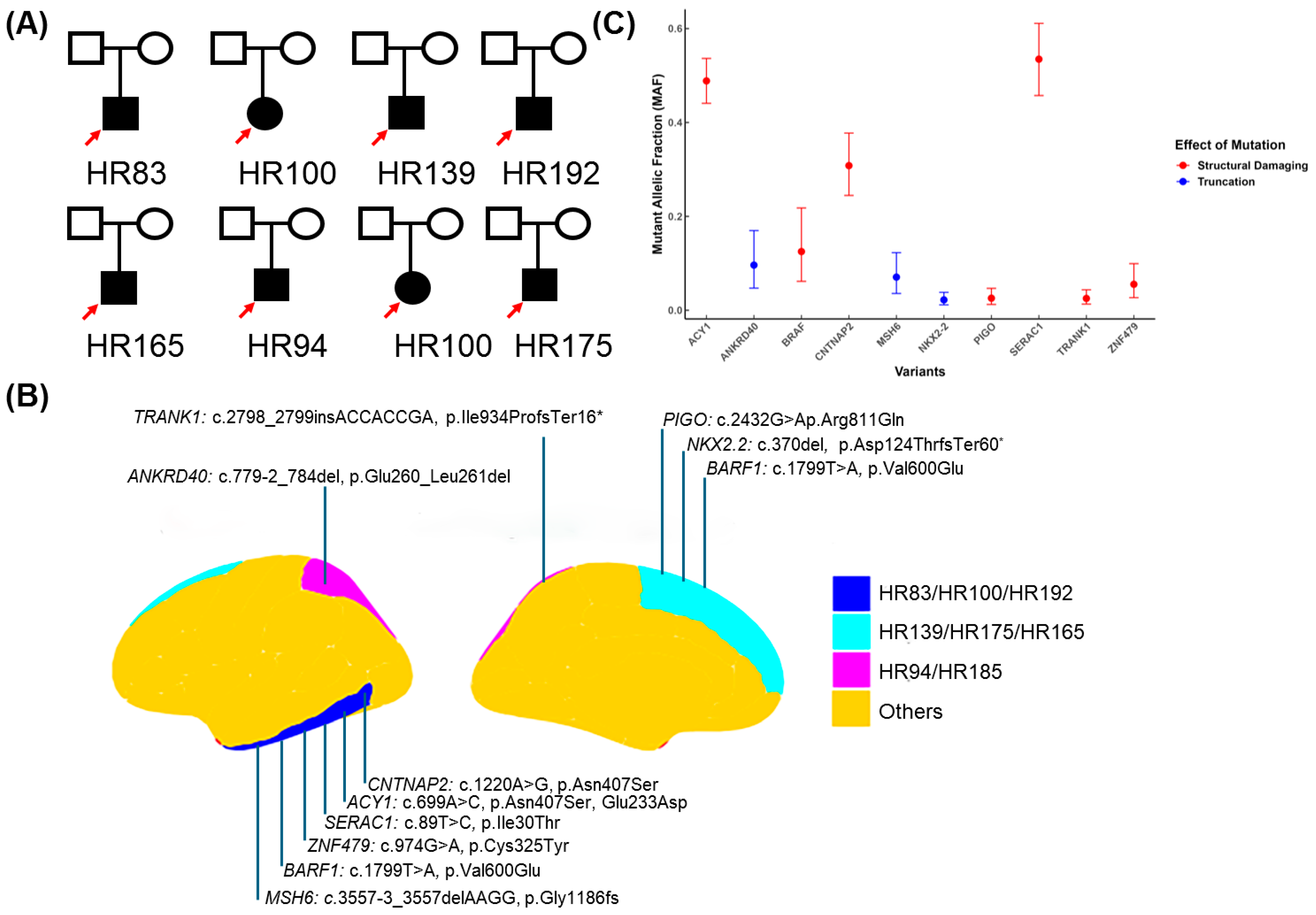
3.2. WES Analyses Identified Ten Novel Potential Pathogenic Candidate Variants in Eight FCDIII Patients

We performed whole-exome sequencing (WES) on both the blood and brain biopsy samples used from FCDIII patients to identify pathogenic mosaic variants. Pairing DNA samples from the brain and blood were sequenced to understand the role somatic variants play in mediating the underlying FCDIII phenotype (Figure 1). Whole-exome sequencing (WES) was performed on paired brain–blood samples from 19 FCDIII patients. After quality control and variant filtration (Figure 1A), all patients harboring high-confidence somatic variants were selected for detailed analysis (Table 1, Figure 2B). Of these, eight patients exhibited pathogenic variants meeting functional and clinical thresholds (Figure 1B). The WES data were then processed with extensive variant filtration pipelines, as shown in Figure 1. The variants were prioritized with frameshift mutations, inframe deletions, missense mutations, stop-gain mutations, splice acceptor variants, or splice donor variants if the protein variant had been previously identified to be pathogenic in functional studies or if the variant was associated with FCDIII or related syndromes. Subsequently, we filtered using pathogenic criteria like CADD, SWIFT, POLYPHEN, and genomic frequency, and we found 35 pathogenic somatic variants.
We considered variants that affected the protein structure/transactions based on this filtration criteria and found 10 potential disease-causing variants in eight patients (Table 2). A total of 6 were found to be missense pathogenic variants (Figure 2B,C); they were (HR83) CNTNAP2, ENST00000361727.3:c.1220A>G and ENSP00000354778.3:p.Asn407Ser; (HR83) ACY1, ENST00000404366.2:c.699A>C and ENSP00000384296.2:p.Glu233Asp; and (HR83) SERAC1, ENST00000367104.3:c.89T>C and ENSP00000356071.3:p.Ile30Thr, associated with FCDbIII in the HR83 patient. ZNF479, ENST00000319636.10:c.974G>A and ENSP00000324518.6:p.Cys325Tyr were identified in HR100 patients with FCDIIIb. BRAF, ENST00000646891.2:c.1799T>A and ENSP00000493543.1:p.Val600Glu were identified in HR139 and HR192 patients with FCDdIII; PIGO, ENST00000378617.4:c.2432G>A and ENSP00000367880.3:p.Arg811Gln were also associated with FCDIIId in HR192 patients. The four potential loss of function/pathogenic frameshift shift truncating variants were found in four patients. TRANK1, ENST00000645898.2:c.2798_2799insACCACCGA and ENSP00000494480.1:p.Ile934ProfsTer16* frameshift variants were identified in HR94 associated with FCDIIIa; MSH6 splice_acceptor_variant, ENST00000234420.11:c.3557-3_3557del in HR(100) with FCDIIIa was found in HR185 associated with FCDIIIb; and for the splice_acceptor_variant ANKRD40, ENST00000285243.7:c.779-2_784del and NKX2-2, ENST00000377142.4:c.370del and ENSP00000366347.4:p.Asp124ThrfsTer60*, a frameshift was found in HR175 and 175 associated with FCDIIId (Figure 1 and Table 2).
The mosaic fraction score of each pathogenic variant was assessed to validate their pathogenicity. The CNTNAP2 c.1220A>G variant showed a mosaic fraction of 62%, suggesting subclonal expansion. The ACY1 c.699A>C variant had a high mosaic fraction of 97.72%, indicating that it is likely a dominant mutation within the tumor and may have arisen early in tumor development. The SERAC1 c.89T>C variant displayed a mosaic fraction exceeding 100% (107%), suggesting tumor-specific amplification, which could be due to either a gain of the mutated allele or a loss of the wild-type allele. Additionally, the mosaic fraction scores for ZNF479 c.974G>A (11%) and BRAF c.1799T>A (18% in HR139 and 54% in HR192) indicated subclonal expansion and moderate clonal expansion. Similarly, the variants PIGO c.2432G>A (5%), TRANK1 c.2798_2799insACCACCGA (5%), MSH6 c.3557-3_3557del (14%), ANKRD40 c.779-2_784del (19%), and NKX2-2 c.370del (4%) also suggested clonal expansion (Table 2).
Furthermore, we also identified several pathogenic variants with clonal status in tumors, based on their mosaic fraction scores. The variants LRPPRC, MYO7A, ACY1, and ABHD14A-ACY1 exhibited mosaic fractions of 102.4%, 98.8%, 97.5%, and 97.5%, respectively, suggesting they may be disease-causing somatic mutations. Additionally, ALMS1 and LRP2 displayed mosaic fractions of 75% and 85%, respectively (Table S3), indicating clonal progression. Additionally, GPN-MSA_SCORES for the following variants indicated high pathogenic potential: PCDHB11I (−9.39), GAB4RA1 (−8.55), and UNC13C (−7.9), all of which have been previously associated with neurological disorder-related phenotypes (Table S2). Overall, these findings support the pathogenicity of these mosaic variants.

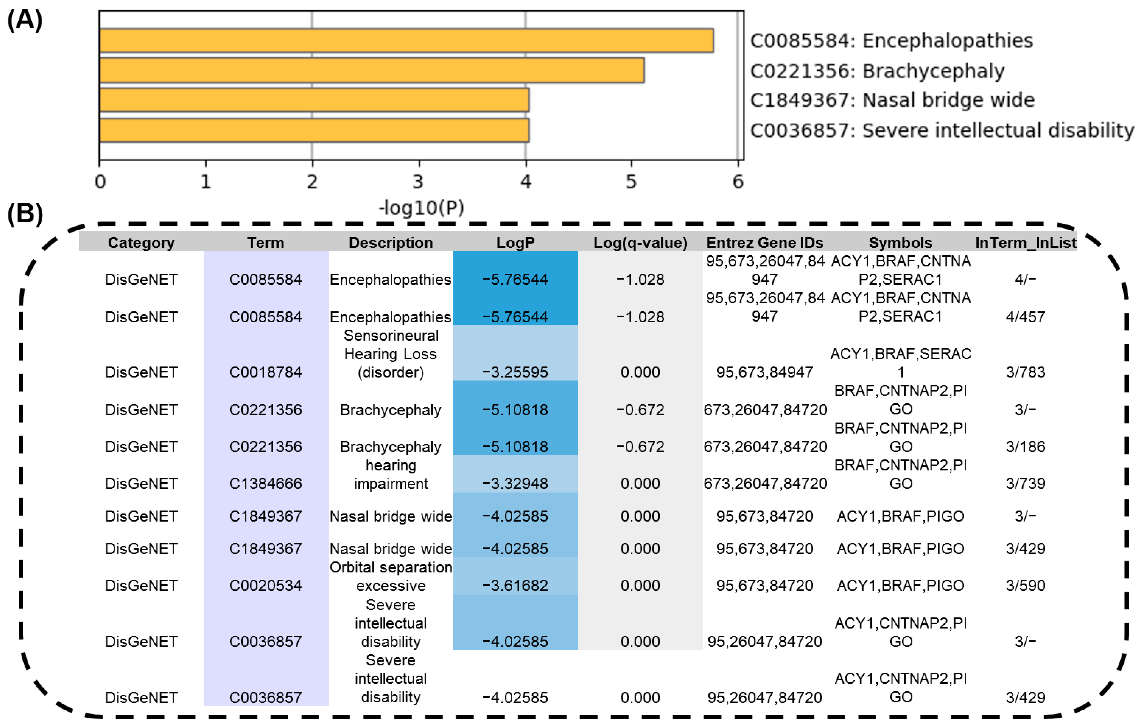
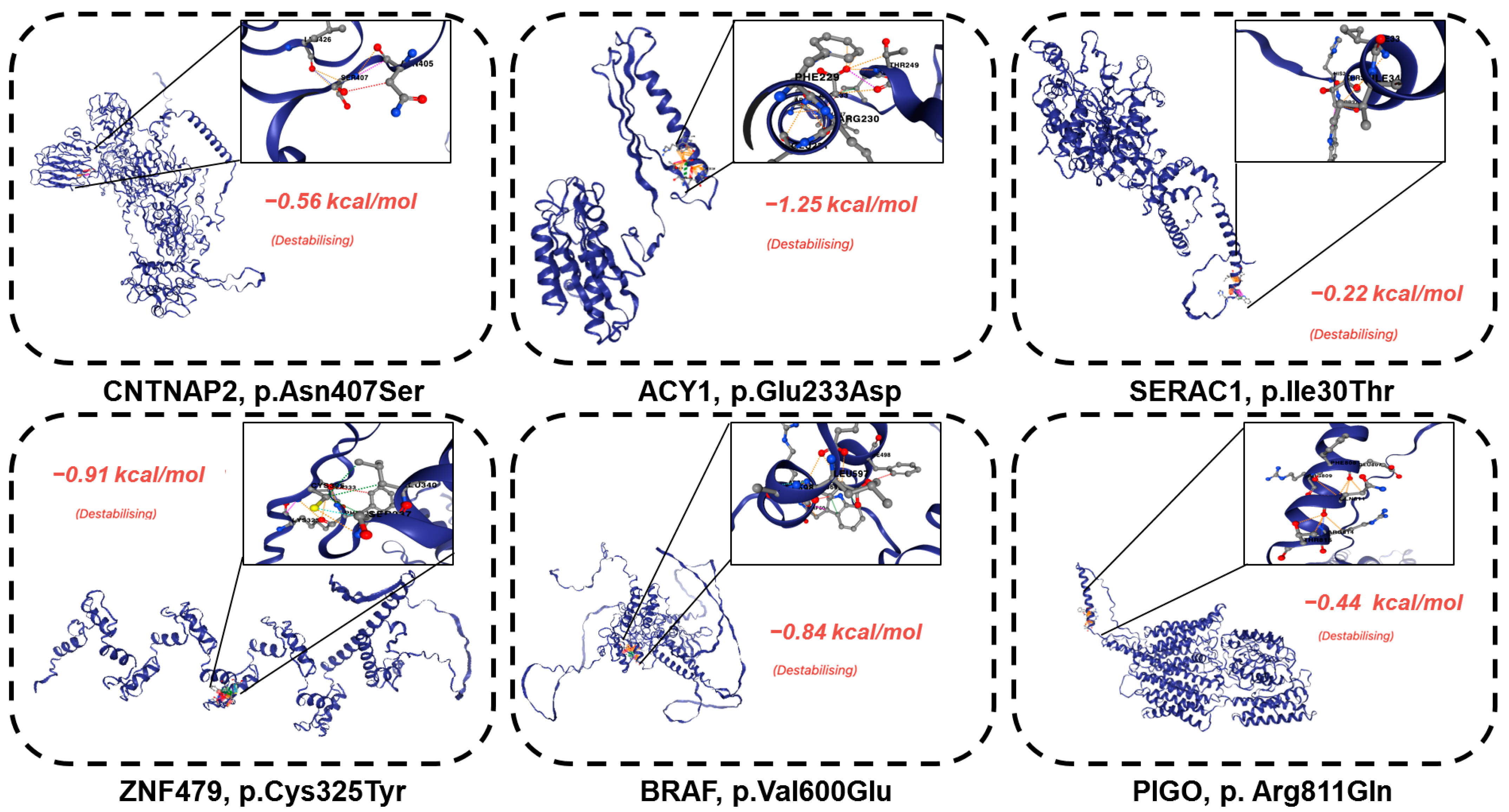
3.3. The Novel Identified Pathogenic Mosaic Variants That Showed Phenotypic Consistency and Severe Protein Structural Damage

The gene ontology enrichment analysis showed how the identified missense variants mosaics were strongly associated with severe encephalopathies and intellectual disabilities, as indicated by high −log10 (p) [26] values (Figure 3A). These findings therefore directly linked genetic variants to patient clinical manifestations, and enriched terms, such as encephalopathies, brachycephaly, and nasal bridge anomalies, suggested an active role in disease etiology (Figure 3B). This analysis highlighted the fundamental contribution of these variants to the patient’s phenotypic abnormalities. In silico analysis was then carried out to evaluate the impacts of these variants on protein structure and stability to further investigate pathogenicity.
Consistent with their severe functional consequences, destabilizing effects were significant for all variants (Figure 4). Key findings included (HR83) CNTNAP2, ENSP00000354778.3:p.Asn407Ser, which exhibited a stability impact of −0.56 kcal/mol; (HR83) ACY1, ENSP00000384296.2:p.Glu233Asp, with −1.25 kcal/mol; SERAC1, ENSP00000356071.3:p.Ile30Thr, with −0.22 kcal/mol; and (HR192&HR139) BRAF, ENSP00000493543.1:p.Val600Glu, showing −0.84 kcal/mol. Similarly, the identification of loss of function/frameshift variants (HR94) TRANK ENSP00000494480.1:p.Ile934ProfsTer16, (HR100) MSH6, (HR185) ANKRD40, (HR175) NKX2-2, and ENSP00000366347.4:p.Asp124ThrfsTer60 may contribute to the disease mechanism by insertion of a premature stop codon that leads to truncation and disrupts the functional integrity of the protein.
These destabilizations indicate that the variants interfere with protein folding or function, further implicating them in the disease process. The results of these structural and gene ontology analyses coupled provide compelling evidence that these mosaic missense variants are damaging. The accumulation of phenotypic and molecular evidence is consistent with the interpretation that these variants are important drivers of the patient’s disease. These results demonstrate how these mosaic variants are functional and structural drivers of the patient’s severe clinical phenotype. As a result, this suggests that their pathogenic potential is more marked for their association with severe intellectual disabilities and with protein instability and that precisely investigating their biological mechanisms and even their possible therapeutic targets are to be further performed.
The functional significance of each gene variant is represented by −log10 (p), ranging from −5 to −1, where higher values indicate greater functional relevance in the disease condition.

4. Discussion

Focal cortical dysplasia type III (FCDIII) represents a rare and challenging subtype of cortical dysplasias associated with drug-resistant epilepsy, necessitating surgical intervention [29]. This condition is typified by cortical lamination abnormalities, often accompanied by additional pathological changes, such as neoplasms or vascular malformations [30,31]. Unlike FCDII (mTOR-associated, e.g., PIK3CA/AKT3) mutations [32] affected by the mTOR pathway, FCDIII exhibits genetic heterogeneity (LRPPRC, UNC13C) and dual pathology (tumors/vascular malformations) with indirect mTOR linkage because mTOR drives FCDII cytopathology (e.g., balloon cells), and FCDIII pathogenesis involves broader somatic and mosaic mechanisms. This distinction underscores the necessity of advanced genetic profiling to disentangle FCDIII-specific pathways [31]. FCD IIIa is associated with hippocampal sclerosis, FCDIIIb with tumor-related lesions (e.g., ganglioglioma, DNT, and PGNT), FCDIIIc with vascular malformations (e.g., Sturge–Weber syndrome), and FCDIIId with early developmental lesions seen in genetic disorders (e.g., diffuse astrocytoma, ganglioglioma, and low-grade neuroepithelial neoplasms). The study’s results emphasize the critical role of advanced genetic approaches, such as next-generation sequencing (NGS) [33], in uncovering somatic and mosaic mutations contributing to the pathophysiology [34] of FCDIII.
Overall, participants with disease-causing somatic variants in neurodevelopmental genes exhibited a range of clinical and neuropsychological phenotypes. One of the significant findings of this research was the identification of 10 pathogenic somatic variants in eight FCDIII patients through whole-exome sequencing (WES).
These findings confirmed the pathogenicity of the identified mosaic variants. The ACY1 variant (97.72%) likely represents an early dominant somatic mutation, while CNTNAP2 (62%) and BRAF (18%) suggest subclonal expansion. The SERAC1 variant (107%) indicates tumor-specific amplification. Lower mosaic fractions in ZNF479, PIGO, TRANK1, MSH6, ANKRD40, and NKX2-2 further support clonal expansion. Together, these results underscore the roles of these variants in tumor development. These variants were located in genes previously unassociated with FCDIII, such as CNTNAP2, ACY1, SERAC1, and BRAF. The discovery of these genetic alterations underscores the complex genetic architecture of FCDIII, wherein both germline and somatic mutations interact to influence disease manifestation. Notably, the mosaic nature of these variants emphasizes the importance of sequencing-affected tissues directly, as opposed to relying solely on peripheral blood samples, which may not capture these low-frequency alterations [35].
The gene ontology analysis [26] further strengthens the connection between the identified variants and the clinical phenotypes observed in FCDIII patients. Variants in genes such as BRAF and CNTNAP2 were associated with severe protein destabilization and phenotypic abnormalities, including encephalopathies and intellectual disabilities [36]. These findings suggest that the pathogenic potential of these variants stems not only from their presence but also from their significant impact on protein function and stability [37]. The identification of pathogenic variants, such as those in TRANK1, MSH6, ANKRD40, and NKX2-2, their association with severe clinical phenotypes, and their molecular consequences represent significant strides toward improving the diagnosis and management of this condition [38].
The correlation between genetic alterations and clinical manifestations highlights the potential for personalized medicine approaches tailored to the specific genetic profile of FCDIII patients. The identification of clonal pathogenic variants through mosaic fraction analysis offers crucial insights into tumorigenesis. Variants in LRPPRC, MYO7A, ACY1, and ABHD14A-ACY1 showed high mosaic fractions (102.4%, 98.8%, 97.5%, and 97.5%), indicating their potential role as early somatic drivers. These variants are linked to processes like mitochondrial regulation and amino acid metabolism [39], critical for tumor survival. In contrast, ALMS1 and LRP2, with mosaic fractions of 75% and 85%, likely contribute to disease progression [40,41], affecting ciliary signaling pathways. GPN-MSA scores highlighted PCDHB11, GABRA4 [42], and UNC13C [43] as high-pathogenicity candidates, suggesting their potential pleiotropic effects. CNTNAP2 has previously been linked to autism and intellectual disability, BRAF to epilepsy, TRANK1 to schizophrenia, and LRPPRC/MYO7A to mitochondrial and ciliary disorders. Our findings highlight potential common pathways, including synaptic dysfunction and mitochondrial deficits, emphasizing overlapping mechanisms between FCDIII and intellectual disabilities. This further indicates a broader phenotypic spectrum of FCDIII. Future studies integrating single-cell sequencing could clarify the mechanistic contributions of these variants. In addition to elucidating the genetic basis of FCDIII, this study highlights the diagnostic and therapeutic potential of NGS. By identifying pathogenic variants that traditional methods might overlook, NGS facilitates a deeper understanding of FCDIII’s molecular mechanisms. This knowledge could pave the way for novel therapeutic strategies, including targeted gene therapies and interventions aimed at stabilizing affected proteins. However, the study also underscores the need for further research to explore the biological mechanisms underlying these genetic alterations and their potential as therapeutic targets.

5. Conclusions

In conclusion, the findings of this study provide critical insights into the genetic etiology of FCDIII, highlighting the importance of integrating advanced genetic technologies into clinical practice. The identification of novel pathogenic variants, their association with severe clinical phenotypes, and the elucidation of their molecular consequences represent significant strides toward improving the diagnosis and management of this challenging condition. Future research should focus on validating these findings in larger cohorts and exploring targeted therapies to address the underlying genetic abnormalities in FCDIII patients.

Supplementary Materials

The following supporting information can be downloaded at: https://www.mdpi.com/article/10.3390/genes16040421/s1, Table S1: Preoperative clinical profile of eight patients; Table S2: Candidate somatic variants; Table S3: Candidate validated mosaic variants in all patients; Table S4: Detailed clinical profiles of all patients in the cohort; Table S5: Patients in the cohort pre-operative clinical investigations.

Author Contributions

Conceptualization: C.A.B.G., M.Z., H.R.M. and X.Y.; methodology: C.A.B.G. and M.Z.; software: M.V.S., S.H.L., I.A.G., R.D.G. and X.Y.; validation: C.A.B.G. and M.V.S.; formal analysis: C.A.B.G. and M.Z.; data curation: C.A.B.G., M.V.S. and V.S.; writing—original draft: M.Z. and C.A.B.G.; writing—review and editing: M.Z., C.A.B.G., S.H.L., I.A.G., J.G.G., M.V.S., H.R.M. and X.Y.; visualization: M.Z. and X.Y.; project administration: C.A.B.G., H.R.M., M.V.S., J.G.G. and X.Y.; funding acquisition: H.R.M., C.A.B.G. and X.Y. All authors have read and agreed to the published version of the manuscript.

Funding

This work was supported by PRONAS (25000.069257/2015-00) (to C.A.B.G. and H.R.M.) and NIH/NICHD R00HD111686 and NIH/NIMH R21MH134401 (to X.Y.).

Institutional Review Board Statement

This study was approved by the Research Ethics Committee of HCFMRP-USP (reference number 6979/2015).

Informed Consent Statement

Informed consent was obtained from all subjects involved in the study.

Data Availability Statement

Data are provided as main and supplemental tables.

Conflicts of Interest

The authors declare no conflicts of interest.

Abbreviations

The following abbreviations are used in this manuscript:
FCDFocal cortical dysplasia
MAFMutant allele fraction
NGSNext-generation sequencing
WESWhole-exome sequencing
VCFVariant call format
OMIMOnline Mendelian Inheritance in Man

References

  1. Guerrini, R.; Barba, C. Focal cortical dysplasia: An update on diagnosis and treatment. Expert Rev. Neurother. 2021, 21, 1213–1224. [Google Scholar] [CrossRef]
  2. Chung, C.; Yang, X.; Bae, T.; Vong, K.I.; Mittal, S.; Donkels, C.; Westley Phillips, H.; Li, Z.; Marsh, A.P.; Breuss, M.W.; et al. Comprehensive multi-omic profiling of somatic mutations in malformations of cortical development. Nat. Genet. 2023, 55, 209–220. [Google Scholar] [CrossRef] [PubMed]
  3. Blümcke, I.; Thom, M.; Aronica, E.; Armstrong, D.D.; Vinters, H.V.; Palmini, A.; Jacques, T.S.; Avanzini, G.; Barkovich, A.J.; Battaglia, G. The clinicopathologic spectrum of focal cortical dysplasias: A consensus classification proposed by an ad hoc task force of the ILAE Diagnostic Methods Commission 1. Comp. Study 2011, 52, 158–174. [Google Scholar] [CrossRef] [PubMed]
  4. Barkovich, A.J.; Guerrini, R.; Kuzniecky, R.I.; Jackson, G.D.; Dobyns, W.B. A developmental and genetic classification for malformations of cortical development: Update 2012. Brain 2012, 135, 1348–1369. [Google Scholar]
  5. Blumcke, I.; Spreafico, R.; Haaker, G.; Coras, R.; Kobow, K.; Bien, C.G.; Pfäfflin, M.; Elger, C.; Widman, G.; Schramm, J. Histopathological findings in brain tissue obtained during epilepsy surgery. N. Engl. J. Med. 2017, 377, 1648–1656. [Google Scholar] [PubMed]
  6. Pinheiro, J.; Honavar, M. Focal cortical dysplasia: Updates. Indian J. Pathol. Microbiol. 2022, 65, S189–S197. [Google Scholar]
  7. Krsek, P.; Pieper, T.; Karlmeier, A.; Hildebrandt, M.; Kolodziejczyk, D.; Winkler, P.; Pauli, E.; Blümcke, I.; Holthausen, H. Different presurgical characteristics and seizure outcomes in children with focal cortical dysplasia type I or II. Epilepsia 2009, 50 (Suppl. S1), 125–137. [Google Scholar]
  8. Specchio, N.; Wirrell, E.; Scheffer, I.; Nabbout, R.; Riney, K.; Samia, P. ILAE classification and definition of epilepsy syndromes with onset in childhood: Position paper by the ILAE task force on nosology and definitions. Epilepsia 2022, 63, 1398–1442. [Google Scholar]
  9. Wan, H.J.; Hu, W.H.; Wang, X.; Zhang, C.; Wang, S.S.; Zheng, Z.; Zhou, F.; Sang, L.; Zhang, K.; Zhang, J.G. Interictal pattern on scalp electroencephalogram predicts excellent surgical outcome of epilepsy caused by focal cortical dysplasia. Epilepsia Open 2022, 7, 350–360. [Google Scholar] [CrossRef]
  10. Lara, C.; JC, S.A. Focal cortical dysplasia and refractory epilepsy. Surgical treatment. Rev. De Neurol. 2001, 32, 738–742. [Google Scholar]
  11. Najm, I.; Lal, D.; Alonso Vanegas, M.; Cendes, F.; Lopes-Cendes, I.; Palmini, A.; Paglioli, E.; Sarnat, H.B.; Walsh, C.A.; Wiebe, S. The ILAE consensus classification of focal cortical dysplasia: An update proposed by an ad hoc task force of the ILAE diagnostic methods commission. Epilepsia 2022, 63, 1899–1919. [Google Scholar] [PubMed]
  12. Shroff, S. Pediatric Epilepsy: A “Histo”-Pathologic Perspective. J. Pediatr. Epilepsy 2016, 5, 153–158. [Google Scholar]
  13. Wasilewska, K.; Gambin, T.; Rydzanicz, M.; Szczałuba, K.; Płoski, R. Postzygotic mutations and where to find them–recent advances and future implications in the field of non-neoplastic somatic mosaicism. Mutat. Res. Rev. Mutat. Res. 2022, 790, 108426. [Google Scholar]
  14. Krochmalnek, E.; Accogli, A.; St-Onge, J.; Addour-Boudrahem, N.; Prakash, G.; Kim, S.-H.; Brunette-Clement, T.; Alhajaj, G.; Mougharbel, L.; Bruneau, E. mTOR pathway somatic pathogenic variants in focal malformations of cortical development: Novel variants, topographic mapping, and clinical outcomes. Neurol. Genet. 2023, 9, e200103. [Google Scholar] [PubMed]
  15. Ye, Z.; McQuillan, L.; Poduri, A.; Green, T.E.; Matsumoto, N.; Mefford, H.C.; Scheffer, I.E.; Berkovic, S.F.; Hildebrand, M.S. Somatic mutation: The hidden genetics of brain malformations and focal epilepsies. Epilepsy Res. 2019, 155, 106161. [Google Scholar]
  16. D’Gama, A.M.; Poduri, A. Precision therapy for epilepsy related to brain malformations. Neurotherapeutics 2021, 18, 1548–1563. [Google Scholar]
  17. Yang, X.; Xu, X.; Breuss, M.W.; Antaki, D.; Ball, L.L.; Chung, C.; Shen, J.; Li, C.; George, R.D.; Wang, Y. Control-independent mosaic single nucleotide variant detection with DeepMosaic. Nat. Biotechnol. 2023, 41, 870–877. [Google Scholar]
  18. Ma, Q.; Chen, G.; Li, Y.; Guo, Z.; Zhang, X. The molecular genetics of PI3K/PTEN/AKT/mTOR pathway in the malformations of cortical development. Genes Dis. 2024, 11, 101021. [Google Scholar]
  19. Benegas, G.; Albors, C.; Aw, A.J.; Ye, C.; Song, Y.S. A DNA language model based on multispecies alignment predicts the effects of genome-wide variants. Nat. Biotechnol. 2025, 1–6. [Google Scholar] [CrossRef]
  20. Andrews, S. FastQC: A Quality Control Tool for High Throughput Sequence Data. Available online: https://www.bioinformatics.babraham.ac.uk/projects/fastqc/ (accessed on 1 March 2010).
  21. Ewels, P.; Magnusson, M.; Lundin, S.; Käller, M. MultiQC: Summarize analysis results for multiple tools and samples in a single report. Bioinformatics 2016, 32, 3047–3048. [Google Scholar]
  22. Li, H.; Durbin, R. Fast and accurate short read alignment with Burrows–Wheeler transform. Bioinformatics 2009, 25, 1754–1760. [Google Scholar] [CrossRef] [PubMed]
  23. McKenna, A.; Hanna, M.; Banks, E.; Sivachenko, A.; Cibulskis, K.; Kernytsky, A.; Garimella, K.; Altshuler, D.; Gabriel, S.; Daly, M. The Genome Analysis Toolkit: A MapReduce framework for analyzing next-generation DNA sequencing data. Genome Res. 2010, 20, 1297–1303. [Google Scholar] [CrossRef]
  24. Cingolani, P.; Platts, A.; Wang, L.L.; Coon, M.; Nguyen, T.; Wang, L.; Land, S.J.; Lu, X.; Ruden, D.M. A program for annotating and predicting the effects of single nucleotide polymorphisms, SnpEff: SNPs in the genome of Drosophila melanogaster strain w1118; iso-2; iso-3. Fly 2012, 6, 80–92. [Google Scholar] [CrossRef]
  25. Béroud, C.; Collod-Béroud, G.; Boileau, C.; Soussi, T.; Junien, C. UMD (Universal mutation database): A generic software to build and analyze locus-specific databases. Hum. Mutat. 2000, 15, 86–94. [Google Scholar] [CrossRef]
  26. Zhou, Y.; Zhou, B.; Pache, L.; Chang, M.; Khodabakhshi, A.H.; Tanaseichuk, O.; Benner, C.; Chanda, S.K. Metascape provides a biologist-oriented resource for the analysis of systems-level datasets. Nat. Commun. 2019, 10, 1523. [Google Scholar] [CrossRef]
  27. Jumper, J.; Evans, R.; Pritzel, A.; Green, T.; Figurnov, M.; Ronneberger, O.; Tunyasuvunakool, K.; Bates, R.; Žídek, A.; Potapenko, A. Highly accurate protein structure prediction with AlphaFold. Nature 2021, 596, 583–589. [Google Scholar] [CrossRef] [PubMed]
  28. Zhou, Y.; Pan, Q.; Pires, D.E.; Rodrigues, C.H.; Ascher, D.B. DDMut: Predicting effects of mutations on protein stability using deep learning. Nucleic Acids Res. 2023, 51, W122–W128. [Google Scholar] [CrossRef] [PubMed]
  29. Ferri, L.; Menghi, V.; Licchetta, L.; Dimartino, P.; Minardi, R.; Davì, C.; Di Vito, L.; Cifaldi, E.; Zenesini, C.; Gozzo, F. Detection of somatic and germline pathogenic variants in adult cohort of drug-resistant focal epilepsies. Epilepsy Behav. 2024, 153, 109716. [Google Scholar] [CrossRef]
  30. Urbach, H.; Kellner, E.; Kremers, N.; Blümcke, I.; Demerath, T. MRI of focal cortical dysplasia. Neuroradiology 2022, 64, 443–452. [Google Scholar] [CrossRef]
  31. Lee, W.S.; Baldassari, S.; Stephenson, S.E.; Lockhart, P.J.; Baulac, S.; Leventer, R.J. Cortical dysplasia and the mTOR pathway: How the study of human brain tissue has led to insights into epileptogenesis. Int. J. Mol. Sci. 2022, 23, 1344. [Google Scholar] [CrossRef]
  32. Baldassari, S.; Ribierre, T.; Marsan, E.; Adle-Biassette, H.; Ferrand-Sorbets, S.; Bulteau, C.; Dorison, N.; Fohlen, M.; Polivka, M.; Weckhuysen, S. Dissecting the genetic basis of focal cortical dysplasia: A large cohort study. Acta Neuropathol. 2019, 138, 885–900. [Google Scholar] [CrossRef] [PubMed]
  33. Schwarz, J.M.; Becker, L.-L.; Wahle, M.; Faßbender, J.; Thomale, U.-W.; Tietze, A.; Morales-Gonzalez, S.; Knierim, E.; Schuelke, M.; Kaindl, A.M. Somatic DNA Variants in Epilepsy Surgery Brain Samples from Patients with Lesional Epilepsy. Int. J. Mol. Sci. 2025, 26, 815. [Google Scholar] [CrossRef]
  34. Jamuar, S.S.; Walsh, C.A. Genomic variants and variations in malformations of cortical development. Pediatr. Clin. 2015, 62, 571–585. [Google Scholar] [CrossRef] [PubMed]
  35. Van Horebeek, L.; Goris, A.; Dubois, B. Somatic Mosaicism in Multiple Sclerosis: Detection and Insights into Disease. Ph.D. Thesis, University of Leuven, Leuven, Belgium, 2022. [Google Scholar]
  36. Auvin, S. Finally, a controversy about neonatal seizure treatment. Epilepsia 2022, 63, 1880–1882. [Google Scholar] [CrossRef]
  37. D’Gama, A.M.; Walsh, C.A. Somatic mosaicism and neurodevelopmental disease. Nat. Neurosci. 2018, 21, 1504–1514. [Google Scholar] [CrossRef]
  38. Rimmerman, N.; Verdiger, H.; Goldenberg, H.; Naggan, L.; Robinson, E.; Kozela, E.; Gelb, S.; Reshef, R.; Ryan, K.M.; Ayoun, L. Microglia and their LAG3 checkpoint underlie the antidepressant and neurogenesis-enhancing effects of electroconvulsive stimulation. Mol. Psychiatry 2022, 27, 1120–1135. [Google Scholar] [CrossRef]
  39. Cui, J.; Wang, L.; Ren, X.; Zhang, Y.; Zhang, H. LRPPRC: A multifunctional protein involved in energy metabolism and human disease. Front. Physiol. 2019, 10, 595. [Google Scholar] [CrossRef]
  40. Jecan-Toader, D.; Trifa, A.; Lucian, B.; Pop, T.L.; Cainap, S.S. Alström syndrome—Wide clinical variability within the same variant: A case report and literature review. Front. Pediatr. 2024, 12, 1463903. [Google Scholar] [CrossRef] [PubMed]
  41. Parcerisas, A.; Rubio, S.E.; Muhaisen, A.; Gomez-Ramos, A.; Pujadas, L.; Puiggros, M.; Rossi, D.; Urena, J.; Burgaya, F.; Pascual, M. Somatic signature of brain-specific single nucleotide variations in sporadic Alzheimer’s disease. J. Alzheimer’s Dis. 2014, 42, 1357–1382. [Google Scholar] [CrossRef]
  42. Sajan, S.A.; Gradisch, R.; Vogel, F.D.; Coffey, A.J.; Salyakina, D.; Soler, D.; Jayakar, P.; Jayakar, A.; Bianconi, S.E.; Cooper, A.H. De novo variants in GABRA4 are associated with a neurological phenotype including developmental delay, behavioral abnormalities and epilepsy. Eur. J. Hum. Genet. 2024, 32, 912–919. [Google Scholar] [CrossRef]
  43. Bujko, M.; Machnicki, M.M.; Grecka, E.; Rusetska, N.; Matyja, E.; Kober, P.; Mandat, T.; Rydzanicz, M.; Płoski, R.; Krajewski, R. Mutational analysis of recurrent meningioma progressing from atypical to Rhabdoid subtype. World Neurosurg. 2017, 97, 754.e1–754.e6. [Google Scholar]
Figure 1. Somatic variants filtering pipeline through WES in FCDII patients. (A) Stepwise process and filtering criteria for somatic variants; (B) Eight patients from eight distinct pedigrees were diagnosed with focal cortical dysplasia type III (FCDIII). MRI neuroimaging of these patients revealed brain lesions, indicated by red arrows in each image.
Genes 16 00421 g001
Figure 2. Novel disease-causing somatic variants identified in focal cortical dysplasia patients. (A) The red arrow highlights the WES-selected patients in each family. Identified novel somatic variants are displayed below each pedigree, with missense variants shown in red and frameshift variants in blue. (B) The identified variants are mapped to specific brain regions, including the temporal, frontal, and multilobar areas. (C) Visualization of the damaging effects of these variants, along with their mutant allele fraction (MAF) values; the error bar shows a 95% binomial confidence interval.
Genes 16 00421 g002
Figure 3. Identified somatic missense variants and their association with disease-related gene ontology through gene enrichment analysis. (A) Gene ontology enrichment analysis using MetaScape highlights the functional roles of each novel gene variant associated with the disease condition. (B) In Metascape, enrichment analyses scores and significance are reported as −log10 (p-value). Larger values indicate stronger enrichment to account for multiple comparisons.
Genes 16 00421 g003
Figure 4. Somatic pathogenic variants destabilize the protein structure. The pathogenic impacts of somatic variants on the protein structures of CNTNAP2 p.Asn407Ser: −0.56 kcal/mol, ACY1 p.Glu233Asp: −1.25 kcal/mol, SERAC1, p.Ile30Thr: −0.22 kcal/mol, ZNF479, Cys325Tyr: −0.91 kcal/mol, BRAFp.Val600Glu: −0.84 kcal/mol, and PIGO, p.Arg811Gln: −0.44 kcal/mol were assessed. The damaging effect was quantified in terms of binding energy change, measured in kcal/mol.
Genes 16 00421 g004
Table 1. Patients’ clinical characteristics.
IDHR83HR100HR139HR175HR185HR192HR165HR94
HOSPITAL ID1207695H0769405C1184599D1073501B0905454I1524999G1432293H1286326H
LOBERIGHT TEMPORALRIGHT/TEMPORALRIGHT/FRONTALLEFT/FRONTALRIGHT/TEMPORAL/PARIETALRIGHT/TEMPORALLEFT/FRONTALRIGHT/MULTILOBAR
WESBLOOD/FROZEN TISSUEBLOOD/FROZEN TISSUEBLOOD/FROZEN TISSUEBLOOD/FROZEN TISSUEBLOOD/FROZEN TISSUEBLOOD/FROZEN TISSUEBLOOD/FROZEN TISSUEBLOOD/FROZEN TISSUE
SexMaleFemaleMaleMaleMaleMaleFemaleMale
Date of Birth12/19/19972005/8/303/29/200703/26/201309/14/200512/21/200609/13/201001/14/2012
Date of Surgery2010/4/703/24/201411/16/201509/28/20152017/2/809/18/201711/30/20172013/4/10
Onset of Epilepsy5 years7 months4 years4 months7 months8 years2 years2 months
Monthly Seizure Frequency/Pre-operative909002.545060540200090
Type of SeizureFocalGeneralized tonic–clonicStructural focalGeneralizedGeneralizedSymptomatic focalStructural focalFocal
ADNPMNNNYYNNY
Neuropsychomotor ImpairmentNYYYYYYY
HemiparesisNNNNNNYN
Ocular Deviation NystagmusNNNNNNYY
MacrocephalyNNNNNNNN
Number of Medications23233142
Treatment ReoperationNYNNNNNN
Engel 1 Year1413131NO DATA
Engel 5 Years1410NO DATANO DATANO DATANO DATA
Table 2. Somatic missense variant was predicted to be highly pathogenic due to its damaging.
Mosaic Missense Pathogenic Variants
IDGeneFCDIII TypeLocationMutationMosaic FractionsHGVScHGVSpSIFTPolyPhenCADD PHRADGPN-MSA_SCORESgnomADe_AFTranscript_IDReference_GenomeGene_ENST
HR83CNTNAP2FCDIIIb7:147132381-147132381missense variant62%ENST00000361727.3:c.1220A>GENSP00000354778.3:p.Asn407Sertolerated (0.73)benign (0.01)8.396−12.630.0006432ENST00000361727GRCh38CNTNAP2-201
HR83ACY1FCDIIIb3:51987188-51987188missense variant97.72%ENST00000404366.2:c.699A>CENSP00000384296.2:p.Glu233Aspdeleterious (0.01)probably_damaging (0.95)22.6−5.980.00004446ENST00000404366GRCh38ACY1-201
HR83SERAC1FCDIIIb6:158158275-158158275missense variant100%(CNV)ENST00000647468.2:c.89T>CENSP00000496731.1:p.Ile30Thrdeleterious (0.01)benign (0.05)17.95−1.630.0009691ENST00000647468GRCh38SERAC1-217
HR100ZNF479FCDIIIb7:57120441-57120441missense variant11%ENST00000319636.10:c.974G>AENSP00000324518.6:p.Cys325Tyrtolerated_low_confidence (1)benign (0)0.02−4.30.0001944ENST00000319636GRCh38ZNF479-201
HR139BRAFFCDIIId7:140753336-140753336missense variant18%ENST00000646891.2:c.1799T>AENSP00000493543.1:p.Val600Gludeleterious (0)probably_damaging (0.963)29.8−12.633.161 × 10−12ENST00000646891GRCh38BRAF-220
HR192BRAFFCDIIId7:140753336-140753336missense variant54%ENST00000646891.2:c.1799T>AENSP00000493543.1:p.Val600Gludeleterious_low_confidence (0)probably_damaging (0.935)29.8−12.636.905 × 10−7ENST00000646891GRCh38BRAF-220
HR165PIGOFCDIIId9:35091455-35091455Missense variant5%ENST00000378617.4:c.2432G>AENSP00000367880.3:p.Arg811Glntolerated (0.2)benign (0.007)18.7−7.390.000591ENST00000378617GRCh38PIGO-203
Mosaic Loss of Function Pathogenic Variants
IDGeneFCDIII TypeLocationMutationMosaic FractionsHGVScHGVSpSIFTPolyPhenCADD PHRADGPN-MSA_SCORESgnomADe_AFTranscript_IDReference_GenomeGene_ENST
HR94TRANK1FCDIIIa3:36856923-36856924frameshift_variant5%ENST00000645898.2:c.2798_2799insACCACCGAENSP00000494480.1:p.Ile934ProfsTer16--19.453.720.00000275ENST00000645898GRCh38TRANK1-205
HR100MSH6FCDIIIb 2:47805615-47805618splice_acceptor_variant,coding_sequence_variant,intron_variant14%ENST00000234420.11:c.3557-3_3557del---393.180.00000138ENST00000234420GRCh38MSH6-201
HR185ANKRD40FCDIIId17:50697116-50697123splice_acceptor_variant,coding_sequence_variant19%ENST00000285243.7:c.779-2_784del---35-0.00000206ENST00000285243GRCh38ANKRD40-201
HR175NKX2-2FCDIIId20:21512375-21512381frameshift_variant4%ENST00000377142.4:c.370delENSP00000366347.4:p.Asp124ThrfsTer60--35-0.0000062ENST00000377142GRCh38NKX2-2-201
Disclaimer/Publisher’s Note: The statements, opinions and data contained in all publications are solely those of the individual author(s) and contributor(s) and not of MDPI and/or the editor(s). MDPI and/or the editor(s) disclaim responsibility for any injury to people or property resulting from any ideas, methods, instructions or products referred to in the content.

