1. Introduction
In addition to tanzanite, the blue-purple gem variety of zoisite that is famous from the region, the gem mines near Merelani, Lelatema Mountains, Simanjiro District, Manyara Region, Tanzania, are host to several other unusually well-crystallized minerals, including tsavorite, the green gem variety of grossular, diopside, prehnite, fluorapatite, and even graphite [
1,
2,
3,
4,
5]. The mines are also host to well-formed and uncommonly large crystals of pyrite, alabandite, and wurtzite as well as several rare sulfides, including clausthalite (PbSe), germanocolusite (Cu
13VGe
3S
16), and merelaniite (Mo
4Pb
4VSbS
15) [
5,
6,
7]. A detailed study of the chemistry of intergrown sphalerite and wurtzite, which included samples the Merelani mines and from the Animas-Chocaya Mine complex, Quechisla district, Bolivia, was recently published [
8]. The Merelani sphalerite and wurtzite are Mn-rich, and were found to contain several trace elements (e.g., Fe, Cu, Se, and Cd) with concentrations greater than 500 ppm and a discernable differentiation between the sphalerite and wurtzite. Noteworthy, 1450 ppm Ga in the wurtzite and 1750 ppm in the sphalerite phases were also reported [
8], with estimated standard deviations of 30 and 80 ppm, respectively. In both the sphalerite and wurtzite phases, trace Ga and Cu concentrations were consistent with the coupled substitution
.
In the course of our ongoing project dealing with the characterization of the Merelani mineralization [
2,
5,
6,
7], we recovered a specimen containing an exceptionally Ga-enriched stannite, with the Ga content indicating a new mineral species. This paper deals with the description of this mineral as new independent species, which was named richardsite. Richardsite is the first gallium-essential sulfide to be described from the Merelani area, joining a very short list of accepted Ga-defined species, of which only three others are sulfides: gallite CuGaS
2, ishiharaite (Cu,Ga,Fe,In,Zn)S, and zincobriartite Cu
2(Zn,Fe)(Ge,Ga)S
4.
The new mineral and its name have been approved by the Commission of New Minerals, Nomenclature and Classification of the International Mineralogical Association (No. 2019-136). It is named in honor of Dr. R. Peter Richards (b. 1943), retired water-quality researcher at Heidelberg College (Tiffin, OH, USA) and consulting editor of the journal
Rocks & Minerals, in recognition of his research and writing, spanning over four decades, on topics related to understanding the genesis of the morphology of minerals. Dr. Richards was a major contributor to the discovery and description of the new minerals carlsonite and huizingite-(Al), and the previously unknown 2
H and 3
R polytypes of sabieite, all from the Huron River shale fire in Huron County, Ohio, USA [
9]. Holotype material is deposited in the collections of the Museo di Storia Naturale, Università degli Studi di Firenze, Via La Pira 4, I-50121, Firenze, Italy, catalogue number 3555/I, and the A. E. Seaman Mineral Museum, 1404 E. Sharon Ave., Houghton, Michigan 49931-1659, USA, catalogue number DM 31876.
3. Analytical Methods
Reflectance values were measured in air using an MPM-200 Zeiss microphotometer (Zeiss, Jena, Germany) equipped with an MSP-20 system processor on a Zeiss Axioplan ore microscope. The filament temperature was approximately 3350 K. An interference filter was adjusted, in turn, to select four wavelengths for measurement (471.1, 548.3, 586.6, and 652.3 nm). Readings were taken for the specimen and the standard (SiC) maintained under the same focus conditions. The diameter of the circular measuring area was 0.05 mm.
Unpolarized micro-Raman spectra were obtained in nearly back-scattered geometry with a Jobin-Yvon Horiba LabRAM HR800 instrument (HORIBA Jobin Yvon, Edison, NJ, USA) equipped with a motorized x–y stage, an Olympus BX41 microscope (Olympus, Tokyo, Japan) with a 100× objective, polarized incident HeNe laser radiation (632.8 nm), and a neutral density filter (D0.3). Spectra were collected through multiple acquisitions with single counting times of 10 s, and repeated on natural and broken surfaces (not polished) of several crystal grains. No damage from the laser was observed on the samples under these conditions.
Quantitative chemical analyses were carried out using a JEOL 8200 microprobe (JEOL, Akishima, Japan), WDS mode, 20 kV, 20 nA, 1 μm beam size, with counting times of 20 s for peak and 10 s for background). For the WDS analyses, the following lines (standards in parentheses) were used: SKα (sphalerite), FeKα (pyrite), CuKα (synthetic Cu2S), ZnKα (sphalerite), GaKα (synthetic Ga2S3), GeKα (synthetic Ge2S3), MnKα (synthetic MnS), and SnLβ (synthetic SnS).
Single-crystal X-ray studies were carried out using a Bruker D8 Venture Photon 100 CMOS (Bruker, Billerica, MA, USA) equipped with graphite-monochromatized Mo
Kα radiation (
λ = 0.71073 Å) operating at 60 kV. The detector-to-crystal distance was 50 mm. Data were collected using
ω and
φ scan modes, in 0.5° slices, with an exposure time of 45 s per frame. Single-crystal X-ray diffraction intensity data were integrated and corrected using the software package APEX3 (Bruker AXS Inc., Madison, WI, USA, [
14]). A total of 955 unique reflections was collected.
X-ray powder diffraction data were collected with a Bruker D8 Venture Photon 100 CMOS using copper radiation (Cu
Kα,
λ = 1.54138 Å). The observed diffraction rings were converted to a conventional powder diffraction pattern using APEX3 [
14].
6. Raman Spectroscopy
The Raman spectrum of richardsite is shown in
Figure 2. The most distinct Raman bands occur at 276, 309, 350, and 366 cm
−1, with the peak at 309 cm
−1 being the narrowest and most intense. Broader and less intense bands occur at 172, 676, and 722 cm
−1. The second-most intense peak in most spectra taken is that at 366 cm
−1, however, the relative intensities of the 366 and 350 cm
−1 peaks tend to vary in spectra taken across the crystal grain and can reach the intensity of the 309 cm
−1 peak in some spectra. Overall, the Raman spectrum of richardsite is similar to that of renierite, (Cu
1+,Zn)
11Fe
4(Ge
4+,As
5+)
2S
16 (RRUFF ID: 050428 514 nm [
15]). The peak at 350 cm
−1 may be due to the presence of a Ge,Ga-rich Cu–Zn sulfide (also containing Fe, Al, Sn, Mn, and Sn) that is sometimes intermixed with richardsite and has a very intense Raman response at this frequency shift.
Based on factor group analysis, richardsite, as a stannite-group mineral, may be expected to have 14 Raman-active modes [
16,
17]. The two A
1-symmetry modes, which involve vibrations of the S atoms, are expected to be the most intense. Definitive symmetry assignments of the Raman peaks would require more detailed experimental studies, such as polarized Raman spectroscopy, checking for resonance effects, and infrared spectroscopy, which are beyond the scope of this paper.
7. Chemical Composition and X-ray Crystallography
A preliminary chemical analysis using energy-dispersive X-ray spectrometry performed on several crystal fragments, including the one used for the structural study, did not indicate the presence of elements (
Z > 9) other than Cu, Zn, Ga, S, and minor amounts of Mn, Sn, Fe, and Ge. Subsequent electron microprobe analyses (
n = 4) revealed the fragment used for the structural study to be homogeneous within analytical error. Microprobe data are presented in
Table 3. Detection limits are <0.01 wt.% for the major elements (Ga, Zn, Cu, S), and <0.02 wt.% for the minor elements (Mn, Sn, Fe, Ge).
The empirical formula, based on 8 atoms per formula unit, is: (Zn1.975Cu0.995Ga0.995Fe0.025Mn0.010Ge0.005Sn0.005)Σ4.010S3.990. The simplified formula is (Zn,Cu)2(Cu,Fe,Mn)(Ga,Ge,Sn)S4, and the ideal formula is Zn2CuGaS4, which requires Zn 33.34, Cu 16.20, Ga 17.77, and S 32.69, totaling 100 wt.%.
Single-crystal X-ray diffraction indicates that richardsite is tetragonal, with
a = 5.3626(2) Å,
c = 10.5873(5) Å,
V = 304.46(2) Å
3, and
Z = 2. It belongs to space group
(#121) and point group
. Least squares refinement of X-ray powder diffraction data (
Table 4) give the tetragonal unit cell-parameter values as
a = 5.3622(3) Å,
c = 10.5844(10) Å, and
V = 304.33(3) Å
3.
The observed tetragonal unit-cell together with the obtained chemical formula suggests that richardsite is a new member of the stannite group. However, two closely related models have been proposed by Hall et al. [
18] for the structure of these quaternary chalcogenides, which are topologically equivalent, but differ in the distributions of metals among the positions at (0,0,0), (0,½,¼), and (0,½,¾) [
19]. In particular, the structure of stannite (Cu
2FeSnS
4) is consistent with the
symmetry, having Fe located at the origin (2
a), Sn located at 2
b (0,0,½), and Cu at 4
d (0,½,¼). In this structure, the Fe and Sn atoms alternate in a chessboard fashion within the layers at
z = 0 and ½, whereas the layers at
z = ¼ and ¾ have only Cu [
20]. The structure of kësterite (Cu
2ZnSnS
4), on the other hand, has one Cu atom at the 2
a (0,0,0) position, and Sn located at 2
b (0,0,½). Zn and the remaining Cu atom are ordered at 2
c (0, ½, ¼) and 2
d (½,0,¼) [equivalent to (0,½,¾)] positions, respectively. This leads to both the Cu,Sn layers (at
z = 0 and ½) and the Zn,Cu layers (at
z = ¼ and ¾) having the metal atoms alternating in a chessboard fashion [
20]. With different atoms occupying the 2
c and 2
d positions in kësterite, the mirror plane parallel to (110) is lost, giving a structure with space group
. In both structural models, S lies on the (110) mirror plane at 8
i (
x,x,z) for stannite, or on the general position 8
g (
x,y,z) for kësterite.
In order to determine the distribution of metal atoms in richardsite without symmetry constraints, the structure was refined in both space groups, and better agreement was obtained in
. The crystal structure was refined using the program SHELXL-97 [
21] up to
R1 = 0.0284 for 655 reflections with
Fo > 4
σ(
Fo) and 14 parameters. The refined mean electron number at the metal sites, using scattering curves for neutral atoms taken from the
International Tables for Crystallography [
22], was 30 (Wyckoff position 4
d), 31 (2
a), and 29 (2
b); thus, given also the observed mean bond distances and the chemical data, Zn, Ga, and Cu were assigned, respectively, to the three tetrahedral sites. Of course, due to the iso-electronic nature of its constituent elements (Cu = 29, Zn = 30, Ga = 31) together with the ambiguity in their valence states, the metal partitioning in richardsite is, however, not straightforward. According to Brese and O’Keeffe [
23], the ideal distance (in Å) in a regular tetrahedron decreases following the sequence: 2.370/Cu
+, 2.346/Zn
2+, 2.288/Ga
3+, 2.116/Cu
2+, and this distribution is in keeping with the site-assignment proposed here for richardsite (
Table 6). Furthermore, the chemical data clearly point to a new mineral species, regardless of the site distribution. Final atomic coordinates and equivalent isotropic displacement parameters are given in
Table 5, and selected metal-sulfur (
Me–S) bond distances are shown in
Table 6. The Crystallographic Information File (CIF) is available as
Supplementary Material.
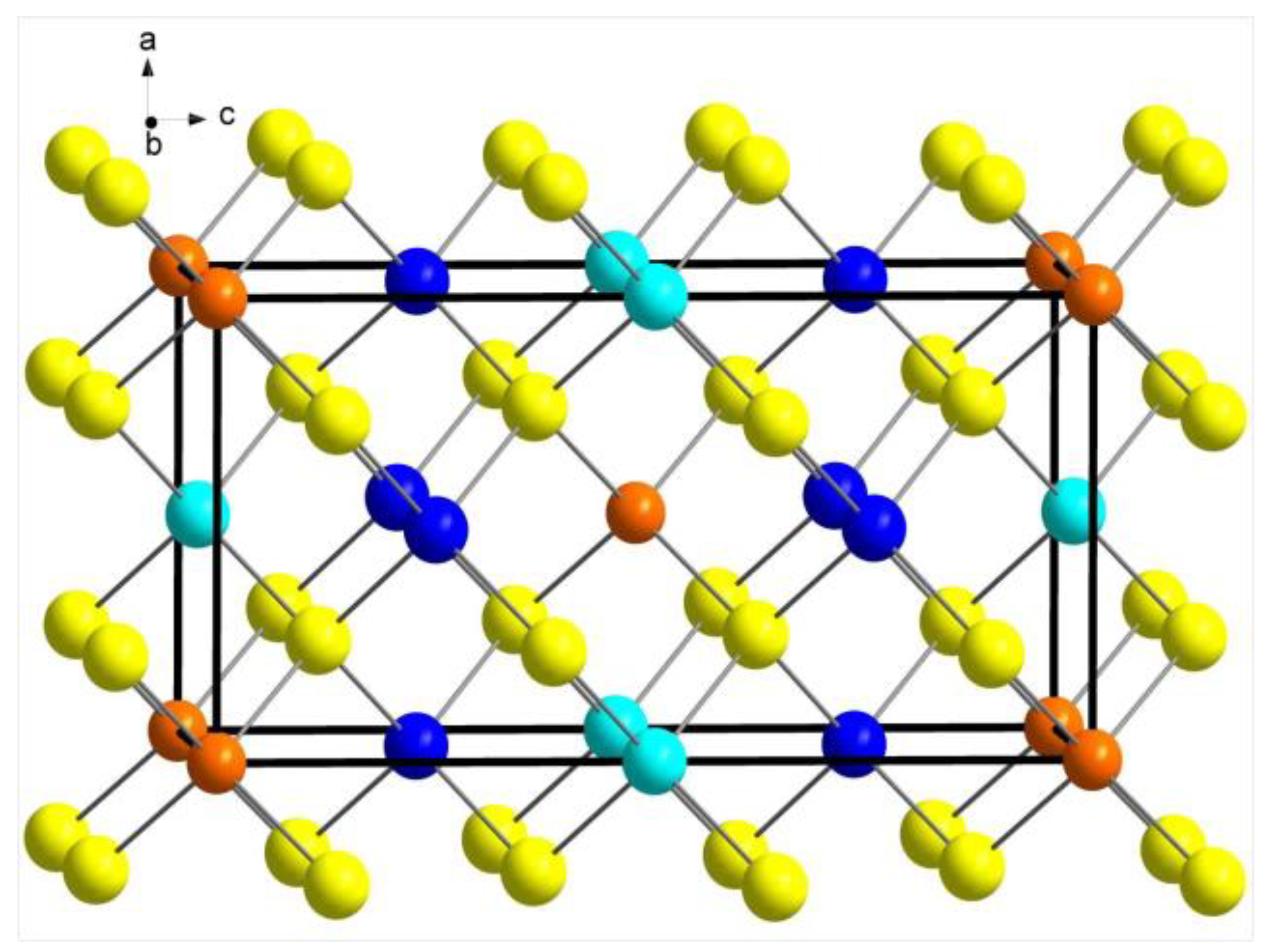
The structure of richardsite consists of a cubic close packing (
ccp) array of sulfur atoms tetrahedrally bonded with metal atoms occupying one half of the
ccp tetrahedral voids (
Figure 3). The ordering of the metal atoms leads to a sphalerite(sph)-derivative tetragonal unit-cell, with
a ≈
asph and
c ≈ 2
asph. The packing of the S atoms slightly deviates from the ideal, however, primarily due to the presence of Ga.
8. Discussion
Minerals of the stannite group are quaternary chalcogenides, typically with the general formula T1
2T2T3X
4, where T1, T2, and T3 correspond to tetrahedrally coordinated cations, and X corresponds to monatomic anions [
24]. Among the mineral species, including richardsite, accepted by the Commission of New Minerals, Nomenclature and Classification of the International Mineralogical Association, T1 = Ag, Cu, Zn; T2 = Ag, Cu, Cd, Fe, Hg, Zn; T3 = As, Ga, Ge, In, Sb, Sn; and X = S, Se. Group members are generally tetragonal but can also be orthorhombic, and their structures can be considered derivatives of the sphalerite (or chalcopyrite) structure type [
20,
25], with the types of cations and their ordering in the tetrahedral sites affecting the resulting overall symmetry of the structures.
Richardsite is the Ga-analogue of UM1985-23-S:CuFeInZn (CuZn
2InS
4) described by Cantinolle et al. [
26] and by Kieft and Damman [
27] as the end-member of the kësterite–sakuraiite series. A similar phase to UM1985-23-S:CuFeInZn (same stoichiometry) but possibly with the sphalerite-type structure, has been reported by Ohta [
28] and Semenyak et al. [
29].
A wide variety of ternary (I–III–VI
2) and quaternary (I
2–II–IV–VI
4) chalcogenides (I = Cu, Ag; II = Zn, Cd, Mn; III = Al, Ga, In; IV = Ge, Sn; VI = S, Se, Te) have been the subject of recent interest for their potential applications in photovoltaic devices, thermoelectric devices, and solar energy conversion materials [
30]. The difficultly of distinguishing between the kesterite and stannite structures, particularly with the high potential for (Cu + Zn) disorder, has been noted for the (Cu,Zn)-containing quaternary phases (see [
30] and references therein). Quaternary chalcogenides containing Ga do not appear to have been synthesized until more recently, as in a study of wurtzite and stannite phases of Cu
2ZnAS
4−x and CuZn
2AS
4 (A = Al, Ga, In) nanocrystals [
31]. These nanocrystals were synthesized using the colloidal hot-injection method as disordered-wurtzite phases. Upon annealing for 2–2.5 h in an N
2 atmosphere at temperatures of 400–450 °C for Cu
2ZnAS
4−x and 500 °C for CuZn
2AS
4, the nanocrystals transformed to ordered stannite phases. Single-crystal X-ray studies and structure refinements have not been carried out on these synthetic materials, however.
First-principles calculations for both Cu
2ZnAS
4−x [
31] and CuZn
2AS
4 [
31,
32] materials indicate that they are direct band gap materials with high absorption coefficients for visible light and, as such, they show initial promise as radiation-absorbing materials for solar cells. First-principles calculations [
32] also show the CuZn
2AS
4 materials to be
p-type semiconductors, and that the stannite-type structure is energetically more stable than the kesterite- and wurtzite-type structures.


