Article

Bioleaching of Gold from Silicate Ore by Macrococcus caseolyticus and Acinetobacter calcoaceticus: Effect of Medium, Amino Acids and Growth Supernatant

by
Kanjana Kudpeng
1,
Parinda Thayanukul
2,† and
Paitip Thiravetyan
1,*
1
School of Bioresources and Technology, King Mongkut’s University of Technology Thonburi, Bangkok 10150, Thailand
2
Department of Environmental Engineering, Faculty of Engineering, King Mongkut’s University of Technology, Thonburi 10140, Thailand
*
Author to whom correspondence should be addressed.
Current address: Department of Biology, Faculty of Science, Mahidol University, Bangkok 10400, Thailand.
Minerals 2021, 11(6), 580; https://doi.org/10.3390/min11060580
Submission received: 12 April 2021 / Revised: 21 May 2021 / Accepted: 25 May 2021 / Published: 30 May 2021
(This article belongs to the Special Issue Bio-Metallurgical Processes and Sustainable Metal Recovery)

Abstract

:
The aims of this work were to study the gold leaching by the isolated bacteria from silicate ore. Three strains were isolated and identified as Macrococcus caseolyticus, Acinetobacter calcoaceticus, and Bacillus sp. MBEA40. However, only M. caseolyticus and A. calcoaceticus were capable of gold bioleaching. In order to examine only the effect of microorganisms involved in the gold bioleaching process, minimal medium and ethanol mineral salt medium without amino acids were used for culturing M. caseolyticus and A. calcoaceticus, respectively. The result showed that the growth supernatant (in the absence of microorganisms) of both strains might be more suitable to leaching gold from ore than leaching by microorganisms (in the presence of microorganisms) directly. This might be due to the fact that there is no interference of gold absorption and metal toxicity in microorganisms in the long-term operation. The result also confirmed that amino acids/peptides/proteins produced by microorganisms might be involved in gold bioleaching, as shown in the high-performance liquid chromatography (HPLC) results. The Fourier transform infrared spectroscopy (FTIR) study also found that amine groups and carboxylic groups played important roles in gold bioleaching by M. caseolyticus and A. calcoaceticus. In addition, the bioleaching process had significantly higher gold leaching than mixed pure amino acids due to the growth supernatant containing mixed amino acids/peptides/proteins and other compounds. Therefore, the growth supernatant of M. caseolyticus and A. calcoaceticus can be applied in gold bioleaching under neutral pH conditions, which is considered to be a safe, not corrosive, and environmentally friendly leaching process. This study is also needed further study in order to increase the percentage of gold bioleaching and decrease times.

1. Introduction

Generally, the gold extraction process from ores is the cyanidation method [1]. This method is the greatest reagent for the gold extraction process and is widely used in various mining areas worldwide. Concurrently, cyanide has high chemical toxicity for human health and causes environmental problems such as the leakage of cyanide into groundwater, which has a strong effect on humans [1]. Moreover, this method is difficult to manage and incurs high operating costs [2]. Alternative methods for gold extraction processes that are less toxic, effective, and inexpensive need to be implemented [3,4].
Various methods have been suggested as the alternative way to extract gold, including using thiocyanate, thiourea, thiosulphate, bromine, and chloride [5,6,7]. However, these methods also have negative effects on gold extraction; for instance, the thiosulphate system has high reagent consumption and leads to gold extraction being performed in a less efficient manner [3,4]. Therefore, the cyanidation process is still used in various mining industries. Another method of interest with a lesser effect on human health and the environment is bioleaching. This method represents the ability of microorganisms to extract gold from various sources, such as electronic waste and mineral ores. Previously, researchers have proposed various strains that were capable of extracting gold, such as Acidithiobacillus, A. ferrooxidans, and A. thiooxidans [8,9,10,11,12,13]. Nevertheless, Acidithiobacillus, A. ferrooxidans, and A. thiooxidans are grown in extreme environments, for example, high temperatures and acidic conditions, which can increase the cost of treatment and need further management of the harmful end product. Thus, the ability of other strains needs to be investigated under mild conditions such as at neutral pH (pH 7) and moderate temperature (30–35 °C).
Another alternative way also interests, which is using amino acids; it has been widely applied to the gold extraction process. Moreover, the mechanism of gold–amino acid bonds has previously been reported by Komnitsas [14] and Reith [15]. The complexes formed between gold and amino acids are mainly through binding with nitrogen atoms in the amine group to form a covalent bond, while the oxygen atom in the carboxylic group could form ionic bonds. On the other hand, Eksteen and Oraby [16,17] and Altinkay [18] also proposed that gold can be bound with glycine, as shown in Equation (1). Oraby’s [19,20] study also confirmed amino acids as a reagent, which can be used to leach gold, replacing traditional methods. In addition, the study of Maneesuwannarat [21,22] found that gallium can be bound with amino acids as produced by Cellulosimicrobium funkei, which was isolated by contaminated soil. However, the real process of gold extraction depends on other factors, particularly pH, oxidizing agents, and oxidation-reduction potential (Eh) of the system. A study by Brown [23] explained that the extraction efficiency might be increased when using hydrogen peroxide as an oxidizing agent in copper extraction.
4Au + 8NH2CH2COOH + 4OH + O2  4Au(NH2CH2COO)2 + 6H2O
Then, the aim of this study was to investigate the isolated strains that had the capability to extract gold from ore, as well as to examine the way in which these strains can leach gold under mild conditions. Firstly, three isolated microorganisms from silicate ore were selected to study an effect on gold bioleaching. Meanwhile, the percentage of gold bioleaching by isolated strains was investigated. High-performance liquid chromatography (HPLC) and Fourier transform infrared spectroscopy (FTIR) were used to study the compounds that these strains produced and the function groups of the compounds that interact with gold, respectively. In addition, gold leaching by mixed synthetic amino acid concentrations found in growth supernatant and growth supernatant was also compared.

2. Materials and Methods

2.1. Ore Sample Preparation

The ore sample was collected from a gold mining site area in Pichit Province, Thailand. The composition of ore was determined by X-ray fluorescence (XRF), as shown in Table 1. Gold content cannot be detected, because it is too low a concentration, which the XRF can detect. Then, one gram of ore was digested by nitric acid (HNO3) and hydrochloric acid (HCl) and analyzed by inductively coupled plasma spectroscopy (ICP-OES JY 2000). The initial gold concentration was approximately 16.54 mg/Kg. The ore was grounded and sieved through a standard 200 mesh screen (particle size smaller than 75 µm). Silicate ore was sterilized by autoclaving before being used in the bioleaching experiment (121 °C for 20 min).

2.2. Screening and Isolate of Microorganisms for Gold Bioleaching

In order to study the gold bioleaching by isolated strains from silicate ore, one gram of ore was inoculated in a Luria Bertani medium (LB: 1% tryptone, 0.5% yeast extract, and 1% sodium chloride) and incubated for 24 h at 150 rpm and 30 °C. A total of 1 × 10⁸ CFU/mL of initial stock was used for the serial dilution method. A total of 1 mL of initial stock was mixed with 9 mL of LB medium including 10−1 to 108 times, respectively. Afterward, 20 µL of 10−3 to 106 was pipetted into the LB agar medium using the spread plate technique and incubated at 30 °C for 3 days. Three different colonies were found and also used to investigate gold bioleaching efficiency in this experiment.

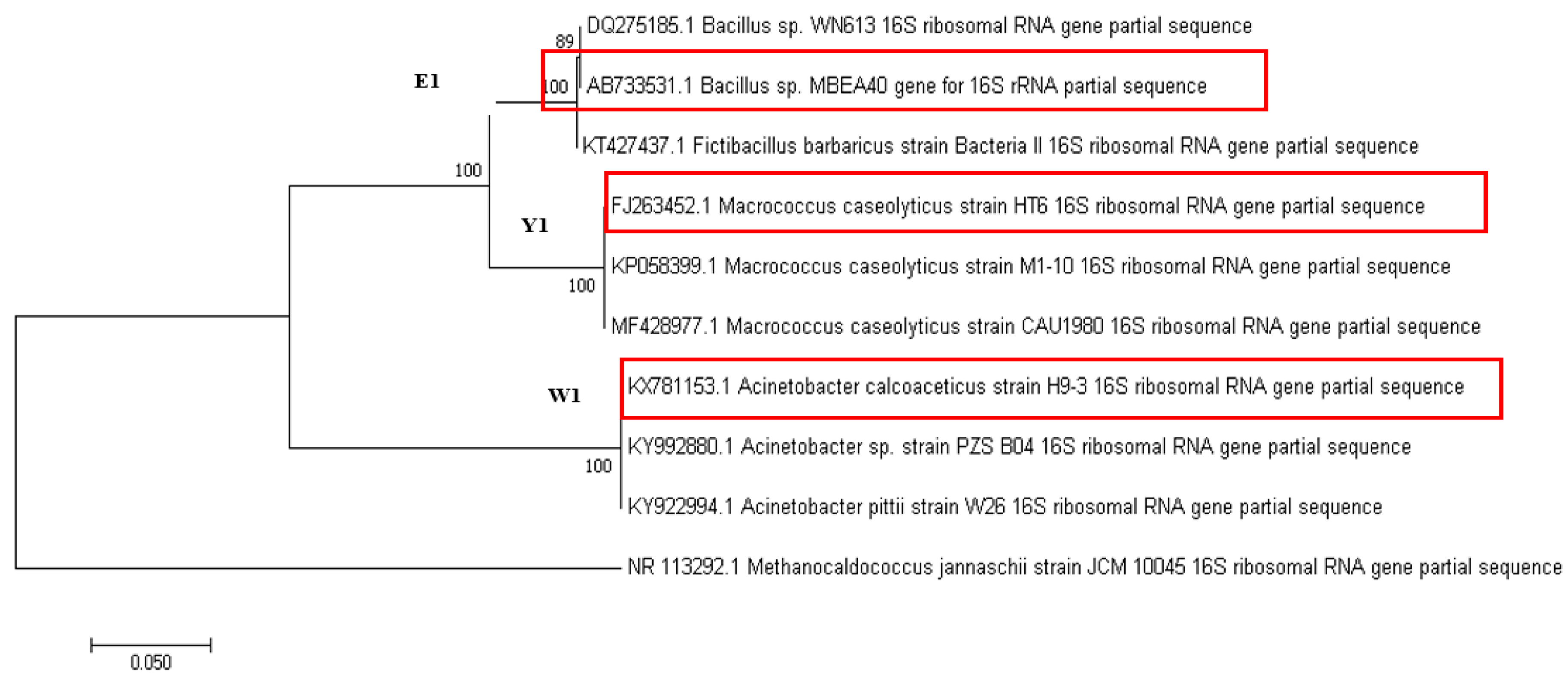
2.3. Microbial Identification

Three microorganisms isolated from silicate ore were identified, and characterization followed the method of [21]. Then, the phylogenetic tree was used for checking the percentage of similarity with the NCBI database, performed by the MEGA7 software (Pennsylvania State University, Centre County, PA, USA).

2.4. Gold Concentration Analysis

After bioleaching experiments, the sample was left for 30 min to precipitate the suspended particles of the ore. Then, a 4 mL liquid sample was collected and filtered by filter paper for measuring by ICP-OES JY 2000. Gold standard for ICP-OES JY 2000 was used (SIGMA-ALDRICH Switzerland). The efficiency of gold bioleaching from ores was calculated according to the following Equation (2):
% Au leaching = CL × 100/CI
where CI is the initial gold concentration = 16.54 mg/Kg, and CL is the gold concentration after the bioleaching process.

2.5. Bioleaching of Gold by Isolated Microorganisms

2.5.1. Bioleaching by Microorganisms (in the Presence of the Microorganism)

Pure culture of three isolated microorganisms (Macrococcus caseolyticus, Acinetobacter calcoaceticus, and Bacillus sp. MBEA40) was prepared in various media, including the LB medium, growth medium, minimal medium, and ethanol mineral salt medium. Composition of the LB medium contained 0.5% yeast extract, 1% tryptone, and 1% sodium chloride, while growth medium contained 0.5% yeast extract, 0.5% glucose, 1% casein peptone, and 0.5% sodium chloride. The composition of the minimal medium was 0.5% glucose, 4 g NH4SO4, 0.3%KH2PO4, 0.6% of Na2HPO4, and 0.05% NaCl [23]. In addition, the ethanol mineral salt medium (500 mL) also contained 10.9 g of K2HPO4.3H2O, 3.57 g of KH2PO4, 2 g of NH4SO4, 0.1 g of MgSO4.7H2O, and 2% of Ethanol. Then, 10% (v/v) of 1 × 10⁸ CFU/mL was added in a 250 mL Erlenmeyer flask containing each medium and one-gram autoclaved ore. The experiments were set in triplicate and shaken at 150 rpm, 30 °C, at various times (1 day, 7 days, 15 days, and 28 days).

2.5.2. Bioleaching by Growth Supernatant (in the Absence of the Microorganism)

The growth supernatant was prepared by adding 10% (v/v) of 1 × 10⁸ CFU/mL as a pure stock isolated microorganism in 250 Erlenmeyer flasks containing each medium including the LB medium, growth medium, minimal medium, and ethanol mineral salt medium. Then, the growth supernatant was taken after 3 days of cultivation and centrifuged at 4000 rpm for 10 min. The growth supernatant was separated by filtering through a membrane filter (CA 0.2 µ Sartorius stedim biotec). Then, 25 mL of growth supernatant was added to the 250 mL Erlenmeyer flask containing 1 g of autoclaved ore. The experiments were set in triplicate and shaken at 150 rpm, 30 °C, at various times (1 day, 7 days, 15 days, and 28 days).

2.6. Amino Acid Analysis

2.6.1. Derivatization of Standards and Samples

Amino acid analysis was performed according to the protocol from Maneesuwannarat [21,22].

2.6.2. Chromatographic Conditions

The samples were run by HPLC. The HPLC system was an Alliance Water Model E2695 Pump with an autosampler (Milford, MA, USA). The condition for analysis of samples was following Maneesuwannarat [21,22]. The mobile phase including phase B was 100% of acetonitrile, phase C was pure water, and phase D was acetate buffer. The wavelength of fluorescence for excitation and emission were set at 250 and 395 nm. The data were analyzed using Millennium software (Millennium Systems International, Parsippany, NJ, USA)

2.7. Gold Leaching by Mixed Amino Acids

In order to confirm that growth supernatant of isolate strain can leach gold from silicate ore, the efficiency of gold bioleaching by growth supernatant containing amino acids/peptides/proteins was compared to mixed synthetic amino acid under the same concentration found in the growth supernatant. Twenty-five milliliters of mixed amino acids (including aspartic acid, serine, glutamic acid, glycine, histidine, arginine, threonine, alanine, proline, cysteine, tyrosine, valine, methionine, lysine, isoleucine, leucine, and phenylalanine) at the same concentration of growth supernatant of both strains was added in the 250 mL Erlenmeyer flask containing 1 g of autoclaved ore. The experiments were set in triplicate and shaken at 150 rpm, 30 °C, for 15 days.

2.8. Fourier Transform Infrared Spectroscopy (FTIR) Analysis

FTIR was used to investigate functional groups involved in gold bioleaching. The sample solution was taken from the growth supernatant of M. caseolyticus and A. calcoaceticus before and after gold bioleaching experiments. These samples were centrifuged at 4000 rpm for 5 min. Then, the samples were filtered using a membrane before use. The clear solutions were filtered again using a 0.2 µm membrane filter. The sample was measured by FTIR (Bruker TENSOR 27 instrument). Analysis was performed using OPUS software version 6.0 and IR mentor PRO. The FTIR spectra were recorded from 4000–500 cm−1.

2.9. Statistical Analysis

Data were analyzed by SPSS statistics version 17 to perform a one-way analysis of variance (ANOVA); significantly different means were assessed by Duncan’s multiple range tests at a 95% confidence level.

3. Results and Discussion

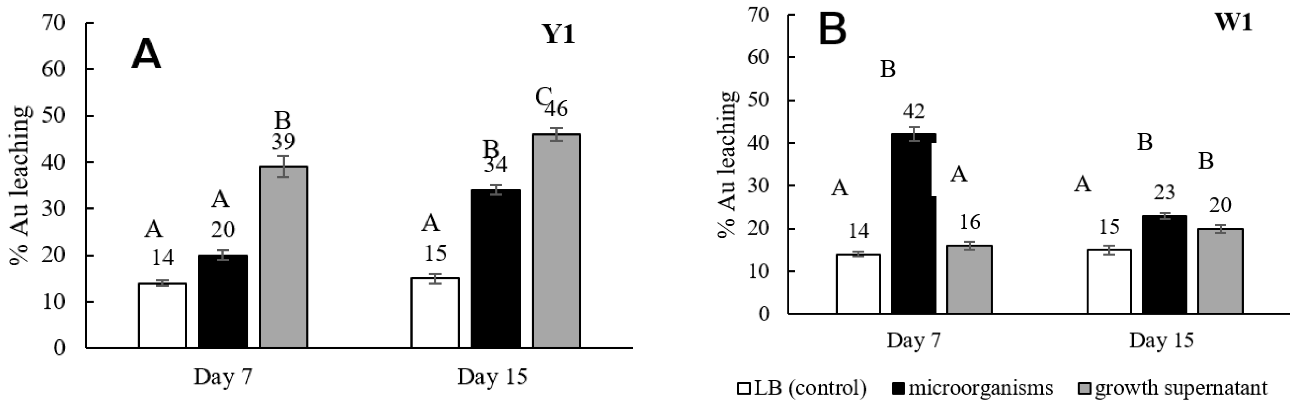
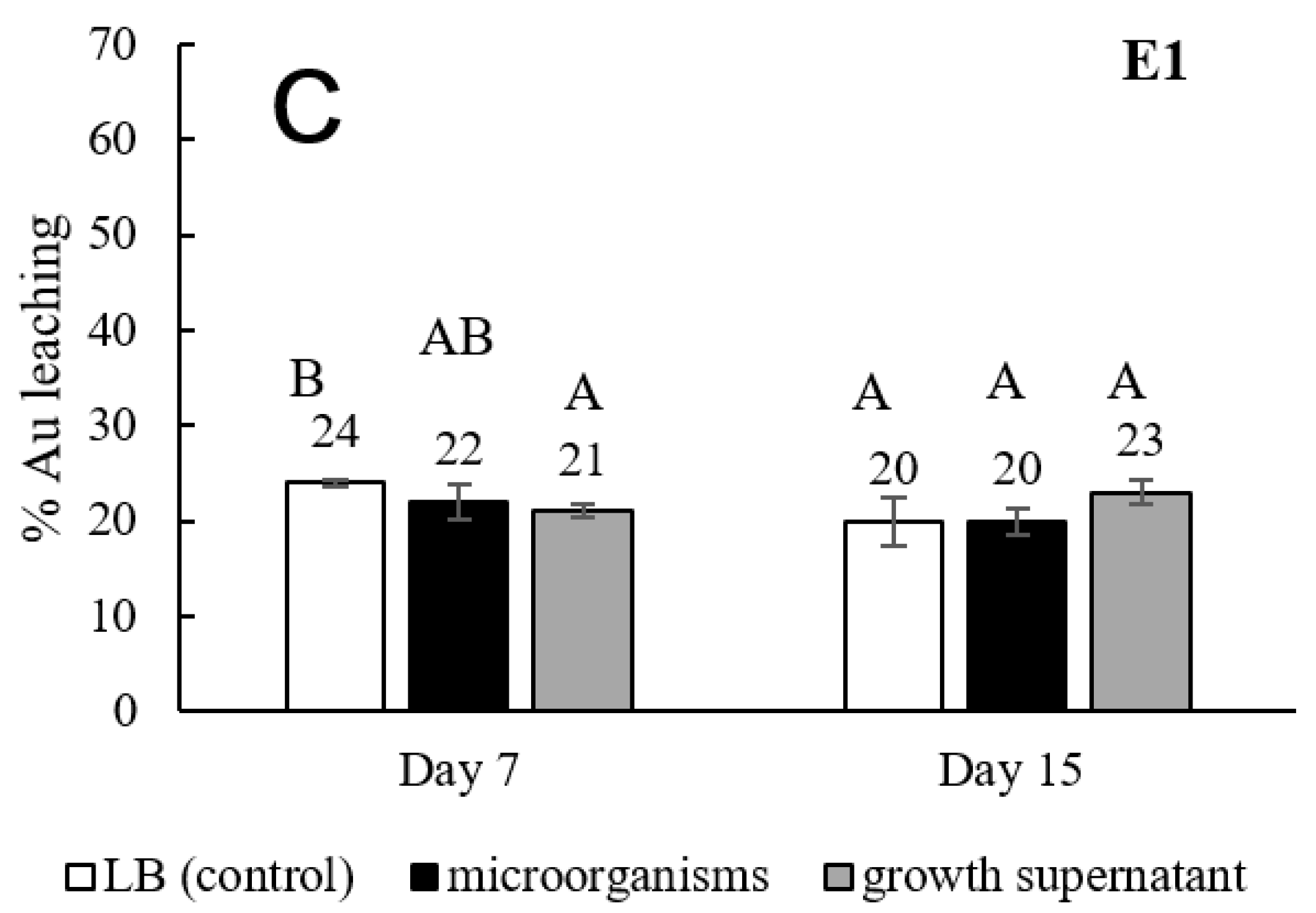
3.1. Screening, Isolation, and Ability of Gold Bioleaching Bacteria

Microorganisms used in this study were screened and isolated from silicate ore. Three strains were isolated including Y1, W1, and E1; all of these were identified by 16S rRNA sequencing analysis. The results indicated that the Y1 strain belonged to Macrococcus caseolyticus, W1 was identified as Acinetobacter calcoaceticus, and E1 was Bacillus sp. MBEA40, respectively, as shown in Figure 1. Afterward, isolated strains were investigated for abilities of gold bioleaching from silicate ore. The results of gold leaching were represented in LB medium individual (as a control), in the presence of the microorganism and growth supernatant (in the absence of the microorganism) as shown in Figure 2. The strain M. caseolyticus (Y1) had the highest efficiency to leach gold from ore approximately 46% after 15 days of cultivation under growth supernatant conditions (Figure 2A), while A. calcoaceticus (W1) was able to extract gold approximately 42% after 7 days of cultivation in the presence of microorganism conditions (Figure 2B). However, after 15 days, gold bioleaching was decreased from 42% to 23% in the presence of microorganism conditions (Figure 2B). It was due to the fact that this organism itself can absorb gold into its cell that gold leaching efficiency was decreased. Interestingly, gold can be leached by the LB medium itself (control), at a level of approximately 14–24% after 15 days (Figure 2). This is consistent with the previous studies by Maneesuwannarat [21,22] showing that amino acids in the LB medium can leach gallium from gallium arsenide. This was possible because amino acid contained in the medium can be affected by gold bioleaching, while the efficiency of gold bioleaching by Bacillus sp MBEA40 had a similar level as the control condition (20%). It might be possible that this strain could not grow well and led to lower ability in gold bioleaching (Figure 2C). Therefore, in this study, the LB medium was changed to other mediums that contained no amino acids in subsequent studies. Then, M. caseolyticus and A. calcoaceticus were chosen for study in the next experiments.

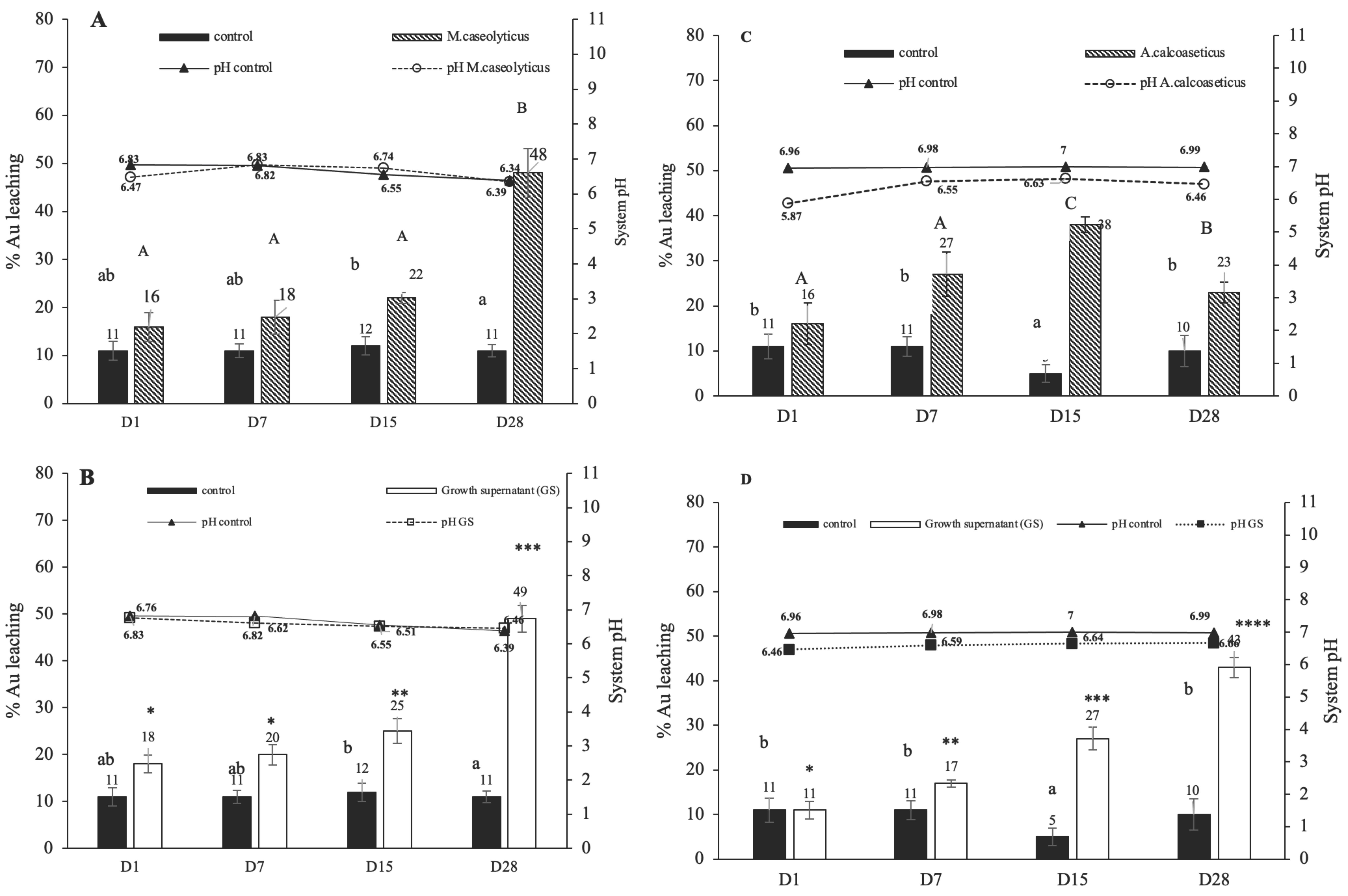
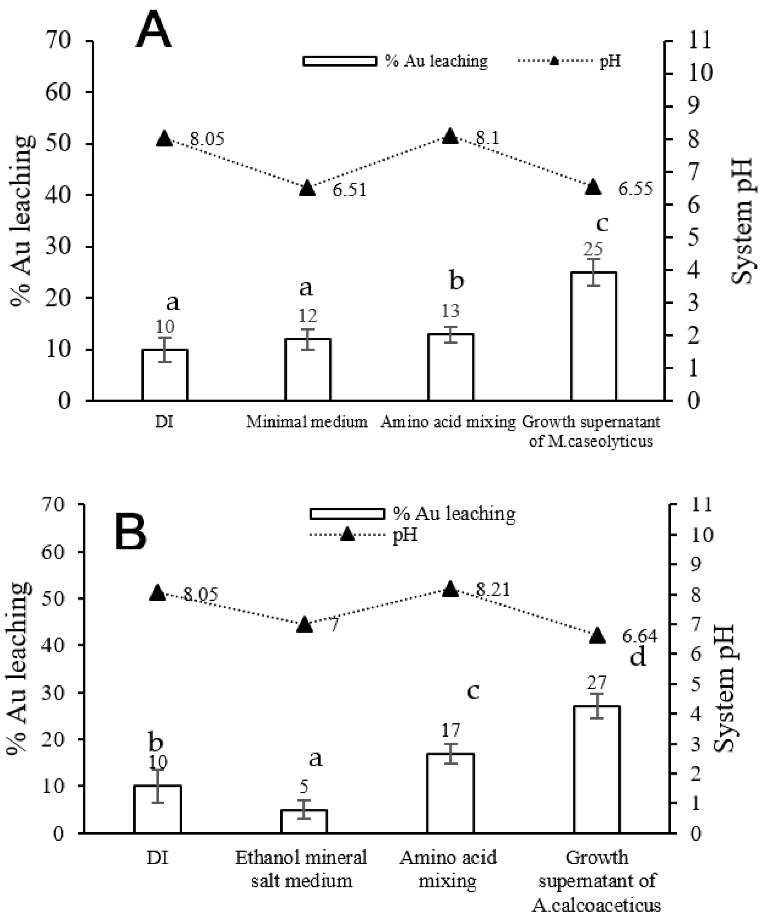
3.2. Effect of Medium Component on Gold Bioleaching from Ores

Based on the result in Figure 2, LB medium solely can leach approximately 14–24% of gold from ore after 7 days. To avoid the effect of the medium, isolated strains (including M. caseolyticus and A. calcoaceticus) under the minimal medium (MMg) were also investigated for the efficiency of gold bioleaching. The result showed that the efficiency of gold bioleaching in the presence of the microorganism and growth supernatant by M. caseolyticus under the minimal medium was approximately 30% and 29%, respectively (Figure 3A), which was not significantly different. This was consistent with gold bioleaching by A. calcoaceticus under the minimal medium (MMg), which was also not significantly different in the presence of cells and growth supernatant condition (Figure 3B).
This was because M. caseolyticus and A. calcoaceticus are able to grow under the minimal medium condition, and it can leach gold in the presence of cells, while the growth supernatant of both strains also leached gold well, probably because M. caseolyticus and A. calcoaceticus can produce some compounds that affect to gold leaching efficiency. However, both strains under the minimal medium had lower capability as compared to other medium (LB medium) conditions (Figure 3A,B). This evidence confirmed that the ingredient of the medium (LB medium) had a direct effect on gold leaching, which was similar to Maneesuwannarat [21,22], and that the medium constituent will be affected by gallium bioleaching.
Thus, the selective medium cultured two isolated strains and evaluated the abilities of gold bioleaching. Our hypothesis was that isolated strains cultured in the medium can grow well and have more ability to produce compounds to increase the efficiency of gold bioleaching. The growth medium was proper for M. caseolyticus and the ethanol mineral salt medium (EMS) was for A. calcoaceticus. The result showed that M. caseolyticus under the growth medium was able to leach gold from silicate ores in the presence of the microorganism (29%) and growth supernatant (38%) conditions (Figure 3A). Surprisingly, growth medium solely (28%) (as a control) showed a similar level to in the presence of the microorganism condition. It implied that microorganisms cannot grow well in the ore under growth medium conditions. The growth medium also contained 0.5% yeast extract, 1% casein peptone, 0.5% glucose, and 0.5% sodium chloride, resulting in containing more amino acids in this medium. This evidence also confirmed that the medium constituent can be affected in gold bioleaching directly. This was also consistent with other previous studies that found that yeast extract in LB medium could affect leach metals such as copper and gallium [21,22,23,24]. Therefore, it was probably because, under growth medium conditions, the efficiency of gold bioleaching was affected by amino acids in the medium, while A. calcoaceticus with ethanol mineral salt medium can leach gold for both conditions, including in the presence of the microorganism (33%) and growth supernatant (28%), as shown in Figure 3B. Interestingly, the growth supernatant of A. calcoaceticus from the EMS medium (28%) showed the efficiency of gold bioleaching higher than from MMg (26%) and LB medium (16%), respectively. As mention previously, the EMS medium was more suitable for A. calcoaceticus and did not contain amino acids [25,26]. This result also confirmed that, under the EMS medium, A. calcoaceticus can produce compounds and lead to an effect on gold bioleaching directly. Therefore, M. caseolyticus cultured in the MMg medium and A. calcoaceticus culture in the EMS medium were investigated regarding their capabilities in gold bioleaching.

3.3. Influence of Amino Acids on Gold Bioleaching

The efficiency of gold bioleaching by M. caseolyticus under the minimal medium and A. calcoaceticus with the ethanol mineral salt medium were investigated for a longer time to understand how isolated strains can leach gold. The result showed that gold leaching in the presence of the microorganism (48%) and growth supernatant (49%) by M. caseolyticus was increased after 28 days (Figure 4A,B). Afterward, the amino acid content in the growth supernatant of this strain was analyzed before and after leaching. It was found that M. caseolyticus produced various amino acids, and after bioleaching for 28 days, amino acids were not found in the system (Table 2). It might be possible that amino acids/peptides/proteins or other compounds that this strain produced were bound with gold directly. This result was similar to Maneesuwannarat [27], in which amino acids found in the supernatant were involved in gallium leaching. Moreover, this result also showed that no amino acids interfered in the initial minimal medium (as control) (Table 2). It demonstrated that the efficiency of gold bioleaching by the growth supernatant of M. caseolyticus cannot be affected by amino acid from the medium. A similar result was also found in the growth supernatant of A. calcoaceticus; gold was leached approximately 43% after 28 days of cultivation (Figure 4D), while in the presence of the microorganism. results showed a decrease after 28 days (23%) (Figure 4C). It might be possible that, on day 28, the organism started to absorb gold into its cell and caused a decrease in gold leaching [28]. Moreover, amino acid content by A. calcoaceticus was also investigated and similar contents to the previous strain (M. caseolyticus) were found, except for aspartic acid, histidine, and cysteine (Table 3). Notably, after bioleaching, amino acids had decreased compared to the initial amino acid content, but they had not disappeared like in the M. caseolyticus. It was due to amino acids, peptides, and protein, which isolated strains produced can bind with gold and also showed lower amino acid contents after bioleaching. Then, this is evidence to evaluate that M. caseolyticus and A. calcoaceticus had capability in gold bioleaching by producing amino acids/peptides or protein. Moreover, from our result, it can be concluded that bacteria and/or amino acids are able to solubilize gold from silicate ore, as shown in Equation (3),because some soluble gold can be dissolved in water under the control condition, as shown in Figure 4. This can be explained by the fact that soluble gold was dissolved from silicate ore to be ions; it is strongly dependent on the value of the soluble product constant (Ksp). For this reason, amino acids can enhance solubility by binding to gold ions to become a more stable form, causing a decrease in soluble ion concentration. According to the principle of Le Châtelier, the depletion of ions affects the equilibrium of the dissolution system. Therefore, more insoluble gold needs to be dissolved to maintain equilibrium.
Au+ (aq) + 8NH2RCHCOO (aq) Au(NH2RCHCOO) (aq)
However, the interaction between gold and the amino acid that is contained in the growth supernatant by M. caseolyticus and A. calcoaceticus needs to be further investigated. To avoid gold absorption and metal toxicity by microorganisms in the long-term operation, the growth supernatant was selected to further study in order to understand how the microorganism can leach gold from ore.

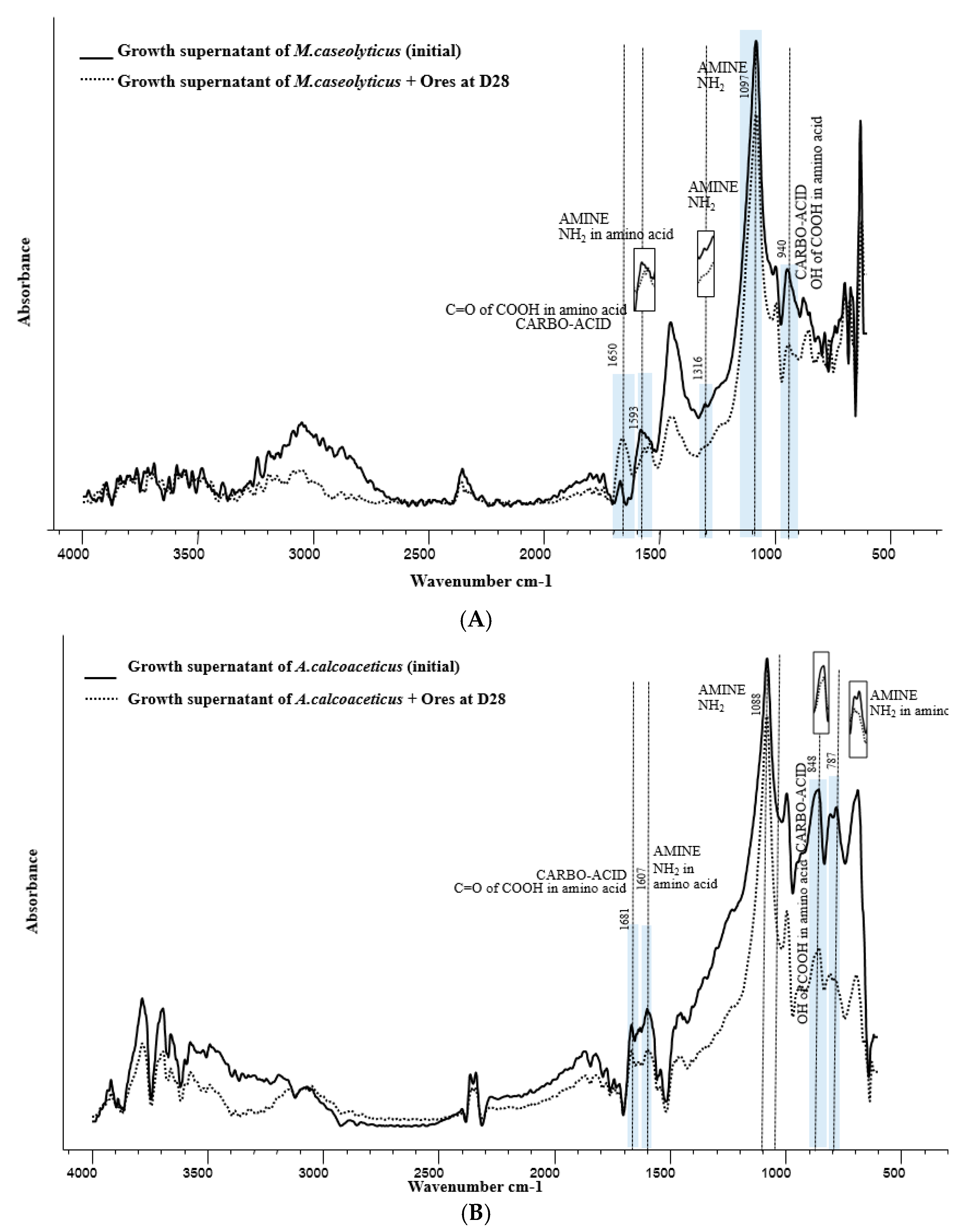
3.4. FTIR Analysis for Functional Groups on Growth Supernatant of M. Caseolyticus and A. Calcoaceticus Involved in Gold Bioleaching

FTIR was used to understand the interaction of gold and amino acids in gold bioleaching by isolated strains. Growth supernatant of M. caseolyticus and A. calcoaceticus were used to confirmed by FTIR analysis. Then, the spectra of growth supernatant as produced by both strains before and after bioleaching were compared. The results showed that the growth supernatant of M. caseolyticus showed a strong peak at 1316 cm−1, which was assigned to the amine group, as represented by NH2 in amino acids. Interestingly, after the bioleaching process, this peak had disappeared (Figure 5A). It might be possible that the amine group of amino acids/peptides/proteins was involved in gold bioleaching. In addition, the growth supernatant of A. calcoaceticus also showed similar results with M. caseolyticus. The result showed that the peak of NH2 occurred at 1607 cm−1 before gold bioleaching, and it had disappeared after 28 days of cultivation (Figure 5B). Moreover, other peaks presented the carboxylic group of the growth supernatant, as produced by both strains at position 848. The height of this peak (COOH) had also decreased after the gold bioleaching process. It was demonstrated that this position could be related to gold bioleaching. Consistent with Reith, Jingrong (15) (26) proposed that amino acids could react with gold through the nitrogen atom of the amine group and the oxygen atom of the carboxylic group. Therefore, this evidence clearly showed that the functional groups of growth supernatant had an influence on the leaching of gold from ores, particularly carboxylic groups and amine groups, which are found in amino acids/peptides/proteins. However, the detailed mechanism of the gold leaching by the amino acids/peptides/proteins in these strains needs to be further investigated.

3.5. Comparison of Gold Bioleaching Efficiency by Growth Supernatant of Isolated Strains and Mixed Amino Acids

It was proven that isolated strains had capability in gold bioleaching by producing amino acids/peptides or protein. Then, the efficiency of gold bioleaching by synthetic mixed amino acids (at the same concentration with growth supernatant) was compared with the growth supernatant of M. caseolyticus and A. calcoaceticus. The result showed that the growth supernatant of M. caseolyticus also had the highest efficiency of gold bioleaching compared with mixed amino acid (Figure 6A). A similar trend was also found in the growth supernatant of A. calcoaceticus (27%); it had significantly higher gold bioleaching than mixed pure amino acid (Figure 6B). This suggested that not only pure amino acids could leach gold, but other peptides, polypeptides, and proteins might be contributing to the gold bioleaching. Therefore, this evidence was to confirm that compounds produced by isolated strains, especially amino acids/peptides or proteins, had a higher ability to leach gold than mixed pure amino acid.

4. Conclusion

M. caseolyticus and A. calcoaceticus as isolated strains from gold silicate ore had the ability to leach gold from ore. Both strains were able to produce compounds, especially amino acids/peptides/proteins, which can bind with gold. The possible interaction of gold and growth supernatant involved the nitrogen atom of amine groups and the oxygen atom of carboxylic groups was confirmed by FTIR. Moreover, the growth supernatant of both strains was more suitable for the extraction of gold from ores than in the presence of microorganism conditions, due to the fact that growth supernatant had no effect on metal toxicity, while, in the presence of the microorganism condition, gold leaching can be re-absorbed through the organism and cause lower leaching. In addition, in a long-term operation, metals can be toxic to the cell and affect bioleaching directly. Our study also confirmed that amino acids/peptides/proteins as produced by microorganisms had higher efficiency than mixed pure amino acid. Therefore, growth supernatants of gold bioleaching by Macrococcus caseolyticus and Acinetobacter calcoaceticus can be applied for use in gold bioleaching; they are safe, environmentally friendly, and not corrosive compared to the traditional cyanidation method. Furthermore, an investigation should be conducted on how to decrease the leaching time and increase the percentage of gold leaching by growth supernatant in order to make leaching constant and more economic.

Author Contributions

Conceptualization, K.K., P.T. (Parinda Thayanukul) and P.T. (Paitip Thiravetyan); methodology, K.K. and P.T.(Paitip Thiravetyan); software, K.K. and P.T. (Paitip Thiravetyan); validation, K.K., P.T. (Parinda Thayanukul) and P.T. (Paitip Thiravetyan); formal analysis, K.K. and P.T. (Paitip Thiravetyan); investigation, K.K.; resources, P.T. (Parinda Thayanukul) and P.T. (Paitip Thiravetyan); data curation, K.K. and P.T. (Paitip Thiravetyan); writing—original draft preparation, K.K. and P.T. (Paitip Thiravetyan); writing—review and editing, K.K., P.T. (Parinda Thayanukul) and P.T. (Paitip Thiravetyan); supervision, P.T. (Paitip Thiravetyan); funding acquisition, P.T. (Paitip Thiravetyan). All authors have read and agreed to the published version of the manuscript.

Funding

This research was funded by King Mongkut’s University of Technology Thonburi (KMUTT) through the Petchara Pra Jom Kloa PhD scholarship No. 15/2558 for financially supporting Kanjana Kudpeng.

Data Availability Statement

Not applicable.

Acknowledgments

The authors would like to thank King Mongkut’s University of Technology Thonburi (KMUTT) through the Petchara Pra Jom Kloa PhD scholarship No. 15/2558 for financially supporting Kanjana Kudpeng.

Conflicts of Interest

The authors declare that they have no conflict of interest.

References

  1. Avraamides, J. Prospects for alternative leaching systems for gold: A review. In Carbon-in-Pulp Technology for the Extraction of Gold; Australasian Institute of Mining and Metallurgy: Carlton, Australia, 1982; pp. 369–391. [Google Scholar]
  2. Perea, C.; Restrepo, O. Use of amino acids for gold dissolution. Hydrometallurgy 2018, 177, 79–85. [Google Scholar] [CrossRef]
  3. Oraby, E.A.; Eksteen, J.J. Gold leaching in cyanide-starved copper solutions in the presence of glycine. Hydrometallurgy 2015, 156, 81–88. [Google Scholar] [CrossRef]
  4. Oraby, E.; Eksteen, J. The leaching of gold, silver and their alloys in alkaline glycine–peroxide solutions and their adsorption on carbon. Hydrometallurgy 2015, 152, 199–203. [Google Scholar] [CrossRef]
  5. Adams, M. Chloride as an Alternative Lixiviant to Cyanide for Gold Ores. In Gold Ore Processing; Elsevier: Amsterdam, The Netherlands, 2016; pp. 525–531. [Google Scholar]
  6. Aylmore, M. Alternative Lixiviants to Cyanide for Leaching Gold Ores. In Gold Ore Processing; Elsevier: Amsterdam, The Netherlands, 2016; pp. 447–484. [Google Scholar]
  7. Aylmore, M. Thiosulfate as an Alternative Lixiviant to Cyanide for Gold Ores. In Gold Ore Processing; Elsevier: Amsterdam, The Netherlands, 2016; pp. 485–523. [Google Scholar]
  8. Senanayake, G. Gold leaching in non-cyanide lixiviant systems: critical issues on fundamentals and applications. Miner. Eng. 2004, 17, 785–801. [Google Scholar] [CrossRef]
  9. Bosecker, K. Bioleaching: Metal solubilization by microorganisms. FEMS Microbiol. Rev. 1997, 20, 591–604. [Google Scholar] [CrossRef]
  10. Olson, G.J. Microbial oxidation of gold ores and gold bioleaching. FEMS Microbiol. Lett. 1994, 119, 1–6. [Google Scholar] [CrossRef]
  11. Kaksonen, A.H.; Mudunuru, B.M.; Hackl, R. The role of microorganisms in gold processing and recovery—A review. Hydrometallurgy 2014, 142, 70–83. [Google Scholar] [CrossRef]
  12. Sun, J.; Kan, F.; Liu, P.; He, S.; Mou, H.; Xue, C.; Mao, X. Screening of Microorganisms from Deep-Sea Mud for Antarctic Krill (Euphausia superba) Fermentation and Evaluation of the Bioactive Compounds. Appl. Biochem. Biotechnol. 2014, 175, 1664–1677. [Google Scholar] [CrossRef]
  13. Korobushkina, E.D.; Karavaiko, G.I.; Korobushkin, I.M. Biochemistry of Gold. Ecological Bulletins. 1983. Available online: https://www.jstor.org/stable/20112867 (accessed on 20 December 2020).
  14. Komnitsas, C.; Pooley, F. Bacterial oxidation of an arsenical gold sulphide concentrate from Olympias, Greece. Miner. Eng. 1990, 3, 295–306. [Google Scholar] [CrossRef]
  15. Reith, F.; Lengke, M.F.; Falconer, D.; Craw, D.; Southam, G. The geomicrobiology of gold. ISME J. 2007, 1, 567–584. [Google Scholar] [CrossRef]
  16. Oraby, E.A.; Eksteen, J.J.; O’Connor, G.M. Gold leaching from oxide ores in alkaline glycine solutions in the presence of permanganate. Hydrometallurgy 2020, 198, 1–10. [Google Scholar] [CrossRef]
  17. Oraby, E.A.; Li, H.; Eksteen, J.J. An Alkaline Glycine-Based Leach Process of Base and Precious Metals from Powdered Waste Printed Circuit Boards. Waste Biomass Valorization 2019, 11, 3897–3909. [Google Scholar] [CrossRef]
  18. Altinkaya, P.; Wang, Z.; Korolev, I.; Hamuyuni, J.; Haapalainen, M.; Kolehmainen, E.; Yliniemi, K.; Lundström, M. Leaching and recovery of gold from ore in cyanide-free glycine media. Miner. Eng. 2020, 158, 106610. [Google Scholar] [CrossRef]
  19. Oraby, E.; Eksteen, J. The selective leaching of copper from a gold–copper concentrate in glycine solutions. Hydrometallurgy 2014, 150, 14–19. [Google Scholar] [CrossRef]
  20. Oraby, E.; Eksteen, J.; Tanda, B. Gold and copper leaching from gold-copper ores and concentrates using a synergistic lixiviant mixture of glycine and cyanide. Hydrometallurgy 2017, 169, 339–345. [Google Scholar] [CrossRef]
  21. Maneesuwannarat, S.; Vangnai, A.S.; Yamashita, M.; Thiravetyan, P. Bioleaching of gallium from gallium arsenide by Cellulosimicrobium funkei and its application to semiconductor/electronic wastes. Process. Saf. Environ. Prot. 2016, 99, 80–87. [Google Scholar] [CrossRef]
  22. Maneesuwannarat, S.; Teamkao, P.; Vangnai, A.S.; Yamashita, M.; Thiravetyan, P. Possible mechanism of gallium bioleaching from gallium nitride (GAN) by Arthrobacter creatinolyticus: Role of amino acids/peptides/proteins bindings with gallium. Process. Saf. Environ. Prot. 2016, 103, 36–45. [Google Scholar] [CrossRef]
  23. Brown, D.; Smith, W.; Fox, P.; Sturrock, R. The reactions of gold(0) with amino acids and the significance of these reactions in the biochemistry of gold. Inorganica Chim. Acta 1982, 67, 27–30. [Google Scholar] [CrossRef]
  24. MacLeod, R.A.; Kuo, S.C.; Gelinas, R. Metabolic injury to bacteria II, Metabolic injury in-duced by distilled water or Cu++ in the plating diluent. J. Bacteriol. 1967, 93, 961–969. Available online: https://jb.asm.org/content/93/3/961/article-info (accessed on 20 December 2020). [CrossRef] [PubMed] [Green Version]
  25. Sar, N.; Rosenberg, E. Emulsifier production byAcinetobacter calcoaceticus strains. Curr. Microbiol. 1983, 9, 309–313. [Google Scholar] [CrossRef]
  26. Jingrong, Z.; Jianjun, L.; Fan, Y.; Jingwei, W.; Fahua, Z. An experimental study on gold solubility in amino acid solution and its geological significance. Chin. J. Geochem. 1996, 15, 296–302. [Google Scholar] [CrossRef]
  27. Maneesuwannarat, S.; Kudpeng, K.; Yingchutrakul, Y.; Roytrakul, S.; Vangnai, A.S.; Yamashita, M.; Thiravetyan, P. A possible protein model involved in gallium arsenide leaching by Cellulosimicrobium funkei. Miner. Eng. 2019, 137, 207–216. [Google Scholar] [CrossRef]
  28. Fein, J.B.; Martin, A.M.; Wightman, P.G. Metal adsorption onto bacterial surfaces: develop-ment of a predictive approach. Geochimica et Cosmochimica Acta 2001, 65, 4267–4273. [Google Scholar] [CrossRef]
Figure 1. Phylogenetic tree of isolated strains from silicate ores, based on 16 rRNA sequencing analysis and using the MEGA7 software for generating the phylogenetic tree.
Minerals 11 00580 g001
Figure 2. Bioleaching of gold from silicate ores by (A) Macrococcus caseolyticus (Y1), (B) Acinetobacter calcoaceticus (W1), and (C) Bacillus sp. MBEA40 (E1) under 7 days and 15 days. Various strains were cultured in Luria Bertani medium (LB) and shaken at 150 rpm, 30 °C (a particle size of ores approximately ≤ 75 µm). Data were expressed as the mean ± SD (n = 3). Data were analyzed by Duncan’s test (p < 0.005). The capital letters compared the percentage of gold bioleaching by three isolated strains (A-Y1, B-W1, and C-E1) under control, in the present of microorganism and growth supernatant.
Minerals 11 00580 g002aMinerals 11 00580 g002b
Figure 3. Bioleaching of gold from silicate ores under various media by (A) M. caseolyticus and (B) A. calcoaceticus under microorganisms and growth supernatant conditions for 7 days. Luria Bertani (LB), minimal medium with glucose (MMg), ethanol mineral salt (EMS), and growth medium. Data were expressed as the mean ± SD (n = 3). The lower case letters, capital letters, and asterisks (*) were compared percentage of gold (Au) leaching by medium (control), in the presence of micro-organism and growth supernatant condition at various time (control: LB/growth medium/minimal medium/ethanol mineral salt medium). Data were analyzed by Duncan’s test (p < 0.005).
Minerals 11 00580 g003
Figure 4. Gold bioleaching from silicate ores by M. caseolyticus in the minimal medium under (A) in the presence of microorganisms and (B) growth supernatant conditions and by A. calcoaceticus in ethanol mineral salt medium under (C) in the presence of microorganisms and (D) growth supernatant conditions. The flasks were shaken at 150 rpm, 30 °C, and the particle size of ores was approximately ≤ 75 µm. Data were expressed as the mean ± SD (n = 3) and analyzed by Duncan’s test (p < 0.005). The lower-case letters, capital letter and asterisks (*) were compared percentage of gold (Au) leaching by medium (control), in the presence of micro-organism and growth supernatant condition at various time.
Minerals 11 00580 g004
Figure 5. FTIR spectra of growth supernatant (GS) of M. caseolyticus (A) and A. calcoalyticus (B) before and after gold bioleaching.
Minerals 11 00580 g005
Figure 6. Comparison of gold bioleaching efficiency by growth supernatant of isolated strains (A) M. caseolyticus and (B) A. calcoaceticus with amino acid mixing (at the same concentration with growth supernatant) within 15 days. The flasks were shaken at 150 rpm, 30 °C, and the particle size of ores was approximately ≤ 75 µm. Data were expressed as the mean ± SD (n = 3) and analyzed by Duncan’s test (p < 0.005). The lower case letters compared the percentage of gold bioleaching by control (DI water), medi-um, amino acid mixing and growth supernatant by (A) M. caseolyticus and (B) A. calcoaceticus.
Minerals 11 00580 g006
Table 1. Chemical component of ores (wt%).
SiO2CaOAl2O3MgOFe2O3K2OSO3MnOTiO2ZnO
659.617.66.674.23.351.690.420.2380.191
Na2OP2O5BaOPbOCuOV2O5AgSrOCr2O3Rb2O
0.1430.09420.08110.04370.03480.0310.02690.00790.007220.00575
Table 2. Amino acid content of growth supernatant by M. caseolyticus before and after gold bioleaching.
Amino AcidMinimal MediumInitial Growth Supernatant of M. caseolyticus (nmol)Growth Supernatant of M. caseolyticus + Ores at Various Time
Day 1Day 7Day 15Day 28
Aspartic acidndndndndndnd
Serinendndndndndnd
Glutamic acidndndndndndnd
Glycinendndndndndnd
Histidinendndndndndnd
Argininend3.96 ± 5.21ndndndnd
Threoninend13.22 ± 10.11ndndndnd
Alaninend54.15 ± 39.52ndndndnd
Prolinendndndndndnd
Cysteinendndndndndnd
Tyrosinend7.09 ± 3.19ndndndnd
Valinend11.89 ± 4.54ndndndnd
Methioninendndndndndnd
Lysinend3.68 ± 5.2ndndndnd
Isoleucinend7.71 ± 4.37ndndndnd
Leucinend11.38 ± 6.01ndndndnd
Phenylalaninend7.33 ± 4.56ndndndnd
nd = not detected. Growth supernatant = supernatant without cells.
Table 3. Amino acid content of growth supernatant by A. calcoaceticus before and after gold bioleaching.
Amino AcidEthanol Mineral Salt MediumInitial Growth Supernatant of A. calcoaceticus (nmol)Growth Supernatant of A. calcoaceticus + Ores at Various Time
Day 1Day 7Day 15Day 28
Aspartic acidndndndndndnd
Serinend104.825ndnd21.131.925
Glutamic acidnd986.7106.0545.736.265.875
Glycinend356.1581.325nd95.125139.425
Histidinendndndndndnd
Argininend110.525ndndndnd
Threoninend130.47516.075ndndnd
Alaninend589.875ndndndnd
Prolinend90.375ndndndnd
Cysteinendndndndndnd
Tyrosinend88.224.4522.8nd35.15
Valinend286.2ndndndnd
Methioninend46.5ndndndnd
Lysinend125.725ndndndnd
Isoleucinend101.025ndndndnd
Leucinend121.125ndndndnd
Phenylalaninend110.85ndndndnd
nd = not detected. Growth supernatant = supernatant without cells.
Publisher’s Note: MDPI stays neutral with regard to jurisdictional claims in published maps and institutional affiliations.

