Review

Micro-Mechanisms and Implications of Continental Red Beds

1
School of Earth Science and Engineering, Sun Yat-sen University, Zhuhai 519080, China
2
Guangdong Geological Survey Institute, Guangzhou 510080, China
*
Author to whom correspondence should be addressed.
Minerals 2022, 12(8), 934; https://doi.org/10.3390/min12080934
Submission received: 29 June 2022 / Revised: 20 July 2022 / Accepted: 22 July 2022 / Published: 25 July 2022
(This article belongs to the Special Issue 3D/4D Geological Modeling for Mineral Exploration)

Abstract

:
Continental red beds, widely formed at various geologic timescales, are sedimentary rocks and sediments with red as the main color. Geoscientists have analyzed the geomorphology, paleomagnetism, paleoenvironments, paleontology, energy, and minerals in continental red beds. Despite the agreement that fine-grained hematite is closely related to the color of continental red beds, controversies and problems still exist regarding the micro-mechanism of their formation. As a review, this paper details the composition and color properties of pigmentation in red beds, analyzes the existence and distribution of authigenic hematite, and summarizes the iron sources and the formation of hematite. In addition, we introduce the fading phenomenon observed in continental red beds, including three types of secondary reduction zones: reduction spots, reduction strips, and reduction areas. Lastly, this paper summarizes the evolution of color in continental red beds, emphasizes the relationship between authigenic hematite and the diagenetic environment, and proposes possible research directions for future red bed-related issues.

1. Introduction

Red beds can be classified as continental or oceanic red beds according to the depositional environment in which they formed. At present, most are continental; oceanic ones are few due to their special formation conditions [1,2,3]. Continental red beds are not lithologically different from ordinary clastic sedimentary rocks, but they are of interest to geologists and geomorphologists because of their bright colors and unique landform types. According to erosion characteristics especially in China, red bed landforms are divided into Danxia landforms, red bed mountains, red bed hills, and red bed plateaus or platforms [4].
Research on continental red beds can be traced back to the 19th century [5,6,7]. Geologists have conducted a wide range of research regarding geomorphology, paleomagnetism, paleontology, energy, and minerals [8,9,10,11,12,13,14,15]. However, debates on the formation mechanism of their color still exist. Early geologists held two views on the origin of red beds. Some researchers believed that red sandstone formed in desert environments and was analogous to the red sand dunes of some modern tropical desert conditions, whereby the color was inherited from the fine-grained hematite film wrapped around the periphery of the sand particles [16], while other researchers suggested that red beds originated from lateritic weathering and formed in warm and humid environments where weathering of the nearby parent rocks produced the color originating from the residual red clay rich in iron oxides [17,18,19,20,21]. Thus, for a long time, the formation of continental red beds was assigned some paleoclimatic significance. However, geologists subsequently found that red beds formed in both arid and humid tropical climates [22,23,24]. The color of the red beds was formed during diagenesis, and the pigment hematite was derived from in situ alterations of iron-bearing mineral fragments or iron oxyhydroxide dihydroxylation [25]. Paleoclimatic conditions were not critical for the formation of the red color [7].
Geologists have confirmed that fine-grained hematite is the main cause of the red color through study of secondary reduction zones and the extraction of pigmentary minerals [21,26,27]. Confusion remains as to its source, existence, and formation process. In recent years, the genesis of red beds has once again become of interest to geoscientists, because the presence of hematite could be indicative of the iron cycle, water storage, and the existence of life on Mars [28,29,30]. Therefore, this paper attempts to review the progress of research on issues related to the continental red beds, summarize the mechanisms of its color formation, and explore future development directions.

2. Red Substance

2.1. Color-Rendering Characteristics

The color of red beds depends mainly on its inherited color and oxidized color, with the inherited color being the color exhibited by the clastic minerals themselves in the rock, while the oxidized color is usually caused by iron oxides or oxyhydroxides contained in its matrix and cement. It is generally accepted that the red color in the red beds originates from hematite, but there is some fluctuation in the hue of the red layer from different places. If the Munsell color system is used to describe the red beds, the color of most red beds usually varies between 5YR and 5 RP [16,31,32,33]. This is mainly because the red beds also include other iron oxides or oxyhydroxides such as goethite, ferrihydrite, and lepidocrocite. Table 1 shows the color variation values of eight synthetic samples measured by Scheinost and Schwertmann (1999) using the Munsell color system [34]. Hematite has the most reddish hue on average and goethite shows a yellowish-brown color, while the remaining iron oxides or oxyhydroxides are in between, which would explain the reddish-brown color of many red beds. However, different iron oxides differ greatly in tinting strength and hiding power. For example, the tinting strength of hematite is much greater than that of goethite at the same particle size and distribution, even though all iron oxides have strong tinting strength [33]. Even a small percentage of hematite mixed with goethite can mask the color of goethite [35]. Moreover, hematite has a greater covering power than goethite (30–60 m2·kg−1 and 15–20 m2·kg−1, respectively) [36]. Therefore, it is necessary to study hematite in the red beds and, thus, its color genesis.
The shape and size, distribution, aggregation, and cation substitution of hematite particles can also lead to changes in color. Franz (1981) showed that acicular hematite has higher reflectance and scattering ability in the long-wavelength region of the spectrum than more symmetrical hematite particles and, therefore, tends to have a more yellowish hue [26]. The tinting strength of hematite powder increases with decreasing particle size, and the strongest coloring ability of hematite is achieved when the particle size decreases to the optimal particle size, i.e., the particle size with the largest scattering cross-section (about 1 μm) [33]. Therefore, even in very brightly colored red beds, hematite is often difficult to detect by XRD. In contrast, large crystals or dense aggregates of hematite are usually dark brown or black, due to much greater absorption than scattered throughout the visible region. The arrangement of hematite aggregates likewise affects the color variation. In the Devonian red beds of Scotland, Turner and Archer (1977) observed oriented aggregates of platelet-like hematite crystals epitaxially growing on altered biotite, and the combination of such small lamellar hematite crystals into oriented aggregates causes a color shift to purple [27,37]. In addition, other metal substitutions of some Fe in the crystals can also modify the color of hematite. For example, manganese- and titanium-substituted hematite is black, while, in the case of aluminum substitution, it shifts to a more reddish hue due to the reduction in particle size [38,39].

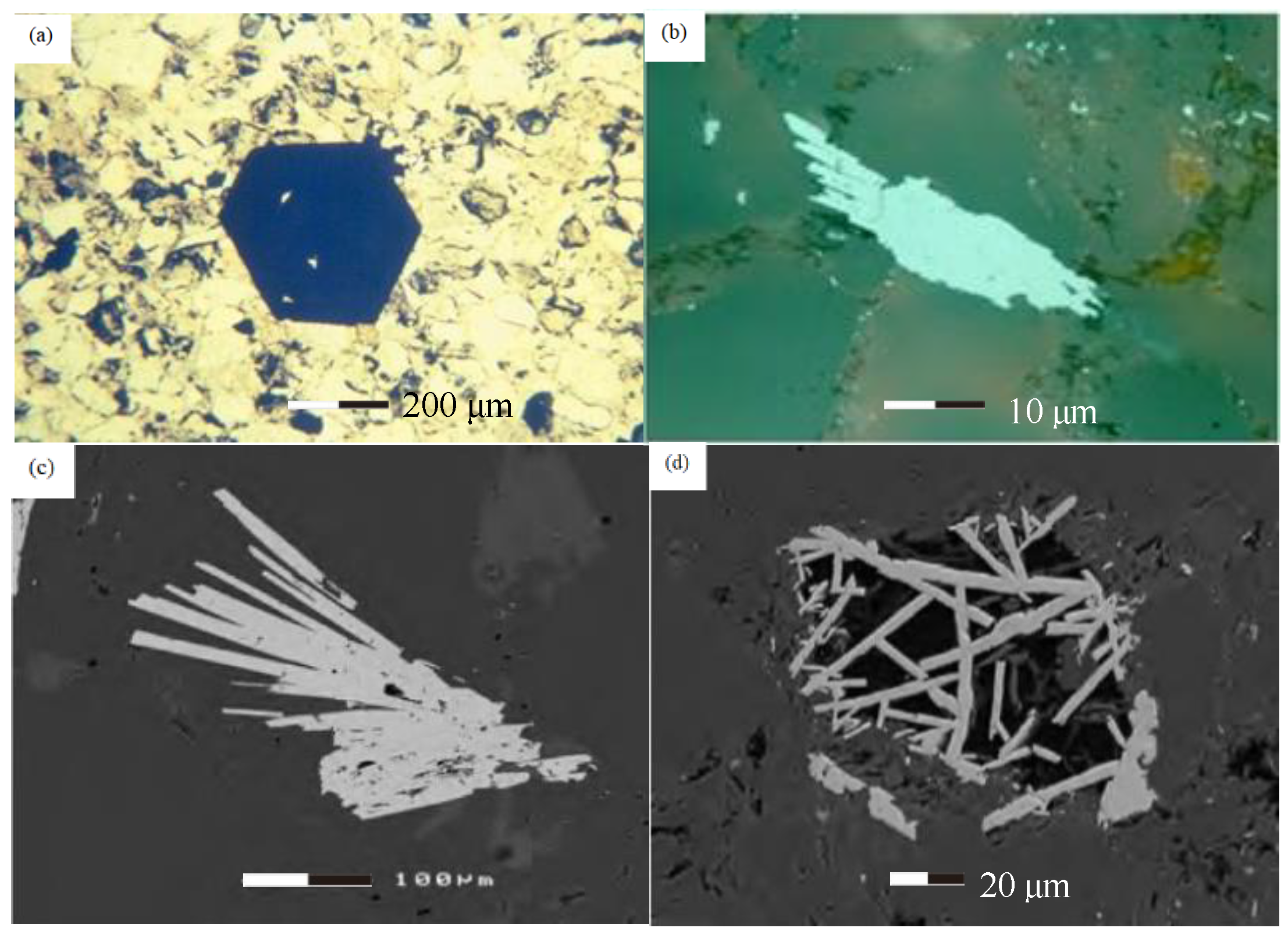
2.2. Distribution and Existence of Hematite

There are two main forms of authigenic hematite in red beds. One is in the form of single or polycrystalline aggregates coexisting with other authigenic minerals around the clastic particles, forming along the cleavage planes of mica (mainly biotite) or filling the pores between the particles; a small number of hematite particles are also present in fractures [40,41,42,43]. The crystal form of this hematite ranges from semi-hedral to euhedral, usually with relatively small quantities but large particles, mainly on the micrometer scale. There are also some submicron crystals. For example, Eren et al. (2013) discovered rhombohedral hematite crystal cross-sections with a grain size of about 0.3 mm in the red sandstones of the Early Cambrian Hüdai Formation in the Aydıncık (Mersin) Zone, central Taurides, southern Turkey (Figure 1a) [44]. Rasmussen and Muhling (2019) found radiolarian aggregates (Figure 1c) and slatted hematite filling granular voids (Figure 1d) in metamorphic red sandstones and shales in the Stirling Formation of Western Australia [45].
Another type of hematite is distributed as microfine grained crystals mixed with clay minerals in clusters disseminated in the matrix and cement of the red beds [46], partially forming a thin film of iron-bearing clay around individual clastic particles; clay films are present on clastic particles in almost all red beds. Films are sometimes developed at the contacts with detrital grains and are covered by quartz secondary overgrowth or wrapped by carbonate cement; clay-containing films formed after quartz overgrowth also exist [42]. This hematite is distributed microscopically and diffusely on montmorillonite and illite/smectite wafers or filled among the micropores between the illite/smectite layers (Figure 2a,b). Most of the hematite is in tiny crystal agglomerates or in individual microcrystals form in the pores of the montmorillonite and exhibit a variety of shapes (Figure 2d) [47]. There are spherical, rod-shaped, leaf-shaped, and well-developed hexagonal flake hematite single crystals, as well as fibrous to lath-shaped, rosette clusters and spherical hematite/goethite aggregates (Figure 2c–f) [47,48]. These morphological features are closely related to the relative abundance of the iron-bearing mineral precursors in the rocks. Hematite particles dispersed in clay minerals are usually small but very abundant and are the main source of coloring of the red beds. The hematite mainly constitutes submicron or even nanoscale crystals; hence, it is difficult to identify it using conventional detection methods [49].
The form of authigenic hematite in the red beds seems to correspond to the age of the red beds. Compared with the hematite in the form of single or polycrystalline aggregates, the latter (mixed with clay minerals) is more widely distributed in time and space, appearing in red beds of all ages. In Mesozoic and Cenozoic red beds, hematite is almost only adsorbed on the surface and/or in the voids of clay minerals [41,48,50]. In older red beds, hematite can be depleted to varying degrees due to later tectonics, groundwater penetration, or the participation of hydrothermal fluids, and then replaced by euhedral to semi-hedral authigenic hematite grains formed in situ and secondary pores or aggregates [42,43]. Residues at the grain contacts or the edges of some reddish iron-bearing clays are surrounded by quartz secondary enlarged edges. Older red beds have a greater proportion of granular hematite particles and a darker overall color.

3. The Source and Formation of Hematite

3.1. Source

Almost all Fe-containing minerals are potential sources of Fe for red-bed chromogenic substances. During oxidative weathering, Fe is released from these minerals, mainly forming Fe-bearing clay minerals and Fe oxides/hydroxides. However, under reducing conditions, Fe carbonates, sulfides, and phosphates may also be formed [33,51]. During the formation of red beds, the alteration of unstable iron-rich particles is an important source of Fe for hematite [24,25,52,53,54].
The commonly altered mineral grains are iron–magnesium silicates (e.g., hornblende, pyroxene, and biotite), volcanic rock fragments and glass, magnetite, and ilmenite [45]. These grains formed at high temperature or high pressure during magmatism and metamorphism became unstable at surface and near-surface conditions in the sedimentary basin. During grain alteration, under reducing conditions, released iron goes into solution as Fe+2 and precipitates as iron oxide or oxyhydroxide under oxidizing conditions [55]. Walker et al. (1967, 1978) [22,56] elaborated on the formation mechanism of red beds during burial diagenesis and pointed out that the alteration of the interior of the iron–magnesium silicate layers by oxygen-containing groundwater during the burial process is the key. They showed that the hydrolysis of iron-bearing detrital minerals follows the Goldich dissolution reaction series and is governed by the Gibbs free energy of the specific reaction [22,56].
Mucke (1994) indicated that alteration after diagenesis can also occur through the oxidation of pyrite and siderite [57].
3O2 + 4FeS2 → Fe2O3 (hematite) + 8S; E = −789 kJ/mol.
O2 + 4FeCO3 → 2Fe2O3 (hematite) + 4CO2; E = −346 kJ/mol.
Dissolution and displacement are the main processes leading to grain alteration in red beds. Partial dissolution of unstable minerals such as amphibole and pyroxene may proceed inward from the grain edges, while minerals such as plagioclase may preferentially disrupt the grain interior [32]. The different morphologies of partially dissolved minerals are an abiotic result of selective dissolution along lattice planes. Xiao et al. (2018), suggested that Fe and Ti-bearing silicate minerals such as biotite gradually release various elements during low-temperature alteration, with water-soluble elements such as K and Na being carried away with the transport of pore water, while water-insoluble iron–titanium oxides are deposited in situ around these altered minerals [49]. Clay mineral replacement of clastic particles is also a common alteration phenomenon in red beds, where the replaced clay forms in situ and shows random sheet crystal orientation [23].
Apart from the inheritance of parent minerals such as ferromagnesian silicates, clastic and clay minerals mechanically permeated by groundwater can also provide a direct source of iron for staining [7,54,58,59]. The mixing of hematite with clay minerals in the form of very fine-grained crystals is an important feature of red beds, and the relative proportions of hematite and clay vary considerably. In some cases, iron-bearing films may consist only of granular crystals of hematite that have precipitated in situ. In other cases, a thicker iron-containing clay film may be present. These Fe-bearing clays may have originated from surface soils or, more likely, from loose sediments formed in the adjacent floodplain [60]. An important feature of mechanical infiltration is that clay is not red when deposited, but reddens over time after contact with oxygenated groundwater [25]. In Cenozoic alluvium, the usual sediment color is yellow or red orange, due to iron oxyhydroxide, of which goethite and ferrihydrite are the main chromogenic minerals. Thus, the reddening of permeable clays represents an early stage of red material formation in red beds.
In addition, Blodgett et al. (1993) suggested that recirculation of red material may be the source of hematite in some red beds [32]. Such red beds would be produced by the erosion and redeposition of pre-existing red beds. These often have irregular color zoning, which is related to an unconformity in the weathering profile. Color boundaries cross lithological contacts and show more intense reddening near unconformities. Secondary red beds formed in this way are representative of late diagenesis, associated with previously deposited uplift, erosion, and surface weathering, requiring similar primary and diagenetic conditions [61].

3.2. Hematite Formation

There are two main potential mineralogical pathways for the formation of hematite during diagenesis: ferrihydrite transformation and ferric oxyhydroxide dehydroxylation. Ferrihydrite is the most common precursor for the occurrence of iron oxides in soils and sediments, and the transformation from ferrihydrite to hematite in red beds is an important process [62]. The conversion of ferrihydrite to hematite appears to start with the internal rearrangement and dehydroxylation of ferrihydrite aggregates, which are converted to hematite over time [63]. This transition may occur faster than the new formation of ferrihydrite and may occur too quickly to be detected in most geological settings [33]. Jiang et al. (2018), proposed a new model for the transformation of ferrihydrite to hematite in soils and sediments, which can be divided into five stages according to the chronological order of transformation: (I) magnetically ordered (or core-shell structured) superparamagnetic (SP) ferrihydrite formation, (II) rapid hematite formation from SP ferrihydrite, (III) relative hematite stabilization, (IV) maghemite nanoparticle neoformation, and (V) completion of the transformation from maghemite to hematite [64]. This model provides a new framework for interpreting the formation of iron oxides and their paleoenvironmental significance.
The transformation of goethite to hematite has long been considered the important pathway for red beds [65,66]. Weibel et al. (1999) observed pseudo-crystals of goethite transforming to hematite in red sandstone samples from the Triassic Skagerak Formation in Denmark. Withing increasing burial depth (550 m to 2500 m), the ground temperature increased from 47 °C to >105 °C [67]. Berner (1969) pointed out that goethite and iron hydroxides are generally unstable relative to hematite, and goethite is easily dehydroxylated under water shortage or high-temperature conditions (2FeOOH (goethite) → Fe2O3 (hematite) + H2O). Experiments show that, at 250 °C, the Gibbs free energy (G) of goethite-hematite reaction is 2.76 kJ/mol [68]. Langmuir (1971) concluded that G becomes more and more negative as particle size decreases. The smaller the mineral particle, the easier it is for goethite to undergo dehydroxylation to form hematite [69]. Dehydroxylation of fine-grained goethite may occur quickly, especially during deposition [7]. Thus, it appears that clastic iron oxyhydroxides (such as goethite) spontaneously dehydrate into red hematite over time. This process helps to explain not only the gradual reddening of the alluvium but also why desert dunes of older geological ages are redder than the newer ones.
In addition, other processes leading to the formation of diagenetic hematite may include the pseudo-crystalline substitution of hematite for minerals such as magnetite [7,70] and microbial interactions [44,71]. It should be noted that usually the formation of hematite in red beds may not be caused by a single mechanism of action but is the result of a combination of mechanisms. On long geological time scales, the origin of hematite in some older red beds may even be more complex because of the possibility of dissolution and reprecipitation of hematite during one or more earlier tectonic or crustal fluid events [43,45].

4. Reduction and Leaching

The local development of light bands of white, gray, or green color in many red-dominated stratigraphic sequences is a typical feature of continental red beds. Many researchers refer to such light bands as secondary reduction zones [7]. Geologists have been observing secondary reduction zones for almost as long as the color has been discussed in the literature [32,72,73,74,75]. According to the mineralogical characteristics and geochemical properties of secondary reduction zones in red beds, two categories have been identified: leaching zones and reduction zones. Rocks in the leaching zone tend to be white, as the iron content is depleted relative to the surrounding red host rock, due to leaching of soluble iron [7,76]. In contrast, rocks in the reduction zone are mainly gray or green and show a slight deficit in iron content relative to the peripheral red host rock. There, Fe2+ /Fe3+ values can be significantly higher and can be attributed to the formation of sulfides, carbonates, or clay minerals (mainly chlorite) [74]. Some red beds are characterized by both leaching and reduction, which are manifested by the coexistence of multiple-colored sediments in such secondary reduction zones.
According to scale of development and morphological characteristics, secondary reduction zones can exhibit three common forms in field outcrops: reduction spots, reduction strips, and reduction areas.

4.1. Reduction Spots

Light-colored zones having a circular or elliptical shape can be referred to as reduction spots or reduction points. Spatially, these reduction spots are a series of irregularly shaped reduction spheres or ellipsoidal spheres, which are early reduction features formed by diffusion of reduced material from the core of reduction spots to the surrounding area. Most reduction spheres in red beds are near-perfect spheres showing clear boundaries with the outer red host rock (Figure 3a,d,e). Some of the ellipsoidal spheres have long axes extending parallel to the stratigraphy (Figure 3e,f), mostly between 1 and 3 cm in diameter [32,59]. Their colors range from white to off-white or light green to blue-green. Reduction spots can occur in both impermeable argillaceous limestone and pore-rich conglomerates; they are like the surrounding red host rock in terms of sediment composition, particle size, roundness, porosity, and sedimentary structure [9]. In terms of chemical composition, the red host rock has a higher total iron content and a lower Fe2+/Fe3+ value than the reduction spot, but the Fe2+ content in the reduction spot is not necessarily higher than the Fe3+ value. Hematite is usually present in the matrix and cement of the red beds as microcrystalline particles or mixed with clay minerals, but is rarely present in reduction spots [7,59,77]. The formation of reduction spots is usually attributed to the in situ reduction of Fe3+ to Fe2+ from the dissolution of iron oxides. Some researchers have suggested that reduction spots form after the crystallization of pigment hematite, i.e., the sediment is red before the formation of reduction spots, and the hematite is converted to the corresponding reduction products by reduction [78,79]. Other researchers have suggested that the area where the reduction spots form never become red, i.e., the spots formed before the surrounding host rocks become pigmented, preventing the precipitation of hematite and preserving the original color of the sediment or reducing it further [7,25,55,59,77,80].
In some red beds, dark cores of black, brown, or red colors are also observed in the center of the reduced spots (Figure 3b). The dark core is often enriched with metallic elements such as Cu, V, U, and Ni and nonmetallic elements such as S and As [74,79,81,82]. Some researchers have suggested that the higher concentration of metallic elements such as copper in the reduction spots may be due to adsorption by organic matter [76,77,78]. More researchers have suggested that the formation of reduction spots containing dark cores is better attributed to the activity of bacteria or microorganisms that use organic matter as an energy source and reducing agent, allowing the dissolution of metallic elements and their precipitation in the cores of the reduction spots through bacterial-mediated reduction [74,75,81,83,84,85].
We note that carbonatite-bearing rock chip conglomerates also commonly appear in the center of the reduction spot in some conglomerate red beds. The formation of such reduction spots is closely related to the composition of the rock chips (Figure 3c,f).

4.2. Reduction Strips

Secondary reduction zones distributed parallel to bedding planes, along with unconformity contacts and structural deformation zones or fractures, usually appear as strips or irregular bands in field outcrops. these are collectively referred to as reduction strips in this paper (Figure 4b,d). Compared with the surrounding red host rocks, the main composition of sediments in the reduction strip is similar, but with greater grain size, porosity, and permeability [59]. Therefore, the reduction strip is often developed on the sides of the relatively coarse-grained laminated units or tectonic fissures. The colors of the reduction strip are mainly white, gray, or off-white, followed by light pink, dark gray, green, etc. [42,54,86,87]. Reduction fluids use the pores or fissures in the reduction strips as channels and leach the free iron oxide from the sediments by reduction [88,89]. According to the chemical composition, reducing fluids can be divided into the following categories: (1) hydrocarbons (hydrocarbon organics associated with hydrocarbons) [90,91]; (2) organic acids (associated with surface facies formation) [54]; (3) acidic reduction fluids such as CO2 and H2S (associated with volcanism and hydrothermal action) [92,93]. In addition, reduction fluids from reservoirs occasionally contain high H2S contents (>5 vol.%) as a result of thermochemical sulfate reduction of methane with capping anhydrite [94]. Most secondary reduction zones in red beds may not be a result of a single reduction, but include a variety of reducing fluids with multiple stages of reduction and leaching, and even the participation of bacteria or other microbial activities [54,87,95].

4.3. Reduction Areas

Reduction areas have a similar genesis mechanism to reduction strips but require an adequate source of reduction fluids to enable the reduction of larger-scale zones and even entire stratigraphic units (Figure 4a,c). Reduction areas must involve the transport and storage of large-scale reduction fluids. The formation of large reductive areas is primarily related to hydrocarbon reservoirs. The reductive fluids are mainly hydrocarbons, followed by CO2. Most Jurassic and Cretaceous red beds in the Colorado Plateau of the United States developed abundant reducing areas with hydrocarbons as the main reducing fluid. These are closely related to nearby oil and gas reservoirs in space [89,96,97,98]. However, in other areas such as the red sandstone reduction zones near Green River, Utah, the reduction fluids were dominated by CO2. Fe and other trace metal elements in the matrix and cement were dissolved in low-pH reduction fluids and eventually reprecipitated near the redox front with fluid transport [93,99,100]. In addition, because the creation of reduction areas usually consumes large amounts of fluids, when they carry dissolved metals such as Cu, U, V, Co, and Au, creating enrich mineralization near redox fronts, geologists often locate copper, uranium, and other sedimentary minerals near the reduction areas [7,15,42,82,101,102,103,104].

5. Conclusions and Future Work

Since the Mesoproterozoic Great Oxidation Event (GOE), the free oxygen content of the atmosphere has increased sharply, and most of the Earth’s surface is generally an oxidizing environment. All iron oxides or oxyhydroxides have high tinting strength and hiding power. Even if their content is less than 1% iron oxides, it is enough to stain the sediment. Therefore, the color of most iron-bearing continental sediments is reddish or yellowish. It is certain that hematite has stronger coloring and covering power than other iron oxides and oxyhydroxides; hence, the color of the red beds is almost dependent on microfine hematite. Thus, the color evolution of the red beds is essentially the evolution of hematite. In the evolution of these red beds, hematite is mainly formed during diagenesis, and iron originates from iron-rich mineral particles such as iron-bearing silicates, sulfides, and oxides, which are released into pore solutions through alterations, and then precipitated under suitable geochemical conditions forming hematite or precursors. Hematite can also be formed by dehydroxylation of iron oxyhydroxides such as goethite, an important pathway for hematite formation during red-bed burial and diagenesis, although the intermediate product of the dehydration process remains controversial [39,105,106,107]. Even though the free energies of the different surfaces of hematite crystals are roughly similar and their relative stability is easily modified by environmental conditions, crystal morphology usually shows great variability [63,108]. Therefore, authigenic hematite in red beds exhibit various shapes such as spherical, rod, flake, and lath, likely caused by different diagenetic environments. The reductive leaching phenomenon of red beds is often accompanied by the interaction between reducing fluids and iron oxides. Thus, the secondary reduction zone not only indicates the redox state of iron ions but also allows the color evolution history of the red beds to be probed by the redistribution traces of iron phases.
According to the progress and existing problems of previous research on the color origin of continental red beds, the authors believe that more in-depth and detailed research related to red beds in the future needs to consider the following topics:
(1)
Distinguishing the difference and connection between hematite formed by alteration of minerals such as iron silicates or oxides and hematite formed by iron-bearing clay to explore the influence of provenance on the color of red beds;
(2)
Combining the thermodynamic behavior of iron oxide in the red beds, as well as the microscopic mechanism of formation of hematite and iron oxyhydroxide, to reveal the microscopic kinetic process of red sediments;
(3)
Detailed quantifying of hematite and other iron oxyhydroxides in red beds to provide a more accurate scientific basis for the definition of red beds and to clarify the similarities and differences between red beds and other sedimentary strata;
(4)
Exploring the relationships among the size, shape, and diagenetic environment of hematite crystals in the red beds;
(5)
Studying the color fading phenomena to further deepen understanding of the history and kinetic process leading to red-bed color formation.

Author Contributions

Conceptualization, W.H. (Wang He) and Z.Y.; methodology, W.H. (Wang He); validation, W.H. (Wang He), Z.Y. and H.D.; investigation, W.H. (Wang He); resources, H.L.; writing—original draft preparation, W.H. (Wang He); writing—review and editing, W.H. (Weisheng Hou), Z.Y. and K.Z.; visualization, H.D. and J.H.; supervision, Z.Y., W.H. (Weisheng Hou) and K.Z. All authors have read and agreed to the published version of the manuscript.

Funding

This study was financially supported by the Guangdong provincial Natural Science Foundation (Grant 2021A1515011472) and the Guangdong Special Fund for National Park Construction (Grant 2021GJGY026).

Conflicts of Interest

The authors declare no conflict of interest.

References

  1. Wang, C.; Hu, X. Cretaceous world and oceanic red beds. Earth Sci. Front. 2005, 12, 11–21. (In Chinese) [Google Scholar]
  2. Hu, X. Distribution, Types and Origins of Phanerozoic Marine Red Beds. Bull. Mineral. Petrol. Geochem. 2013, 32, 335–342. (In Chinese) [Google Scholar]
  3. Lyu, X.; Liu, Z. Distribution, compositions and significance of oceanic red beds. Adv. Earth Sci. 2017, 32, 1307–1318. (In Chinese) [Google Scholar]
  4. Peng, H.; Pan, Z.; Yan, L.; Scott, S. A review of the research on red beds and Danxia landform. Acta Geogr. Sin. 2013, 68, 1170–1181. (In Chinese) [Google Scholar]
  5. Russell, I.C. Subaerial Decay of Rocks and Origin of the Red Color of Certain Formations; US Government Printing Office: Washington, DC, USA, 1889; pp. 537–597.
  6. Dorsey, G.E. The origin of the color of red beds. J. Geol. 1926, 34, 131–143. [Google Scholar] [CrossRef]
  7. Turner, P. Continental Red Beds; Elsevier Scientific Publishing Company: Amsterdam, The Netherlands, 1980. [Google Scholar]
  8. Migoń, P. Geomorphology of conglomerate terrains—Global overview. Earth-Sci. Rev. 2020, 208, 103302. [Google Scholar] [CrossRef]
  9. Zhu, C.; Wu, L.; Zhu, T.; Hou, R.; Hu, Z.; Tan, Y.; Sun, W.; Jia, T.; Peng, H. Experimental studies on the Danxia landscape morphogenesis in Mt. Danxiashan, South China. J. Geogr. Sci. 2015, 25, 943–966. [Google Scholar] [CrossRef]
  10. Dill, H.G.; Kadirov, O.; Tsoy, Y.; Usmanov, A. Palaeogeography of Neogene red bed sequences along the Aksa-Ata River in the Parkent-Nurekata intermontane basin (Tien Shan Mountains, Uzbekistan): With special reference to the magnetic susceptibility of siliciclastic rocks. J. Asian Earth Sci. 2007, 29, 960–977. [Google Scholar] [CrossRef]
  11. Jiang, Z.; Liu, Q.; Dekkers, M.J.; Zhao, X.; Roberts, A.P.; Yang, Z.; Jin, C.; Liu, J. Remagnetization mechanisms in Triassic red beds from South China. Earth Planet. Sci. Lett. 2017, 479, 219–230. [Google Scholar] [CrossRef]
  12. Thibal, J.; Etchecopar, A.; Pozzi, J.P.; Barthes, V.; Pocachard, J. Comparison of magnetic and gamma ray logging for correlations in chronology and lithology; example from the Aquitanian Basin (France). Geophys. J. Int. 1999, 137, 839–846. [Google Scholar] [CrossRef]
  13. Sheldon, N.D. Do red beds indicate paleoclimatic conditions? A Permian case study. Palaeogeogr. Palaeoclimatol. Palaeoecol. 2005, 228, 305–319. [Google Scholar] [CrossRef]
  14. Bai, H.; Kuang, H.; Liu, Y.; Peng, N.; Chen, X.; Wang, Y. Marinoan-aged red beds at Shennongjia, South China: Evidence against global-scale glaciation during the Cryogenian. Palaeogeogr. Palaeoclimatol. Palaeoecol. 2020, 559, 109967. [Google Scholar] [CrossRef]
  15. Gao, R.; Xue, C.; Zhao, X.; Chen, X.; Li, Z.; Symons, D. Source and possible leaching process of ore metals in the Uragen sandstone-hosted Zn-Pb deposit, Xinjiang, China: Constraints from lead isotopes and rare earth elements geochemistry. Ore Geol. Rev. 2019, 106, 56–78. [Google Scholar] [CrossRef]
  16. Folk, R.L. Reddening of desert sands: Simpson Desert, N. T., Australia. J. Sediment. Petrol. 1976, 46, 604–615. [Google Scholar] [CrossRef]
  17. Krynine, P.D. The origin of red beds. Trans. N. Y. Acad. Sci. 1949, 11, 60–68. [Google Scholar] [CrossRef]
  18. Krynine, P.D. Petrology, Stratigraphy, and Origin of the Triassic Sedimentary Rocks of Connecticut; State Geological and Natural History Survey: Berkeley, CA, USA, 1950; p. 247.
  19. Van Houten, F.B. Climatic significance of red beds. Descr. Paleoclimatology 1961, 21, 89–139. [Google Scholar]
  20. Van Houten, F.B. Origin of red beds—Some unsolved problems. In Problems in palaeoclimatology, Proceedings of the NATO Paleoclimate Conference, Newcastle upon Tyne, UK, 7–12 January 1963; Inter-Science Pub. Inc.: New York, NY, USA, 1964; pp. 647–660. [Google Scholar]
  21. van Houten, F.B. Iron oxides in red beds. Geol. Soc. Am. Bull. 1968, 79, 399–416. [Google Scholar] [CrossRef]
  22. Walker, T.R. Formation of red beds in modern and ancient deserts. Geol. Soc. Am. Bull. 1967, 78, 353–368. [Google Scholar] [CrossRef]
  23. Walker, T.R. Color of recent sediments in tropical Mexico: A contribution to the origin of red beds. Geol. Soc. Am. Bull. 1967, 78, 917–920. [Google Scholar] [CrossRef]
  24. Walker, T.R. Formation of red beds in moist tropical climates: A hypothesis. Geol. Soc. Am. Bull. 1974, 85, 633–638. [Google Scholar] [CrossRef]
  25. Walker, T.R. Diagenetic origin of continental red beds. In The Continental Permain in Central, West, and South Europe; Springer: Berlin/Heidelberg, Germany, 1976; pp. 240–282. [Google Scholar]
  26. Hund, F. Inorganic Pigments: Bases for Colored, Uncolored, and Transparent Products. Angew. Chem. Int. Ed. Engl. 1981, 20, 723–730. [Google Scholar] [CrossRef]
  27. Torrent, J.; Schwertmann, U. Influence of hematite on the color of red beds. J. Sediment. Petrol. 1987, 57, 682–686. [Google Scholar] [CrossRef]
  28. Chan, M.A.; Beitler, B.; Parry, W.T.; Ormo, J.; Komatsu, G. A possible terrestrial analogue for haematite concretions on Mars. Nature 2004, 429, 731–734. [Google Scholar] [CrossRef] [PubMed]
  29. Chan, M.A.; Bowen, B.B.; Parry, W.; Ormö, J.; Komatsu, G. Red rock and red planet diagenesis. GSA Today 2005, 15, 4–10. [Google Scholar] [CrossRef]
  30. Chen, S.A.; Heaney, P.J.; Post, J.E.; Fischer, T.B.; Eng, P.J.; Stubbs, J.E. Superhydrous hematite and goethite: A potential water reservoir in the red dust of Mars? Geology 2021, 49, 1343–1347. [Google Scholar] [CrossRef]
  31. McBride, E.F. Significance of color in red, green, purple, olive, brown, and gray beds of Difunta Group, northeastern Mexico. J. Sediment. Petrol. 1974, 44, 760–773. [Google Scholar] [CrossRef]
  32. Blodgett, R.H.; Crabaugh, J.P.; McBride, E.F. The color of red beds—A geologic perspective. Soil Color 1993, 31, 127–159. [Google Scholar]
  33. Cornell, R.M.; Schwertmann, U. The Iron Oxides: Structure, Properties, Reactions, Occurrences, and Uses; Wiley-vch Weinheim: Weinheim, Germany, 2003; Volume 2. [Google Scholar]
  34. Scheinost, A.C.; Schwertmann, U. Color identification of iron oxides and hydroxysulfates: Use and limitations. Soil Sci. Soc. Am. J. 1999, 63, 1463–1471. [Google Scholar] [CrossRef]
  35. Barrón, V.; Torrent, J. Use of the Kubelka—Munk theory to study the influence of iron oxides on soil colour. J. Soil Sci. 1986, 37, 499–510. [Google Scholar] [CrossRef]
  36. Fuller, C.W. Iron oxides, synthetics. In Chemical and Process Technology Encyclopedia; Considine, D.M., Ed.; McGraw-Hill Book Company: New York, NY, USA, 1974; p. 645. [Google Scholar]
  37. Turner, P.; Archer, R. The role of biotite in the diagenesis of red beds from the Devonian of northern Scotland. Sediment. Geol. 1977, 19, 241–251. [Google Scholar] [CrossRef]
  38. Scheinost, A.C.; Schulze, D.G.; Schwertmann, U. Diffuse reflectance spectra of Al substituted goethite: A ligand field approach. Clays Clay Miner. 1999, 47, 156–164. [Google Scholar] [CrossRef]
  39. Jiang, Z.; Liu, Q.; Roberts, A.P.; Dekkers, M.J.; Barrón, V.; Torrent, J.; Li, S. The Magnetic and Color Reflectance Properties of Hematite: From Earth to Mars. Rev. Geophys. 2022, 60, e2020RG000698. [Google Scholar] [CrossRef]
  40. Rasmussen, B.; Muhling, J.R. Monazite begets monazite: Evidence for dissolution of detrital monazite and reprecipitation of syntectonic monazite during low-grade regional metamorphism. Contrib. Mineral. Petrol. 2007, 154, 675–689. [Google Scholar] [CrossRef]
  41. Eren, M.; Kadir, S.; Kapur, S.; Huggett, J.; Zucca, C. Colour origin of Tortonian red mudstones within the Mersin area, southern Turkey. Sediment. Geol. 2015, 318, 10–19. [Google Scholar] [CrossRef]
  42. Bankole, O.M.; El Albani, A.; Meunier, A.; Rouxel, O.J.; Gauthier-Lafaye, F.; Bekker, A. Origin of red beds in the Paleoproterozoic Franceville Basin, Gabon, and implications for sandstone-hosted uranium mineralization. Am. J. Sci. 2016, 316, 839–872. [Google Scholar] [CrossRef]
  43. Mahmud, S.A.; Hall, M.W.; Almalki, K.A. Mineralogy and spectroscopy of Owen Group sandstones, Australia: Implications for the provenance, diagenesis, and origin of coloration. Geosci. J. 2018, 22, 765–776. [Google Scholar] [CrossRef]
  44. EREN, M. Colour origin of red sandstone beds within the Hüdai Formation (Early Cambrian), Aydıncık (Mersin), southern Turkey. Turk. J. Earth Sci. 2013, 22, 563–573. [Google Scholar] [CrossRef]
  45. Rasmussen, B.; Muhling, J.R. Syn-tectonic hematite growth in Paleoproterozoic Stirling Range “red beds”, Albany-Fraser Orogen, Australia: Evidence for oxidation during late-stage orogenic uplift. Precambrian Res. 2019, 321, 54–63. [Google Scholar] [CrossRef]
  46. Luo, X.; Yang, Z.; Zhang, K.; Wang, H.; Du, H.; He, W. A Mineralogical study of red origin in Mount Danxia, Guangdong Province. Acta Mineral. Sin. 2021, 41, 1–10. (In Chinese) [Google Scholar]
  47. Tan, C.; Yu, B.; Yuan, X.; Liu, C.; Wang, T.; Zhu, X. Color Origin of the Lower Triassic Liujiagou and Heshanggou Formations Red Beds in the Ordos Basin. Geoscience 2020, 34, 769–783. (In Chinese) [Google Scholar] [CrossRef]
  48. Al Juboury, A.I.; Hussain, S.H.; McCann, T.; Aghwan, T.A. Clay mineral diagenesis and red bed colouration: A SEM study of the Gercus Formation (Middle Eocene), northern Iraq. Geol. J. 2020, 55, 7977–7997. [Google Scholar] [CrossRef]
  49. Xiao, Y.; Li, Y.; Ding, H.; Li, Y.; Lu, A. The Fine Characterization and Potential Photocatalytic Effect of Semiconducting Metal Minerals in Danxia Landforms. Minerals-Basel 2018, 8, 554. [Google Scholar] [CrossRef] [Green Version]
  50. Singh, S.; Awasthi, A.K.; Khanna, Y.; Kumari, A.; Singh, B.; Kumar, A.; Popli, C. Sediment colour as recorder of climate and tectonics: Cenozoic continental red beds of the Himalayan foreland basin in NW India. Catena 2021, 203, 105298. [Google Scholar] [CrossRef]
  51. Schindler, M.; Michel, S.; Batcheldor, D.; Hochella, M.F. A nanoscale study of the formation of Fe-(hydr)oxides in a volcanic regolith; implications for the understanding of soil forming processes on Earth and Mars. Geochim. Cosmochim. Acta 2019, 264, 43–66. [Google Scholar] [CrossRef]
  52. Walker, T.R. Red beds in the western interior of the United States. US Geol. Surv. Prof. Pap. 1975, 853, 49–56. [Google Scholar]
  53. Miki, T.; Matsueda, H.; Yim, W.W.S. Petrography and geochemistry of Cretaceous (?) red beds in Hong Kong. J. Southeast Asian Earth Sci. 1990, 4, 99–106. [Google Scholar] [CrossRef]
  54. Aehnelt, M.; Hilse, U.; Pudlo, D.; Heide, K.; Gaupp, R. On the origin of bleaching phenomena in red bed sediments of Triassic Buntsandstein deposits in Central Germany. Geochemistry 2021, 81, 125736. [Google Scholar] [CrossRef]
  55. Weibel, R. Diagenesis in oxidising and locally reducing conditions—An example from the Triassic Skagerrak Formation, Denmark. Sediment. Geol. 1998, 121, 259–276. [Google Scholar] [CrossRef]
  56. Walker, T.R.; Waugh, B.; Grone, A.J. Diagenesis in first-cycle desert alluvium of Cenozoic age, southwestern United States and northwestern Mexico. Geol. Soc. Am. Bull. 1978, 89, 19–32. [Google Scholar] [CrossRef]
  57. MüCke, A. Part i. postdiagenetic ferruginization of sedimentary rocks (sandstones, oolitic ironstones, kaolins and bauxites)—Including a comparative study of the reddening of red beds. In Developments in Sedimentology; Elsevier: Amsterdam, The Netherlands, 1994; Volume 51, pp. 361–395. [Google Scholar]
  58. Muchez, P.; Viaene, W.; Dusar, M. Diagenetic control on secondary porosity in flood plain deposits: An example of the Lower Triassic of northeastern Belgium. Sediment. Geol. 1992, 78, 285–298. [Google Scholar] [CrossRef]
  59. Bensing, J.P.; Mozley, P.S.; Dunbar, N.W. Importance of Clay in Iron Transport and Sediment Reddening: Evidence from Reduction Features of the Abo Formation, New Mexico, U.S.A. J. Sediment. Res. 2005, 75, 562–571. [Google Scholar] [CrossRef]
  60. Matlack, K.S.; Houseknecht, D.W.; Applin, K.R. Emplacement of clay into sand by infiltration. J. Sediment. Petrol. 1989, 59, 77–87. [Google Scholar] [CrossRef]
  61. Johnson, S.A.; Glover, B.W.; Turner, P. Multiphase reddening and weathering events in Upper Carboniferous red beds from the English West Midlands. J. Geol. Soc. Lond. 1997, 154, 735–745. [Google Scholar] [CrossRef]
  62. Chukhrov, F.V. On mineralogical and geochemical criteria in the genesis of red beds. Chem. Geol. 1973, 12, 67–75. [Google Scholar] [CrossRef]
  63. Guo, H.; Barnard, A.S. Thermodynamic modelling of nanomorphologies of hematite and goethite. J. Mater. Chem. 2011, 21, 11566–11577. [Google Scholar] [CrossRef]
  64. Jiang, Z.; Liu, Q.; Roberts, A.P.; Barrón, V.; Torrent, J.; Zhang, Q. A new model for transformation of ferrihydrite to hematite in soils and sediments. Geology 2018, 46, 987–990. [Google Scholar] [CrossRef] [Green Version]
  65. Al-Rawi, Y. Origin of red color in the gercus formation (eocene), Northeastern Iraq. Sediment. Geol. 1983, 35, 177–192. [Google Scholar] [CrossRef]
  66. Stel, H. Diagenetic crystallization and oxidation of siderite in red bed (Buntsandstein) sediments from the Central Iberian Chain, Spain. Sediment. Geol. 2009, 213, 89–96. [Google Scholar] [CrossRef]
  67. Weibel, R.; Grobety, B. Pseudomorphous transformation of goethite needles into hematite in sediments of the Triassic Skagerrak Formation, Denmark. Clay Miner. 1999, 34, 657. [Google Scholar] [CrossRef]
  68. Berner, R.A. Goethite stability and the origin of red beds. Geochim. Cosmochim. Acta 1969, 33, 267–273. [Google Scholar] [CrossRef]
  69. Langmuir, D. Particle size effect on the reaction goethite = hematite + water. Am. J. Sci. 1971, 271, 147–156. [Google Scholar] [CrossRef]
  70. Zhao, J.; Brugger, J.; Pring, A. Mechanism and kinetics of hydrothermal replacement of magnetite by hematite. Geosci. Front. 2019, 10, 29–41. [Google Scholar] [CrossRef]
  71. García-Rivas, J.; Suárez, M.; Torres, T.; Sánchez-Palencia, Y.; García-Romero, E.; Ortiz, J. Geochemistry and Biomarker Analysis of the Bentonites from Esquivias (Toledo, Spain). Minerals-Basel 2018, 8, 291. [Google Scholar] [CrossRef] [Green Version]
  72. Chen, G. A solution to the origin of white spots in red rock formations. Geol. Rev. 1941, Z3, 393–396. (In Chinese) [Google Scholar]
  73. Hofmann, R.A. Mineralogy and geochemistry of reduction spheroids in red beds. Mineral. Petrol. 1991, 44, 107–124. [Google Scholar] [CrossRef]
  74. Spinks, S.C.; Parnell, J.; Bowden, S.A. Reduction spots in the Mesoproterozoic age: Implications for life in the early terrestrial record. Int. J. Astrobiol. 2010, 9, 209–216. [Google Scholar] [CrossRef]
  75. Fox, D.C.M.; Spinks, S.C.; Thorne, R.L.; Barham, M.; Aspandiar, M.; Armstrong, J.G.T.; Uysal, T.; Timms, N.E.; Pearce, M.A.; Verrall, M.; et al. Mineralogy and geochemistry of atypical reduction spheroids from the Tumblagooda Sandstone, Western Australia. Sedimentology 2019, 67, 677–698. [Google Scholar] [CrossRef]
  76. Hartmann, M. Einige geochemische Untersuchungen an Sandsteinen aus Perm und Trias. Geochim. Cosmochim. Acta 1963, 27, 459–499. [Google Scholar] [CrossRef]
  77. Durrance, E.M.; Meads, R.E.; Ballard, R.; Walsh, J.N. Oxidation state of iron in the Littleham Mudstone Formation of the New Red Sandstone Series (Permian-Triassic) of southeast Devon, England. Geol. Soc. Am. Bull. 1978, 89, 1231–1240. [Google Scholar] [CrossRef]
  78. Mykura, H.; Hampton, B.P. On the mechanism of formation of reduction spots in the Carboniferous/Permian red beds of Warwickshire. Geol. Mag. 1984, 121, 71–74. [Google Scholar] [CrossRef]
  79. Hofmann, B.A. Reduction spheroids from northern Switzerland: Mineralogy, geochemistry and genetic models. Chem. Geol. 1990, 81, 55–81. [Google Scholar] [CrossRef]
  80. Picard, M.D. Iron oxides and fine-grained rocks of Red Peak and Crow Mountain Members, Chugwater (Triassic) Formation, Wyoming. J. Sediment. Res. 1965, 35, 464–479. [Google Scholar] [CrossRef]
  81. Parnell, J.; Brolly, C.; Spinks, S.; Bowden, S. Metalliferous biosignatures for deep subsurface microbial activity. Orig. Life Evol. Biosph. 2016, 46, 107–118. [Google Scholar] [CrossRef] [PubMed] [Green Version]
  82. Parnell, J.; Still, J.; Spinks, S.; Bellis, D. Gold in Devono-Carboniferous red beds of northern Britain. J. Geol. Soc. Lond. 2016, 173, 245–248. [Google Scholar] [CrossRef] [Green Version]
  83. Spinks, S.C.; Parnell, J.; Still, J.W. Redox-controlled selenide mineralization in the Upper Old Red Sandstone. Scott. J. Geol. 2014, 50, 173–182. [Google Scholar] [CrossRef]
  84. Taylor, B.; Beaudoin, G. Sulphur Isotope Stratigraphy of the Sullivan Pb-Zn-Ag Deposit, B. C.: Evidence for Hydrothermal Sulphur, and Bacterial and Thermochemical Sulphate Reduction; Mineral Deposits Division of the Geological Association of Canada: Ottawa, ON, Canada, 2000; Volume 1, pp. 696–719. [Google Scholar]
  85. Dill, H.G.; Berner, Z.A. Sedimentological and structural processes operative along a metalliferous catena from sandstone-hosted to unconformity-related Pb-Cu-Zn deposits in an epicontinental basin, SE Germany. Ore Geol. Rev. 2014, 63, 91–114. [Google Scholar] [CrossRef]
  86. Parry, W.T.; Chan, M.A.; Beitler, B. Chemical bleaching indicates episodes of fluid flow in deformation bands in sandstone. AAPG Bull. 2004, 88, 175–191. [Google Scholar] [CrossRef]
  87. Xie, D.; Yao, S.; Cao, J.; Hu, W.; Wang, X.; Zhu, N. Diagenetic alteration and geochemical evolution during sandstones bleaching of deep red-bed induced by methane migration in petroliferous basins. Mar. Pet. Geol. 2021, 127, 104940. [Google Scholar] [CrossRef]
  88. Chan, M.A.; Parry, W.T.; Bowman, J.R. Diagenetic hematite and manganese oxides and fault-related fluid flow in Jurassic sandstones, southeastern Utah. AAPG Bull. 2000, 84, 1281–1310. [Google Scholar]
  89. Beitler, B.; Chan, M.A.; Parry, W.T. Bleaching of Jurassic Navajo Sandstone on Colorado Plateau Laramide highs; evidence of exhumed hydrocarbon supergiants? Geology (Boulder) 2003, 31, 1041–1044. [Google Scholar] [CrossRef]
  90. Rainoldi, A.L.; Franchini, M.; Beaufort, D.; Patrier, P.; Giusiano, A.; Impiccini, A.; Pons, J. Large-Scale Bleaching of Red Beds Related To Upward Migration of Hydrocarbons: Los Chihuidos High, Neuquen Basin, Argentina. J. Sediment. Res. 2014, 84, 373–393. [Google Scholar] [CrossRef]
  91. Zhang, L.; Liu, C.; Lei, K. Green altered sandstone related to hydrocarbon migration from the uranium deposits in the northern Ordos Basin, China. Ore Geol. Rev. 2019, 109, 482–493. [Google Scholar] [CrossRef]
  92. Rushton, J.C.; Wagner, D.; Pearce, J.M.; Rochelle, C.A.; Purser, G. Red-bed bleaching in a CO2 storage analogue: Insights from Entrada Sandstone fracture-hosted mineralization. J. Sediment. Res. 2020, 90, 48–66. [Google Scholar] [CrossRef] [Green Version]
  93. Mortezaei, K.; Amirlatifi, A.; Ghazanfari, E.; Vahedifard, F. Potential CO2 leakage from geological storage sites: Advances and challenges. Environ. Geotech. 2021, 8, 3–27. [Google Scholar] [CrossRef]
  94. Dill, H.G.; Wehner, H.; Blum, N. The origin of sulfide mineralization in arenaceous rocks beneath carbonaceous horizons in fluvial depositions of late Paleozoic through Cenozoic age (SE Germany). Chem. Geol. 1993, 104, 159–173. [Google Scholar] [CrossRef]
  95. Konhauser, K.O.; Kappler, A.; Roden, E.E. Iron in microbial metabolisms. Elements 2011, 7, 89–93. [Google Scholar] [CrossRef]
  96. Garden, I.R.; Guscott, S.C.; Burley, S.D.; Foxford, K.A.; Walsh, J.J.; Marshall, J. An exhumed palaeo-hydrocarbon migration fairway in a faulted carrier system, Entrada Sandstone of SE Utah, USA. Geofluids 2001, 1, 195–213. [Google Scholar] [CrossRef]
  97. Petrovic, A.; Khan, S.D.; Chafetz, H.S. Remote detection and geochemical studies for finding hydrocarbon-induced alterations in Lisbon Valley, Utah. Mar. Pet. Geol. 2008, 25, 696–705. [Google Scholar] [CrossRef]
  98. Eichhubl, P.; Davatz, N.C.; Becker, S.P. Structural and diagenetic control of fluid migration and cementation along the Moab fault, Utah. AAPG Bull. 2009, 93, 653–681. [Google Scholar] [CrossRef] [Green Version]
  99. Wigley, M.; Kampman, N.; Dubacq, B.; Bickle, M. Fluid-mineral reactions and trace metal mobilization in an exhumed natural CO2 reservoir, Green River, Utah. Geology (Boulder) 2012, 40, 555–558. [Google Scholar] [CrossRef]
  100. Jung, N.; Han, W.S.; Watson, Z.T.; Graham, J.P.; Kim, K. Fault-controlled CO2 leakage from natural reservoirs in the Colorado Plateau, East-Central Utah. Earth Planet. Sci. Lett. 2014, 403, 358–367. [Google Scholar] [CrossRef]
  101. Parnell, J.; Spinks, S.; Brolly, C. Tellurium and selenium in Mesoproterozoic red beds. Precambrian Res. 2018, 305, 145–150. [Google Scholar] [CrossRef] [Green Version]
  102. Parnell, J.; Wang, X.; Raab, A.; Feldmann, J.; Brolly, C.; Michie, R.; Armstrong, J. Metal Flux from Dissolution of Iron Oxide Grain Coatings in Sandstones. Geofluids 2021, 2021, 1–14. [Google Scholar] [CrossRef]
  103. Dill, H.G. The “chessboard” classification scheme of mineral deposits: Mineralogy and geology from aluminum to zirconium. Earth-Sci. Rev. 2010, 100, 1–420. [Google Scholar] [CrossRef]
  104. Castillo-Oliver, M.; Melgarejo, J.C.; Torró, L.; Villanova-de-Benavent, C.; Campeny, M.; Díaz-Acha, Y.; Amores-Casals, S.; Xu, J.; Proenza, J.; Tauler, E. Sandstone-Hosted Uranium Deposits as a Possible Source for Critical Elements: The Eureka Mine Case, Castell-Estaó, Catalonia. Minerals 2020, 10, 34. [Google Scholar] [CrossRef] [Green Version]
  105. Ozdemir, O.; Dunlop, D.J. Intermediate magnetite formation during dehydration of goethite. Earth Planet. Sci. Lett. 2000, 177, 59–67. [Google Scholar] [CrossRef]
  106. Prasad, P.S.R.; Shiva Prasad, K.; Krishna Chaitanya, V.; Babu, E.V.S.S.; Sreedhar, B.; Ramana Murthy, S. In situ FTIR study on the dehydration of natural goethite. J. Asian Earth Sci. 2006, 27, 503–511. [Google Scholar] [CrossRef]
  107. Zhang, W.; Huo, C.; Feng, G.; Li, Y.; Wang, J.; Jiao, H. Dehydration of goethite to hematite from molecular dynamics simulation. J. Mol. Struct. THEOCHEM 2010, 950, 20–26. [Google Scholar] [CrossRef]
  108. Guo, H.; Barnard, A.S. Naturally occurring iron oxide nanoparticles: Morphology, surface chemistry and environmental stability. J. Mater. Chem. A 2013, 1, 27–42. [Google Scholar] [CrossRef]
Figure 1. (a) Hematite in a thin section illustrating a euhedral hexagonal crystal form; (b) hematite outgrowths aligned in the fabric defined by strain fringes on quartz grains; (c) hematite plates partly filling a grain-shaped cavity; (d) hematite grain with large radiating blades. (a) is reprinted with permission from Ref. [44]. 2013, Turkish Journal of Earth Sciences; (bd) are reprinted with permission from Ref. [45]. 2019, Elsevier.
Minerals 12 00934 g001
Figure 2. (a) Hematite scattered over illite/smectite layers; (b) fine and dispersed hematite distributed among clay minerals; (c) hematite pigment showing a patchy coating on detrital grains; (d) plated hexagonal hematite crystals distributed on the surface of a detrital mineral; (e) spherical hematite particles; (f) rod-shaped (or bacillus-shaped) hematite particles disseminated on the grain surface. (a,b,d) are reprinted with permission from Ref. [47]. 2020, Geoscience; (c,e,f) are reprinted with permission from Ref. [44]. 2013, Turkish Journal of Earth Sciences.
Minerals 12 00934 g002
Figure 3. (a) A large perfectly round white reduction spot in reddish-brown sandstone; (b) many reduction spots of different sizes in reddish-brown sandstone (note red core exists at center of a white reduction spot); (c) reduction ring around carbonate fragments in red sandstone; (d) sub-circular gray-white reduced spot in the red bed; (e) sub-elliptical light green reduction spot in red conglomerate (note its long axis is parallel to bedding); (f) gravelly debris in center of sub-elliptical reduction spot and crossed by late-forming fractures. Reduction spot photo from Danxia Mountain, Guangdong, China.
Minerals 12 00934 g003
Figure 4. (a) Coal measures within the green facies; (b) green facies and relicts of red facies in outcrop showing bleaching of red sandstone to green; (c) gray sandstones and conglomerates separated by red mudstone interbeds; (d) reduction strips in the red bed are distributed along the bedding plane. (a,b) are reprinted with permission from Ref. [15]. 2019, Elsevier; (c) is reprinted with permission from Ref. [91]. 2019, Elsevier; (d) is from Danxia Mountain, Guangdong, China.
Minerals 12 00934 g004
Table 1. Munsell colors of the Fe oxides (median and range) (Reprinted with permission from Ref. [34]. 1999, John Wiley and Sons).
NHueValueChroma
Hematite591.2 YR3.65.2
3.5 R–4.1 YR2.4–4.41.5–7.9
Goethite820.4 Y6.06.9
7.3 YR–1.6 Y4.0–6.86.0–7.9
Lepidocrocite326.8 YR5.58.2
4.9 YR–7.9 YR4.6–5.97.1–9.9
Ferrihydrite596.6 YR4.96.3
2.8 YR–9.2 YR2.3–6.31.9–7.3
Akaganéite85.2 YR3.85.8
1.2 YR–6.8 YR2.8–4.34.4–7.3
Schwertmannite168.5 YR5.96.9
6.2 YR–0.3 Y4.7–6.74.0–9.1
Feroxyhyte104.2 YR3.86.0
3.7 YR–5.4 YR3.4–4.75.5–7.0
Maghemite78.3 YR3.13.2
6.2 YR–9.4 YR2.5–3.62.5–4.1
Note: N = number of samples.
Publisher’s Note: MDPI stays neutral with regard to jurisdictional claims in published maps and institutional affiliations.

