Article

Mineralogy and Distribution of REE in Oxidised Ores of the Mount Weld Laterite Deposit, Western Australia

1
School of Chemical Engineering, The University of Adelaide, Adelaide, SA 5005, Australia
2
Adelaide Microscopy, The University of Adelaide, Adelaide, SA 5005, Australia
3
Lynas Rare Earths Ltd., Perth, WA 6000, Australia
*
Author to whom correspondence should be addressed.
Minerals 2023, 13(5), 656; https://doi.org/10.3390/min13050656
Submission received: 11 March 2023 / Revised: 7 May 2023 / Accepted: 8 May 2023 / Published: 10 May 2023

Abstract

:
The Mount Weld rare earth element (REE) deposit, Western Australia, is one of the largest of its type on Earth. Current mining exploits the high-grade weathered goethite-bearing resource that lies above, and which represents the weathering product of a subjacent carbonatite. The mineralogy, petrography, deportment of lanthanides among the different components, and variation in mineral speciation, textures, and chemistry are examined. Microanalysis, involving scanning electron microscope (SEM) imaging, electron probe microanalysis (EPMA) and laser ablation inductively coupled-plasma mass spectrometry (LA-ICP-MS), was conducted on sized fractions of three crushed and ground laterite ore samples from current and planned production, and a representative sample from the underlying carbonatite. High-magnification imaging of particles in laterite samples show that individual REE-bearing phases are fine-grained and extend in size well below the micron-scale. Nanoscale inclusions of REE-phosphates are observed in apatite, Fe-(Mn)-(hydr)oxides, and quartz, among others. These have the appearance, particularly in fluorapatite, of pervasive, ultrafine dusty domains. Apart from the discrete REE minerals and abundant nano- to micron-scale inclusions in gangue, all ore components analysed by LA-ICP-MS contain trace to minor levels of REEs within their structures. This includes apatite, where low levels of REE are confirmed in preserved igneous apatite, but also Fe- and Mn-(hydr)oxides in which concentrations of hundreds, even thousands of ppm are measured. This is significant given that Fe-(Mn)-(hydr)oxides are the most abundant component of the laterite and points to extensive mobility and redistribution of REEs, and especially HREE, during progressive lateritisation. Late-formed minerals, notably tiny grains of cerianite, reflect a shift to oxidising conditions. REE-fluorocarbonates are the main host for REEs in carbonatite and are systematically replaced by hydrated, Ca-bearing REE-phosphates (largely rhabdophane). The latter displays varied compositions but is characteristically enriched in HREE relative to monazite in the same sample. Fine-grained, compositionally heterogeneous rhabdophane is accompanied by minor amounts of other paragenetically late, hydrated phosphates with enhanced MREE/HREE relative to LREE (although still LREE-dominant). Minor, relict xenotime and zircon are significant HREE carriers. Ilmenite and pyrochlore group members contain REE but contribute only negligibly to the overall REE budget. Although the proportions of individual mineral species differ, the chemistry of key ore components are similar in different laterite samples from the current resource. Mineral signatures are, however, subtly different in the lower grade southeastern part of the deposit, including higher concentrations of HREE relative to LREE in monazite, rhabdophane, florencite and Fe-(Mn)-(hydr)oxides.

1. Introduction

Global interest in rare earth elements (REEs) has accelerated in recent years as these metals have found applications in a growing range of rapidly developing ‘green’ technologies. More than 60% of REE mine production in 2021 comes from China [1], leading to global efforts to establish a secure, sustainable mining and refining REE supply chain from other jurisdictions, including Australia [2,3].
The Mount Weld mine, located about 30 km south of Laverton, Western Australia, began operation in 2011. It currently ranks among the highest-grade REE deposits on Earth and contains a proven ore reserve of 13.2 million tonnes at an average grade of 8.3% TREO (total rare earth oxides expressed as the sum of La2O3, CeO2, Pr6O11, Nd2O3, Sm2O3, Eu2O3, Gd2O3, Tb4O7, Dy2O3, Ho2O3, Er2O3, Tm2O3, Yb2O3, Lu2O3, and Y2O3), and a probable reserve of 5 million tonnes @ 4.4% TREO. The total (measured + indicated + inferred) resource is given as 54.5 million tonnes at an average grade of 5.2% TREO, using a cut-off grade of 2.5% REE2O3, giving 2.8 million tonnes of contained REO [4]. Within the current resource, REEs are contained within largely secondary phosphate and aluminophosphate minerals that are interpreted as the goethite-rich laterite weathering product of the underlying Proterozoic Mount Weld carbonatite [5].
The orebodies at Mount Weld were discovered in 1988 and represent paleosoils and regolith horizons that blanket the carbonatite to depths of up to 60 m from the surface. The so-called Central Lanthanide Deposit (CLD), in which a majority of the current REE resource is located, occurs at the centre of the carbonatite pipe. The importance of weathering processes for upgrading REE grades at Mount Weld is emphasised by Smith et al. [6]. Background on production history is given by Hellman and Duncan [7]. The Mount Weld carbonatite also features enrichment in niobium and tantalum.
This contribution combines high-magnification scanning electron microscopy (SEM), electron probe microanalysis (EPMA) and laser ablation inductively coupled-plasma mass spectrometry (LA-ICP-MS) to address the speciation and distribution of REE minerals and their compositional variation within the current goethite-rich laterite resource. We aim to identify: (1) the main mineral hosts for REEs and the chondrite-normalised distributions of REEs in individual minerals, with emphasis on high-value heavy REEs, notably Dy and Tb; (2) the role of volumetrically minor mineral components of the ore as disproportionally greater carriers of heavy rare earth elements (HREE); and (3) whether common gangue minerals such as fluorapatite and Fe-(Mn)-(hydr)oxides can host measurable concentrations of REEs and implicitly, whether these make a significant contribution to the overall REE budget. This work also compares the deportment of REEs in different samples from the deposit, and in a sample of less weathered carbonatite from immediately beneath the centre of the deposit. Our observations and quantitative data also provide valuable information on the mineralogy of associated rare metals (Nb, Ta, Zr, Sc, etc.) as well as an improved understanding of the mineral locations for non-target components of the laterite (U, Th, Pb, etc.).

2. Background

2.1. Geological Setting and Ore Genesis

The mid-Palaeoproterozoic Mount Weld carbonatite lies within the Laverton greenstone belt, Western Australia, a complex domain comprising three major cycles of ultramafic to mafic volcanic rocks. The carbonatite, dominantly sövite, but intruded by rauhaugite and carbonate-rich veins [8,9], is hosted within greenstones bound by a variety of granitoids with intrusive contacts. The carbonatite and its host sequence are overprinted by greenschist facies metamorphism [9]. The Mount Weld carbonatite belongs to a regional alkaline province represented by alkaline ultramafic-mafic igneous rocks, kimberlites, and carbonatites. Isotopic data from oxide minerals, and carbonatite, kimberlite and melonite rock types, yield a precise Re-Os isochron of 2025 ± 10 Ma [10]. All these rocks are strongly enriched in incompatible elements.
Genetic and spatial relationships between the alkaline ultramafic and carbonatitic melts appear to be controlled by the depth and composition of the sub-continental lithospheric mantle, without significant crustal contamination [10]. The Mount Weld carbonatite itself has been dated at between 2021 ± 13 Ma and 2090 ± 10 Ma (unpublished data from various sources using different isotopic methods reported and compiled by Hoatson et al. [11]). The latter authors state that the Mount Weld carbonatite comprises a ~3 to 4 km in diameter, steeply plunging cylindrical body of primary carbonatite, with an outer 500 m wide annulus of fenitic glimmerite alteration. The glimmerite annulus includes strongly brecciated ferriphlogopite. A new Th-Pb age for monazite-(Ce) from carbonatite (2056 ± 67 Ma, [12]) provides further constraints on the age of primary mineralisation.

2.2. The Mount Weld Orefield

In the Mount Weld resource, rare earths are contained within secondary REE-phosphate minerals that are finely disseminated within various iron oxides, -hydroxides, and oxyhydroxides, such as goethite and limonite [5,9]. The REE-phosphates within the central part of the deposit (formerly referred to as the CLD) are predominantly LEE-enriched: CeO2 (46.7%), La2O3 (25.5%), Nd2O3 (18.5%), Pr6O11 (5.32%), Sm2O3 (2.27%), to Eu2O3 (0.443%), together with minor HREE (0.124% Dy2O3 and 0.068% Tb4O7 (Lawrence, 2006 cited in [13]). A simplified, schematic map and cross-section of the deposit, adapted from [13], is shown as Figure 1.
Prior descriptions of the Mount Weld deposit and its geological setting [5,9,14] has summarised the supergene enrichment processes that operated in the host lateritic rocks in terms of leaching and removal of carbonate by groundwater activity leading to progressive accumulation of primary igneous apatite and minor oxides, sulphides, and silicates. This was accompanied by the replacement, decomposition, and oxidation of primary igneous minerals, crystallisation of secondary minerals, and the formation of ferruginous cap rocks. These complex weathering-hydrological processes resulted in the formation of a mineralogically and chemically zoned laterite profile. The base of this profile is defined by a relatively sharp, karst-like interface with the underlying carbonatite. A residual zone containing relict igneous apatite, magnetite, ilmenite, pyrochlore, monazite, and silicates concentrated by the removal of carbonate directly overlies the unweathered carbonatite. The residual zone is overlain by a supergene-enriched zone containing abundant insoluble phosphates, aluminophosphates, clays, crandallite-group minerals, and Fe- and Mn-bearing oxides, hydroxides, and oxyhydroxides that contain elevated REE, Y, U, Th, Nb, Ta, Zr, Ti, V, Cr, Ba, and Sr. Extreme conditions of lateritic weathering that prevailed in the supergene zone over a protracted period ensured that REE-bearing minerals of igneous origin are often obliterated.
Very high-grade lanthanide concentrations (up to 45% combined lanthanide oxides) in the regolith are attributed to secondary monazite in polycrystalline aggregates that often pseudomorph apatite. This monazite is particularly rich in light rare earth elements (LREE) and low in Th. The weathered monazite contains only 0.07% Th and 0.003% U [5]. Other REE-bearing minerals include crandallite, rhabdophane, cerianite, and churchite. Supergene churchite contains considerable amounts of high-grade yttrium (up to 2.5% Y2O3) and was considered an important host for HREE [15]. Niobium- and Ta-bearing pyrochlore, ilmenite, and niobian rutile in the primary carbonatite are concentrated in the apatite and magnetite-rich residual zone. Grades are typically variable and locally high. Higher grades of niobium (up to 6% Nb2O5) characterise the supergene zone where crandallite and goethite have been partly derived from the lacustrine fluviatile sedimentary rocks. Lottermoser [5] and Lottermoser and England [16] discuss the mineralogy and spatial distribution of REE-bearing minerals in the laterite profile, noting that HREE and Y are preferentially concentrated in xenotime and churchite at depth in the deposit. The recent publication of Zhukova et al. [12] has outlined the complex REE systematics of carbonatites and their weathering products at Mount Weld, highlighting the significant role played by bio-assisted processes in the supergene environment.
The undeveloped, lower grade segment in the southeast of the REE deposit, referred to as the separate Duncan deposit in older sources and maps, contains about 25% of the total REO resource, but has a relatively higher component of the more valuable HREE (hosted within crandallite, xenotime, and other relatively minor minerals). The Crown Deposit in the northern part of the complex has economic resources of Nb and Ta, Ti, Zr and REE. Indicated + inferred resources [11] are given as 37.7 Mt @ 1.07% Nb2O5, 1.16% TlnO (total lanthanide oxide), 0.09% Y2O3, 0.3% ZrO, 0.024% Ta2O5, 7.99% P2O5. The Swan Deposit is a phosphate resource (indicated + inferred resource of 77 Mt @ 13.5% P2O5 [11].
Until relatively recently, the mineralogy of the current Mount Weld resource has only been selectively documented in publicly accessible literature. The following minerals are among those previously reported [5]. Minerals containing essential REE, or lesser concentrations as substituents are phases of the monazite group, monazite-(La) and –(Ce), “rhabdophane”, xenotime, cerianite-(Ce), churchite-(Y), beudantite, crandallite, plumbogummite, “florencite”, “fluocerite”, “pyrochlore group minerals”, phases of the gorceixite-goyazite series, and apatite group minerals. Lottermoser [5] describes the discovery of churchite, while Lottermoser and England [15] describe compositional variation in pyrochlore group minerals. Zhukova et al. [12] provide valuable data on carbonatite, regolith and ferricrete whole-rock major and trace element geochemistry and new EPMA and LA-ICP-MS mineral chemistry data covering many of the minerals investigated here. A companion paper by Zhukova et al. [17] highlighted the utility of Raman spectroscopy to differentiate rhabdophane from non-hydrous phosphates based on a sample from Mount Weld. Additional work on pyrochlores and other Nb-(Ta)-bearing minerals at Mount Weld is provided by Aral and Bruckard [18].
The gangue minerals reported previously at Mount Weld include carbonates (calcite, dolomite, and strontianite), sulfides (pyrite, sphalerite and galena), sulphates (baryte), halides (atacamite, fluorite and frankdicksonite), oxides and hydroxides (magnetite, ilmenite, rutile, anatase, baddeleyite, hollandite, lithiophorite, limonite and lepidocrocite). Silicates include olivines, biotite, phlogopite and tetraferriphlogopite, aegirine, magnesio-riebeckite, various white micas, vermiculite, kaolinite, montmorillonite, and clay minerals of the smectite group.

3. Sample Suite

Three distinct metallurgical samples representative of current and planned production from the laterite zone were supplied by Lynas Rare Earths Ltd., Perth, Australia “AP RC Comp” and “CZ RC Comp”; and a sample from the relatively low REO grade, but HREE enriched southeastern part of deposit formerly known as the Duncan deposit (hereafter named “Duncan 6%”). The three laterite samples are all composites of several RC (Reverse Circulation) drill holes, spread over the resource/deposit. Each composite sample (>100 kg) was crushed to 3 mm and ostensibly created to test processing performance.
The use of crushed metallurgical test material offers the significant advantage of a much larger volume than any grab or drill core sample. A split of the larger sample will be representative of a greater volume of ore and the variation within it, thus avoiding any potential bias introduced by sampling of small, selective samples from drill core. Because the material is crushed, however, some petrographic information is inevitably lost. Assay data for head samples (unsized), as provided by Lynas Rare Earths and conducted by Bureau Veritas (Perth), is tabulated in Table 1.
These metallurgical samples were complemented by a sample (LWB-26) of carbonatite from the transition zone immediately underlying the current resource and composited from a 50 m-long drillcore interval. This section was logged as ‘transition ore’, but the composited chips were quite competent and displayed only minimal weathering.
All samples were crushed. Imaging and microanalysis were undertaken on the +53, −53 +20, and −20 μm size fractions of each sample.

4. Analytical Methodology

Samples were mounted in epoxy, polished and initially examined under reflected light and in backscatter electron mode using an FEI Quanta 450 Field Emission Gun scanning electron microscope equipped with a silicon-drift energy-dispersive X-ray spectrometer at Adelaide Microscopy, The University of Adelaide.
Quantitative compositional data was obtained using a CAMECA SX-Five electron microprobe equipped with five tuneable wavelength-dispersive spectrometers in the same laboratory. The instrument is running PeakSite v6.2 software for microscope operation, and Probe for EPMA software, version 13 (distributed by Probe Software Inc., Eugene, OR, USA) for all data acquisition and processing. The instrument was operated at an accelerating voltage of 20 kV and a beam current of 20 nA. Oxygen was calculated by stoichiometry. Matrix corrections of Armstrong-Love/Scott φ (ρz) [19] and Henke MACs were used for data reduction. Beam damage and alkali element migration were minimised via use of a defocused electron beam, (set at a nominal 5 μm diameter) in addition to use of the Mean Atomic Number (MAN) background correction (e.g., [20,21]) over traditional two-point background interpolation. This allows single point analysis time to be greatly shortened, reducing the aforementioned effects of beam damage and element migration and their subsequent impact on the quality of analysis.
X-ray lines and standards used were as follows: Ca and Si (Kα, wollastonite), U (Mα, uraninite), La (Lα, synthetic lanthanum phosphate), Ce (Lα, synthetic cerium phosphate), F (Kα, MgF2), Fe and Al (Kα, almandine garnet), Ho (Lα, synthetic holmium phosphate), Yb (Lα, synthetic ytterbium phosphate), Tm (Lα, synthetic thulium phosphate), Er (Lα, synthetic erbium phosphate), Gd (Lα, synthetic gadolinium phosphate), Dy (Lα, synthetic dysprosium phosphate), Tb (Lα, synthetic terbium phosphate), Sm (Lα, synthetic samarium phosphate), Eu (Lα, synthetic europium phosphate), Nd (Lα, synthetic neodymium phosphate), Pr (Lα, synthetic praseodymium phosphate), Na (Kα, albite), Sr and S (Lα and Kα, celestine), P (Kα, apatite), Cl (Kα, tugtupite), Th (Mα, huttonite), Pb (Mα, synthetic standard K227), Y (Lα, synthetic yttrium phosphate), Nb (Lα, niobium metal), Zr (NMNH-117288-3 Zircon), Mn (Kα, rhodonite), Sc (Kα, scandium metal), Mg (Kα, NMNH-113312-44 olivine), Lu (Lα, synthetic lutetium phosphate). Typical minimum limits of detection (in wt.%) are CaO 0.013, UO2 0.030, La2O3 0.027, Ce2O3 0.031, F 0.023, FeO 0.052, Ho2O3 0.124, Yb2O3 0.069, Tm2O3 0.064, Sm2O3 0.062, Er2O3 0.061, Gd2O3 0.106, Dy2O3 0.058, Tb2O3 0.051, Sm2O3 0.091, Eu2O3 0.051, Nd2O3 0.086, Pr2O3 0.097, Na2O 0.038, SiO2 0.032, Al2O3 0.012, SrO 0.018, P2O5 0.031, SO3 0.029, Cl 0.015, ThO2 0.035, PbO 0.035, Y2O3 0.025, Nb2O5 0.028, ZrO2 0.032, MnO 0.046, Sc2O3 0.028, MgO 0.027, and Lu2O3 0.073.
LA-ICP-MS trace element spot analysis and mapping was undertaken using a Resonetics LR 193 nm Excimer laser coupled to an Agilent 8900 Quadrupole ICP mass spectrometer. Methodology followed practices established for other REE-bearing minerals conducted in the same laboratory (e.g., [22,23]).
Two distinct set up routines were used. A consistent spot size of 43 μm was used for Fe- and Mn-(hydr)oxides. The isotope suite comprised 46 measured isotopes (dwell times in ms and average minimum limits of detection, mdl, in ppm, are given in brackets), 24Mg (5, 0.1), 27Al (5, 0.4), 29Si (5, 100), 31P (5, 18), 43Ca (5, 150), 45Sc (10, 0.1), 49Ti (10, 0.2), 51V (10, 0.2), 53Cr (10, 0.3), 55Mn (10, 0.2), 57Fe (5, 5), 59Co (10, 0.04), 60Ni (10, 0.1), 65Cu (10, 0.2), 66Zn (10, 0.15), 69Ga (10, 0.05), 75As (10, 0.4), 88Sr (10, 0.05), 89Y (20, 0.002), 90Zr (10, 0.003), 93Nb (20, 0.004), 95Mo (10, 0.001), 118Sn (20, 0.05), 137Ba (10, 0.002), 139La, 140Ce, 141Pr, 146Nd, 147Sm, 153Eu (all 20, 0.001), 157Gd (20, 0.01), 159Tb, 163Dy, 165Ho, 166Er, 169Tm, 172Yb and 175Lu (all 20, 0.001), 178Hf and 181Ta (both 10, 0.001), 182W (20, 0.001), 206Pb, 207Pb and 208Pb (all 20, 0.002), 232Th (10, 0.001), and 238U (20, 0.001). Quantitative data were calculated by calibration against Fe as the internal standard assuming a composition of goethite (62.85% Fe). External reference materials used were GSD-1G and NIST-610.
A consistent spot size of 30 μm was used for all REE-minerals, fluorapatite, coronadite group minerals, and pyrochlores. Forty-two isotopes were measured (dwell times in ms given in brackets), 23Na (5), 24Mg (5), 27Al (5), 29Si (5), 31P (5), 34S (5), 39K (5), 43Ca (5), 45Sc (10), 49Ti (10), 51V (10), 55Mn (10), 57Fe (10), 66Zn (10), 88Sr (10), 89Y (10), 90Zr (10), 93Nb (10), 118Sn (10), 137Ba (10), 139La (20), 140Ce (20), 141Pr (20), 146Nd (20), 147Sm (20), 153Eu (20), 157Gd (20), 159Tb (20), 163Dy (20), 165Ho (20), 166Er (20), 169Tm (20), 172Yb (20), 175Lu (20), 178Hf (10), 181Ta (10), 182W (10), 206Pb (10), 207Pb (10), 208Pb (10), 232Th (10), and 238U (10). Minimum detection limits were comparable to those given above; mdl for the added elements Na and K were ~5 ppm in both cases. Quantitative data were calculated by normalisation to 100% wt.% element-oxide totals, the established method for minerals such as these. Concentration data for S and P are not quantitative; these elements were measured only to provide information on the presence or absence of these elements.

5. Results

The petrographic imaging detailed in the following sections was undertaken on particles containing monazite, rhabdophane, REE-fluorocarbonates, fluorapatite, and several minor REE-minerals, as well as various gangue and accessory minerals. Electron probe compositional data and LA-ICP-MS trace element data were obtained on selected minerals. Complete analytical datasets are available in Supplementary Materials Tables S1–S41. Electron probe microanalysis and calculation of formulae was significantly compromised by an abundance of inclusions or other sub-micron-scale mixtures in all minerals analysed. Particularly in the case of rhabdophane, we note the presence of several wt.% FeO, indicating mixed analyses with Fe-(hydr)oxides, necessitating extensive quality control and discarding of 30%–50% of all data. Inclusion-induced phenomena are amplified in LA-ICP-MS datasets that also require careful scrutiny and deletion of suspect data.

5.1. Monazite

Monazite occurs throughout both laterite and carbonatite in a wide variety of different textures, including relatively coarse euhedral/subhedral grains (Figure 2A), sub-rounded and/or replaced varieties (Figure 2B), euhedral or dense ‘matted’ masses of acicular grains (Figure 2C,D), and clusters/rosettes of acicular grains (Figure 2E). Grain size also varies from <1 μm to >100 μm. Monazite is hosted within almost all other minerals (including Mn-oxides and quartz), but the most common hosts are Fe-(hydr)oxides and fluorapatite (Figure 2F,G). Orbicular textures with radial or empty cores are formed by sub-micron-scale monazite within apatite (and less commonly quartz) are particularly characteristic (Figure 2H,I). The replacement of monazite by rhabdophane is widespread (see next section). Monazite displays a range of relationships with other minerals (Figure 2J–L).
EPMA compositional data for monazite from laterite (sample AP) shows remarkably similar compositions irrespective of grain morphology, grain size or stage (including new growth), possibly implying in situ recrystallisation in a locally closed system. Ce-dominant species are by far the most abundant, typically with Ce > La > Nd and more rarely Ce > Nd > La, but La- and Nd-dominant species were also noted. There is negligible intra-grain zoning (only very faint fluctuation of Ce:La ratio was noted in a few cases). Most monazite analysed contains minor Sr (~0.3%) and Ca (~0.2%), together with variable Fe (mean 0.5%). Mean Th content is 0.31 ± 0.19 wt.% (Supplementary Materials, Table S1). Compositions of analysed monazite in the CZ sample are similar, with majority Ce > La > Nd and minority Ce > Nd > La. Thorium content is 0.36 ± 0.14 wt.% and Sr slightly higher than in sample AP. Compositions of monazite grains in the “Duncan 6%” sample are also similar, although slightly over half the analyses showed Ce > Nd > La. Th content is 0.25 ± 0.12 wt.%. In carbonatite, monazite is also of uniform composition irrespective of textural appearance. All analysed grains were Ce-dominant, with only minor differences in Ce:La:Nd ratio. Minor components include Ca and Sr (ca. 0.3 wt.% each). Th content is 0.15 ± 0.03 wt.%.
Trace element concentration data for monazite-(Ce) in the four samples is documented in Supplementary Materials, Tables S2–S5. All are conspicuously LREE-rich (especially Ce) and correspondingly poor in middle rare earth elements (MREE) and HREE. Chondrite-normalised REE fractionation trends are shown as Figure 3, separated by sample. Fractionation trends appear remarkably consistent within each sample with little difference in fractionation trend noted with respect to texture or grain morphology. The monazite structure will always preferentially incorporate LREE over MREE or HREE. Monazite from the carbonatite (n = 31) shows chondrite-normalised concentrations of LREE that are up to six orders of magnitude higher than those of HREE, giving a steeply downwards-sloping trend characterised by a negative Y anomaly. Monazite in laterite (sample AP, n = 101) shows very similar steeply dipping trends across the lanthanide series, apart from a couple of outliers that have elevated MREE-HREE. The absence of a Y anomaly in these outliers may suggest sub-micron admixtures with xenotime or other HREE-bearing phases. Monazites from the CZ and “Duncan 6%” samples (n = 102 and n = 25, respectively) are subtly distinct, in that HREE are relatively enriched compared to LREE, giving a notably less steeply sloping trend, and possibly indicating either formation of primary monazite at different conditions, or simply reflecting bulk LREE:MREE:HREE ratios in the different ore domains.
Very few Nd- or La-dominant REE-phosphates are sufficiently large to obtain high-quality trace element concentration data (Supplementary Materials, Tables S6 and S7). Chondrite normalised REE fractionation trends for monazite-(Nd) and a La-Nd-dominant phosphate (probably monazite) are given in Figure 3 (lower right).

5.2. Rhabdophane

In laterite, monazite is commonly replaced by a hydrated, Ca-bearing REE-phosphate that characteristically appears darker on BSE images (Figure 4A–C), here assigned to rhabdophane, (Ce,La,….)PO4·(H2O). The same phase also replaces Ca-bearing REE-fluorocarbonates, including parisite, röntgenite, synchysite (Figure 4D–F). Grain morphologies vary extensively and include patchy, acicular, lath-like varieties (Figure 4G–O), often containing growth of new monazite or themselves replaced by other phases. Compositional heterogeneity is common with Ce-, La-, and Nd-dominant varieties often observed within the same particle.
EPMA data give analytical totals of ~91%–93%. In laterite, Ca contents range between 2 and as much as 5 wt.%, a feature reported for rhabdophane from other localities (e.g., [25,26]). Many grains in our samples contain up to ~1% F, supporting origin via replacement of pre-existing REE-fluorocarbonates. The vast majority are Ce-dominant but both La- and Nd-dominant varieties are noted. Almost all contain a few wt.% Fe, again indicative of replacement and intergrowths with Fe-(hydr)oxides. In rhabdophane-(Ce) from sample AP, we note mean concentrations of 0.45% Gd, 1.14% Sm, 0.26% Eu, 0.26% Ho, 0.23% Yb, 0.07% Er, 0.12% Dy, and 0.08% Tb. Mean Th content is 0.28%. Analyses also include ~0.4% Sr and usually, ~0.1% S (Supplementary Materials, Table S8). In the “Duncan 6%” sample, analytical totals are on average slightly higher (93%). A proportion of grains, typically those occurring as fine acicular crystals (rosettes) show Ce > Nd > La, mirroring similar trends for coexisting monazite. Mean concentrations of most MREE/HREE are higher than in sample AP: 0.90% Gd, 1.46% Sm, 0.38% Eu, 0.24% Ho, 0.12% Yb, 0.14% Er, 0.47% Dy, and 0.15% Tb. Mean Th content is 0.22%. Sr is omnipresent (~0.4 wt.%), as is S (mean 0.43 wt.%).
Chondrite-normalised fractionation trends for rhabdophane in different samples generated by LA-ICP-MS are given in Supplementary Materials, Tables S9–S12 and illustrated in Figure 5. The relatively small population (n = 6) in the carbonatite sample, in which rhabdophane is observed to replace parisite shows a steep, downwards-sloping trend that is little different from monazite (Figure 3), apart from a less pronounced negative Y-anomaly and slightly higher HREE concentrations. The pattern shown by the rhabdophane population in laterite (sample AP, n = 47) resembles that in the carbonatite and shows a remarkable consistency from analysis to analysis. Fractionation trends exhibited by rhabdophane in the CZ and “Duncan 6%” samples are, in contrast, subtly different and noteworthy. The downwards slope is less steep, and especially in the latter sample, MREE and HREE concentrations are an order of magnitude higher than in rhabdophane from sample AP. In both cases, these HREE values are also significantly higher than in coexisting monazite, possibly inferring that either the rhabdophane structure can better accommodate MREE/HREE than monazite, or there is an upgrading associated with the transformation process.

5.3. REE-Fluorocarbonates

Members of the bastnäsite-synchysite group of fluorocarbonates are prominent components of the carbonatite sample (Figure 6A–G). While some REE-fluorocarbonate grains appear homogeneous and have compositions corresponding to named phases from across the bastnäsite-synchysite group, others are visibly heterogeneous at the (sub)-micron scale and yield compositions by EPMA that extend, almost continuously, across the entire compositional range between bastnäsite and synchysite, including compositions close to parisite and röntgenite but also extending to known, yet unnamed polytypes “B2S”, CaCe3(CO3)4F3, and “BS4”, Ca4Ce5(CO3)9F7 (Figure 7A), where B and S represent bastnäsite and synchysite modules, respectively, following established polytype nomenclature for the series [27]. Many of these grains display evidence of overprinting and decomposition, characteristically by a poorly crystalline Ca-bearing (hydrated?) REE-phosphate. Most analysed grains are Ce-dominant, i.e., they can formally be named as bastnäsite-(Ce), parisite-(Ce), synchysite-(Ce) etc. A small minority are, however, La-dominant, i.e., synchysite-(La) with small epitaxial grains of synchysite-(Ce) at the margins. No Nd-dominant species were observed. Electron probe data suggests that that all analysed grains are F-dominant (i.e., not hydrated). Traces of Cl were noted.
Our ongoing nanoscale characterisation of REE-fluorocarbonates from Mount Weld, to be prepared for future publication elsewhere, has revealed extensive disordered intergrowths of structural units, as well as regular repeats over hundreds of nm of the [BS4] polytype and, intriguingly, also its isochemical variant combining regular units of the [BS3] and [BS5] polytypes. Together with our observations of nanoscale disorder in the bastnäsite-synchysite group from other localities [28,29], we propose that lattice-scale disordering in the group may be far more common than previously believed.
In laterite, REE-fluorocarbonates are relatively sparse and correspond exclusively to bastnäsite and synchysite end members (Figure 6H,I). Neither parisite nor röntgenite are observed, consistent with their limited stabilities, and potentially also the aforementioned lattice scale disorder that renders them more susceptible to supergene replacement. Synchysite-(Ce) is the most abundant (Ce > La > Nd), typically as well-preserved grains. Bastnäsite-(Ce) is variably replaced by a mass of Ca-bearing REE-phosphates. A small minority are La-dominant, i.e., bastnäsite-(La) and synchysite-(La). No Nd-dominant species were observed.
REE-fluorocarbonate compositions range across the series with a majority close to parisite, albeit not perfectly stoichiometric, with smaller populations between parisite and röntgenite or in the bastnäsite-B2S compositional domains (Figure 7A). All analysed grains (Supplementary Materials, Table S13) are Ce-dominant (Ce > La > Nd). There is no correlation between HREE content and Ca content (position in the bastnäsite-synchysite series). There is almost always 1–3 wt.% Fe present, considered not to be structurally bound but rather as Fe-oxides located along cleavage planes. LA-ICP-MS analysis (Supplementary Materials, Table S14) reveals that parisite displays a typical downwards-sloping REE fractionation trend, with strong LREE enrichment (Figure 7B). A single good analysis of bastnäsite from laterite sample AP (Supplementary Materials, Table S15) gives a similar fractionation pattern, albeit with a subtly slightly different slope.

5.4. Apatite Group Minerals

Fluorapatite [Ca5(PO4)3F] is a major component of both laterite and subjacent carbonatite. Primary igneous fluorapatite is largely ‘clean’, especially in carbonatite, and does not contain REE at concentrations detectable by an electron microprobe. Hydrothermal apatite in all samples contains abundant inclusions of monazite and rhabdophane (Figure 8). These inclusions range in size from a few microns down to the sub-micrometre scale. Moreover, fine dustings of REE-mineral inclusions as well as characteristic rosette-like concentrations of fine-grained REE-phosphate minerals are abundant (Figure 8). The fine dustings are also commonplace in the carbonatite and might be best explained as originating via biogenic mediation during progressive lateritisation, even if an inorganic origin cannot be ruled out.
All analysed grains correspond to fluorapatite. Chlorapatite and hydroxlapatite components are negligible. Measurable Sr is noted, most typically <1 wt.% but as high as 4% in some grains (Supplementary Materials, Table S16). Apatite containing dusty inclusions or rosette-like masses of different REE-minerals has a Ce-dominant signature.
Apatite group minerals have the capacity to accommodate a wide range of trace elements, including REE. Variability among chondrite-normalised fractionation trends for distinct sub-populations of apatite group minerals can represent a valuable petrogenetic tool (e.g., [30,31]). Supplementary Materials, Tables S17–S19 summarise trace element data for fluorapatite in the carbonatite and the laterite samples AP, and “Duncan 6%”. The data show REE concentrations typically in the range of hundreds to thousands of ppm ΣREE. There are some subtle differences in REE concentrations among analysed apatite grains in the carbonatite. LA-ICP-MS analysis targeted grains that were free of inclusions. The resultant fractionation trends (Trend 1, T1, left on Figure 9A) are downwards sloping and defined by a flat segment from La to Sm. A smaller population (Trend 2, T2, centre on Figure 9A, n = 8) display a steeper downwards-sloping trend that mimics that of monazite and is best attributed to inclusions of the LREE-enriched phosphate. A second smaller population (Trend 3, T3, right on Figure 9A, n = 5) has a varied but flatter trend, with some relative enrichment in HREE. These analyses correspond to apatite grains that contain fine dustings of nanoscale inclusions of more varied speciation.
Comparable REE fractionation patterns can be identified among analysed apatite grains in the ore sample AP (Figure 9B). Clean apatite grains without visible inclusions (Trend 1, T1, left on Figure 9B, n = 18) show the same downwards-sloping trend with a flat segment from La to Sm. Smaller populations (Trend 2, T2, centre on Figure 9B, n = 8, and Trend 3, T3, right on Figure 9B, n = 10) display trends consistent with inclusions of monazite (T2) and fine dustings of nanoscale REE-mineral inclusions (T3).
Apatite is somewhat less abundant in the “Duncan 6%” sample and fewer LA-ICP-MS spot analyses were thus made. REE concentrations are quite varied in terms of absolute values (Figure 9C) but at least some chondrite-normalised fractionation patterns show a convex pattern and relative enrichment in MREE and HREE. This observation can be explained in two ways: either primary (igneous) apatite has a subtly distinct REE signature, in line with similar trends seen in other minerals from the southeastern part of the deposit and caused by different fluids/conditions at the time of initial carbonatite crystallisation, or alternatively, that included phases include species that are relatively enriched in MREE/HREE.
Collectively, the LA-ICP-MS data for apatite confirm the significance of the mineral as a REE carrier, at relatively low levels in solid solution but also in abundant grains of phosphate inclusions, either visible monazite, or the fine nanoscale dustings that include rhabdophane and other species alongside monazite.

5.5. Florencite

Although a common component of the laterite, florencite seldom occurs as the major phase of individual particles. Where present, it is characterised by abundant inclusions of monazite (Figure 10A). EDS analysis indicates considerable compositional variation, including high to very high Fe content (admixtures with Fe-hydroxides?) or lower REE relative to ideal stoichiometry, compensated by increased Ca content, giving a formula closer to crandallite, CaAl3(PO4)2(OH)5·(H2O). LA-ICP-MS datasets are limited due to the small size and intergrown character of florencite (Supplementary Materials, Tables S20 and S21). Figure 10B highlights chondrite-normalised REE fractionation patterns in florencite from two samples. Both have similar downward-sloping patterns but as seen in other REE-minerals, the “Duncan 6%” sample shows higher HREE relative to LREE.

5.6. Fluocerite

Fluocerite is a minor component of the laterite, most typically occurring as narrow (<3 μm) rims on complex, intergrown particles (Figure 11A).

5.7. Cerianite and Cerite

Like fluocerite, most of the cerianite (ideally Ce4+O2) observed is extremely fine-grained and occurs as a minor part of a larger particle, often along mineral boundaries, as inclusions in rhabdophane (Figure 11B,C), and in one case (Figure 11D), as a spherical grain that appears paragenetically late and may have a biogenic or colloidal aggregation origin. LA-ICP-MS analysis was restricted to particles that contained only cerianite in a matrix of Fe-(hydr)oxides (Supplementary Materials, Table S22). Chondrite-normalised fractionation trends are unlike those of other REE-minerals, with the expected Ce anomaly but also showing significant concentrations of HREE (Figure 11D).
Some REE-phosphate particles also contain minor amounts of sub-micrometric cerite, Ce9Fe3+(SiO4)6(SiO3)(OH)4. Their size presents a challenge in terms of analysis by EPMA or LA-ICP-MS.

5.8. Xenotime

Xenotime-(Y) is a trace mineral in the samples. Grains are often patchy or corroded (Figure 12A,B), with textures interpreted as indicative of partial replacement or poor preservation during lateritisation. EPMA analysis of xenotime gives a mean Y2O3 content of 32.15%, with 1.11% Ho2O3, 1.17% Yb2O3, 0.30% Tm2O3, 2.03% Er2O3, 3.33% Dy2O3 and 0.44% Tb2O3. Also noted are 0.33% Sc2O3 and 0.58% ThO2.
Although xenotime is not especially abundant in the laterite and is often not well preserved, LA-ICP-MS analysis was able to confirm it is an important carrier of Y and MREE/HREE (Supplementary Materials, Table S23). The characteristic enrichment in MREE and HREE relative to LREE can be seen clearly on the chondrite-normalised REE fractionation pattern (Figure 12C). The mineral also hosts mean U and Th concentrations of 1141 and 342 ppm, respectively. The uranium concentration is higher than in other U-minerals. The mean Sc content of xenotime (>2500 ppm) is also higher than other REE-minerals at Mount Weld.

5.9. Zircon

Zircon is a sparse component of the samples and is thus not considered to play any major role in the distribution of minor elements, except for Zr and Hf. Where seen, it is typically euhedral, metamict (radial cracks), and with compositional zoning expressed as brighter rims on darker cores in BSE images (Figure 13A).
Trace element data is given in (Supplementary Materials, Table S24). Zircon will always incorporate REE and is normally preferentially enriched in HREE relative to LREE. This is also the case for Mt. Weld zircon (mean Dy and Er concentrations of 2839 and 2049 ppm, respectively. Almost 2% Y, 2400 ppm Sc and 7100 ppm Hf are also present. Chondrite-normalised rare earth fractionation trends for zircon are shown in Figure 13B), on which the relative enrichment in HREE is clearly seen. Mean U and Th concentrations are 409 and 3761 ppm, respectively.

5.10. Fe-(hydr)oxides

Goethite, limonite, and other Fe-(hydr)oxides are the dominant gangue component of the laterite samples. No attempt has been made to distinguish individual species, measure Fe2+ and Fe3+, or ascertain H2O contents. Fe-(hydr)oxides display an extremely diverse range of textures. Many relationships and textures are suggestive of organic-mediated processes, although other processes (e.g., [32] cannot be excluded. Indeed, some of the Fe-(hydr)oxides may be poorly crystalline, if not amorphous. Although many Fe-(hydr)oxide particles appear clean of inclusions and relatively homogeneous, grains of Fe-(hydr)oxides containing fine to very fine inclusions of REE minerals or fine dustings similar to those in fluorapatite are abundant (Figure 14) and may be a significant contributor to the overall REE budget.
LA-ICP-MS analysis was undertaken to quantify REE concentrations in Fe minerals from the Mount Weld laterite, and secondly, to determine if lattice-hosted REE exist alongside nanometre- to micrometre inclusions of discrete REE-minerals. For simplicity, all concentration data was calculated assuming a goethite composition, Fe3+O(OH). A majority of the grains selected for analysis did not contain visible inclusions of REE-minerals and appeared compositionally homogeneous. Supplementary Materials, Tables S25–S27 summarise trace element data for Fe-(hydr)oxides within three laterite samples. There is a marked variation in absolute concentrations, as would be expected from the proportions of different included species and the distinct appearance of different Fe-(hydr)oxide phases. All analysed grains contained measurable REE, at concentrations ranging from some tens of ppm ΣREY (sum of all lanthanides + yttrium) to more than 1%.
Time-resolved LA-ICP-MS downhole depth profiles provide additional information (Figure 15). In some cases, profiles clearly show the presence of included REE phases (Figure 15A). Smooth signals, on the other hand (Figure 15B), do not support the presence of inclusions but rather structurally bound REEs. Some otherwise smooth downhole profiles so, however, indicate the presence of nanoscale inclusions of REE-minerals that are too small to be seen on BSE images (Figure 15C).
Chondrite-normalised fractionation trends for Fe-(hydr)oxides divided by sample (Figure 16) show, perhaps expectedly, a considerable range of patterns, all downwards sloping, and all with a small negative Y anomaly. Two distinct signatures can be recognised in the AP dataset (Trends 1 and 2 on Figure 16). T2 is conspicuous by the positive Ce anomaly, which is best explained by tiny crystals of new-formed cerianite or cerian pyrochlore that form very late during lateritisation under highly oxidising conditions.
Although standard deviations are high relative to means in all cases and the analysed populations are highly heterogeneous with respect to petrography as much as REE content, we note some subtle differences in absolute trace element concentrations in Fe-(hydr)oxides between samples (e.g., overall higher mean concentrations for most elements in Fe-(hydr)oxides from the “Duncan 6%” sample).
Coupled with the petrographic observations, the LA-ICP-MS data lead us to conclude that REE are contained in Fe-(hydr)oxides at a wide range of concentrations (ΣREY varying over several orders of magnitude, up to ΣREY >1 wt.%), both bound within the host phase and as inclusions. Fe-(hydr)oxides primarily concentrate LREE over HREE, with chondrite-normalised REY fractionation trends characteristically downwards-sloping.
All analysed Fe-(hydr)oxides contain a wide range of other trace elements: Co, Ni, As, Sn, and U (tens of ppm); V, Cr, Cu, Sr, Zr, Mo, Ba, Pb, Th (tens to hundreds of ppm); and hundreds to thousands of ppm Mg, Al, Si, Ca, Ti, Mn, Zn, and Nb. Among these, the Nb concentrations stand out and are consistent with the observation of newly formed microscopic pyrochlore within Fe-(hydr)oxides.

5.11. Mn- and Mn-Fe-(hydr)oxides

Although less abundant than Fe-(hydr)oxides in all samples, Mn-(hydr)oxides also display diverse secondary textures, including textures that can be interpreted as of bioorganic origin and the co-existence of multiple species (manganite, groutite?). Like their Fe-bearing analogues, Mn-(hydr)oxide minerals also host micron-sized inclusions of REE-phosphates (Figure 17). K-, Ba-, and Pb-bearing species of the coronadite group are addressed separately below (Section 5.14).
LA-ICP-MS analysis of a small population of Mn- and Mn-Fe-(hydr)oxides (Supplementary Materials, Table S28) indicates concentration ranges for REE that are comparable to those in the Fe-(hydr)oxides (i.e., a few hundred ppm ΣREY on average, LREE >> HREE). Concentrations of some transition metals (notably Co), as well as Zn, Sr, Ba, and Pb, are typically higher in Mn-(hydr)oxides compared to Fe-(hydr)oxides.

5.12. Ilmenite

Ilmenite, FeTiO3, is a minor component of all samples, typically occurring within composite grains containing Fe-(hydr)oxides, Mn-oxides, fluorapatite, and often also REE-phosphates (Figure 18A,B). Ilmenite is a common carrier of both divalent cations, substituting for Fe (typically Mg, Mn and Zn, and more rarely, Co), as well as tri-, tetra-, and pentavalent ions substituting for Ti, notably Zr, Hf, Nb, Ta, and Th, and less commonly, also V, Cr, and Sn. Rare earths are considered incompatible in ilmenite and will partition into melt in igneous environments. The trace element geochemistry of low-temperature ilmenite is less well understood.
Our limited LA-ICP-MS dataset for ilmenite (n = 14, drawn from all samples; Supplementary Materials, Table S29) confirms the presence of many minor elements, albeit at concentrations that range over several orders of magnitude, reflecting the presence of included phases in at least some of the analysed grains. These include up to 4% Nb (mean 10,600 ppm), and Ta (mean 212 ppm) as well as Mn (up to 2 wt.%) and Zn (typically 1–2 wt.%, as much as 15% in one analysis. Ilmenite is probably a major Zn-carrier in the laterite and possibly the second most important host for Nb after pyrochlore group minerals. Ilmenite is a very minor host of Zr, Hf, V, Sn, U, and Th. Steady LA-ICP-MS signals (Figure 18C) indicate these elements are hosted in the ilmenite structure.
REE are present in ilmenite at concentrations ranging over several orders of magnitude. Higher concentrations (ΣREY up to a few thousand ppm) can be readily related to tiny inclusions of monazite and other phases. Ilmenite would appear to be a rather minor host for REE on the deposit scale, although all analysed grains, including those without visible inclusions, do contain measurable REE concentrations. Chondrite-normalised REE fractionation patterns for ilmenite (Figure 18D) are all downward-sloping (LREE >> HREE).

5.13. Pyrochlore Group Minerals

Pyrochlore group minerals have the general formula A2Nb2(O,OH)6Z, where A is Na, Ca, Sn2+, Sr, Pb2+, Sb3+, Y, U4+, H2O or ◻ (vacancy), and Z is OH, F, O, H2O, or ◻, and Ti, Ta and probably Fe3+ can substitute for Nb, and ◻ = vacancy [33]. Pyrochlores are common accessory components of all of samples studied by us and are typically idiomorphic and <50 microns in size (Figure 19A–H). Lottermoser and England [15] described Na-Ca-pyrochlore, strontian pyrochlore, strontiopyrochlore, ceriopyrochlore, cerian strontiopyrochlore, strontian ceriopyrochlore, and plumbian pyrochlore from Mount Weld (using the nomenclature of the time of publication). These authors provided partial structural formulae and interpreted the extensive compositional variation in terms of leaching and reprecipitation processes, with the relative mobilities of different elements playing a significant role.
Like previous work, we identified substantial compositional variation, and varieties in which Na, Ca, Sr, and Ce are the major measurable cations and in which A-site vacancies approach 1 a.p.f.u. based on a preliminary set of EPMA data (Supplementary Materials, Table S30). A smaller population of pyrochlores contains significant concentrations (~10 wt.%) of Pb. A majority of species identified by us are inferred to contain OH and/or H2O but a minority would appear to have near complete occupancy of the Z site by F. Many pyrochlore grains observed by us display weak compositional zoning and evidence of replacement by finely intergrown Fe- and Al-phosphates, various crandallite group minerals and Fe-(hydr)oxides.
Small grain size, abundant inclusions and advanced replacement all hamper generation of extensive trace element datasets by LA-ICP-MS. A small number of pyrochlores in which Sr and Ce are the main measurable A-site cations were however successfully analysed by LA-ICP-MS (Supplementary Materials, Tables S31–S33). Figure 19I shows chondrite-normalised REY fractionation trends for pyrochlores and confirms, as expected, that concentrations of Ce far exceed those of other REE in “cerian pyrochlore” but not in the Sr-dominant varieties. None of the pyrochlore group minerals analysed by us harbour more than a few ppm HREE. The presence of Zr in Ce-bearing pyrochlore (0.5%–1.0%), Ta (150–500 ppm) and Sn (hundreds of ppm) is confirmed. We also note the presence of measurable Sc, Zn, Th and U, as well as lesser concentrations of V and W.
In view of the extraordinary petrographic and compositional complexity of pyrochlore group minerals at Mount Weld and the presence of as-yet unnamed phases, we are currently preparing a comprehensive description of pyrochlore group mineral petrography and composition. Ongoing work also addresses the diversity of species that occur as nanoscale inclusions within pyrochlore, including euxenite-(Y), and woodhousite [34]. A publication in review addresses the trigonal symmetry (as opposed to the more common cubic structure) we observe in the Sr- and Ce-bearing species [35].

5.14. Hollandite, Cryptomelane and Coronadite

Hollandite, Ba(Mn++++,Mn++)8O16, the Ba-dominant endmember of the coronadite supergroup [36], cryptomelane, K(Mn++++,Mn++)8O16, the K-dominant endmember, and coronadite, Pb(Mn++++,Mn++)8O16, the Pb-dominant endmember, occur throughout the samples (Figure 20A–D). Hollandite occurs as homogeneous platy grains readily identifiable by their brightness in reflected light. Cryptomelane and coronadite are, in contrast, more commonly observed as inclusions in, or as intergrowths with REE-minerals, or Fe-(Mn)-(hydr)oxides. Energy-dispersive X-ray data demonstrate extensive solid solution between hollandite and cryptomelane. Most grains of hollandite and cryptomelane contain 2–3 wt.% Pb, and several wt.% Fe, indicative of intergrowths with, and inclusions of Fe-(hydr)oxides. Coronadite, in contrast, is consistently close to end-member composition.
The presence of LREE in all three species at concentrations that are conspicuously above EPMA minimum levels of detection prompted us to undertake LA-ICP-MS analysis that could confirm the presence of REE (Supplementary Materials, Tables S34–S39). Measured concentrations are, at least in part, attributable to inclusions and intergrowths with other minerals. REE concentrations are, however, lower than in many other minerals, inferring that coronadite supergroup minerals are unlikely to play a major role in controlling REE distributions. Chondrite-normalised patterns for hollandite and cryptomelane are broadly comparable (Figure 20E,F) and display relative enrichment in LREE, albeit at a range of values; the single grain of homogeneous coronadite analysed shows a flatter trend. A subset of hollandite/cryptomelane spot analyses display a conspicuous positive Ce anomaly (Figure 20G), which is best explained by included crystals of new-formed cerian pyrochlore or cerianite (Figure 20H). Other elements consistently present in hollandite/cryptomelane include Mg (0.1%–2%, higher in hollandite from non-lateritised carbonatite), Zn (0.2%–2%), Sr (0.1%–2.6%), and Nb (200–8000 ppm). Uranium concentrations are consistently <100 ppm.

5.15. Sulphides and Sulphates

Pyrite, galena, chalcopyrite, bornite, and sphalerite are relatively sparse components of partially lateritised carbonatite. Galena may be relatively coarse but also occurs as bleb-like inclusions within pyrite and sphalerite (Figure 21A,B,E). (Supergene?) bornite is observed to corrode and replace pyrite (Figure 21D). No sulphides are observed in laterite samples. Acicular monazite is associated with pyrite (Figure 21F). Barite, typically containing measurable traces of Sr is, however, a minor component of the current resource.

5.16. Carbonate Minerals, Quartz, and Other Minerals

Quartz is a ubiquitous component of the laterite. Ca-(Mg)-carbonates are virtually absent in laterite but a major component of the carbonatite sample. Although most are clean, a minority contains inclusions or a fine dusting of nanocrystalline Ce-dominant REE-phosphate aggregates similar in appearance to those observed in fluorapatite (e.g., Figure 12). Most Ca-(Mg)-carbonates are compositionally homogeneous, but compositional zoning can sometimes be observed (Figure 22A), expressed in terms of changing Fe:Mg ratios. BSE imaging shows darker domains that contain greater Mg (~12%–16%) and correspondingly less Fe (~1%–2%) than brighter domains (~7%–10% Fe, ~9%–10% Mg). Ca and Mn contents are roughly the same (21% and 1%, respectively) in both zones. Carbonates can contain 1–3 wt.% Sr, although many seen by us appeared Sr-free. LA-ICP-MS analysis of Ca-(Mg) carbonates from the carbonatite (Supplementary Materials, Tables S40 and S41) shows measurable concentrations of LREE (tens to low hundreds of ppm). Irregular downhole profiles (Figure 22B) point to REE occurring as nanoscale inclusions rather than within the crystal structure. Sparse amounts of other silicate minerals are noted, including chlorite.
Our systematic mineralogical investigation has identified several other minerals in minor amounts. These include columbite (Fe2+Nb2O6) and ferrosilite (FeSiO3).

6. Discussion

The new results corroborate previous observations and interpretations concerning rare earth mineralogy at Mount Weld e.g., [5,12]. The data shows that laterite ores are complex, fine-grained, intimately intergrown down to the sub-micron scale, and emphasise the extensive evidence for multiple (re)-mobilisation of minor/trace elements accompanying replacement and new growth events during lateritisation. The abundance of orbicular textures, common in weathering environments is also considered evidence for bio-mediated processes. Assemblages reflect a combination of igneous, metasomatic, and superimposed weathering-biogenic stages, as illustrated by the textural and compositional variety displayed by all REE-minerals. The observed compositional variation in key mineral groups agrees with prior work elsewhere on low temperature alteration processes (e.g., pyrochlore group, [37,38]), or rhabdophane [39,40]).
The exceptionally high REE grades at Mount Weld are attributable to weathering and supergene enrichment that favour lanthanide dissolution and transport [5,6,11]. Monazite and rhabdophane (largely a low-temperature replacement product of monazite and REE-fluorocarbonates) are important carriers of rare earths in all samples yet are only two of many contributors to the overall REE budget. Moreover, monazite, and indeed REE-fluorocarbonates in the carbonatite protore beneath the laterite, are LREE-enriched and cannot account for 100% of the more valuable HREE, such as Dy or Er, for which a range of minor minerals play a disproportionately large role.
In addition to the above, our new data, importantly combining detailed petrography with mineral-specific trace element analysis, enable a series of new interpretations and raise additional, hitherto unreported additional perspectives on rare earth mineralogy that can be expected to impact on processing and recovery of value components at Mount Weld. Detailed characterisation of this type is also relevant for exploration and exploitation of analogous REE deposits hosted by lateritised carbonatite in other parts of the world, e.g., from the Kovdor, Sokli, Bukusu, Catalão I and Glenover ores [41], Bayan Obo (China), Serra Verde and associated deposits (Goiás, Brazil), Tomtor (Russia), and many others [6]. Characterisation of this type can also underpin understanding REE behavior in laterite profiles more generally (e.g., [42]).

6.1. REE Deportment: The Role of Gangue Minerals

Our observations show that nano- to micron-scale inclusions of REE-minerals are ubiquitous in common minerals, notably fluorapatite and secondary Fe-oxides and -hydroxides, but also extending to quartz, ilmenite and coronadite group minerals. REE hosted within these gangue components may be difficult or impossible to recover. Moreover, our LA-ICP-MS data show that the same gangue minerals likely contain REE at levels of tens to thousands of ppm within their crystal structures. There is, in fact, hardly a component of the laterite that does not contain measurable REE—a list that includes phases normally considered of little value in a REE resource, such as ilmenite or coronadite group minerals. Although the concentrations of REE they contain is relatively low, their metallurgical importance is amplified in an ore in which 90% of the volume is composed of gangue. Our study highlights that a deportment study should examine all components in the system, even those that might be considered irrelevant.
A pertinent case in point is the presence of REE in Fe-(hydr)oxides, by far the most abundant mineral in the laterite. Our data point to the presence of REE-mineral inclusions, fine disseminations of REE-bearing phases, and indirectly, to REE that may be structurally incorporated. Recent work by the present authors has shown that hematite, α-Fe2O3, and secondary Fe-oxides/hydroxides readily accommodate a wide range of minor and trace elements at concentrations up to wt.% levels. Our work has shown that the range of incorporated elements include heavy metals such as Mo, Sn, W and U [43,44] but more recently, that both hematite and magnetite can incorporate REE at concentrations of thousands of ppm [45]. Direct substitution of REE into the crystal structures is often accompanied by formation of nanometre- to micrometre inclusions of discrete REE-minerals. This points at a trace/minor element release from a host structure during overprinting, a process that can lead to ore upgrading. The speciation of those inclusions, each with its own characteristic signature, will influence the shape of chondrite-normalised fractionation patterns. Similar phenomena are, perhaps not unexpectedly, also seen in the Mount Weld samples (e.g., relative HREE enrichment in Fe-(hydr)oxides that results from nanoscale inclusions, Figure 15C).

6.2. The Imperative of Nanoscale Characterisation, and the Hunt for HREE-Specific Minerals

High-magnification imaging has shown the extremely fine-grained nature of the laterite, ubiquitous intergrowths among minerals, the presence of diverse inclusions that impact on mineral chemistry at the scale it can be analysed, as well as widespread evidence for partial or total replacement. These phenomena, together with the extensive grain-scale chemical heterogeneity of both ore and gangue minerals, hamper application of conventional automated mineralogical techniques that have a spatial resolution of a few microns (thus potentially missing inclusions or other small features), or which may also not recognise compositions that correspond to complex solid solutions, e.g., in the beudandite group and other alunite supergroup phases that carry REE [46]. Although accurate, robust quantitative REE distribution models for laterite ores may be possible in the future, they will require acknowledgement of intergrowths among phases at the micron-scale (and often well below that), as much as an improved understanding of the chemical and bio-mediated processes involved in lateritisation.
The present contribution has focused on linking petrography and ore textures with mineral compositions and trace element data, and as such there is scant space to explore our ongoing work targeting some of the many questions that can only be resolved through a dedicated nanoscale approach. This work, covering nanoscale intergrowths in bastnäsite-synchysite group minerals, the diverse chemistry of pyrochlore group minerals and the inclusions they host, or the monazite-to-rhabdophane transformation, will be published elsewhere in due course.
We show that most common REE-minerals contain concentrations of HREE that are typically subordinate to LREE (e.g., monazite, rhabdophane or fluorapatite; Figure 3, Figure 5 and Figure 9). Xenotime and zircon (Figure 12 and Figure 13) concentrate HREE and play a disproportionate role as hosts for the more valuable HREE even if they are only minor accessories. Key HREE-bearing phases are mostly small, difficult to spot during routine investigation, and typically intergrown with more common minerals. These include a spectrum of paragenetically late hydrated phosphates with enhanced MREE/HREE relative to LREE (Figure 3, bottom right).

6.3. Deposit-Scale Differences in REE Distributions and Mechanisms of REE Re-Distribution

A third major finding of our work is that although the three laterite samples display comparable mineralogy, albeit with differing proportions of component phases, the REE deportment in major minerals such as monazite, rhabdophane, and apatite, shows marked differences in their signatures, concordant with whole-rock composition. The major REE-minerals in the “Duncan 6%” sample from the southeastern part of the deposit, notably monazite, rhabdophane and fluorapatite, but also Fe-(Mn)-(hydr)oxides, have elevated concentrations of HREE relative to LREE compared to their counterparts from central parts of the deposit represented by the AC and CZ samples. There are several plausible interpretations for this. These include differences in the primary composition of the carbonatite, the intensity of the weathering process, and conditions during weathering that accelerate HREE enrichment. Despite its overall lower REO grade, the sample from the southeastern part of the deposit analysed by us would also appear to contain larger amounts of minor species such as those of the pyrochlore group, and those minerals tend to display greater compositional variety.
Our observations point to extensive mobility and redistribution of REE during progressive lateritisation. Rare earths are dissolved from their primary hosts and (re)-incorporated into newly formed minerals, a process likely occurring multiple times. Enrichment in MREE/HREE is associated with replacement of primary REE-fluorcarbonates by hydrated Ca-bearing REE-phosphates, largely rhabdophane. The latter displays varied compositions but show a characteristic subtle enrichment in HREE relative to monazite in the same sample. Rhabdophane is conspicuously fine-grained, compositionally heterogeneous (e.g., patchy domains with different La:Ce:Nd ratios in the same grain), and contains abundant inclusions of other minerals.
In contrast, monazite displays remarkably consistent, LREE-rich chondrite-normalised REE fractionation trends that vary little with ore texture (e.g., preserved blocky grains vs. new growth of rosette-like acicular crystals) or paragenesis (igneous vs. secondary). Cerianite is common although extremely fine-grained relative to other REE-minerals and almost always included in other minerals. The finer grained phases replacing other components, notably florencite and beudantite/crandallite group minerals, are LREE-rich. Minor, relict xenotime and zircon are observed to be significant HREE carriers. Ilmenite and pyrochlore group minerals contain REE (particularly the cerian pyrochlores) but only contribute negligibly to the overall REE budget.

7. Conclusions and Implications

Detailed petrography and high-magnification imaging of particles in laterite samples shows that individual REE minerals are generally fine-grained and often well below the micron-scale. Inclusions of REE-phosphates are observed in fluorapatite, Fe-(Mn)-(hydr)oxides, and other minerals, including quartz. Especially in fluorapatite, these have the appearance of pervasive, ultrafine dusty domains. Apart from the (sub)micron-scale inclusions, all minerals analysed by LA-ICP-MS contain trace to minor levels of REE within their structures. This includes fluorapatite, where low levels of REE are confirmed in preserved grains of igneous origin, but also the REE concentrations (hundreds, even thousands of ppm) measured in Fe-(Mn)-(hydr)oxides. This finding is significant given that Fe- and Mn-(hydr)oxides are by far the most abundant component of the ores. Although beyond the scope of the present manuscript, it would be worthwhile to investigate if the Fe- and Mn-(hydr)oxides or other minerals can ‘host’ ionic REE adsorbed onto their surfaces, or indeed if such phenomena are induced or amplified during processing.
The size and complex intergrown nature of minerals shows the limitations of traditional approaches that rely on assay and automated mineralogy and emphasise the need for studies that can bridge the meso-, micron- and nanoscale scales of observation to discern sufficient detail. Such studies should preferably combine geological and metallurgical samples investigated in parallel. The Mount Weld ores represent a rich target for continuing research which is generic (i.e., aiming at a better understanding of the mineral transformations that take place during progressive lateritisation and associated upgrading of REE grade in analogous lateritised carbonatite ores), and site-specific (i.e., ore deposit knowledge that can drive optimised processing and identify reasons for recovery issues at Mount Weld). Last but not least, the Mount Weld ores offer a wealth of opportunities to better understand REE minerals, their crystal structures, and chemistry, especially if a micron-scale approach is extended to the nanoscale.

Supplementary Materials

The following supporting information can be downloaded at: https://www.mdpi.com/article/10.3390/min13050656/s1, Tables S1–S41. Summary EPMA compositional data and LA-ICP-MS trace/minor element data for selected minerals.

Author Contributions

Conceptualization, N.J.C.; formal analysis, B.P.W. and S.E.G.; investigation, N.J.C. and C.L.C.; Resources, R.A.; writing—original draft, N.J.C.; writing—review & editing, C.L.C., B.P.W. and S.E.G. All authors have read and agreed to the published version of the manuscript.

Funding

This research was funded by Lynas Rare Earths carried out by the University of Adelaide on a research contract basis.

Data Availability Statement

Data supporting this work is available from the authors upon request are subject to any confidentiality restrictions imposed by the research sponsor.

Acknowledgments

The authors thank the staff of Adelaide Microscopy for their assistance with microanalysis. We also appreciate the constructive comments from four reviewers that helped us clarify aspects of this contribution.

Conflicts of Interest

The authors declare no conflict of interest. The project sponsors approve publication of the manuscript.

References

  1. United States Geological Survey. Mineral Commodity Summaries. 2022. Available online: https://pubs.usgs.gov/periodicals/mcs2022/mcs2022.pdf (accessed on 10 May 2022).
  2. European Commission. Study on the EU’s List of Critical Raw Materials; Publications Office of the European Union: Luxembourg, 2020; ISBN 978-92-76-21050-4. Available online: https://rmis.jrc.ec.europa.eu/uploads/CRM_2020_Report_Final.pdf (accessed on 10 May 2022).
  3. Australian Government, Department of Industry, Science, Energy and Resources. Critical Minerals Strategy. 2022. Available online: https://www.industry.gov.au/sites/default/files/March%202022/document/2022-critical-minerals-strategy.pdf (accessed on 10 May 2022).
  4. Lynas Corporation Ltd. Annual Report. 2020. Available online: https://www.lynascorp.com/wp-content/uploads/2020/10/LYC_AR20-30Sep20-LODGE-2122450.pdf (accessed on 4 February 2023).
  5. Lottermoser, B.G. Rare earth element mineralisation within the Mt. Weld carbonatite laterite, Western Australia. Lithos 1990, 24, 151–167. [Google Scholar] [CrossRef]
  6. Smith, M.P.; Moore, K.; Kavecsánszki, D.; Finch, A.A.; Kynicky, J.; Wall, F. From mantle to critical zone: A review of large and giant sized deposits of the rare earth elements. Geosci. Front. 2016, 7, 315–334. [Google Scholar] [CrossRef]
  7. Hellman, P.L.; Duncan, R.K. Evaluation of rare earth element deposits. Appl. Earth Sci. 2014, 123, 107–117. [Google Scholar]
  8. Middlemost, E. Mineralogy and petrology of the rauhaugites of the Mt Weld carbonatite complex of Western Australia. Mineral. Petrol. 1990, 41, 145–161. [Google Scholar] [CrossRef]
  9. Duncan, R.K.; Willett, G.C. Mount Weld Carbonatite. In Geology of the Mineral Deposits of Australia & Papua New Guinea; Hughes, F.E., Ed.; Australasian Institute of Mining & Metallurgy Monograph 14: Carlton, VIC, Australia, 1990; pp. 591–597. [Google Scholar]
  10. Graham, S.; Lambert, D.; Shee, S. The petrogenesis of carbonatite, melonite and kimberlite from the Eastern Goldfield Province, Yilgarn Craton. Lithos 2004, 76, 519–533. [Google Scholar] [CrossRef]
  11. Hoatson, D.M.; Jaireth, S.; Miezitis, Y. The Major Rare-Earth-Element Deposits of Australia: Geological Setting, Exploration, and Resources; Geoscience Australia: Symonston, ACT, Australia, 2011; 204p. [Google Scholar]
  12. Zhukova, I.A.; Stepanov, A.S.; Jiang, S.-Y.; Murphy, D.; Mavrogenes, J.; Allen, C.; Chen, W.; Bottrill, R. Complex REE systematics of carbonatites and weathering products from uniquely rich Mount Weld REE deposit, Western Australia. Ore Geol. Rev. 2021, 139B, 104539. [Google Scholar] [CrossRef]
  13. Jaireth, S.; Hoatson, D.M.; Miezitis, Y. Geological setting and resources of the major rare-earth-element deposits in Australia. Ore Geol. Rev. 2014, 62, 72–128. [Google Scholar] [CrossRef]
  14. Pirajno, F.; Gonzalez-Alvarez, I.; Border, A.; Porter, M. Mount Weld and Gifford Creek rare earth elements carbonatites. In Australian Ore Deposits, 6th ed.; Phillips, N., Ed.; Monograph 32; Australasian Institute of Mining and Metallurgy: Melbourne, Australia, 2017; pp. 163–166. [Google Scholar]
  15. Lottermoser, B. Churchite from the Mt Weld Carbonatite Laterite Western Australia. Mineral. Mag. 1987, 51, 468–469. [Google Scholar] [CrossRef]
  16. Lottermoser, B.G.; England, B.M. Compositional variation in pyrochlores from the Mt Weld carbonatite laterite, Western Australia. Mineral. Petrol. 1988, 38, 37–51. [Google Scholar] [CrossRef]
  17. Zhukova, I.A.; Stepanov, A.S.; Korsakov, A.V.; Jiang, S.-Y. Application of Raman spectroscopy for the identification of phosphate minerals from REE supergene deposit. J. Raman Spectr. 2022, 53, 485–496. [Google Scholar] [CrossRef]
  18. Aral, H.; Bruckard, W.J. Characterisation of the Mt Weld (Western Australia) niobium ore. Miner. Process. Extr. Metall. Trans. Inst. Min. Metall. Sect. C 2008, 117, 193–204. [Google Scholar] [CrossRef]
  19. Armstrong, J.T. Quantitative analysis of silicate and oxide minerals: Comparison of Monte Carlo, ZAF, and φ(ρz) procedures. In Microbeam Analysis; Newbury, D.E., Ed.; San Francisco Press: San Francisco, CA, USA, 1988; pp. 239–246. [Google Scholar]
  20. Donovan, J.J.; Tingle, T.N. An Improved Mean Atomic Number Background Correction for Quantitative Microanalysis. Microsc. Microanal. 1996, 1, 1–7. [Google Scholar] [CrossRef]
  21. Donovan, J.J.; Singer, J.W.; Armstrong, J.T. A new EPMA method for fast trace element analysis in simple matrices. Am. Mineral. 2016, 101, 1839–1853. [Google Scholar] [CrossRef]
  22. Schmandt, D.S.; Cook, N.J.; Ciobanu, C.L.; Ehrig, K.; Wade, B.P.; Gilbert, S.; Kamenetsky, V.S. Rare earth element fluorocarbonate minerals from the Olympic Dam Cu-U-Au-Ag deposit, South Australia. Minerals 2017, 7, 202. [Google Scholar] [CrossRef]
  23. Schmandt, D.S.; Cook, N.J.; Ciobanu, C.L.; Ehrig, K.; Wade, B.P.; Gilbert, S.; Kametetsky, V.S. Rare earth element phosphate minerals from the Olympic Dam Cu-U-Au-Ag deposit, South Australia: Recognizing temporal-spatial controls on REE mineralogy in an evolved IOCG systems. Can. Mineral. 2019, 57, 1–22. [Google Scholar] [CrossRef]
  24. Warr, L.N. IMA-CNMNC approved mineral symbols. Mineral. Mag. 2021, 85, 291–320. [Google Scholar] [CrossRef]
  25. Berger, A.; Gnos, E.; Janots, E.; Fernandez, A.; Giese, J. Formation and composition of rhabdophane, bastnäsite and hydrated thorium minerals during alteration: Implications for geochronology and low-temperature processes. Chem. Geol. 2008, 254, 238–248. [Google Scholar] [CrossRef]
  26. Belogub, E.V.; Shilovskikh, V.V.; Novoselov, K.A.; Blinov, I.A.; Filippova, K.A. Authigenic rhabdophane from brown iron ore of the oxidation zone of the Babaryk massive sulfide occurrence (South Urals): Scanning electron microscope (SEM) and electron backscattered diffraction (EBSD) study. Eur. J. Mineral. 2021, 33, 605–620. [Google Scholar] [CrossRef]
  27. Van Landuyt, J.; Amelinckx, S. Multiple Beam Direct Lattice Imaging of New Mixed-Layer Compounds of the Bastnaesite-Synchisite Series. Am. Mineral. 1975, 60, 351–358. [Google Scholar]
  28. Ciobanu, C.L.; Kontonikas-Charos, A.; Slattery, A.; Cook, N.J.; Ehrig, K.; Wade, B.P. Short-range stacking disorder in mixed-layer compounds: A HAADF STEM study of bastnäsite-parisite intergrowths. Minerals 2017, 7, 227. [Google Scholar] [CrossRef]
  29. Ciobanu, C.L.; Cook, N.J.; Slattery, A.; Ehrig, K.; Liu, W.Y. Nanoscale intergrowths in the bastnäsite-synchysite series record transition towards thermodynamic equilibrium. MRS Bull. 2022, 47, 250–257. [Google Scholar] [CrossRef]
  30. Krneta, S.; Ciobanu, C.L.; Cook, N.J.; Ehrig, K.; Kontonikas-Charos, A. Rare earth element behaviour in apatite from the Olympic Dam Cu-U-Au-Ag deposit, South Australia. Minerals 2017, 7, 135. [Google Scholar] [CrossRef]
  31. Krneta, S.; Ciobanu, C.L.; Cook, N.J.; Ehrig, K.J. Numerical modelling of REE fractionation patterns in fluorapatite from the Olympic Dam deposit (South Australia). Minerals 2018, 8, 342. [Google Scholar] [CrossRef]
  32. Sokol, E.V.; Kokh, S.N.; Kozmenko, O.A.; Nekipelova, A.V.; Rudmin, M.; Khvorov, P.V.; Artemyev, D.A. Geochemistry and mineralogy of rare earth elements in high-phosphorus ooidal ironstones: A case study of the Kamysh-Burun deposit (Azov–Black Sea iron Province). Ore Geol. Rev. 2020, 127, 103827. [Google Scholar] [CrossRef]
  33. Atencio, G. Pyrochlore-Supergroup Minerals Nomenclature: An Update. Front. Chem. 2021, 9, 713368. [Google Scholar] [CrossRef]
  34. Slattery, A.; Ciobanu, C.L.; Cook, N.J.; Ehrig, K. iDPC STEM as a tool for atomic-scale imaging of light elements and beam-sensitive minerals. In Proceedings of the 27th Australian Conference on Microscopy and Microanalysis, Perth, Australia, 29 January–2 February 2023; p. 134. [Google Scholar]
  35. Cook, N.J.; Ciobanu, C.L.; Slattery, A.D.; Wade, B.P. Cerian-, strontian- and sodium-calcium pyrochlores from Mount Weld, Western Australia: A nanoscale study. Mineral. Mag. 2023. to be submitted. [Google Scholar]
  36. Biagioni, C.; Capalbo, C.; Pasero, M. Nomenclature tunings in the hollandite supergroup. Eur. J. Mineral. 2013, 25, 85–90. [Google Scholar] [CrossRef]
  37. Lumpkin, G.R.; Ewing, R.C. Geochemical alteration of pyrochlore group minerals: Pyrochlore subgroup. Am. Mineral. 1995, 80, 732–743. [Google Scholar] [CrossRef]
  38. Geisler, T.; Berndt, J.; Meyer, H.-W.; Pollok, K.; Putnis, A. Low-temperature aqueous alteration of crystalline pyrochlore: Correspondence between nature and experiment. Mineral. Mag. 2004, 68, 905–922. [Google Scholar] [CrossRef]
  39. Ochiao, A.; Utsunomiya, S. Crystal chemistry and stability of hydrated rare-earth phosphates formed at room temperature. Minerals 2017, 8, 84. [Google Scholar] [CrossRef]
  40. Ondrejka, M.; Bačík, P.; Sobocký, T.S.; Uher, P.; Škoda, R.; Mikuš, T.; Luptáková, J.; Konečný, P. Minerals of the rhabdophane group and the alunite supergroup in microgranite: Products of low-temperature alteration in a highly acidic environment from the Velence Hills, Hungary. Mineral. Mag. 2018, 82, 1277–1300. [Google Scholar] [CrossRef]
  41. Broom-Fendley, S.; Siegfried, P.R.; Wall, F.; O’Neill, M.; Brooker, R.A.; Fallon, E.K.; Pickles, J.R.; Banks, D.A. The origin and composition of carbonatite-derived carbonate-bearing fluorapatite deposits. Min. Depos. 2021, 56, 863–884. [Google Scholar] [CrossRef]
  42. Berger, A.; Janots, E.; Gnos, E.; Frei, R.; Bernier, F. Rare earth element mineralogy and geochemistry in a laterite profile from Madagascar. Appl. Geochem. 2014, 41, 218–228. [Google Scholar] [CrossRef]
  43. Verdugo-Ihl, M.R.; Ciobanu, C.L.; Cook, N.J.; Ehrig, K.; Courtney-Davies, L.; Gilbert, S. Textures and U-W-Sn-Mo signatures in hematite from the Cu-U-Au-Ag orebody at Olympic Dam, South Australia: Defining the archetype for IOCG deposits. Ore Geol. Rev. 2017, 91, 173–195. [Google Scholar] [CrossRef]
  44. Ilton, E.S.; Collins, R.N.; Ciobanu, C.L.; Cook, N.J.; Verdugo-Ihl, M.; Slattery, A.D.; Paterson, D.J.; Mergelsberg, S.T.; Bylaska, E.J.; Ehrig, K. Pentavalent Uranium Incorporated in the Structure of Proterozoic Hematite. Environm. Sci. Technol. 2022, 56, 11857–11864. [Google Scholar] [CrossRef]
  45. Cook, N.J.; Ciobanu, C.L.; Ehrig, K.; Slattery, A.D.; Gilbert, S.E. Micron- to atomic-scale investigation of rare earth elements in iron oxides. Front. Earth Sci. 2022, 10, 967189. [Google Scholar] [CrossRef]
  46. Owen, N.D.; Cook, N.J.; Rollog, M.; Ehrig, K.; Schmandt, D.S.; Ram, R.; Brugger, J.; Ciobanu, C.L.; Wade, B.; Guagliardo, P. REE-, Sr- Ca-aluminum-phosphate-sulfate minerals of the alunite supergroup and their role as hosts for radionuclides. Am. Mineral. 2019, 104, 1806–1819. [Google Scholar] [CrossRef]
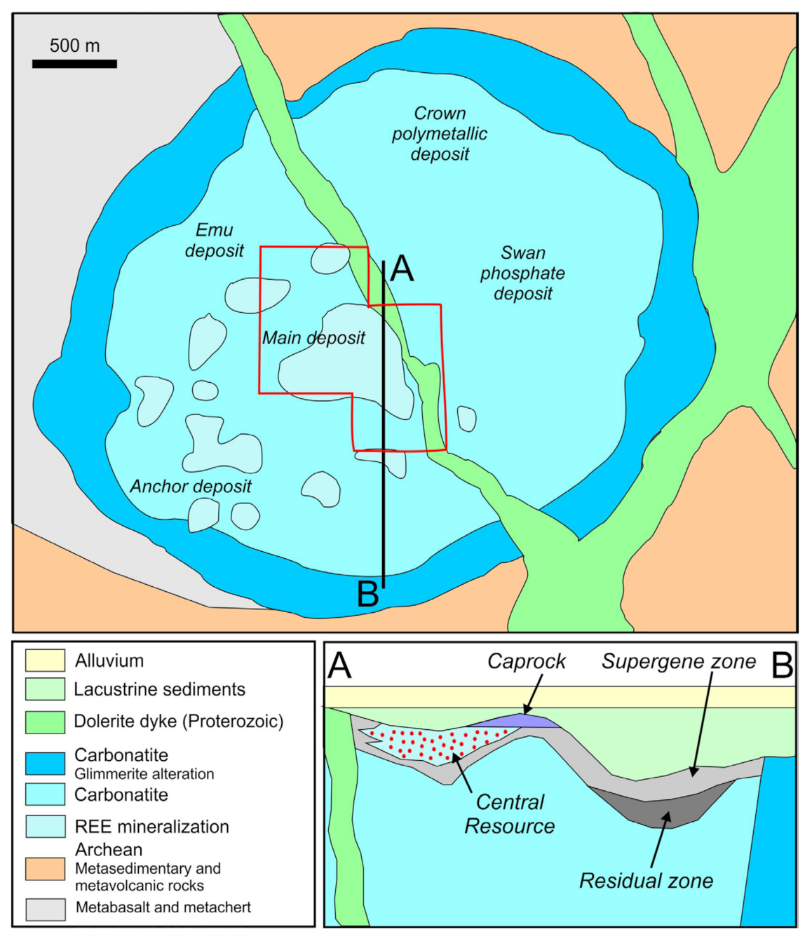
Figure 1. Simplified, schematic map and cross-section of the Mount Weld REE deposit, adapted from [13].
Minerals 13 00656 g001
Figure 2. BSE images illustrating aspects of monazite in laterite (sample AP, +53 μm size fraction). (A) relatively coarse, ‘blocky grains of monazite-(Ce), Ce > La > Nd. (B) Rounded to sub-rounded fragments of monazite-(Ce) (Mnz-Ce) in a matrix of Fe-(hydr)oxides (Fe-hydr.). (C) Euhedral, acicular monazite-(Ce) (Mnz-Ce) within quartz (Qz). (D) Dense ‘matted’ networks, rosettes and irregular arrangements of fine-grained acicular monazite-(Ce). Qz–quartz. (E) Monazite-(Ce) making up rosette-like structures within Fe-(hydr)oxides (Fe-hydr., lighter grey) and fluorapatite (Fap, darker grey). (F) Sub-rounded to acicular monazite-(Ce) enclosing orbicular Fe-(hydr)oxide (Fe-hydr.). (G) Fine, acicular monazite-(Ce) forming oriented, dense or less dense arrays within Fe-(hydr)oxide. (H) Characteristic rosettes of fine-grained monazite-(Ce) (Mnz-Ce) in fluorapatite (Fap). (I) Monazite-(Ce) (Mnz-Ce) making up rosette-like structures within quartz (Qz). (J) Fine-grained acicular monazite-(Ce) (Mnz-Ce), often oriented or rosette-like, growing onto Fe-(hydr)oxides (Fe-hydr.). (K) Particle with new-formed monazite-(Ce) (Mnz-Ce, bright) overgrowing patchy rhabdophane-(Ce) (Rha-Ce). (L) Particle composed of monazite-(Nd) and churchite-(Nd). The latter slightly appears slightly darker on the BSE image. Various shades of grey on all images are attributable to subtly different Ce:La:Nd ratios. Abbreviations on this and other figures are IMA-approved [24].
Minerals 13 00656 g002
Figure 3. Chondrite-normalised REE fractionation trend for monazite-(Ce) in the four samples and for monazite-(Nd) in black and a La-Nd-phosphate in green (both from CZ sample, +53 μm size fraction). Note that the monazites in the “Duncan 6%” sample, and to some extent also in the CZ sample, are slightly higher in HREE relative to the AP sample. Also note the relatively flat, HREE-enriched trend of the La-Nd-phosphate.
Minerals 13 00656 g003
Figure 4. BSE images illustrating rhabdophane morphology and the relationships of rhabdophane with monazite (A,B,M) and with REE-fluorocarbonates (E,F). (A) Relict monazite-Ce (bright, Mnz-Ce) in a matrix of rhabdophane-(Ce) (Rha-Ce). (B) Rosette-like grains of monazite-(Ce) (Mnz-Ce) within quartz (Qz). Each is enclosed and partially corroded by new growth of rhabdophane-(Ce) (Rha-Ce)on the surface of the pre-existing monazite. (C) Characteristic aggregate composed of rhabdophane-(Ce) (grey) intergrown with Mn-oxides (darker on image, arrowed), interpreted as possibly an organic-assisted replacement reaction. Note the typical rosette texture of rhabdophane-(Ce) nucleated onto the cores of Mn-oxides. (D) Compositionally heterogeneous rhabdophane in which the darker domains have Ce > Nd > La (but greater H2O content); lighter domains are Ce > La > Nd. (E) Synchysite-(Ce) (Syn-Ce) replaced by rhabdophane-Ce (~4% Ca, Rha-Ce) along the lower part of the grain. (F) Orbicular rhabdophane-(Ce) (Rha-Ce) replacing Ca-bearing REE-fluorocarbonate compositionally close to synchysite-(Ce) (Syn-Ce). (G) Composite particle containing rhabdophane-(Ce) with 3% Ca, Ce > Nd > La, 1%–2% Y and ca. 1% S. Note the rim (arrowed) of the very fine-grained, newly formed rhabdophane. (H) High-contrast image of REE-phosphates with varying Ca content and different proportions of Nd, La, and Ce. The fine cluster of acicular grains on right of image are rhabdophane-(Nd) (Rha-Nd). (I) Acicular rhabdophane-(Nd) (Rha-Nd). SEM-EDX analysis (normalised to 100%, excluding 7%–8% H2O) gives 31% Nd, 30% La, 6.9% Pr, 5% Ce, and 4% Ca, with traces of HREE and ~1% S. Fe-hydr.–Fe-(hydr)oxides. (J) Composite particle comprising blocky lath-like grains of rhabdophane-(Ce) (Rhab-Ce), Ce > Nd > La, 5% Ca and ~2% S. Darker phases between laths are Fe-(hydr)oxides (Fe-hydr.). (K) Network-like rhabdophane (Rha, both La- and Nd-dominant varieties are noted) within Mn-(hydr)oxides (Mn-hydr.). (L) Particle containing at least two fine-grained, compositionally distinct, and intimately intergrown REE-phosphates. The brighter phase contains less Ca (3%–4%) than the darker (6%–8%). (M) New growth of stubby monazite-(Ce) grains (Mnz-Ce) in a matrix of rhabdophane-(Ce) (Rha-Ce). Traces of S are noted in both phases. (N) Grains of relatively coarse acicular rhabdophane-(Ce). (O) Distinctive boxwork-like texture displayed by rhabdophane-(Ce) (~8% Ca, Ce > Nd > La). Images A, B, and F are from laterite sample AP, C from carbonatite, D and E from CZ sample, and G–O from “Duncan 6%” (all +53 μm size fractions).
Minerals 13 00656 g004
Figure 5. Chondrite-normalised REE fractionation trend for rhabdophane in the four samples. Note the similar trends of the carbonatite and ‘AP’ populations but slightly higher MREE and HREE relative to LREE in the CZ sample, and substantially higher MREE and HREE (less steep) relative to LREE in the “Duncan 6%” sample.
Minerals 13 00656 g005
Figure 6. BSE images illustrating aspects of REE-fluorocarbonates in carbonatite (+53 μm size fraction). (A) Banded grain of REE-fluorocarbonate where brighter domains correspond closest to bastnäsite-(Ce) and darker domains to parisite-(Ce). Darker inclusions and grains at margin are fluorapatite (Fap). (B) Grain showing oscillatory banding of different REE-fluorocarbonates in the compositional range parisite-röntgenite-synchysite. Dusty dark inclusions are hematite and/or hydrated Fe-oxides. (C) Synchysite-(Ce) (syn-Ce) with brighter domains corresponding to parisite-(Ce) (Pst-Ce). Bright inclusions are monazite-(Ce) (Mnz-Ce). (D) Parisite-(Ce) (Pst-Ce) with characteristic euhedral inclusions of monazite-(Ce) (Mnz-Ce). (E) Particle largely composed of synchysite-(Ce) (Syn-Ce), with euhedral micron-scale inclusions of monazite-(Ce) (Mnz-Ce) that appear brighter on image. Lighter parts of REE-fluorocarbonate are compositionally closer to parisite-(Ce) or röntgenite-(Ce). Note the presence of dusty Fe-(hydro)oxides. (F) Micron-scale oscillatory intergrowths of parisite (light grey) and synchysite (medium grey). Replacement by secondary Fe-hydroxides (darkest grey, arrowed) is ubiquitous. (G) Lamellar intergrowth of bastnäsite-(Ce) (Bsn-Ce) and parisite-(Ce). Fap–fluorapatite. (H,I) Bastnäsite and synchysite in laterite (sample AP, +53 μm size fraction). (H) Relatively coarse-grained synchysite-(Ce) intergrown with bastnäsite shows a lesser replacement by secondary phases. (I) Relict bastnäsite-(Ce) (brighter shade of grey, arrowed) preserved within rhabdophane-(Ce).
Minerals 13 00656 g006
Figure 7. (A) Compositional data for minerals of the bastnäsite-synchysite group expressed in terms of Ca vs. REE [Σ(La − Lu) + Y]. Note that the bastnäsite and synchysite were analysed in laterite, whereas intermediate compositions are from carbonatite. Compositions of named minerals bastnäsite (Bsn), parisite (Pst), röntgenite (Rng) and synchysite (Syn) are shown as red stars, as well as two unnamed polytypes, [B2S] and [BS4] (green stars). B and S represent component bastnäsite and synchysite modules, respectively. (B) Chondrite-normalised REE fractionation trend for REE-fluorocarbonates. Parisite from the carbonatite sample in shown in blue, bastnäsite from laterite sample AP in red.
Minerals 13 00656 g007
Figure 8. BSE images illustrating aspects of fluorapatite in carbonatite (AD), and laterite (sample AP in E-H and sample CZ in I, all +53 μm size fraction. (A) Euhedral fluorapatite (Fap) in a matrix of carbonate (Carb). Both fluorapatite and carbonate show grain-scale zonation/compositional heterogeneity. In fluorapatite, core-to-rim zonation is largely expressed by varying Sr content (1.0 to ~3.5%), with high-Sr varieties appearing brighter on the BSE image. (B) Fluorapatite (Fap) with fine-grained skeletal REE-phosphates (bright) and included quartz (Qz). (C) Skeletal, rosette-like REE-phosphates (bright, arrowed) in fluorapatite (Fap) containing 3% Sr. Carb–Ca-Mg carbonate. (D) Compositionally zoned apatite with Fe-(hydr)oxides and REE-phosphates (bright). (E) Fluorapatite (Fap) in Fe-(hydr)oxide (0.5% Mn, 2% Zn). Note the weak compositional zoning in fluorapatite with respect to Sr content (1%–3%) (arrowed). (F) Two distinct types of fluorapatite (Fap): Euhedral, ‘clean’ grains (lower left) and apatite with conspicuous dustings of REE phosphates (right). (G) Characteristic concentric REE-phosphates (bright) within fluorapatite matrix. At higher magnification (H), the phosphates can be seen as individual sub-micron crystals. Their growth within fluorapatite is probably linked to supergene processes. (I) Fluorapatite (Fap) in laterite sample CZ. Note the textural similarity with (G,H) in terms of domains containing REE phosphates.
Minerals 13 00656 g008
Figure 9. Chondrite-normalised REE fractionation patterns for (A) fluorapatite in carbonatite, (B) fluorapatite in laterite ore (sample AP), and (C) fluorapatite in “Duncan 6%”. Note the division into three distinct trends for carbonatite and laterite (AP) samples.
Minerals 13 00656 g009
Figure 10. (A) Backscatter electron image of florencite-crandallite grain (Flo-Cdl) containing inclusions of monazite-(Ce) (Mnz-Ce) (“Duncan 6%” sample, +53 μm fraction). (B) Chondrite-normalised REE fractionation trends for florencite. Note the subtle differences between the two sub-populations in the Gd-Lu segment.
Minerals 13 00656 g010
Figure 11. (AC) BSE images illustrating aspects of fluocerite and cerianite. (A). Bright rim of fluocerite-(Ce) (Fcrt-Ce) on an aggregate comprised of fine-grained REE-phosphates replacing apatite (dark). (B) Cerianite-(Ce) and CeO2 (Cei-Ce) occurring as bright white domains within a particle containing fine-grained complex intergrowths among REE-phosphates and Mn-minerals (notably surrounding REE-phosphate bottom left of image, yellow arrow). Larger grey phosphate grains (labelled ‘a’) on lower left of image could not be definitively identified. EDS analysis gave give 2%–3% Ca, Nd > La > Ce, and 2%–3% Y. Darker domains on lower right of image are Mn-hydroxides (Mn-hydr.) replacing hollandite. The darker grey phosphate grains at bottom left of image (labelled ‘c’) could not be definitively identified. EDS analysis gives Nd > La > Ce (Ce only 3%) and 11% Y. (C) Narrow (sub-μm scale) rims of cerianite-(Ce) (Cei-Ce) around replaced grains in a particle made up of various secondary REE-phosphates. (D) ‘Oolitic’ (?) cerianite-(Ce) (Cei-Ce); please note the fine rhythmic banding. Darker phases are Ce-Ca-Fe phosphates. (E) Chondrite-normalised REE fractionation trend for cerianite. Note the prominent positive Ce-anomaly.
Minerals 13 00656 g011
Figure 12. BSE images illustrating aspects of xenotime-(Y). (A) Relatively coarse xenotime-(Y) (Xtm-Y) with corroded, porous appearance. (B) Skeletal xenotime-(Y) (Xtm-Y). Both images are from the “Duncan 6%” sample. (C) Chondrite-normalised REE fractionation trend for xenotime. Note the relative enrichment in MREE/HREE and modest positive Ce anomaly.
Minerals 13 00656 g012
Figure 13. (A) BSE image of metamict zircon (Zrc, “Duncan 6%” sample, +53 μm fraction). Note the brighter rim enclosing the slightly larger core domain. Fe-hydr.–Fe-hydroxide; Pyc–pyrochlore. (B) Chondrite-normalised REE fractionation trend for zircon. Note the upward-sloping pattern characteristic for zircon and modest positive Ce-anomaly.
Minerals 13 00656 g013
Figure 14. BSE images illustrating aspects of REE-mineral inclusions in Fe-(hydr)oxides in laterite (sample AP, 20–53 μm and +53 μm fractions). (A) Inclusions of monazite-(Ce) (Mnz-Ce) on grain margin of otherwise homogeneous Fe-(hydr)oxide (Fe-hydr.). (B) Micron-scale, subhedral monazite-(Ce) (Mnz-Ce) in Fe-(hydr)oxide host. (C) Micron-sized inclusions of monazite-(Ce) (Mnz-Ce) scattered throughout Fe-(hydr)oxide (Fe-hydr.). (D) ‘Wispy’ REE-phosphates (rhabdophane, Rha) in Fe-(hydr)oxide (Fe-hydr.). (E) Fine disseminations of rhabdophane (Rha) in darker matrix of Fe-(hydr)oxide. (F) Fine-grained dustings of Ce-dominant REE-phosphate (bright) in Fe-(hydr)oxide (Fe-hydr.). (G) Euhedral grains and crustiform monazite-(Ce) (Mnz-Ce) in matrix of Fe-(hydr)oxide. (HK) Heterogeneous Fe-(hydr)oxide particles displaying complex textures suggestive of supergene and/or bacteria-mediated processes. (L) Characteristic skeletal growth of Fe-(hydr)oxides (bright) within matrix of quartz (Qz, dark).
Minerals 13 00656 g014
Figure 15. Selected time-resolved LA-ICP-MS downhole profile for representative Fe-(hydr)oxide grains (CZ sample). In (A), note irregular signals for REE suggestive of included phases. In contrast, (B) shows smooth profiles over the duration of ablation, suggestive of trace elements, including REE, in solid solution. In this case, concentrations (in ppm) are relatively high: 2990 La, 6410 Ce, 525 Pr, 1900 Nd, 275 Sm, 69 Eu, 140 Gd, 11.4 Tb, 44 Dy, 6.4 Ho, 15 Er, 1.6 Tm, 9.3 Yb, 1.3 Lu. The third example (C) shows an essentially smooth profile but nanoscale inclusions of a HREE-bearing phase (red arrow on Yb signal).
Minerals 13 00656 g015
Figure 16. Chondrite-normalised REE fractionation trends for Fe-(hydr)oxides divided by sample. Note the two sub-populations (Trend 1 and Trend 2) in spot analyses from laterite sample AP, whereby T2 has a distinct positive Ce anomaly.
Minerals 13 00656 g016
Figure 17. BSE images illustrating representative Mn-(hydr)oxide particles in laterites. (A) Complex assemblage of different Mn-(hydr)oxides in various grey shades; note minor acicular monazite (arrowed). (B) Grain in centre of image shows fibrous, boxwork-like texture composed of Mn-(hydr)oxides, also with minor monazite (arrowed). (C) Mn-(hydr)oxide with orbicular (white arrow) and fibrous (yellow arrow) textures of supergene and/or organic-mediated origin. (D) Grain in centre of image shows Fe-(Mn) –(hydr)oxide with ‘oolitic’ texture suggestive of colloidal aggregation or an organic-mediated origin.
Minerals 13 00656 g017
Figure 18. (A,B) BSE images illustrating compositionally homogeneous ilmenite (Ilm) associated with ‘agglomerate’ of fine-grained REE-phosphates and Fe-(hydr)oxides. (C) Time-resolved LA-ICP-MS downhole profile for representative ilmenite grain (CZ sample) containing >1% Nb and measurable concentrations of REE and other elements of interest. (D) Chondrite-normalised REE fractionation trends for ilmenite (all samples). Note the common slope (highly LREE rich) but large range of absolute values.
Minerals 13 00656 g018
Figure 19. (AE) BSE images illustrating aspects of pyrochlore group minerals from the “Duncan 6%” sample (20–53 μm and +53 μm size fractions). (A) Cluster of small, subhedral or fragmented, and partially replaced grains of Ce-dominant pyrochlore (Pyc). EDS analysis gives 16% Ce, 4% Na, 2% Ti, minor La, Ca, Sr) in matrix of Fe-(hydr)oxides (Fe-hydr). (B) Extensively replaced pyrochlore (Pyc) containing 6%–8% Sr, 5%–7% Ce, 8%–10% Pb, and 1%–2% Ti, with traces of Fe, Ce, Ca, and Na (EDS data). (C) Almost totally replaced Ce-Pb-(Na)-dominant pyrochlore (Pyc) in matrix of Fe-(hydr)oxides. Infill of the replaced pyrochlore is a mixture of Fe-Al-phosphate phase and Fe-(hydr)oxides, with conspicuous microcrystals of fluocerite-(Ce) (Fcrt-Ce). (D) Euhedral-subhedral crystals of Ce-a fluorinated Ca-Na-dominant pyrochlore in matrix of Fe-(hydr)oxides. (E) Euhedral-subhedral crystals of Ce-bearing pyrochlore in matrix of florencite (darker) and Fe-(hydr)oxides (lighter). (F) Ce-Pb pyrochlore (15 wt.% Pb, 9.5 wt.% Ce, EDS data) within a Fe-(hydr)oxide matrix. (G) Compositionally zoned Ce-dominant pyrochlore. EDX element maps show infill to be an undefined Al-phosphate. (H) Fragment of larger pyrochlore crystal showing fine (sub-micron)-scale oscillatory zoning. EDS analysis indicates this phase is Pb-Ce-dominant, with measurable Ti, Sr, Na, Ca, and Fe. Zoning is expressed by varying concentrations of Ce and Pb. (I) Chondrite-normalised REE fractionation trend for pyrochlore group minerals (cerian pyrochlore in red, strontian pyrochlore in blue). Note the strong positive Ce-anomaly in the Ce-pyrochlore.
Minerals 13 00656 g019
Figure 20. (AD) BSE images illustrating aspects of coronadite group minerals in laterite ores. (A) Composite grain containing coronadite (Cor), fluorapatite (Fap) and monazite-(Ce) (Mnz-Ce). (B) Fine intergrowth among members of hollandite-cryptomelane-coronadite series (different grey shades reflecting varying Ba:K ratios and Pb content). Fe-hydr–Fe-hydroxides. (C) Wispy REE-phosphates (bright) in matrix of coronadite (Cor). (D) Euhedral and dusty monazite (bright) in a matrix of cryptomelane (Cml). (EG) Chondrite-normalised REE fractionation trend for hollandite (E), cryptomelane and coronadite (black line on F). Note the presence of varied concentrations of REE. The fractionation trend for coronadite is distinctly flatter than for hollandite or cryptomelane. Fractionation trend in (G) is a subset of spot analyses that display a conspicuous positive cerium anomaly. This is best explained by tiny crystals of new-formed Ce-bearing pyrochlore (Pcl) within hollandite (Hol) on the BSE image, H) or cerianite within the Mn-minerals.
Minerals 13 00656 g020
Figure 21. BSE images illustrating aspects of sulphides in carbonatite (+53 μm size fraction). (A) Subhedral pyrite (Py) with inclusions of galena (bright, Gn). Syn-Ce–synchysite-(Ce). (B) Sphalerite (Sp) with inclusions of galena (bright) and minor chalcopyrite (Ccp). (C) Relatively coarse galena grain. (D) Pyrite replaced and corroded by bornite (Bn). Fe-hydr: Fe-(hydr)oxide. (E) Assemblage of sphalerite, galena, and pyrite (in this case containing ~5% Co). (F) Intergrowth of pyrite and monazite-(Ce) (Mnz) in particle also containing Ca-Mg carbonate (Carb) and Fe-oxide/hydroxide (Fe-ox).
Minerals 13 00656 g021
Figure 22. (A) BSE image illustrating compositionally zoned Ca-Mg-Fe-carbonate particle in carbonatite. (B) Chondrite-normalised REE fractionation trend for Ca-Mg-carbonates. Absolute concentrations range from very low (preserved homogeneous grains of igneous origin) to moderate (tens to hundreds of ppm). The steeper trends are clearly linked to inclusions of monazite and other phosphates.
Minerals 13 00656 g022
Table 1. Assay data for the three metallurgical samples (in ppm unless otherwise given).
SampleSiO2 %Fe2O3 %MnO %MgO %P2O5 %Th %K2O %SO3 %Na2O %BaO %TiO2 %Zn %Zr %ΣREO %
AP RC4.0537.483.720.9015.20.0600.120.0820.180.150.7230.6260.0412.25
CZ RC2.7652.182.610.468.40.1150.050.1550.110.292.0700.4040.0815.22
Duncan 6%6.5051.412.761.017.00.0400.160.6790.270.411.0900.1760.037.44
SampleNb2O5Ta2O5CaO %Al2O3 %SrO %PbO %ScUCuZnSrOAs2O3SbY2O3
AP RC0.2690.00514.50.680.5140.06814421.63281604890400.6404
CZ RC0.5250.0050.753.340.4350.05622222.712250604070601.2790
Duncan 6%0.361−0.0053.655.150.3880.02914661.4176225037706013090
SampleLa2O3 %CeO2 %Pr6O11 %Nd2O3 %Sm2O3Eu2O3Gd2O3Tb2O3Dy2O3Ho2O3Er2O3Tm2O3Yb2O3Lu2O3
AP RC3.105.820.6312.170.289587105068.417418.831.43.0914.51.83
CZ RC3.687.250.7942.750.373804153011131536.461.55.9427.83.24
Duncan 6%1.483.160.3871.450.242661170022598313726325.11069.51
Disclaimer/Publisher’s Note: The statements, opinions and data contained in all publications are solely those of the individual author(s) and contributor(s) and not of MDPI and/or the editor(s). MDPI and/or the editor(s) disclaim responsibility for any injury to people or property resulting from any ideas, methods, instructions or products referred to in the content.