
Development of High-Energy µ-X-ray Fluorescence and X-ray Absorption Fine Structure for the Distribution and Speciation of Rare Earth Elements in Natural Samples


Abstract
:1. Introduction
2. Materials and Methods
2.1. Samples
2.2. Micro-XRF Analysis
2.3. Micro-XAFS Analysis
3. Results and Discussion
3.1. Optical Setup to Cover Wide Energy Range
3.2. HE-XRF Analysis
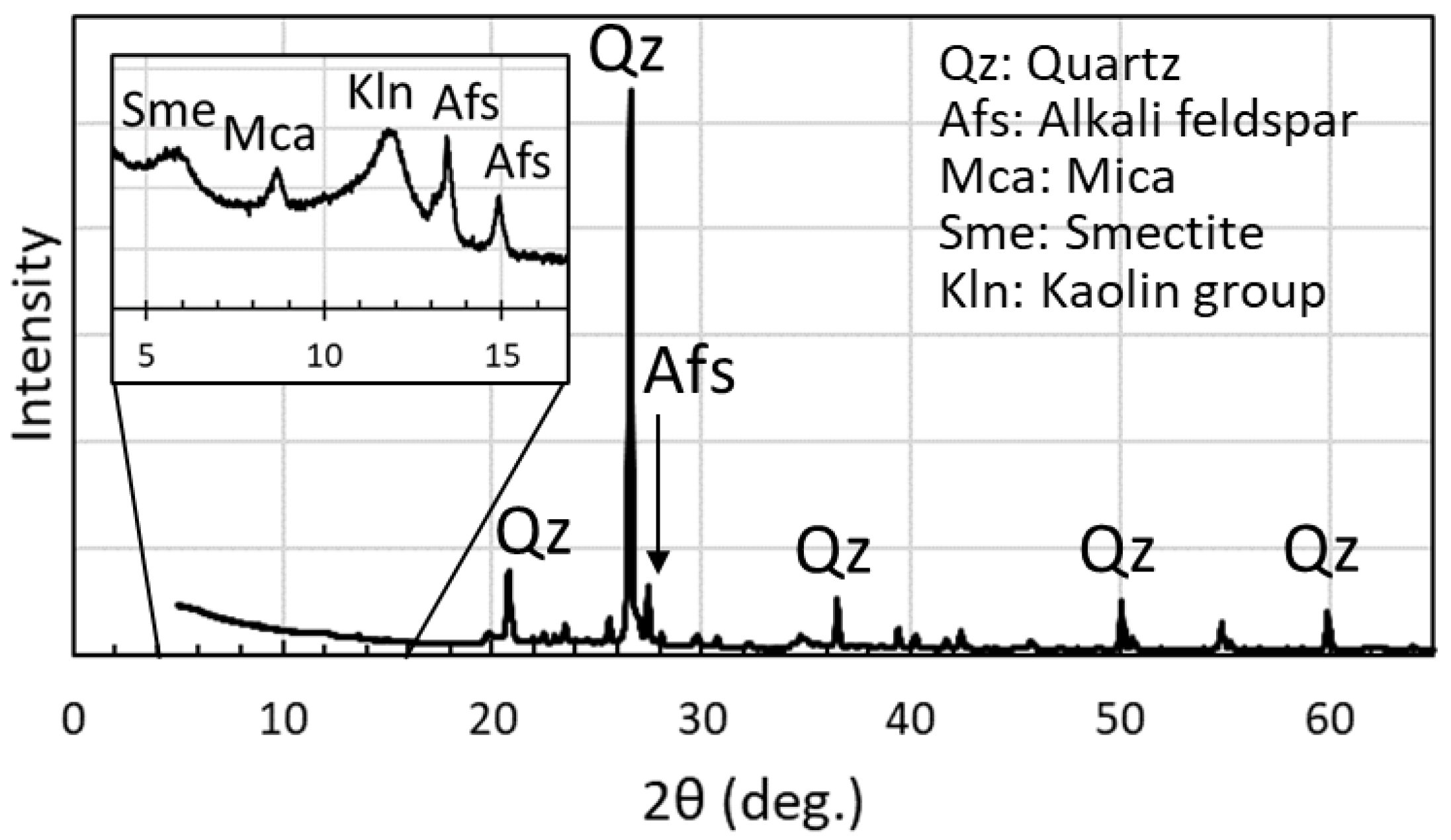
3.3. Micro-XRF Analysis
3.4. Micro-XAFS

| Sample | k Range (Å−1) | FT Range (Å) | Shell | CN | R (Å) | ΔE0 (eV) | σ2 (×10−3 Å2) | Residual (%) |
|---|---|---|---|---|---|---|---|---|
| Bulk (Point 1) | 2.35–7.25 | 1.19–2.57 | La-O | 9 * | 2.559 ± 0.017 | −3.40 ± 1.70 | 9.8 ± 2.6 | 2.1 |
| Point 2 | 2.35–7.25 | 0.89–2.55 | La-O | 9 * | 2.515 ± 0.026 | −5.39 ± 2.51 | 16.4 ± 4.1 | 1.3 |
| 1.65–6.05 | 0.58–2.52 | Nd-O | 9 * | 2.348 ± 0.028 | −12.3 ± 2.22 | 18.8 ± 5.5 | 1.8 | |
| 0.05 M La(NO3)3 solution | 2.35–7.25 | 1.26–2.58 | La-O | 9 * | 2.560 ± 0.025 | −3.73 ± 2.57 | 9.8 ± 4.0 | 0.91 |
| La-adsorbed mnt | 2.35–7.25 | 1.32–2.58 | La-O | 9 * | 2.561 ± 0.025 | −1.54 ± 2.56 | 8.5 ± 3.9 | 0.95 |
| La-adsorbed apatite | 2.35–7.25 | 1.41–2.39 | La-O | 9 * | 2.495 ± 0.022 | −2.43 ± 2.11 | 14.2 ± 3.6 | 0.057 |
| LaPO4 | 2.35–7.25 | 1.29–2.42 | La-O | 9 * | 2.523 ± 0.024 | −2.03 ± 2.22 | 15.4 ± 3.7 | 0.59 |
| Monazite | 2.35–7.25 | 1.27–2.49 | La-O | 9 * | 2.511 ± 0.016 | −2.88 ± 1.40 | 20.2 ± 2.6 | 1.1 |
4. Conclusions
Author Contributions
Funding
Data Availability Statement
Acknowledgments
Conflicts of Interest
References
- McSween, H.Y.; Richardson, S.M.; Uhle, S.M. Geochemistry: Pathways and Processes; Columbia University Press: New York, NY, USA, 2003. [Google Scholar]
- Henderson, P. Rare Earth Element Geochemistry; Elsevier: Amsterdam, The Netherlands, 2013. [Google Scholar]
- Takahashi, Y.; Manceau, A.; Geoffroy, N.; Marcus, M.A.; Usui, A. Chemical and structural control of the partitioning of Co, Ce, and Pb in marine ferromanganese oxides. Geochim. Cosmochim. Acta 2007, 71, 984–1008. [Google Scholar] [CrossRef]
- Goodenough, K.M.; Wall, F.; Merriman, D. The Rare Earth Elements: Demand, Global Resources, and Challenges for Resourcing Future Generations. Nat. Resour. Res. 2018, 27, 201–216. [Google Scholar] [CrossRef]
- Kashiwabara, T.; Toda, R.; Nakamura, K.; Yasukawa, K.; Fujinaga, K.; Kubo, S.; Nozaki, T.; Takahashi, Y.; Suzuki, K.; Kato, Y. Synchrotron X-ray spectroscopic perspective on the formation mechanism of REY-rich muds in the Pacific Ocean. Geochim. Cosmochim. Acta 2018, 240, 274–292. [Google Scholar] [CrossRef]
- Borst, A.M.; Smith, M.P.; Finch, A.A.; Estrade, G.; Villanova-de-Benavent, C.; Nason, P.; Marquis, E.; Horsburgh, N.J.; Goodenough, K.M.; Xu, C.; et al. Adsorption of rare earth elements in regolith-hosted clay deposits. Nat. Commun. 2020, 11, 4386. [Google Scholar] [CrossRef]
- Yamaguchi, A.; Honda, T.; Tanaka, M.; Tanaka, K.; Takahashi, Y. Discovery of ion-adsorption type deposits of rare earth elements (REE) in southwest Japan with speciation of REE by extended X-ray absorption fine structure spectroscopy. Geochem. J. 2018, 52, 415–425. [Google Scholar] [CrossRef]
- Nagasawa, M.; Qin, H.; Yamaguchi, A.; Takahashi, Y. Local structure of rare earth elements (REE) in marine ferromanganese oxides by extended X-ray absorption fine structure and its comparison with REE in ion-adsorption type deposits. Chem. Lett. 2020, 8, 909–911. [Google Scholar] [CrossRef]
- Takahashi, Y.; Shimizu, H.; Usui, A.; Kagi, H.; Nomura, M. Direct observation of tetravalent cerium in ferromanganese nodules and crusts by X-ray-absorption near-edge structure (XANES). Geochim. Cosmochim. Acta 2000, 64, 2929–2935. [Google Scholar] [CrossRef]
- Takahashi, Y.; Kolonin, G.R.; Shironosova, G.P.; Kupriyanova, I.I.; Uruga, T.; Shimizu, H. Determination of the Eu(II)/Eu(III) ratios in minerals by X-ray absorption near-edge structure (XANES) and its application to hydrothermal deposits. Mineral. Mag. 2005, 69, 179–190. [Google Scholar] [CrossRef]
- Rakovan, J.; Newville, M.; Sutton, S. Evidence of heterovalent europium in zoned llallgua apatite using wavelength dispersive XANES. Am. Mineral. 2001, 86, 697–700. [Google Scholar] [CrossRef]
- Janots, E.; Bernier, F.; Brunet, F.; Muñoz, M.; Trcera, N.; Berger, A.; Lanson, M. Ce(III) and Ce(IV) (re)distribution and fractionation in a laterite profile from Madagascar: Insights from in situ XANES spectroscopy at the Ce LIII-edge. Geochim. Cosmochim. Acta 2015, 153, 134–148. [Google Scholar] [CrossRef]
- Konagaya, R.; Kawamura, N.; Yamaguchi, A.; Takahashi, Y. Highly-sensitive Analysis of Fluorescence XANES at Europium (Eu) LIII -edge for the Determination of Oxidation State for Trace Amount of Eu in Natural Samples by Bragg-type Crystal Analyzer System. Chem. Lett. 2021, 50, 1570–1572. [Google Scholar] [CrossRef]
- Takahashi, Y.; Yamamoto, M.; Yamamoto, Y.; Tanaka, K. EXAFS study on the cause of enrichment of heavy REEs on bacterial cell surfaces. Geochim. Cosmochim. Acta 2010, 74, 5443–5462. [Google Scholar] [CrossRef]
- Manceau, A.; Lanson, M.; Takahashi, Y. Mineralogy and crystal chemistry of Mn, Fe, Co, Ni, and Cu in a deep-sea Pacific polymetallic nodule. Am. Mineral. 2014, 99, 2068–2083. [Google Scholar] [CrossRef]
- Marcus, M.A.; Toner, B.M.; Takahashi, Y. Forms and distribution of Ce in a ferromanganese nodule. Mar. Chem. 2018, 202, 58–66. [Google Scholar] [CrossRef]
- Kirkpatrick, P.; Baez, A.V. Formation of optical images by X-rays. J. Opt. Soc. Am. 1948, 38, 766–774. [Google Scholar] [CrossRef]
- Nitta, K.; Sekizawa, O. BL37XU (Trace Element Analysis). In SPring-8/SACLA Annual Report FY2018; RIKEN/JASRI: Hyogo, Japan, 2019; Volume 18, pp. 64–66. [Google Scholar]
- Rosa, A.D.; Kupenko, I.; Hernandez, J.A.; Forestier, A.; Muñoz, M.; Morard, G.; Bouhifd, M.A.; Lomachenko, K.A.; Torchio, R.; Chumakov, A.; et al. New Opportunities for Earth Science at the Extremely Brilliant Source of the European Synchrotron Radiation Facility. Synchrotron Radiat. News 2022, 35, 8–16. [Google Scholar] [CrossRef]
- De Pauw, E.; Tkalcec, B.J.; Tack, P.; Vekemans, B.; Di Michiel, M.; Brenker, F.E.; Vincze, L. High energy synchrotron X-ray fluorescence trace element study of a millimeter-sized asteroidal particle in preparation for the Hayabusa2 return sample analyses. Spectrochim. Acta Part B At. Spectrosc. 2022, 188, 106346. [Google Scholar] [CrossRef]
- Quartieri, S.; Dalconi, M.C.; Boscherini, F.; Oberti, R.; D’Acapito, F. Changes in the local coordination of trace rare-earth elements in garnets by high-energy XAFS: New data on dysprosium. Phys. Chem. Miner. 2004, 31, 162–167. [Google Scholar] [CrossRef]
- Pearce, N.J.G.; Perkins, W.T.; Westgate, J.A.; Gorton, M.P.; Jackson, S.E.; Neal, C.R.; Chenery, S.P. A Compilation of New and Published Major and Trace Element Data for NIST SRM 610 and NIST SRM 612 Glass Reference Materials. Geostand. Geoanal. Res. 1997, 21, 115–144. [Google Scholar] [CrossRef]
- Badanina, E.V.; Trumbull, R.B.; Dulski, P.; Wiedenbeck, M.; Veksler, I.V.; Syritso, L.F. The behavior of rare-earth and lithophile trace elements in rare-metal granites: A study of flourite, melt inclusions and host rocks from the Khangilay complex, Transbaikalia, Russia. Can. Mineral. 2006, 44, 667–692. [Google Scholar] [CrossRef]
- Tanaka, K.; Takahashi, Y.; Shimizu, H. Determination of rare earth element in carbonate using laser-ablation inductively-coupled plasma mass spectrometry: An examination of the influence of the matrix on laser-ablation inductively-coupled plasma mass spectrometry analysis. Anal. Chim. Acta 2007, 583, 303–309. [Google Scholar] [CrossRef] [PubMed]
- Zhu, Y.; Mongelli, G.; Sinisi, R.; Yim, Y.H. Editorial: Analytical chemistry of rare earth elements (REEs). Front. Chem. 2020, 10, 1631. [Google Scholar] [CrossRef]
- Wallrich, B.M.; Miller, C.F.; Gualda, G.A.R.; Miller, J.S.; Hinz, N.H.; Faulds, J.E. Volcano-pluton connection: Perspectives on material and process linkages, Searchlight pluton and Highland Range volcanic sequence, Nevada, USA. Earth-Sci. Rev. 2023, 238, 104361. [Google Scholar] [CrossRef]
- Terashima, S.; Usui, A.; Imai, N. 2 new GSJ geochemical reference samples—Syenite JSY-1 and manganese-nodule JMN-1. Geostand. Newsl. 1995, 19, 221–229. [Google Scholar] [CrossRef]
- Verlaan, P.A.; Cronan, D.S. Origin and variability of resource-grade marine ferromanganese nodules and crusts in the Pacific Ocean: A review of biogeochemical and physical controls. Geochemistry 2022, 82, 125741. [Google Scholar] [CrossRef]
- Nagasawa, M.; Takahashi, Y. Vertical migration and enrichment of rare earth elements (REEs) in ion-adsorption type mineralization in Japan based on REE speciation analysis. 2023; in press. [Google Scholar]
- Takahashi, Y.; Yoshida, H.; Sato, N.; Hama, K.; Yusa, Y.; Shimizu, H. W- and M-type tetrad effects in REE patterns for water-rock systems in the Tono uranium deposit, central Japan. Chem. Geol. 2002, 184, 311–335. [Google Scholar] [CrossRef]
- The Center for X-Ray Optics (CXRO). X-Ray Interactions with Matter. Available online: https://henke.lbl.gov/optical_constants/ (accessed on 19 May 2023).
- Solé, V.A.; Papillon, E.; Cotte, M.; Walter, P.; Susini, J. A multiplatform code for the analysis of energy-dispersive X-ray fluorescence spectra. Spectrochim. Acta Part B At. Spectrosc. 2007, 62, 63–68. [Google Scholar] [CrossRef]
- Anders, E.; Grevesse, N. Abundances of the elements—Meteoritic and solar. Geochim. Cosmochim. Acta 1989, 53, 197–214. [Google Scholar] [CrossRef]
- Nakada, R.; Tanimizu, M.; Takahashi, Y. Difference in the stable isotopic fractionations of Ce, Nd, and Sm during adsorption on iron and manganese oxides and its interpretation based on their local structures. Geochim. Cosmochim. Acta 2013, 121, 105–119. [Google Scholar] [CrossRef]
- Zabinsky, S.I.; Rehr, J.J.; Ankudinov, A.; Albers, R.C.; Eller, M.J. Multiple-scattering calculations of X-ray-absorption spectra. Phys. Rev. B 1995, 52, 2995–3009. [Google Scholar] [CrossRef]
- O’Day, P.A.; Rehr, J.J.; Zabinsky, S.I.; Brown, G.E. Extended X-ray absorption fine structure (EXAFS) analysis of disorder and multiple scattering in complex crystalline solids. J. Am. Chem. Soc. 1994, 116, 2938–2949. [Google Scholar] [CrossRef]
- Li, W.; Yamada, S.; Hashimoto, T.; Okumura, T.; Hayakawa, R.; Nitta, K.; Sekizawa, O.; Suga, H.; Uruga, T.; Ichinohe, Y.; et al. High-sensitive XANES analysis at Ce L2-edge for Ce in high-Ti geological samples using transition-edge sensors. Anal. Chim. Acta 2023, 1240, 340755. [Google Scholar] [CrossRef]
- Abe, Y.; Iizawa, Y.; Terada, Y.; Adachi, K.; Igarashi, Y.; Nakai, I. Detection of uranium and chemical state analysis of individual radioactive microparticles emitted from the fukushima nuclear accident using multiple synchrotron radiation X-ray analyses. Anal. Chem. 2014, 86, 8521–8525. [Google Scholar] [CrossRef] [PubMed]
- Matsushita, T.; Hashizume, H. X-Ray Monochromators. Handb. Synchrotron Radiat. 1983, 1, 261–314. [Google Scholar]
- Boone, M.N.; Van Assche, F.; Vanheule, S.; Cipiccia, S. Full-field spectroscopic measurement of the X-ray beam from a multilayer monochromator using a hyperspectral X-ray camera research papers. J. Synchrotron Rad. 2020, 27, 110–118. [Google Scholar] [CrossRef] [PubMed]
- Firester, A.H.; Heller, M.E.; Sheng, P. Knife-edge scanning measurements of subwavelength focused light beams. Appl. Opt. 1977, 16, 1971. [Google Scholar] [CrossRef]
- Takeuchi, A.; Suzuki, Y.; Uesugi, K. Differential-phase-contrast knife-edge scan method for precise evaluation of X-ray nanobeam. Jpn. J. Appl. Phys. 2015, 54, 092401. [Google Scholar] [CrossRef]
- Takahashi, Y.; Shimizu, H.; Kagi, H.; Yoshida, H.; Usui, A.; Nomura, M. A new method for the determination of CeIII/CeIV ratios in geological materials; application for weathering, sedimentary and diagenetic processes. Earth Planet. Sci. Lett. 2000, 182, 201–207. [Google Scholar] [CrossRef]
- Sanematsu, K.; Watanabe, Y. Characteristics and genesis of ion adsorption-type rare earth element deposits. Rev. Econ. Geol. 2016, 18, 55–79. [Google Scholar]
- Liu, X.; Tournassat, C.; Grangeon, S.; Kalinichev, A.G.; Takahashi, Y.; Marques Fernandes, M. Molecular-level understanding of metal ion retention in clay-rich materials. Nat. Rev. Earth Environ. 2022, 3, 461–476. [Google Scholar] [CrossRef]
- Frietsch, R.; Perdahl, J.A. Rare earth elements in apatite and magnetite in Kiruna-type iron ores and some other iron ore types. Ore Geol. Rev. 1995, 9, 489–510. [Google Scholar] [CrossRef]
- Nishihata, Y.; Kamishima, O.; Kubozono, Y.; Maeda, H.; Emura, S. XAFS in the high-energy region. J. Synchrotron Radiat. 1998, 5, 1007–1009. [Google Scholar] [CrossRef]
- Ohta, A.; Kagi, H.; Nomura, M.; Shitsuno, H.; Kawabe, I. Coordination study of rare earth elements on Fe oxyhydroxide and Mn dioxides: Part ii. Correspondence of structural change to irregular variations of partitioning coeffcients and tetrad effect variations appearing in interatomic distances. Am. Mineral. 2009, 94, 476–486. [Google Scholar] [CrossRef]
- Kumar, Y.; Tripathi, S.; Nand, M.; Jangir, R.; Srihari, V.; Das, A.; Singh, R.; Deshpande, U.; Jha, S.N.; Arya, A. Structural and optical properties of Nd doped LaPO4. J. Alloys Compd. 2022, 925, 166772. [Google Scholar] [CrossRef]
- Cole, J.M.; Newport, R.J.; Bowron, D.T.; Pettifer, R.F.; Mountjoy, G.; Brenna, T.; Saunders, G.A. A rare-earth K-edge XAFS study of rare-earth pohsphate glasses, (R2O3)x(P2O5)1-x, x = 0.187-0.239, R = La, Nd, Sm, Eu. Gd, Dy, Er. J. Phys. Condens. Matter 2001, 13, 6659–6674. [Google Scholar] [CrossRef]
- Sanematsu, K.; Kon, Y.; Imai, A. Influence of phosphate on mobility and adsorption of REEs during weathering of granites in Thailand. J. Asian Earth Sci. 2015, 111, 14–30. [Google Scholar] [CrossRef]
- Mukai, H.; Kon, Y.; Sanematsu, K.; Takahashi, Y.; Ito, M. Microscopic analyses of weathered granite in ion-adsorption rare earth deposit of Jianxi Province, China. Sci. Rep. 2020, 10, 20194. [Google Scholar] [CrossRef] [PubMed]
- Kowall, T.; Foglia, F.; Helm, L.; Merbach, A.E. Molecular dynamics simulation study of lanthanide ions Ln3+ in aqueous solution including water polarization. Change in coordination number from 9 to 8 along the series. J. Am. Chem. Soc. 1995, 117, 3790–3799. [Google Scholar] [CrossRef]
- Kanazawa, Y.; Kamitani, M. Rare earth minerals and resources in the world. J. Alloys Compd. 2006, 408, 1339–1343. [Google Scholar] [CrossRef]
- Habenschuss, A.; Spedding, F.H. The coordination (hydration) of rare earth ions in aqueous chloride solutions from X-ray diffraction. III. SmCl3, EuCl3, and series behavior. J. Chem. Phys. 1980, 73, 442–450. [Google Scholar] [CrossRef]


| Element | SWG Sample | NIST SRM 610 | JMn-1 |
|---|---|---|---|
| SiO2 | 69.7 (wt.%) | 72 (wt.%) | 14.11 (wt.%) |
| Al2O3 | 16.2 | 2.0 | 4.30 |
| Fe2O3 | 1.34 | 0.0654 | 14.4 |
| MnO | 0.03 | 0.0560 | 33.09 |
| MgO | 0.36 | 0.0772 | 3.12 |
| CaO | N.D. | 0.12 | 2.91 |
| Na2O | 0.19 | 0.12 | 2.80 |
| K2O | 3.87 | 0.0691 | 0.94 |
| TiO2 | 0.03 | 0.0724 | 1.06 |
| P2O5 | N.D. | 0.0785 | 0.54 |
| Y | 2040 (mg/kg) | 450 (mg/kg) | 111 (mg/kg) |
| Ba | 49.7 | 424 | 1714 |
| La | 189 | 457 | 122 |
| Ce | 21.2 | 448 | 277 |
| Pr | 65.0 | 430 | 31.4 |
| Nd | 280 | 431 | 137 |
| Sm | 123 | 451 | 30.2 |
| Eu | 0.660 | 461 | 7.6 |
| Gd | 227 | 420 | 29.8 |
| Tb | 46.6 | 443 | 4.8 |
| Dy | 340 | 427 | 28.3 |
| Ho | 66.8 | 449 | 5.8 |
| Er | 169 | 426 | 14.6 |
| Tm | 22.3 | 420 | 2.1 |
| Yb | 139 | 462 | 13.8 |
| Lu | 19.4 | 435 | 2.1 |
| Element | JMn-1 (ppm) | Point 1 (ppm) | Point 2 (ppm) |
|---|---|---|---|
| La | 140 ± 3 | 119 ± 5 | 8797 ± 114 |
| Ce | 280 ± 4 | 7.60 ± 1.72 | 10433 ± 115 |
| Pr | 23.0 ± 1.5 | 27.7 ± 2.7 | 863 ± 31 |
| Nd | 114 ± 3 | 176.9 ± 5.5 | 2706 ± 54 |
| Sm | 29.3 ± 1.4 | 83.1 ± 3.7 | 329 ± 28 |
| Eu | N.D. | N.D. | N.D. |
| Gd | 34.9 ± 1.2 | 156 ± 5 | 173 ± 15 |
| Tb | N.D. | 30.9 ± 3.0 | N.D. |
| Dy | 37.6 ± 1.2 | 217 ± 6 | 121 ± 4 |
Disclaimer/Publisher’s Note: The statements, opinions and data contained in all publications are solely those of the individual author(s) and contributor(s) and not of MDPI and/or the editor(s). MDPI and/or the editor(s) disclaim responsibility for any injury to people or property resulting from any ideas, methods, instructions or products referred to in the content. |
© 2023 by the authors. Licensee MDPI, Basel, Switzerland. This article is an open access article distributed under the terms and conditions of the Creative Commons Attribution (CC BY) license (https://creativecommons.org/licenses/by/4.0/).
Share and Cite
Nagasawa, M.; Sekizawa, O.; Nitta, K.; Kashiwabara, T.; Takahashi, Y. Development of High-Energy µ-X-ray Fluorescence and X-ray Absorption Fine Structure for the Distribution and Speciation of Rare Earth Elements in Natural Samples. Minerals 2023, 13, 746. https://doi.org/10.3390/min13060746
Nagasawa M, Sekizawa O, Nitta K, Kashiwabara T, Takahashi Y. Development of High-Energy µ-X-ray Fluorescence and X-ray Absorption Fine Structure for the Distribution and Speciation of Rare Earth Elements in Natural Samples. Minerals. 2023; 13(6):746. https://doi.org/10.3390/min13060746
Chicago/Turabian StyleNagasawa, Makoto, Oki Sekizawa, Kiyofumi Nitta, Teruhiko Kashiwabara, and Yoshio Takahashi. 2023. "Development of High-Energy µ-X-ray Fluorescence and X-ray Absorption Fine Structure for the Distribution and Speciation of Rare Earth Elements in Natural Samples" Minerals 13, no. 6: 746. https://doi.org/10.3390/min13060746