Article

Carbonate Nanoparticles Formed by Water–Rock Reactions in Groundwater: Implication of Carbonate Rock Weathering in Carbonate Aquifers

by
Gang Tao
1,2,3,
Rui Liu
1,4,*,
Peng Zhang
4,
Yaqin Wang
4,
Lei Zuo
4 and
Xiaoheng Zhang
4
1
Fundamental Science on Nuclear Wastes and Environmental Safety Laboratory, Mianyang 621010, China
2
School of Environment and Resource, Southwest University of Science and Technology, Chengdu 610213, China
3
Tianfu Institute of Research and Innovation, Southwest University of Science and Technology, Chengdu 610218, China
4
School of Resources and Environmental Engineering, Shandong University of Technology, Zibo 255000, China
*
Author to whom correspondence should be addressed.
Minerals 2024, 14(10), 980; https://doi.org/10.3390/min14100980 (registering DOI)
Submission received: 27 July 2024 / Revised: 23 September 2024 / Accepted: 26 September 2024 / Published: 28 September 2024
(This article belongs to the Special Issue Mineral Evolution and Mineralization during Weathering)

Abstract

:
Carbonate rocks are highly reactive and exhibit higher ratios of chemical weathering compared to most other rock types. A chemo-mechanical mechanism, which is particularly effective in groundwater due to higher ion concentrations, is common in fine-grained carbonates at the nanoscale. As a result, the weathering of carbonate aquifers produces a substantial number of carbonate nanoparticles (CNPs). In this study, we utilized high-resolution transmission electron microscopy (HRTEM) to analyze CNPs formed by water–rock reactions in two types of groundwater from Shandong Province, China. Our findings reveal a significant presence of naturally occurring CNPs in groundwater. The HRTEM results show that CNPs display spherical, cubic, hexagonal, and irregular shapes, with some forming aggregates. Energy-dispersive spectrometry indicates that most nanoparticles contain O, C, Ca, and Fe, with some also containing Si, Mg, S, Sr, and Cl. Selected area electron diffraction (SAED) patterns show that CNPs are mainly amorphous, with some crystalline forms. The diverse shapes and complex compositions of these CNPs suggest that they are not man-made but formed through the weathering of carbonate minerals via chemo-mechanical mechanisms. This discovery provides new insights into carbonate mineral evolution and mineralization during weathering. Given their widespread presence, CNPs in groundwater could represent the transportation of elements in the form of particles.

1. Introduction

Nanominerals and mineral nanoparticles on Earth are formed through various physical, chemical, and biological processes, including (bio)chemical weathering, photo-oxidation, redox, and precipitation reactions, as well as (bio)mineralization. Weathering is a crucial inorganic process that leads to nanoparticle formation [1].
Although nanoparticle formation is typically linked to precipitation from a solution, recent studies have demonstrated that metal-carbonate nanoparticles can also be mechanically released from mineral surfaces due to repulsive forces between grains that contribute to surface retreat [2,3,4,5]. Additionally, the dissolution of mineral matrices containing nanoparticles can release them into solutions, as observed with chromite nanoparticles released from clinochlore under various pH conditions [6]. Nanoparticles may also form by precipitation from supersaturated fluids, controlling the fate, transport, and availability of key elements in hydrothermal environments [7]. For instance, the chemical weathering of silicate minerals often results in the in situ precipitation of amorphous silica nanoparticles, forming altered surface layers. Moreover, pyrite weathering releases sulfate and ferrous iron, which, upon oxidation, precipitate as iron (III) oxyhydroxide nanoparticles [8].
Groundwater, in comparison to surface water, has higher ion concentrations due to its longer residence time and higher water–rock ratios [9,10,11]. The elevated CO2 partial pressure and temperature of groundwater can intensify chemical weathering. For instance, Rad et al. [9,12] initially took the dissolved cations in groundwater into consideration when studying weathering fluxes on the volcanic islands of the Lesser Antilles. By integrating groundwater data with surface water assessments, they discovered that the estimated chemical weathering rate significantly increased from 100–120 t/km2·year to 365–1300 t/km2·year. Similarly, Calmels et al. [13] investigated steep, highly erosive mountainous catchments in Taiwan and found that although groundwater accounted for only 16% of the total runoff volume, it contributed 40% of the total cation flux. This emphasizes the crucial role of groundwater in significantly enhancing the overall chemical weathering rates, particularly in rugged, erosion-prone landscapes. Thus, groundwater is a vital factor in the chemical weathering process.
Due to its outstanding biocompatibility, carbonate nanoparticles (CNPs) have attracted significant attention for biomedical applications, including cosmetics, drug delivery, and tissue engineering (e.g., CaCO3 nanoparticles [12]). However, natural nanoparticles, originating from geochemical and biotic processes, are also abundant in groundwater. This study examines the characteristics of CNPs in the groundwater of Shandong Province, China. Specifically, the main research questions to be addressed are associated with (i) the ultrastructural features of CNPs, including their morphology, size, crystal type, crystalline state, and aggregation state; (ii) the presence of natural CNPs formed by water–rock interactions in groundwater; (iii) the role of CNPs in element transport, mineral evolution, and mineralization during weathering.

2. Sampling and Analytical Methods

2.1. Sampling Site

We chose two types of groundwater from Shandong Province, China: karst groundwater from Jinan city, and geothermal water from Zibo city and Jinan city (Figure 1). The karst groundwater aquifer in Jinan city is mainly composed of Cambrian and Ordovician carbonate formations, while the geothermal reservoirs in Zibo and Jinan cities are primarily made up of aquifers within Ordovician limestone fracture karst zones.

2.2. Sampling Method

Groundwater samples were collected from wells using a 50 mL volumetric flask. For each sample collection, the flask was rinsed first. Then, more than 30 mL of groundwater was filled into the flask. Subsequently, the flask was sealed and stored securely.

2.3. Analytical Methods

Before the test, a clean pipette was used to absorb the stored sample and slowly drip it onto a clean blank grid. The grid was allowed to dry naturally before being put into transmission electron microscopy (TEM) for observation. Nanoparticle TEM, with a maximum acceleration voltage of 200 kV, was primarily used for analysis. TEM foils were prepared and attached to Cu grids via Pt welding, then thinned to 50–70 nm. High-angle annular dark-field scanning transmission electron microscopy (HAADF-STEM) imaging was conducted using an ultra-high-resolution FEI Titan Themis TEM. High-resolution transmission electron microscopy (HRTEM) images were processed using Gatan’s Digital Micrograph software (version 3.7.4, Gatan, San Diego, CA, USA), and elemental distribution was determined by energy-dispersive X-ray spectrometry (EDS). All the analyses were carried out at the Sinoma Institute of Materials Research in Guangzhou, China.

3. Results

The TEM analysis disclosed the existence of numerous nanoparticles in the groundwater samples, with sizes ranging from 20 nm to 500 nm. The elemental compositions differed, but all the nanoparticles contained C (Table 1), suggesting their carbonate nature. The representative CNPs were analyzed in detail.

3.1. CNPs in Karst Groundwater from Jinan City

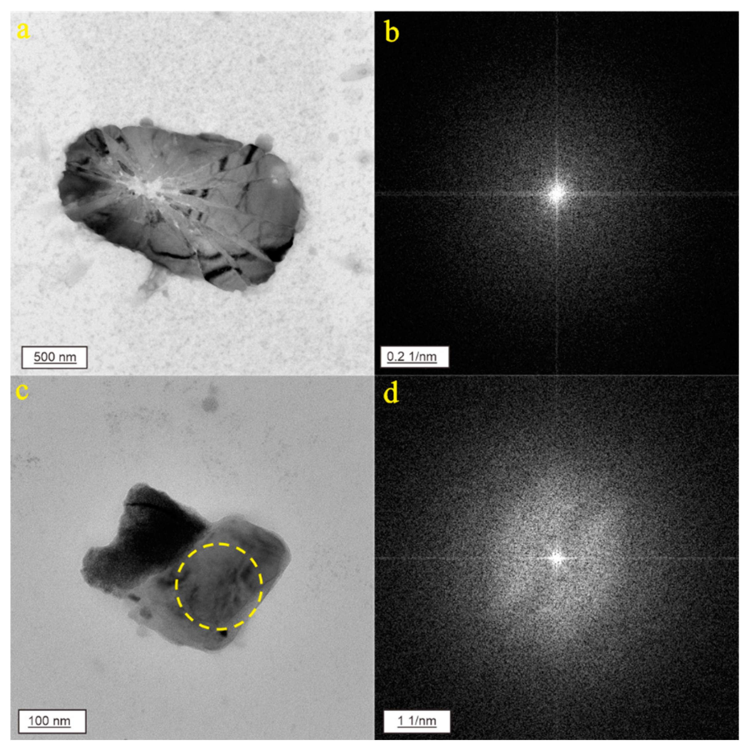
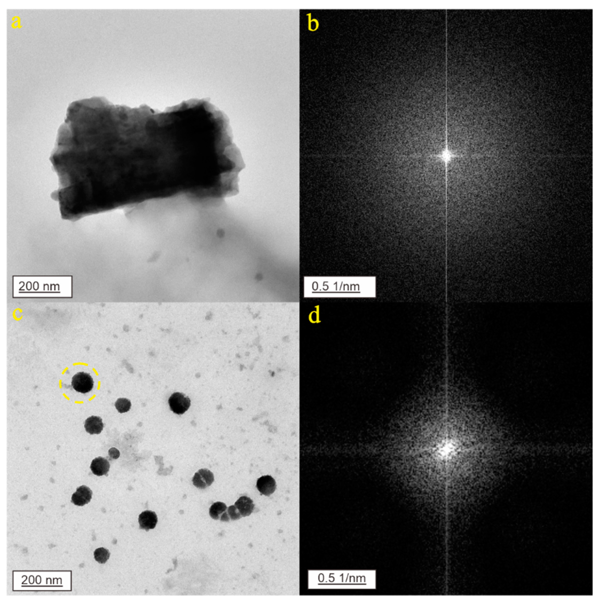
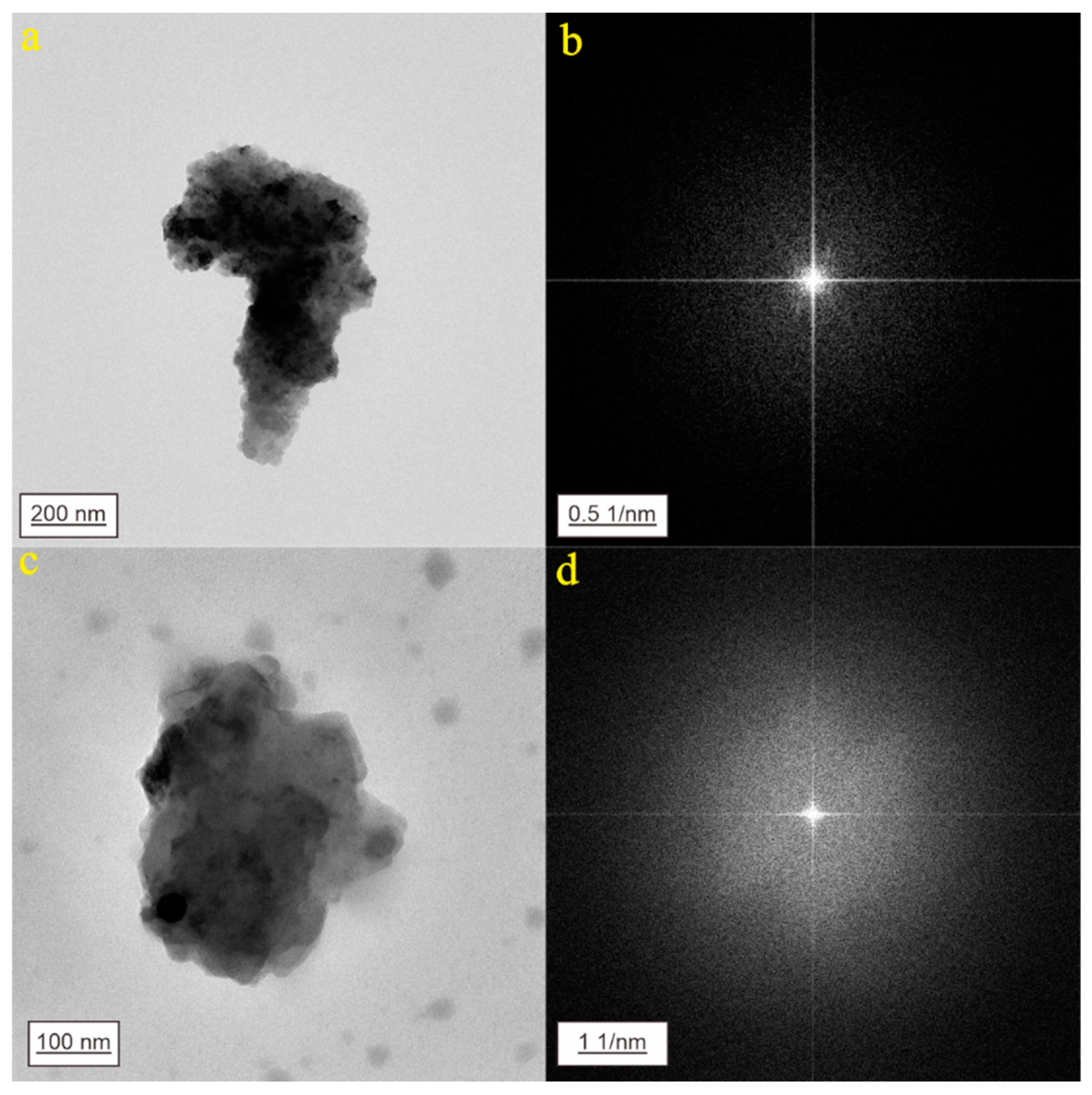
The nanoparticle in Figure 2a is irregular in shape and appears in an amorphous form in the diffraction pattern (Figure 2b). It is primarily composed of C (35.45%), O (40.95%), Ca (10.35%), and S (8.99%), along with trace elements such as Si, Na, and Cl. The nanoparticle in Figure 2c also has an irregular shape and an amorphous form in the diffraction pattern (Figure 2d). Its main components are C (47.44%), O (36.84%), and Ca (13.87%), with impurities including Mg, Na, and Si. Figure 3a shows a nanoparticle with an irregular shape. In the diffraction pattern (Figure 3b), this nanoparticle appears in an amorphous form. It is mainly composed of C (30.97%), O (38.39%), and Ca (27.94%), along with trace elements such as Mg, Na, and Si. In Figure 3c, the nanoparticles are approximately spherical in shape. The diffraction pattern in Figure 3d shows that these nanoparticles are in an amorphous form. They are primarily composed of C (40.18%), O (34.30%), and Ca (19.78%), with a small amount of S (2.26%), as well as trace elements such as Mg and Si. The nanoparticle in Figure 4a is irregular in shape. The diffraction pattern indicates that this nanoparticle is in an amorphous form (Figure 4b). It is mainly composed of C (48.75%), O (32.57%), and Ca (16.67%), with trace amounts of elements such as Mg, S, and Si. Figure 4c shows another nanoparticle that is irregular in shape. The diffraction pattern in Figure 4d reveals that this nanoparticle is in an amorphous state. It primarily consists of C (44.50%), O (32.71%), and Fe (15.08%), with impurities including Al and Si.

3.2. CNPs in Geothermal Water from Jinan City

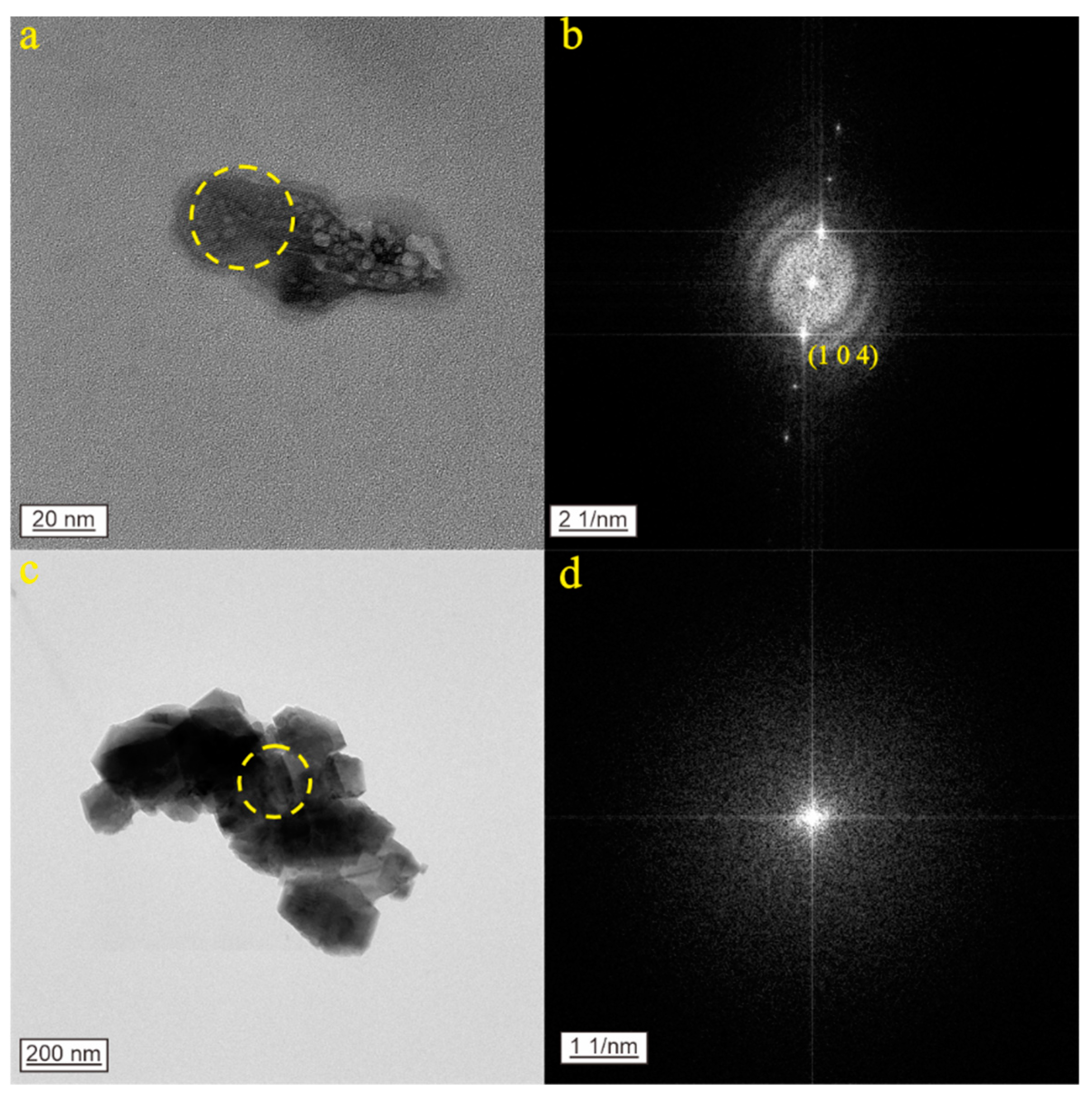
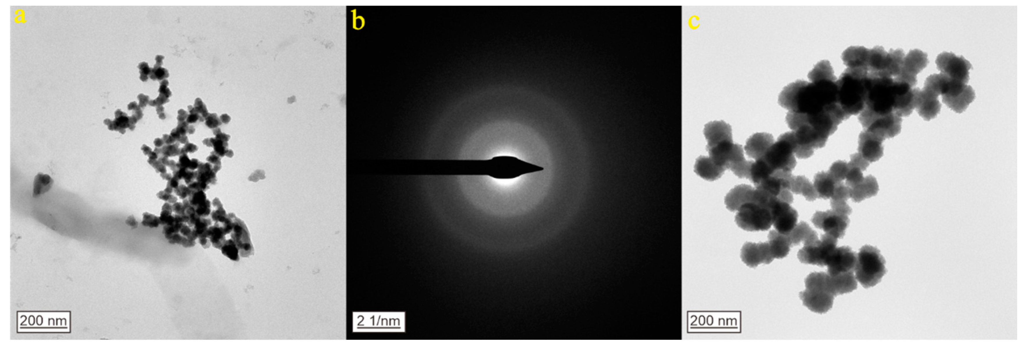
As shown in Figure 5a, the nanoparticles display a bubbly appearance and form aggregates. The selected area electron diffraction (SAED) pattern within the dashed circle reveals that these nanoparticles possess a relatively ordered structure and exist as single crystals (Figure 5b). The interplanar spacing has been accurately measured at 3.04 Å (1 0 4). The energy-dispersive X-ray spectroscopy (EDS) analysis indicates that the nanoparticles mainly consist of O (41.66%), C (29.78%), Ca (13.76%), and S (12.61%), along with minor amounts of Sr and Cl. Moreover, the data match PDF card 05-0586. Based on this comprehensive analysis, it can be inferred that the nanoparticles are calcite (CaCO3). Figure 5c shows a nanoparticle aggregate which has an irregular shape. The diffraction pattern in Figure 5d indicates that the nanoparticles are in an amorphous form. These nanoparticles are primarily composed of C (25.75%), O (46.32%), and Ca (25.60%), along with trace amounts of elements such as Mg, S, and Si. The nanoparticles in Figure 6a exist as chain-like aggregates. In the diffraction pattern, concentric diffuse rings with blurred boundaries between each ring can be observed, indicating an amorphous form (Figure 6b). These nanoparticles are primarily composed of C (25.94%), O (38.39%), and Fe (28.80%), along with trace amounts of impurities such as Mg, Ca, Si, and S. The nanoparticles in Figure 6c are primarily composed of C (15.70%), O (40.31%), and Fe (36.85%), along with trace amounts of impurities such as Ca and Si.

3.3. CNPs in Geothermal Water from Zibo City

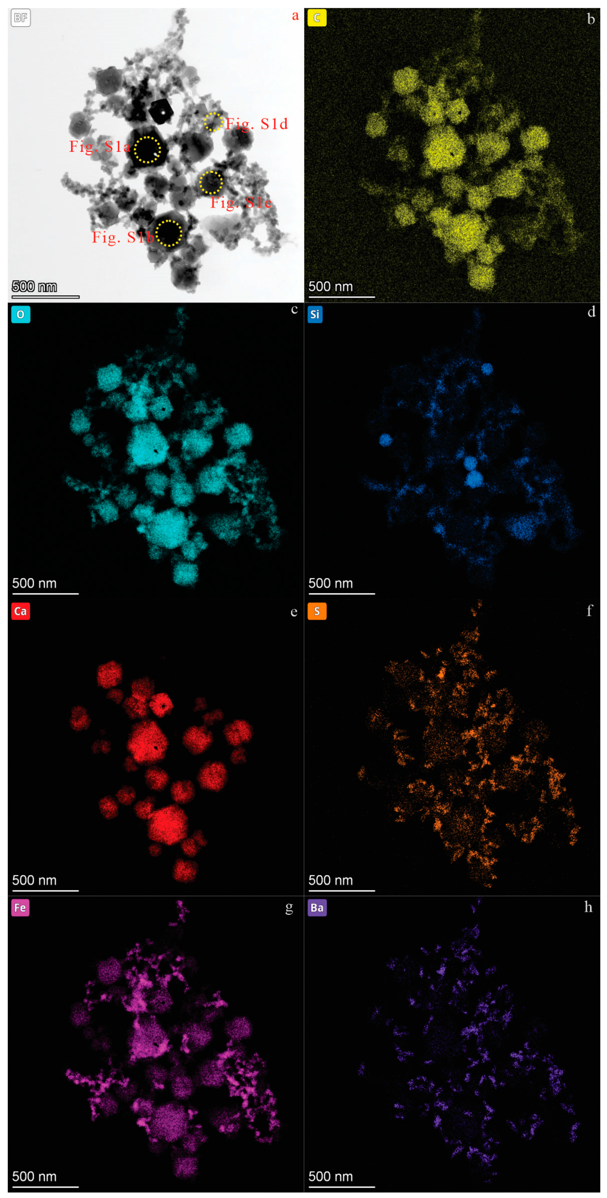
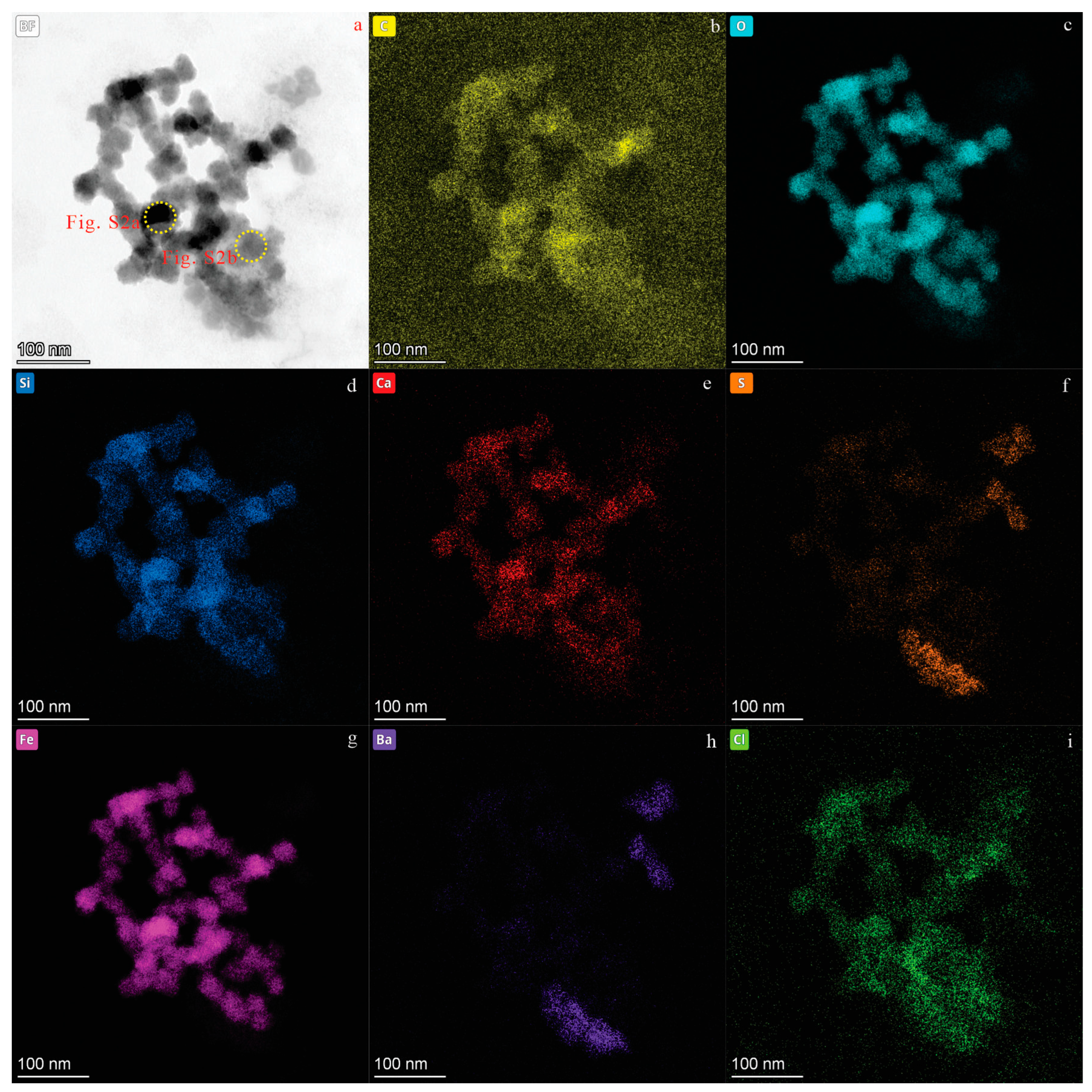
Figure 7 and Figure 8, respectively, show two nanoparticle aggregates, each made up of numerous small nanoparticles. The nanoparticles range in size from 40 to 200 nm. These nanoparticles display a variety of shapes, including spherical, cubic, hexagonal, and irregular forms. EDS mapping reveals that the main elements present in these nanoparticles are C, O, Ca, Si, Fe, S, Cl, and Ba. In the diffraction pattern (Figures S1 and S2), the absence of distinct diffraction spots or rings indicates that these nanoparticles are mainly in an amorphous state.

4. Discussion

Our experiment is conducted at the nanoscale, and the spatial scale of the experimental field is much larger. However, based on the following points, we believe that our experimental results are still valid for large-scale research: First, although the spatial scale is small, the principles are equally applicable in a larger-scale field. Although they may be affected by more external factors, the fundamental principles remain unchanged. Second, in small-scale experiments, we can control and observe the changes in this factor more accurately. Although the spatial scale of the field is larger, the law of action of this key factor will not change. Small-scale experiments can provide a basis for understanding its behavior in large-scale settings. Last, although the scale of the experiment is small, we ensure that the design and parameter selection of the experiment can simulate the key features in the large-scale field. By establishing similar physical, chemical, or biological conditions, we can infer the possible situations in the large-scale field.

4.1. The Genesis of CNPs in Groundwater

Synthetic carbonate nanoparticles (CNPs) typically have a relatively simple composition and are usually spherical with a narrow size distribution (e.g., [14,15,16,17]). In contrast, the CNPs examined in this study showed varying diameters and compositions, suggesting that they were naturally formed rather than artificially produced.
In general, CO32 and HCO3 have high solubility in water, suggesting that CNPs could originate from the precipitation and crystallization of carbonate. However, based on our observation, we propose that this is unlikely for the following reasons: Firstly, nanoparticles formed through precipitation and crystallization typically have regular and uniform shapes and pure compositions [18,19,20], such as spherical SiO2 nanoparticles [21] and square calcite nanoparticles [22]. In contrast, all the CNPs in our study are diverse in shape and have complex compositions, indicating they were not formed by these processes. Secondly, nanoparticles formed by precipitation and crystallization are usually crystalline, whereas almost all the CNPs in our study are amorphous. Finally, previous studies have indicated that nanoparticles in water have a relatively high solubility [23]. Additionally, many other nanoparticles are discovered in groundwater [24,25,26,27,28,29,30]. Therefore, the likely source of the CNPs is particle ejection from the existing primary or secondary carbonate minerals in the surrounding carbonate zone. The aquifers in this study contain primary carbonate minerals such as calcite [CaCO3] and likely secondary carbonates such as dolomite [CaMg(CO3)2]. Ca and Mg can bind with carbonate to form nanoparticles in the groundwater.
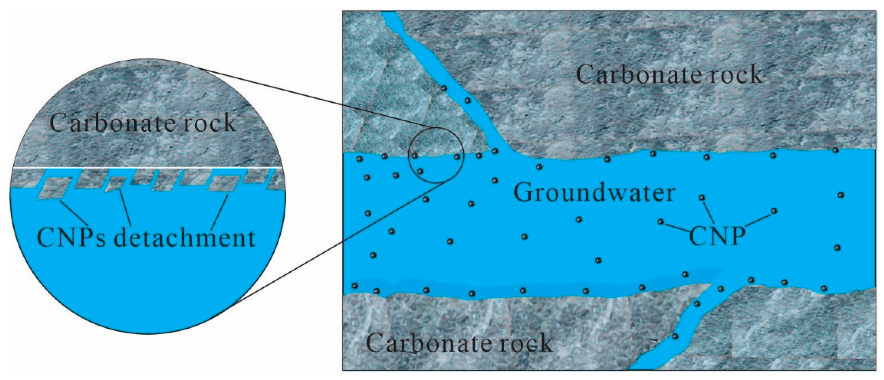
In limestones, which are known to be highly fractured, these fractures play a crucial role. Fractures can occur as a result of various geological processes such as tectonic forces, stress relaxation, or differential thermal expansion. The presence of fractures in limestones can enhance the porosity and permeability of the rock mass. This increased permeability enables groundwater to flow more freely through the limestone formation. Fractures can range in size from microscopic fissures to large cracks. Recent experiments have demonstrated that weathering in carbonate rocks is primarily driven by a combination of chemical dissolution and mechanical processes in the fractured area [31,32]. These processes can even extend to the nanoscale, where rapid chemical dissolution along grain boundaries is followed by the mechanical detachment of tiny grains from the carbonate rock surfaces [31]. This detachment is likely due to fluid shearing or repulsive forces between the calcite grains. This mechanism may be a major source of CNPs in groundwater. Importantly, this chemo-mechanical process could be common in fine-grained carbonates, significantly accelerating weathering rates and releasing numerous CNPs into groundwater. Therefore, we propose that CNPs are likely generated through the following sequence: the weathering of carbonate minerals in groundwater → water–rock interaction → crystal repulsion → particle detachment → solution entrainment → limited dissociation during transport, rather than through crystal formation in solution with mineral-phase saturation ([5]; Figure 9).

4.2. The Role of CNPs in Carbonate Mineral Evolution and Mineralization during Weathering

Carbonate minerals in rocks typically interact with water and dissolved CO2, producing calcium and bicarbonate ions in hydrological systems [32,33,34]. However, numerous studies have shown that the weathering of carbonate minerals can also release CNPs into groundwater [5,25,27,30,35]. Estimates indicate that CNPs can contribute between 10% [36] and over 90% [31] to the overall weathering rates of carbonate rocks. Therefore, the formation and transport of geogenic CNPs in groundwater are of considerable concern due to their potential for widespread distribution. Furthermore, CNPs could be a significant product of the decomposition and transformation of carbonate rocks and minerals in aquifers during weathering.
Compared to macromineral crystals, nanoparticles are more likely to agglomerate in natural environments due to their small size and high surface energy. This agglomeration and subsequent migration are of crucial importance for mineralization processes. Nanoparticles can disperse through various media, such as gasses, liquids, or solids, and form aerosols, colloids, solid solutions, or minerals [37,38]. In the absence of microcracks, this dispersion can lead to mineralization [39]. For example, colloform or botryoidal sphalerite aggregates, which are optically anisotropic and have a disordered structure or hexagonal habit, are formed through the coalescence and agglomeration of colloidal nanocrystalline particles [40]. Similarly, the irregularly shaped platy micronuggets of gold result from the agglomeration of isolated semispherical nanoparticles [41], as evidenced by the studies of the growth history of native gold particles. CNPs exhibit metastability in natural settings, with surface or structurally bound water molecules contributing to their stabilization. These nanoparticles often have structures similar to their bulky counterparts but are highly disordered, with significant rotations and translations of CO3 groups; the smallest particles can be essentially amorphous [42]. This observation is in line with the phenomena described in the article. Furthermore, CNP structures typically transform with dewatering, gradually reorganizing into macrominerals or crystals. For instance, amorphous silica nanoparticles can evolve into opal-CT, opal-C, moganite, cristobalite, and eventually quartz [43,44]. Consequently, CNPs make a significant contribution to the mineralization of carbonate minerals.

4.3. Implication of CNPs for the Migration of Carbon and Associated Elements during Weathering

The weathering of rocks is of crucial importance for the geochemical cycling of elements. Numerous studies have established a connection between the mobility of elements and the breakdown of minerals during progressive weathering [45,46,47,48,49,50]. Traditionally, research has been centered around the mobility of ions and molecules, particularly soluble cations like K+, Na+, Ca2+, Mg2+, and CO2, which play a significant role in chemical weathering. However, for insoluble elements such as Si, Al, and Fe, their migration as ions or molecules is more intricate. As a consequence, it is essential to consider other forms of elemental mobility in the weathering process.
This study reveals that carbonate nanoparticles (CNPs) associated with other elements in groundwater provide a significant mechanism for element migration during weathering, especially for insoluble elements like Si, Al, and Fe. Traditionally, it is known that carbon primarily migrates in the form of HCO3 during carbonate rock weathering [51]. However, this study shows that CNPs also play a key role in carbon migration. Additionally, carbonate nanoparticles can carry soluble elements such as Ca, Na, and K, suggesting that these elements can migrate as nanoparticles during carbonate weathering. The migration forms of poorly soluble elements like Si, Fe, and Al have been a subject of debate. These elements are believed to migrate in various forms: (1) as detrital particles [52,53]; (2) as colloidal particles [54]; or (3) as both detrital and colloidal particles [55]. This study offers a new perspective, indicating that these elements can also migrate as nanoparticles during carbonate weathering and potentially other geological processes. Previous research has indicated that elements such as Si, Al, and Au can achieve high solubility in geological fluids when present as nanoparticles, providing an explanation for their extensive migration [54,56,57,58,59]. Furthermore, Yi et al. [60] discovered that rare earth elements (REEs) can migrate and accumulate as nanoparticles during the chemical weathering of granite. These findings support the idea that nanoparticles are a crucial factor in the migration of elements during weathering processes.
Some CNPs also contain non-metal elements such as S and Cl. These non-metal elements may originate from the minerals in carbonate rocks, including sulfide minerals, sulfate minerals, and Cl-bearing minerals. Chlorine might also come from groundwater sources. These findings imply that CNPs play a role in the migration of non-metal elements during carbonate rock weathering. In addition to accelerating weathering, the mechanisms involving CNPs could have an impact on the evolution of hydrocarbon reservoirs. They provide a natural flux of colloids capable of transporting elements through groundwater.

5. Conclusions

The analysis of the carbonate nanoparticles (CNPs) in the groundwater from Zibo and Jinan cities in Shandong Province unveils that these nanoparticles can manifest as single particles or aggregates, with sizes spanning from 20 nm to 500 nm. They display diverse compositions, mainly consisting of C, O, and Ca, and occasionally containing traces of S, Fe, Mg, and other trace elements. The irregular shapes and amorphous nature of these CNPs suggest that they are naturally occurring rather than synthetic. Additionally, the irregular morphology and complex composition of the CNPs imply that they are not merely products of precipitation or crystallization. Instead, their formation is likely propelled by weathering processes, where the chemical dissolution and mechanical detachment of carbonate minerals contribute to their generation. CNPs play a significant role in the weathering and mineralization of carbonate minerals. Their ability to agglomerate and disperse in various media highlights their importance in the transformation and distribution of carbonate minerals within groundwater systems. Furthermore, the presence of CNPs indicates their contribution to the overall weathering rates of carbonate rocks. They facilitate the migration of both soluble elements such as C, Ca, Na, and K and poorly soluble elements like Si, Al, and Fe. This presents a new perspective on the migration of these elements and their potential for extensive distribution in geological processes, underscoring the significant role of CNPs in the weathering and transport of elements.

Supplementary Materials

The following supporting information can be downloaded at: https://www.mdpi.com/article/10.3390/min14100980/s1, Figure S1. Select diffraction patterns (a–d) of CNPs in Figure 7a from geothermal water from Zibo city; Figure S2. Select diffraction patterns (a and b) of CNPs in Figure 8a from geothermal water from Zibo city.

Author Contributions

Conceptualization, G.T.; methodology, L.Z.; software, L.Z.; validation, X.Z.; investigation, P.Z.; data curation, Y.W.; writing—original draft preparation, G.T.; writing—review and editing, R.L.; funding acquisition, G.T. and R.L. All authors have read and agreed to the published version of the manuscript.

Funding

This work was jointly supported by the National Natural Science Foundation of China (Grant Nos. 42102076 and 42077129), Shandong Province Natural Science Foundation under Grant No. ZR2021QD037 and ZR2020ZD19, SDUT and Zibo City Integration Development Project (NO. 2021SNPT0012), project 21kfhk03 supported by Fundamental Science on Nuclear Wastes and Environmental Safety Laboratory, and project 18zx7115 supported by Southwest University of Science and Technology Doctor Foundation.

Data Availability Statement

The data presented in this study are available upon request from the corresponding author due to privacy.

Conflicts of Interest

The authors declare no conflicts of interest.

References

  1. Sharma, V.K.; Filip, J.; Zboril, R.; Varma, R.S. Natural Inorganic Nanoparticles-Formation, Fate, and Toxicity in the Environment. Chem. Soc. Rev. 2015, 44, 8410–8423. [Google Scholar] [CrossRef] [PubMed]
  2. Emmanuel, S.; Anovitz, L.M.; Day-Stirrat, R.J. Effects of Coupled Chemo-Mechanical Processes on the Evolution of Pore-Size Distributions in Geological Media. Rev. Mineral. Geochem. 2015, 80, 45–60. [Google Scholar] [CrossRef]
  3. Levenson, Y.; Emmanuel, S. Quantifying Micron-Scale Grain Detachment during Weathering Experiments on Limestone. Geochim. Cosmochim. Acta 2016, 173, 86–96. [Google Scholar] [CrossRef]
  4. Levenson, Y.; Emmanuel, S. Repulsion between Calcite Crystals and Grain Detachment during Water-Rock Interaction. Geochem. Perspect. Lett. 2017, 3, 133–141. [Google Scholar] [CrossRef]
  5. Langman, J.B.; Moberly, J.G. Weathering of a Mined Quartz-Carbonate, Galena-Sphalerite Ore and Release and Transport of Nanophase Zinc Carbonate in Circumneutral Drainage. J. Geochem. Explor. 2018, 188, 185–193. [Google Scholar] [CrossRef]
  6. Schindler, M.; Lussier, A.J.; Principe, E.; Mykytczuk, N. Dissolution Mechanisms of Chromitite: Understanding the Release and Fate of Chromium in the Environment. Am. Mineral. 2018, 103, 271–283. [Google Scholar] [CrossRef]
  7. Findlay, A.J.; Estes, E.R.; Gartman, A.; Yücel, M.; Kamyshny, A.; Luther, G.W. Iron and Sulfide Nanoparticle Formation and Transport in Nascent Hydrothermal Vent Plumes. Nat. Commun. 2019, 10, 1597. [Google Scholar] [CrossRef]
  8. Alekseyev, V.A. Nanoparticles and Nanofluids in Water–Rock Interactions. Geochem. Int. 2019, 57, 357–368. [Google Scholar] [CrossRef]
  9. Rad, S.; Louvat, P.; Gorge, C.; Gaillardet, J.; Allègre, C.J. River Dissolved and Solid Loads in the Lesser Antilles: New Insight into Basalt Weathering Processes. J. Geochem. Explor. 2006, 88, 308–312. [Google Scholar] [CrossRef]
  10. Bailly-Comte, V.; Martin, J.B.; Screaton, E.J. Time variant cross correlation to assess residence time of water and implication for hydraulics of a sink-rise karst system. Water Resour. Res. 2011, 47, W05547. [Google Scholar] [CrossRef]
  11. Lorenzi, V.; Banzato, F.; Barberio, M.D.; Goeppert, N.; Goldscheider, N.; Gori, F.; Lacchini, F.; Manetta, M.; Medici, G.; Rusi, S.; et al. Tracking flowpaths in a complex karst system through tracer test and hydrogeochemical monitoring: Implications for groundwater protection (Gran Sasso, Italy). Heliyon 2024, 10, e24663. [Google Scholar] [CrossRef] [PubMed]
  12. Rad, S.D.; Allègre, C.J.; Louvat, P. Hidden Erosion on Volcanic Islands. Earth Planet. Sci. Lett. 2007, 262, 109–124. [Google Scholar] [CrossRef]
  13. Calmels, D.; Galy, A.; Hovius, N.; Bickle, M.; West, A.J.; Chen, M.C.; Chapman, H. Contribution of Deep Groundwater to the Weathering Budget in a Rapidly Eroding Mountain Belt, Taiwan. Earth Planet. Sci. Lett. 2011, 303, 48–58. [Google Scholar] [CrossRef]
  14. Cai, W.Y.; Feng, L.D.; Liu, S.H.; Zhu, J.J. Hemoglobin-CdTe-CaCO3@ Polyelectrolytes 3D Architecture: Fabrication, Characterization, and Application in Biosensing. Adv. Funct. Mater. 2008, 18, 3127–3136. [Google Scholar] [CrossRef]
  15. Ueno, Y.; Futagawa, H.; Takagi, Y.; Ueno, A.; Mizushima, Y. Drug-Incorporating Calcium Carbonate Nanoparticles for a New Delivery System. J. Control. Release 2005, 103, 93–98. [Google Scholar] [CrossRef]
  16. Wang, C.Y.; He, C.Y.; Tong, Z.; Liu, X.X.; Ren, B.Y.; Zeng, F. Combination of Adsorption by Porous CaCO3 Microparticles and Encapsulation by Polyelectrolyte Multilayer Films for Sustained Drug Delivery. Int. J. Pharm. 2006, 308, 160–167. [Google Scholar] [CrossRef]
  17. Peng, C.Y.; Zhao, Q.H.; Gao, C.Y. Sustained Delivery of Doxorubicin by Porous CaCO3 and Chitosan/Alginate Multilayers-Coated CaCO3 Microparticles. Colloids Surf. A Physicochem. Eng. Asp. 2010, 353, 132–139. [Google Scholar] [CrossRef]
  18. Sozer, N.; Kokini, J.L. Nanotechnology and its applications in the food sector. Trends Biotechnol. 2009, 27, 82–89. [Google Scholar] [CrossRef]
  19. Mcneil, S.E. Unique benefits of nanotechnology to drug delivery and diagnostics. Methods Mol. Biol. 2011, 697, 3–8. [Google Scholar] [PubMed]
  20. Sonkaria, S.; Ahn, S.H.; Khare, V. Nanotechnology and its impact on food and nutrition: A review. Recent Patents Food Nutr. Agric. 2012, 4, 8–18. [Google Scholar]
  21. Nabeshi, H.; Yoshikawa, T.; Matsuyama, K.; Nakazato, Y.; Arimori, A.; Isobe, M.; Tochigi, S.; Kondoh, S.; Hirai, T.; Akase, T. Size-Dependent Cytotoxic Effects of Amorphous Silica Nanoparticles on Langerhans Cells. Die Pharm. Int. J. Pharm. Sci. 2010, 65, 199–201. [Google Scholar] [CrossRef]
  22. Nakayama, M.; Kajiyama, S.; Kumamoto, A.; Ikuhara, Y.; Kato, T. Bioinspired Selective Synthesis of Liquid-Crystalline Nanocomposites: Formation of Calcium Carbonate-Based Composite Nanodisks and Nanorods. Nanoscale Adv. 2020, 2, 2326–2332. [Google Scholar] [CrossRef] [PubMed]
  23. Yu, J.; Dong, H.W.; Shi, L.T.; Tang, X.Y.; Liu, J.R.; Shi, J.H. Reproductive Toxicity of Perchlorate in Rats. Food Chem. Toxicol. 2019, 128, 212–222. [Google Scholar] [CrossRef]
  24. Wu, C.Y.; Martel, J.; Wong, T.Y.; Young, D.; Liu, C.C.; Lin, C.W.; Young, J.D. Formation and Characteristics of Biomimetic Mineralo-Organic Particles in Natural Surface Water. Sci. Rep. 2016, 6, 28817. [Google Scholar] [CrossRef]
  25. Liu, R.; Zhao, Z.Q.; Lv, M.H.; Wang, H.W.; Li, L.X.; Gang, S.T.; Zuo, L.; Zhang, P.; Wang, Y.Q.; Li, C.S.; et al. Discovery of Environmental Nanoparticles in a Mineral Water Spring from Yiyuan County, Shandong Province, Eastern China: A New Form of Elements in Mineral Water. Water 2023, 15, 3497. [Google Scholar] [CrossRef]
  26. Shao, G.Y.; Li, C.S.; Liu, R.; Zhang, P.; Zuo, L.; Wang, Y.Q. Silicified Microorganisms and Microorganism-like Particles in the Groundwater of an Abandoned Coal Mine. Mine Water Environ. 2023, 42, 489–499. [Google Scholar] [CrossRef]
  27. Zuo, L.; Zhang, P.; Wang, Y.Q.; Liu, R.; Ma, G.X. Characteristics and Research Significance of Micro-Nanoparticles in Geothermal Fluids in the Central Area of Shandong Province. Water 2023, 15, 3737. [Google Scholar] [CrossRef]
  28. Zuo, L.; Li, C.S.; Zhang, P.; Wang, Y.Q.; Gao, S.; Sun, B.; Liu, R. Characteristics of Natural Ti-Bearing Nanoparticles in Groundwater within Karst Areas of Northern China. Water 2024, 16, 650. [Google Scholar] [CrossRef]
  29. Chen, Z.Y.; Liu, R.; Wang, Y.Q.; Lu, B.; Cui, Y.X.; Zuo, L.; Zhang, P.; Wang, Y.Q.; Cao, C. Biomimetic Fe-Bearing Nanoparticles in Hot Spring: Morphology, Origin and Potential Bioavailable Fe. Front. Earth Sci. 2024, 12, 1404788. [Google Scholar] [CrossRef]
  30. Hu, C.P.; Liu, R.; Zhang, P.; Wang, Y.Q.; Zuo, L.; Zhang, X.H.; Li, C.S. Characteristics and Significance of Natural Nanoparticles in the Groundwater of the Baotu Spring Area in Jinan, Shandong Province, Eastern China. Water 2024, 16, 1820. [Google Scholar] [CrossRef]
  31. Emmanuel, S.; Levenson, Y. Limestone Weathering Rates Accelerated by Micron-Scale Grain Detachment. Geology 2014, 42, 751–754. [Google Scholar] [CrossRef]
  32. Liu, Z.; Zhao, J. Contribution of Carbonate Rock Weathering to the Atmospheric CO2 Sink. Environ. Geol. 2000, 39, 1053–1058. [Google Scholar] [CrossRef]
  33. Basak, C.; Martin, E.E. Antarctic Weathering and Carbonate Compensation at the Eocene–Oligocene Transition. Nat. Geosci. 2013, 6, 121–124. [Google Scholar] [CrossRef]
  34. Komar, N.; Zeebe, R.E.; Dickens, G.R. Understanding Long-Term Carbon Cycle Trends: The Late Paleocene through the Early Eocene. Paleoceanography 2013, 28, 650–662. [Google Scholar] [CrossRef]
  35. Tan, X.B.; Bo, B.Y.; Zhang, P.; Shao, G.Y.; Liu, R.; Wang, K. Carbonaceous Nanoparticles in Zibo Hot Springs: Implications for the Cycling of Carbon and Associated Elements. Environ. Chem. Lett. 2021, 19, 4009–4014. [Google Scholar] [CrossRef]
  36. Vleugels, G.; Van Grieken, R. Suspended Matter in Run-off Water from Limestone Exposure Setups. Sci. Total Environ. 1995, 170, 125–132. [Google Scholar] [CrossRef]
  37. Wang, X.Q.; Zhang, B.M.; Lin, X.; Xu, S.F.; Yao, W.S.; Ye, R. Geochemical Challenges of Diverse Regolith-Covered Terrains for Mineral Exploration in China. Ore Geol. Rev. 2016, 73, 417–431. [Google Scholar] [CrossRef]
  38. Wang, X.Q.; Zhang, B.M.; Ye, R. Nanogeochemistry for Mineral Exploration through Covers. Bull. Mineral. Petrol. Geochem. 2016, 35, 43–51. [Google Scholar] [CrossRef]
  39. Ju, Y.W.; Huang, C.; Sun, Y.; Wan, Q.; Lu, X.C.; Lu, S.F.; He, H.P.; Wang, X.Q.; Zou, C.N.; Wu, J.G.; et al. Nanogeosciences: Research History, Current Status, and Development Trends. J. Nanosci. Nanotechnol. 2017, 17, 5930–5965. [Google Scholar] [CrossRef]
  40. Wu, S.H.; Mao, J.W.; Yuan, S.D.; Dai, P.; Wang, X.D. Mineralogy, Fluid Inclusion Petrography, and Stable Isotope Geochemistry of Pb–Zn–Ag Veins at the Shizhuyuan Deposit, Hunan Province, Southeastern China. Miner. Depos. 2018, 53, 89–103. [Google Scholar] [CrossRef]
  41. Wierchowiec, J.; Mikulski, S.Z.; Zieliński, K. Supergene Gold Mineralization from Exploited Placer Deposits at Dziwiszów in the Sudetes (NE Bohemian Massif, SW Poland). Ore Geol. Rev. 2021, 131, 104049. [Google Scholar] [CrossRef]
  42. Cooke, D.J.; Elliott, J.A. Atomistic Simulations of Calcite Nanoparticles and Their Interaction with Water. J. Chem. Phys. 2007, 127, 104706. [Google Scholar] [CrossRef] [PubMed]
  43. Herdianita, N.R.; Browne, P.R.L.; Rodgers, K.A.; Campbell, K.A. Mineralogical and Textural Changes Accompanying Ageing of Silica Sinter. Miner. Depos. 2000, 35, 48–62. [Google Scholar] [CrossRef]
  44. Icopini, G.A.; Brantley, S.L.; Heaney, P.J. Kinetics of Silica Oligomerization and Nanocolloid Formation as a Function of PH and Ionic Strength at 25 °C. Geochim. Cosmochim. Acta 2005, 69, 293–303. [Google Scholar] [CrossRef]
  45. Colman, S.M. Chemical Weathering of Basalts and Andesites; Evidence from Weathering Rinds; U.S. G.P.O.: Washington, DC, USA, 1982. [CrossRef]
  46. Eggleton, R.A.; Foudoulis, C.; Varkevisser, D. Weathering of Basalt: Changes in Rock Chemistry and Mineralogy. Clays Clay Miner. 1987, 35, 161–169. [Google Scholar] [CrossRef]
  47. Nesbitt, H.W.; Wilson, R.E. Recent Chemical Weathering of Basalts. Am. J. Sci. 1992, 292, 740–777. [Google Scholar] [CrossRef]
  48. Gavshin, V.M.; Shcherbov, B.L.; Bobrov, V.A.; Solotchina, E.P.; Sukhorukov, F.V.; Mel’gunov, M.S. Behavior of Trace Elements in the Process of Formation of a Weathering Profile on Granites. Russ. Geol. Geophys. C/C Geol. Geofiz. 1997, 38, 1264–1276. [Google Scholar]
  49. Hill, I.G.; Worden, R.H.; Meighan, I.G. Yttrium: The Immobility-Mobility Transition during Basaltic Weathering. Geology 2000, 28, 923–926. [Google Scholar] [CrossRef]
  50. Patino, L.C.; Velbel, M.A.; Price, J.R.; Wade, J.A. Trace Element Mobility during Spheroidal Weathering of Basalts and Andesites in Hawaii and Guatemala. Chem. Geol. 2003, 202, 343–364. [Google Scholar] [CrossRef]
  51. Freeze, R.A.; Cherry, J.A. Groundwater; Prentice Hall: Englewood Cliffs, NJ, USA, 1979. [Google Scholar]
  52. Öztürk, H.; Hein, J.R.; Hanilçi, N. Genesis of the Doğankuzu and Mortaş Bauxite Deposits, Taurides, Turkey: Separation of Al, Fe, and Mn and Implications for Passive Margin Metallogeny. Econ. Geol. 2002, 97, 1063–1077. [Google Scholar] [CrossRef]
  53. Yang, S.J.; Huang, Y.X.; Wang, Q.F.; Deng, J.; Liu, X.F.; Wang, J.Q. Mineralogical and Geochemical Features of Karst Bauxites from Poci (Western Henan, China), Implications for Parental Affinity and Bauxitization. Ore Geol. Rev. 2019, 105, 295–309. [Google Scholar] [CrossRef]
  54. Gamaletsos, P.N.; Godelitsas, A.; Kasama, T.; Church, N.S.; Douvalis, A.P.; Göttlicher, J.; Steininger, R.; Boubnov, A.; Pontikes, Y.; Tzamos, E.; et al. Nano-Mineralogy and -Geochemistry of High-Grade Diasporic Karst-Type Bauxite from Parnassos-Ghiona Mines, Greece. Ore Geol. Rev. 2017, 84, 228–244. [Google Scholar] [CrossRef]
  55. He, H.Z.; Zhang, Q.Z. Analysis on Geological Anomaly of Karst Accumulative Bauxite Deposit and Delimitation of Favorable Area for Prospecting Based on GIS in Western Guangxi. Miner. Resour. Geol. 2007, 21, 436–439. [Google Scholar]
  56. Perry, C.C.; Keeling-Tucker, T. Biosilicification: The Role of the Organic Matrix in Structure Control. JBIC J. Biol. Inorg. Chem. 2000, 5, 537–550. [Google Scholar] [CrossRef] [PubMed]
  57. Pancost, R.D.; Pressley, S.; Coleman, J.M.; Benning, L.G.; Mountain, B.W. Lipid Biomolecules in Silica Sinters: Indicators of Microbial Biodiversity. Environ. Microbiol. 2005, 7, 66–77. [Google Scholar] [CrossRef] [PubMed]
  58. Reich, M.; Utsunomiya, S.; Kesler, S.E.; Wang, L.; Ewing, R.C.; Becker, U. Thermal Behavior of Metal Nanoparticles in Geologic Materials. Geology 2006, 34, 1033–1036. [Google Scholar] [CrossRef]
  59. Tobler, D.J.; Stefánsson, A.; Benning, L.G. In-situ Grown Silica Sinters in Icelandic Geothermal Areas. Geobiology 2008, 6, 481–502. [Google Scholar] [CrossRef]
  60. Yi, Z.; Fu, W.; Zhao, Q.; Lu, H.; Fu, X.; Li, P.; Luo, P.; Han, Z.; Tan, Z.; Xu, C. Characterization of Nano-Minerals and Nanoparticles in Supergene Rare Earth Element Mineralization Related to Chemical Weathering of Granites. Am. Mineral. 2023, 108, 1461–1475. [Google Scholar] [CrossRef]
Figure 1. Locations of sampling sites.
Minerals 14 00980 g001
Figure 2. Carbonate nanoparticles in the karst groundwater from Jinan city. (a,c) are high-resolution morphology images (the yellow circle in (c) is the location of select diffraction), and (b,d) are select diffraction patterns.
Minerals 14 00980 g002
Figure 3. Carbonate nanoparticles in the karst groundwater from Jinan city. (a,c) are high-resolution morphology images (the yellow circle in (c) is the location of select diffraction), and (b,d) are select diffraction patterns.
Minerals 14 00980 g003
Figure 4. Carbonate nanoparticles in the karst groundwater from Jinan city. (a,c) are high-resolution morphology images, and (b,d) are select diffraction patterns.
Minerals 14 00980 g004
Figure 5. Carbonate nanoparticles in the geothermal water from Jinan city. (a,c) are high-resolution morphology images (the yellow circle in (a) and (c) is the location of select diffraction), and (b,d) are select diffraction patterns.
Minerals 14 00980 g005
Figure 6. Carbonate nanoparticle aggregates in geothermal water from Jinan city. (a,c) are high-resolution morphology images, and (b) is select diffraction pattern.
Minerals 14 00980 g006
Figure 7. High-resolution morphology image (a) and the elemental mapping of the carbonate nanoparticle aggregate (bh) in the geothermal water from Zibo city. The yellow circles in (a) are the location of select diffraction.
Minerals 14 00980 g007
Figure 8. High-resolution morphology image (a) and the elemental mapping of the carbonate nanoparticle aggregate (bi) in the geothermal water from Zibo city. The yellow circles in (a) are the location of select diffraction.
Minerals 14 00980 g008
Figure 9. Schematic representation of process to form CNPs in groundwater (modified from [5]).
Minerals 14 00980 g009
Table 1. EDS results (wt.%) of CNPs in karst groundwater and geothermal water from Jinan city.
LocationGroundwater SamplesCOCaSClSiMgFeAlNaK
Jinankarst groundwaterKG-135.45 40.95 10.35 8.990.891.010.17 2.35
KG-247.44 36.84 13.870.12 1.340.13 0.25
KG-330.97 38.39 27.940.56 0.960.80
KG-440.18 34.30 19.78 2.26 0.742.73
KG-548.75 32.57 16.67 0.46 0.71 0.220.29
KG-644.5032.71 1.37 0.761.42 2.55 2.08 15.081.421.23 0.78
geothermal waterGW-129.78 41.66 13.76 12.61
GW-225.7546.3225.600.80 0.36 0.26
GW-325.94 38.39 1.510.53 4.110.71 28.800.40
GW-415.7040.310.70 0.90 36.850.44
Disclaimer/Publisher’s Note: The statements, opinions and data contained in all publications are solely those of the individual author(s) and contributor(s) and not of MDPI and/or the editor(s). MDPI and/or the editor(s) disclaim responsibility for any injury to people or property resulting from any ideas, methods, instructions or products referred to in the content.