Communication

Synthesis of an Antipyrine-Based Fluorescent Probe with Synergistic Effects for the Selective Recognition of Zinc Ion

1
Henan Institute of Metallurgy Co., Ltd., Zhengzhou 450053, China
2
Henan Province Research Center of Metallurgical Waste Heat and Solid Waste Resource Utilization Engineering Technology, Zhengzhou 450053, China
3
School of Chemistry and Chemical Engineering, Jiangxi University of Science and Technology, Ganzhou 341000, China
*
Authors to whom correspondence should be addressed.
Minerals 2024, 14(7), 649; https://doi.org/10.3390/min14070649
Submission received: 10 May 2024 / Revised: 20 June 2024 / Accepted: 24 June 2024 / Published: 25 June 2024
(This article belongs to the Special Issue Recent Advances in Extractive Metallurgy)

Abstract

:
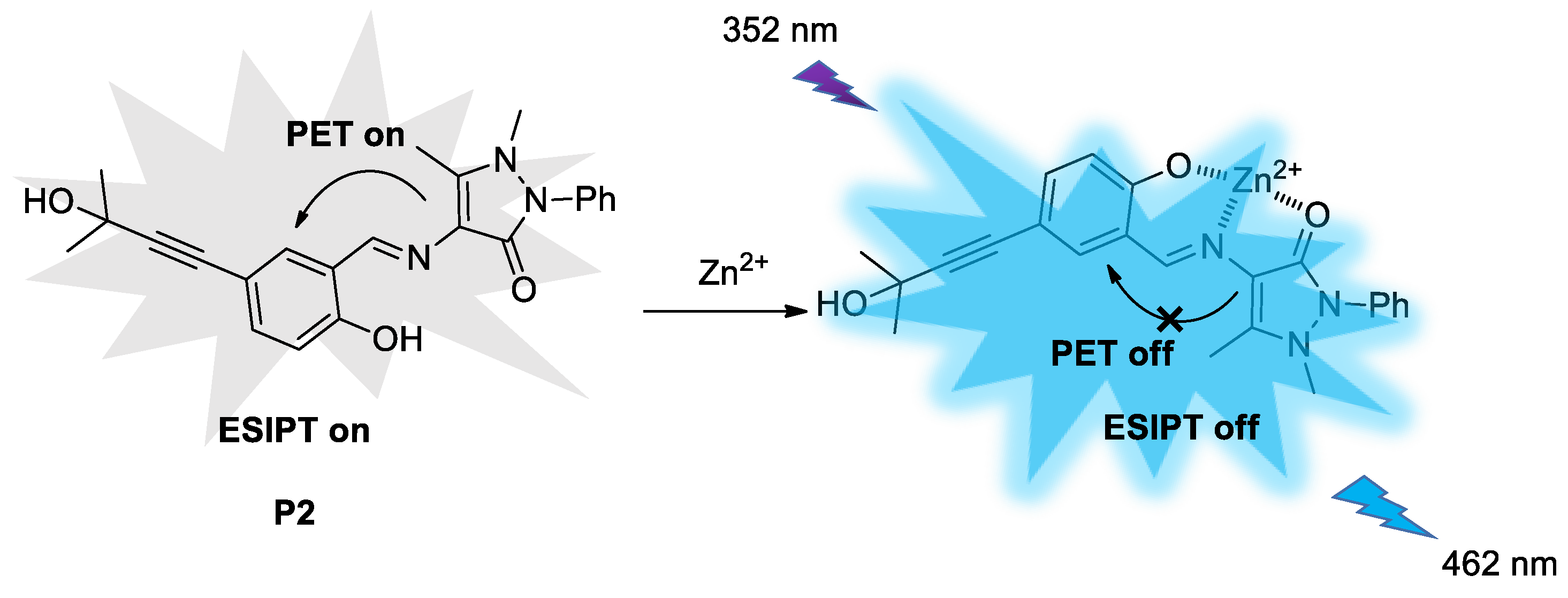
A novel fluorescent probe containing an imine structure was synthesized through a condensation reaction based on the skeleton of antipyrine. Due to the synergistic effect of photoinduced electron transfer (PET), excited-state intramolecular proton transfer (ESIPT), and E/Z isomerization, the probe itself has weak fluorescence. When zinc ions are added to the ethanol solution of the probe, the formed complex inhibits PET, ESIPT, and E/Z isomerization while activating chelation-enhanced fluorescence (CHEF), resulting in fluorescent “turn-on” at 462 nm. Under optimal detection conditions, the probe can rapidly respond to zinc ions within 3 min, with a linear range of 60–220 μM and a lower limit of detection (LOD) of 0.63 μM. It can also specifically identify zinc ions in the presence of 13 common metal ions.

1. Introduction

Metal ions play a pivotal role in human health, yet an overabundance of essential metal ions such as iron and zinc can induce various maladies, including Parkinson’s disease, Alzheimer’s disease, muscular dystrophy, ischemic stroke, etc. [1,2,3,4]. Consequently, the rapid and efficient selective detection of metal ions has remained a focal point of research endeavors. Up to now, a myriad of assays, including traditional titrimetry [5], atomic absorption spectroscopy (AAS) [6], ultraviolet–visible spectrophotometry (UV–vis) [7], inductively coupled plasma (ICP) [8], ion selective electrode (ISE) [9], biosensors [10], and fluorescence analysis (FA) have been developed for metal ion detection. Among them, fluorescent probes have attracted considerable attention due to their exceptional sensitivity, rapid response rate, uncomplicated equipment requirements, and user-friendly operation, enabling them to facilitate “naked eye” detection [11,12,13,14,15]. Additionally, by meticulously modifying the probe molecules chemically, one can achieve targeted and specific recognition of the desired analytes.
The presence of a ketone group and two nitrogen atoms within the molecular structure of antipyrine derivatives endows them with exceptional biological activity and coordination capabilities. These attributes make them valuable as ligands in synthetic applications, where they demonstrate distinct characteristics in the optical, electrical, and biological domains [16,17,18]. Particularly, 4-aminoantipyrine derivatives are frequently employed as chromogenic agents for the detection of heavy metal ions. For example, Gupta’s group reported that the condensation products of 4-aminoantipyrine with α-diones and β-diones can serve in the colorimetric detection of aluminum and chromium ions [19]. Das’s group synthesized a 4-aminoantipyrine Schiff base derived from 1-formylpyrene, which exhibits a selective blue shift in the UV–vis spectrum specifically for copper ions [20]. Furthermore, Kim et al. developed a 4-aminoantipyrine Schiff base obtained from 4-diethylaminosalicylaldehyde, enabling the dual colorimetric detection of copper and fluoride ions [21]. Additionally, Kumar’s team crafted an antipyrine-based hydrazone fluorescent probe that allows for the rapid detection of copper ions [22]. It is clear that the identification of metal ions using antipyrine compounds often relies on discernible colorimetric responses.
Given that the sensitivity of fluorescence detection is two orders of magnitude superior to that of colorimetry, the enhancement in fluorescent probe functionalities would be significant for practical applications. Luxami’s group introduced a sulfonamide probe created from 4-aminoantipyrine and dansyl chloride. When this probe complexes with copper ions, it inhibits the photoinduced electron transfer (PET) from the dimethylamino group to the antipyrine moiety, thereby releasing fluorescence and enabling the selective fluorometric detection of copper ions [23]. John’s team designed and synthesized a perilyl antipyrine fluorescent probe that activates fluorescence by suppressing the PET process through the coordination of Th(IV) with imines [24]. In 2020, Maity et al. synthesized an aggregation-induced emission enhancement (AIEE) probe based on the condensation reaction between ortho-vanillin and 4-aminoantipyrine for the dual sensing detection of Al3+ and Zn2+ [25]. Chattopadhyay et al. utilized a benzotriazole framework in conjunction with antipyrine to construct a zinc ion fluorescent probe exhibiting an aggregation-induced enhancement (AIE) effect [26]. In addition to the aforementioned work, several articles and patents have also reported fluorescence probes based on the antipyrine motif [27,28,29].
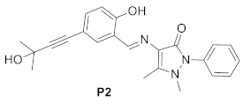
Herein, we designed and synthesized a novel antipyrine derivative P2 that employs the phenolic hydroxyl group within the molecule as a proton donor and the Schiff base structure (-CH=N-) as a proton acceptor. Based on the excited-state intramolecular proton transfer (ESIPT) effect, it achieves precise recognition of zinc ions; meanwhile, the integration of the methyl butynol structure within the molecule extends the conjugated region of the compound, serving as a zinc ion probe with heightened sensitivity.

2. Materials and Methods

2.1. Instruments and Reagents

In this study, we used FA1004 Electronic Analytical Balance (Shanghai Liangping Instrument & Meter Co., Ltd., Shanghai, China), DF-101Z Heat Collection Constant Temperature Heating Magnetic Stirrer (Jintan Fuhua Instrument Co., Ltd., Changzhou, China), RE-201 Rotary Evaporator (Shanghai Hongguan Instrument Equipment Co., Ltd., Shanghai, China), DZG-6020 Vacuum Drying Oven (Shanghai Senxin Laboratory Instrument Co., Ltd., Shanghai, China), and F-4600 Fluorescence Spectrophotometer (Hitachi Technology Corporation, Tokyo, Japan). Nuclear magnetic resonance spectra were obtained using a Bruker AV 400 NMR instrument, and FT-IR spectra were recorded on a Bruker Tensor 27 spectrophotometer (Bruker Corporation, Billerica, MA, USA).
We used 5-bromosalicylaldehyde, 2-methyl-3-butyn-2-ol, 4-aminoantipyrine, CuI, PPh3, Pd(PPh3)2Cl2, tetrahydrofuran, triethylamine, anhydrous ethanol, petroleum ether (PE), ethyl acetate (EA), and metal salts (Shanghai Aladdin Biochemical Technology Co., Ltd., Shanghai, China); all reagents used were of analytical purity.

2.2. Synthesis and Characterization of Probe P2

Synthesis of P1: Under a nitrogen atmosphere, 5-bromosalicylaldehyde (2.01 g, 10 mmol), CuI (0.038 g, 0.2 mmol), PPh3 (0.052 g, 0.2 mmol), Pd(PPh3)2Cl2 (0.07 g, 0.1 mmol), 15 mL THF, and 20 mL Et3N were added into a 50 mL three-necked flask. After stirring for 15 min, 2-methyl-3-butyn-2-ol (1.26 g, 15 mmol) was injected into the reaction mixture with a syringe. The reaction was conducted under reflux for 11 h and monitored using thin-layer chromatography until the starting material was completely consumed. Then, the reaction mixture was cooled to room temperature and filtered. The filtrate was concentrated to obtain a black oily substance and purified by silica gel column chromatography (VPE:VEA = 10:1) to give 2-hydroxy-5-(3-hydroxy-3-methylbut-1-yn-1-yl)benzaldehyde (intermediate P1, 1.43 g) as a pale yellow solid, with a yield of 70.3%. 1H NMR (400 MHz, CDCl3) δ 11.08 (s, 1H), 9.85 (s, 1H), 7.65–7.54 (m, 2H), 6.94 (d, J = 8 Hz, 1H), 1.62 (s, 6H). IR(KBr), ν, cm−1 3132, 2250, 1666, 689.
Synthesis of P2: P1 (0.25 g, 1.2 mmol) and 4-aminoantipyrine (0.25 g, 1.2 mmol) were added to a 50 mL three-necked flask, followed by the addition of 25 mL of anhydrous ethanol. The reaction was carried out at 75 °C for 2.5 h. After cooling to room temperature, the reaction mixture was filtered, and the filter residue was washed with anhydrous ethanol and then recrystallized from anhydrous ethanol to obtain (E)-4-((2-hydroxy-5-(3-hydroxy-3-methylbut-1-yn-1-yl)benzylidene)amino)-1,5-dimethyl-2-phenyl-1H-pyrazol-3(2H)-one (probe P2, 0.32 g) as an orange-red crystal, with a yield of 68.4%. 1H NMR (400 MHz, DMSO-d6) δ 13.03 (s, 1H), 9.66 (s, 1H), 7.62–7.49 (m, 3H), 7.45–7.34 (m, 3H), 7.31 (dd, J = 8.5, 2.1 Hz, 1H), 6.90 (d, J = 8.5 Hz, 1H), 5.43 (s, 1H), 3.22 (s, 3H), 2.41 (s, 3H), 1.46 (s, 6H). 13C NMR (101 MHz, DMSO-d6) δ 159.63, 159.47, 156.33, 150.82, 134.63, 133.71, 129.71, 127.79, 125.52, 121.16, 117.45, 114.46, 113.79, 94.86, 80.40, 64.07, 35.56, 32.17, 10.33. IR(KBr), ν, cm−1 3473, 2250, 1658, 762.

2.3. Spectral Analysis

For fluorescent spectra measurement, the excitation wavelength was set to 352 nm, and the emission spectrum was scanned from 360 nm to 650 nm, with both excitation and emission slit widths set to 10 nm.
For the preparation of stock solutions, the stock solutions of P2 (1 mM) were prepared in ethanol (EtOH), dimethyl sulfoxide (DMSO), and N,N-dimethylformamide (DMF), separately. The stock solutions of metal ions (10 mM) were prepared in deionized water.
For the solvent screening experiment, stock solutions (EtOH, DMSO, DMF) of probe P2 and Zn2+ were added into a 1 mL cuvette and diluted with the corresponding organic solvent/water mixture to form the test solution with P2 (100 μM) and Zn2+ (100 μM). The fluorescence spectra were measured 3 min later,.
For the water content test, various amounts of Zn2+ and P2 stock solutions were added into dilutes with H2O/EtOH ratios ranging from (10:0) to (0:10), diluting to the testing solution consisting of P2 (100 μM) and Zn2+ (100 μM). The fluorescence spectra were measured 3 min later to determine the optimal water content of the system.
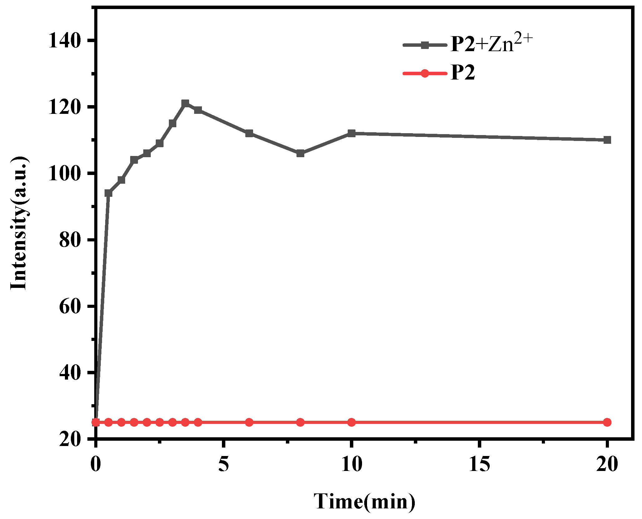
For the response time test, the Zn2+ stock solution was mixed with the probe solution and diluted with 30% water content in EtOH to be the test solution, in which the concentration of zinc ions and P2 were both 100 μM. Fluorescence spectra measurements were taken every 30 s to determine the optimal incubation time. At the same time, a blank group without zinc ions was set up to eliminate the fluorescence interference of the detection system itself.
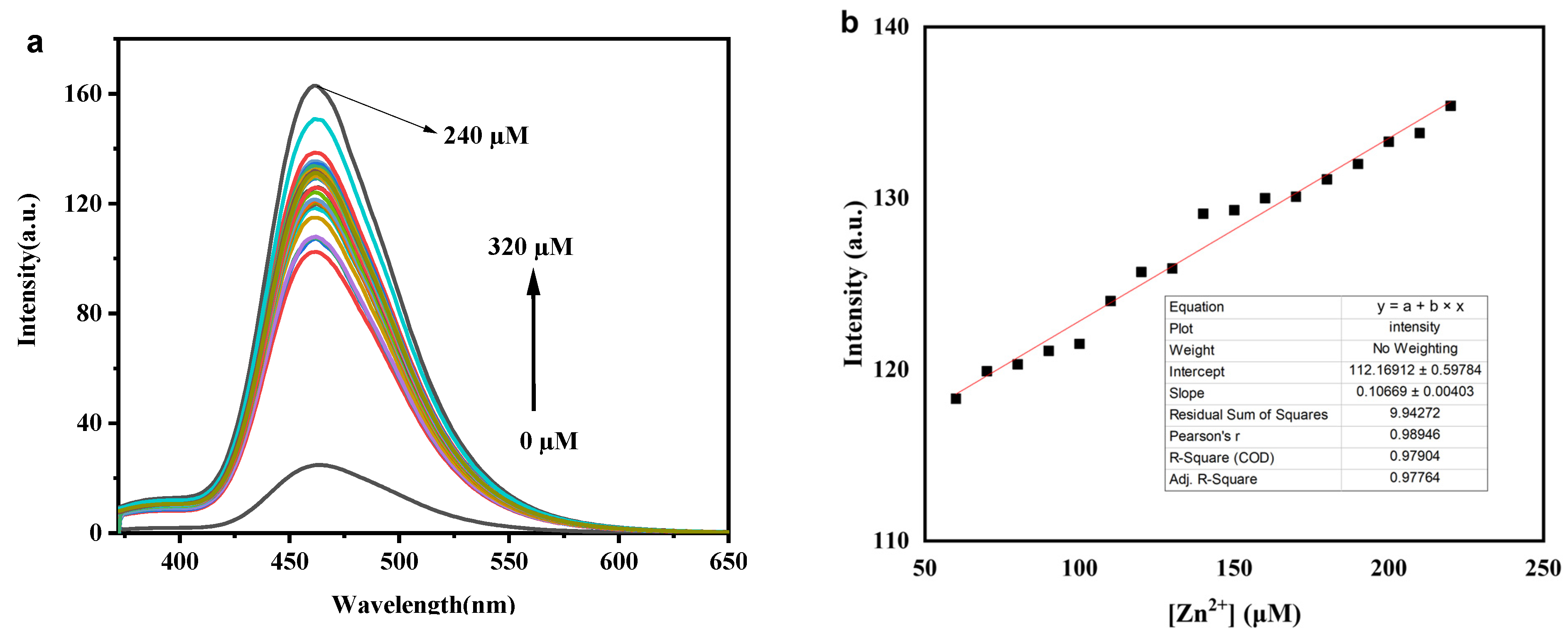
For fluorescence titration, we mixed 10 μL P2 stock solution and various amounts of Zn2+ stock solution in a 1 mL cuvette. After diluting with 30% water content in EtOH, a series of test solutions with different concentrations were prepared, where the concentration of P2 was 100 μM, and the concentration of Zn2+ ranged from 0 to 320 μM with a concentration gradient of 10 μM. The fluorescence spectra of the test solutions were analyzed 3 min later. At the same time, a fluorescence titration curve was plotted, and the limit of detection (LOD) was calculated.
For the anti-interference test, CuSO4·5H2O, MgSO4, ZnSO4, CoCl2·6H2O, Al2(SO4)3, Fe2(SO4)3·9H2O, Ce(NO3)3·6H2O, NiSO4·6H2O, Pb(NO3)2, AgNO3, Sr(NO3)2, Eu(NO3)3·6H2O, Pr(NO3)3·6H2O, Gd(NO3)3·6H2O were dissolved in deionized water to prepare separate 100 μM metal ion solutions. In a cuvette, 1 mL of interfering metal ion solution was mixed with 1 mL P2 solution, and then the fluorescence spectrum of each mixed solution as the control group was measured. Subsequently, 0.5 mL of interfering metal ion solution was mixed with 1 mL of P2 solution in a cuvette, followed by adding 0.5 mL of zinc ion solution and measuring the fluorescence spectrum.

3. Results and Discussion

3.1. Synthesis of Probe P2

The synthesis procedure of P2 is shown in Scheme 1. Similar to a previous report [30], P1 was synthesized via Sonogashira coupling, and then P1 was reacted with 4-aminoantipyrine via condensation to generate probe P2. The structural identification of intermediate P1 and probe P2 was confirmed by NMR and FT-IR spectroscopies (Figures S1–S5).

3.2. Influence of Solvents on Fluorescence Detection

Given the propensity for the luminescence of most fluorophores to be impacted by the surrounding environment, an initial assessment was conducted to evaluate the solvent’s impact on the detection system. The data presented in Figure 1 elucidate that different aqueous solvents—EtOH, DMSO, and DMF—when employed as media for the probe, result in varying levels of fluorescence response. The effectiveness of these aqueous solvents in enhancing fluorescence followed the hierarchy EtOH > DMSO > DMF. Consequently, 70% EtOH emerged as the solvent of choice due to its superior ability to enhance fluorescence, thereby optimizing the performance of the detection probe.

3.3. Influence of Water Content on Fluorescence Detection

The tolerance of the detection system to water content is crucial for assessing its suitability for in situ detection within biological systems. To evaluate this, Zn2+ ions were added to the detection system with varying water contents and allowed to stand for 3 min before measuring the fluorescence changes. A plot was created with the water content as the x-axis and the relative fluorescence intensity I/I0 at 462 nm as the y-axis. Figure 2 reveals that the relative fluorescence intensity increased with the water content up to 30%, reaching a maximum at this level. However, a further increase in water content led to a decrease in relative fluorescence intensity, which could be attributed to the reduced probe solubility in the mixed solvents with excessively high water proportions. Consequently, for subsequent detections, a 30% water content in ethanol was determined to be the optimal solvent system for the detection setup.

3.4. Influence of Incubation Time on Fluorescence Detection

Considering the importance of incubation time in sensing systems, we investigated the detection efficiency of the system toward zinc ions under different incubation durations. The concentrations of the probe and zinc ions were both 100 μM. A graph was plotted with reaction time on the x-axis and the fluorescence intensity at 462 nm on the y-axis. As seen in Figure 3, in the absence of Zn2+, the fluorescence intensity of the probe remained stable over extended reaction times, indicating that the probe was stable in a 30% water content ethanol solvent. Upon addition of Zn2+ to the system and commencement of the timing test, it was observed that the fluorescence intensity gradually increased with longer reaction times. The fluorescence intensity peaked at 3 min, with a 5-fold enhancement. Afterward, the fluorescence intensity stabilized. Based on these results, we established an optimal incubation time of 3 min for the system, which was then used as the optimal response time in subsequent experiments.

3.5. Fluorescence Titration

To establish a fluorescence titration curve, we prepared test solutions with varying concentrations of Zn2+ and conducted fluorescence spectroscopy measurements. As shown in Figure 4a, under an excitation wavelength of 352 nm, the probe alone exhibited a weak characteristic emission peak. With the addition of zinc ions, the fluorescence intensity of the probe at 462 nm gradually increased. When the concentration of zinc ions reached 240 μM, the fluorescence intensity peaked, enhancing by approximately seven times. After the zinc ion concentration exceeded 240 µM, the fluorescence intensity at 462 nm slightly decreased instead (Figure S5). Based on the fluorescence spectroscopy data, a working curve was fitted with zinc ion concentration on the x-axis and the fluorescence intensity at 462 nm on the y-axis, as shown in Figure 4b. Within the range of zinc ion concentrations from 60 to 220 μM, there is a good linear relationship (R2 = 0.9776), indicating that this detection system can be used for the quantitative detection of zinc ions.
Furthermore, the sensitivity of fluorescence detection determines the application prospects of this detection system, so we needed to test the system’s detection sensitivity toward zinc ions. Using the detection limit formula reported in the literature, LOD = 3σ/k, where σ is the standard deviation of the blank signal, and k is the slope of the calibration curve, we determined that at a signal-to-noise ratio (S/N) of three, the detection limit for zinc ions using the probe was 0.63 μM. Additionally, the performance of typical Zn2+ sensors are summarized in Table 1 in terms of the solvent system, limit of detection (LOD), and linear range. In comparison to reports in the literature, our method demonstrates superior sensitivity, a lower detection limit, and an expanded linear range. Notably, our analytical approach offers significant benefits, including straightforward operation, convenience, and cost-effectiveness.

3.6. Anti-Interference Experiments

The impact of coexisting ions on the performance of P2 is illustrated in Figure 5. Although several ions, such as Ag+, Sr2+ Al3+, and Ce4+, caused a slight increase in the fluorescence intensity at 462 nm, the addition of Zn2+ to the system resulted in a significant enhancement in fluorescence intensity. For other ions, aside from the test solutions containing Pb2+ and Gd3+, which significantly reduced the fluorescence intensity at 462 nm, noticeable fluorescence enhancements were observed with the rest of the test solutions. This indicates that the probe has a strong anti-interference capability for the recognition of Zn2+ in the presence of common metal ions.

3.7. Job’s Plot Curve and Recognition Mechanism

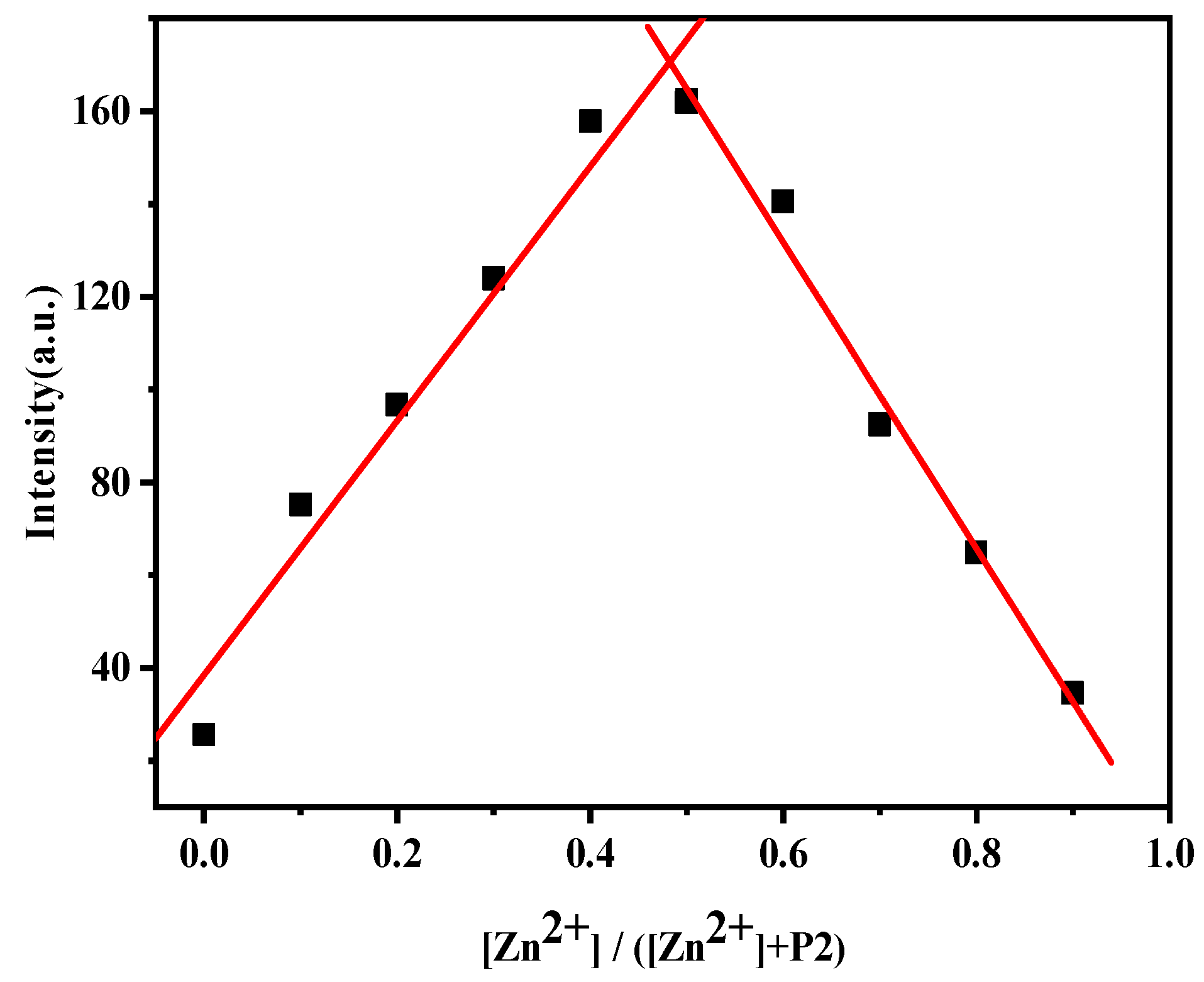
To further investigate the coordination mode between P2 and Zn2+, test solutions were prepared with a total concentration of 200 μM and a [Zn2+]/([Zn2+] + [P2]) ratio ranging from 0 to 0.9. Fluorescence spectroscopy measurements were conducted, and the fluorescence intensity at 462 nm was plotted to create a Job’s plot, as shown in Figure 6. The plot reveals that the fluorescence intensity was maximized when the coordination ratio of Zn2+ to the probe was 1:1. Based on this observation, it can be inferred that the complexation mechanism involves Zn2+ coordinating with the oxygen of the phenolic hydroxyl group, the nitrogen of the imine group, and the carbonyl group in the antipyrine structure within the probe molecule, leading to the formation of a complex with both six-membered and five-membered ring structures. In this process, on the one hand, the PET effect and ESIPT effect of P2 are simultaneously inhibited; on the other hand, the coordination of the probe with metal ions results in the Schiff base double bond being unable to rotate freely, thus inhibiting E/Z isomerization. Based on the above-mentioned synergistic effect, after the probe is excited, energy is released through fluorescence emission, which results in an enhancement in fluorescence (Figure 7).

3.8. Practical Application

The probe P2 was utilized for the determination of Zn2+ in real water samples. Tap water, Ganjiang river water, and rainwater were gathered and pretreated with the methods described in the literature [36,37]. The introduction of exogenous Zn2+ (100 μM) into these samples led to a substantial increase in fluorescent intensity. Table 2 illustrates that the recovery rates for tap water and rainwater were 95.72% and 96.30%, respectively, with corresponding relative standard deviations of 0.88% and 0.72%. These findings indicate that the fluorescence probe P2 holds promise for the monitoring of Zn2+ levels in real water samples.

4. Conclusions

In summary, this work developed an imine-based fluorescent sensor built on the antipyrine scaffold for the selective detection of Zn2+ in ethanol/water mixtures. In the presence of Zn2+, the probe P2 achieves a fluorescence turn-on through the inhibition of photoinduced electron transfer (PET), excited-state intramolecular proton transfer (ESIPT), and E/Z isomerization, along with the activation of chelation-enhanced fluorescence emission (CHEF). The probe can specifically identify Zn2+ in the presence of 13 common interfering cations, offering a broad linear detection range and a low detection limit. It was successfully applied to the quantitative determination of Zn2+ in real water samples.

Supplementary Materials

The following supporting information can be downloaded at: https://www.mdpi.com/article/10.3390/min14070649/s1, Figure S1: 1H NMR spectrum of P1; Figure S2: FT-IR spectrum of P1; Figure S3: 1H NMR spectrum of P2; Figure S4: 13C NMR spectrum of P2; Figure S5: FT-IR spectrum of P2; Figure S6: Fluorescent intensity change of P2 with Zn2+ concentration.

Author Contributions

Conceptualization, J.L. and K.L.; methodology, Y.G.; validation, Y.L. and H.Y.; formal analysis, D.C.; writing—original draft preparation, Y.G.; writing—review and editing, J.L; supervision, K.L. All authors have read and agreed to the published version of the manuscript.

Funding

This research was funded by the National Key R&D Program of China (2023YFC2908100), the National Natural Science Foundation (52064018), Distinguished Professor Program of Jinggang Scholars in Institutions of Higher Learning, Henan Province Science and Technology Research Plan (242102240016), and National College Students’ Innovation and Entrepreneurship Training Program (202110407006).

Data Availability Statement

Data are contained within the article and Supplementary Materials.

Conflicts of Interest

Yan Gao and Dezheng Chang are employees of Henan Institute of Metallurgy Co., Ltd. This paper reflects the views of the scientists and not the company.

References

  1. Jomova, K.; Makova, M.; Alomar, S.Y.; Alwasel, S.H.; Nepovimova, E.; Kuca, K.; Rhodes, C.J.; Valko, M. Essential metals in health and disease. Chem. Biol. Interact. 2022, 367, 110173. [Google Scholar] [CrossRef]
  2. Aulakh, S.K.; Varma, S.J.; Ralser, M. Metal ion availability and homeostasis as drivers of metabolic evolution and enzyme function. Curr. Opin. Genet. Dev. 2022, 77, 101987. [Google Scholar] [CrossRef]
  3. Zoroddu, M.A.; Aaseth, J.; Crisponi, G.; Medici, S.; Peana, M.; Nurchi, V.M. The essential metals for humans: A brief overview. J. Inorg. Biochem. 2019, 195, 120–129. [Google Scholar] [CrossRef]
  4. Wang, X.-Q.; Teng, Y.-F.; Ji, C.-L.; Wu, H.-F.; Li, F. Critical target identification and human health risk ranking of metal ions based on mechanism-driven modeling. Chemosphere 2022, 301, 134724. [Google Scholar] [CrossRef]
  5. Zhai, J.-Y.; Xie, X.-J.; Cherubini, T.; Bakker, E. Ionophore-Based Titrimetric Detection of Alkali Metal Ions in Serum. ACS Sens. 2017, 2, 606–612. [Google Scholar] [CrossRef]
  6. Narin, I.; Soylak, M.; Elçi, L.; Doğan, M. Determination of trace metal ions by AAS in natural water samples after preconcentration of pyrocatechol violet complexes on an activated carbon column. Talanta 2000, 52, 1041–1046. [Google Scholar] [CrossRef]
  7. Zengin, H.B.; Gürkan, R. Application of a novel poly(SMAm)-Tris-Fe3O4 nanocomposite for selective extraction and enrichment of Cu(I)/Cu(II) from beer, soft drinks and wine samples, and speciation analysis by micro-volume UV–Vis spectrophotometry. Talanta 2021, 224, 121789. [Google Scholar] [CrossRef]
  8. Smirnova, S.V.; Ilin, D.V.; Pletnev, I.V. Extraction and ICP-OES determination of heavy metals using tetrabutylammonium bromide aqueous biphasic system and oleophilic collector. Talanta 2021, 221, 121485. [Google Scholar] [CrossRef]
  9. Ali, T.A.; Abd-Elaal, A.A.; Mohamed, G.G. Screen printed ion selective electrodes based on self-assembled thiol surfactant-gold-nanoparticles for determination of Cu(II) in different water samples. Microchem. J. 2021, 160, 105693. [Google Scholar] [CrossRef]
  10. Bereza-Malcolm, L.T.; Mann, G.; Franks, A.E. Environmental sensing of heavy metals through whole cell microbial biosensors: A synthetic biology approach. ACS Synth. Biol. 2015, 4, 535–546. [Google Scholar] [CrossRef]
  11. Jiang, D.-D.; Zheng, M.-H.; Ma, X.-F.; Zhang, Y.-Z.; Jiang, S.-H.; Li, J.-H.; Zhang, C.-M.; Liu, K.-M.; Li, L.-Q. Rhodamine-anchored polyacrylamide hydrogel for fluorescent naked-eye sensing of Fe3+. Molecules 2023, 28, 6572. [Google Scholar] [CrossRef]
  12. Gao, L.; Zheng, M.-H.; Liu, H.-Y.; Xiao, J.; Gong, T.-H.; Liu, K.-M.; Li, J.-H.; Liu, J.-B. A novel HBI-based ratiometric fluorescent probe for rapid detection of trifluoroborate. RSC Adv. 2023, 13, 23812. [Google Scholar]
  13. Li, L.-Q.; Zheng, M.-H.; Yan, X.-Y.; Huang, H.; Cao, S.-X.; Liu, K.-M.; Liu, J.-B. Quantitative detection of H2O2 with a composite fluorescent probe of 8-quinoline boronic acid-Al(III). J. Photoch. Photobio. A Chem. 2022, 432, 114069. [Google Scholar] [CrossRef]
  14. Jiang, D.-D.; Zheng, M.-H.; Yan, X.-Y.; Huang, B.; Huang, H.; Gong, T.-H.; Liu, K.-M.; Liu, J.-B. A “turn-on” ESIPT fluorescence probe of 2-(aminocarbonyl)phenylboronic acid for the selective detection of Cu(II). RSC Adv. 2022, 12, 31186. [Google Scholar] [CrossRef]
  15. Ye, Q.-X.; Ren, S.-F.; Huang, H.; Duan, G.-G.; Liu, K.-M.; Liu, J.-B. Fluorescent and Colorimetric Sensors Based on the Oxidation of o-Phenylenediamine. ACS Omega 2020, 5, 20698–20706. [Google Scholar] [CrossRef]
  16. Arumugam, A.; Shanmugam, R.; Munusamy, S.; Muhammad, S.; Algarni, H.; Sekar, M. Study of the crystal architecture, optoelectronic characteristics, and nonlinear optical properties of 4-amino antipyrine Schiff bases. ACS Omega 2023, 8, 15168–15180. [Google Scholar] [CrossRef]
  17. Hassanien, A.M.; Altalhi, T.A.; Atta, A.A.; AlHazaa, A.N.; Alsawat, M.; Mersal, G.A.M.; Adam, A.M.A.; Refat, M.S. Studying spectroscopic, cyclic voltammetry, and electrical properties of novel 4-amino antipyrine derivative for photonic applications. J. Mol. Struct. 2023, 1272, 134201. [Google Scholar] [CrossRef]
  18. Manjunath, M.; Kulkarni, A.D.; Bagihalli, G.B.; Malladi, S.; Patil, S.A. Bio-important antipyrine derived Schiff bases and their transition metal complexes: Synthesis, spectroscopic characterization, antimicrobial, anthelmintic and DNA cleavage investigation. J. Mol. Struct. 2017, 1127, 314–321. [Google Scholar] [CrossRef]
  19. Kumawat, L.K.; Mergu, N.; Asif, M.; Gupta, V.K. Novel synthesized antipyrine derivative based “Naked eye” colorimetric chemosensors for Al3+ and Cr3+. Sens. Actuators B Chem. 2016, 231, 847–859. [Google Scholar] [CrossRef]
  20. Chakraborty, N.; Chakraborty, A.; Das, S. A pyrene based fluorescent turn on chemosensor for detection of Cu2+ ions with antioxidant nature. J. Lumin. 2018, 199, 302–309. [Google Scholar] [CrossRef]
  21. Yu, J.-N.; Ye, W.-C.; Jin, Y.-Y.; Park, K.; Chang, P.; Kim, C. Dual-channel detection of Cu2+ and F with a simple Schiff-based colorimetric and fluorescent sensor. Spectrochim. Acta A Mol. Biomol. Spectrosc. 2015, 136, 1649–1657. [Google Scholar]
  22. Leyana, K.S.; Kumar, S.K.A. Antipyrine derived Schiff’s base as a colorimetric probe for the rapid and selective detection of Cu2+ ions. Inorg. Chem. Commun. 2021, 134, 109037. [Google Scholar]
  23. Luxami, V.; Gupta, A.S.; Paul, K. Dansyl–antipyrine dyad as a fluorescent sensor for Cu2+ and F: Sequential XNOR logic operation. New J. Chem. 2014, 38, 2841–2846. [Google Scholar] [CrossRef]
  24. Rani, B.K.; John, S.A. Pyrene–antipyrine based highly selective and sensitive turn-on fluorescent sensor for Th(IV). New J. Chem. 2017, 41, 12131. [Google Scholar] [CrossRef]
  25. Maity, S.; Shyamal, M.; Maity, R.; Mudi, N.; Hazra, P.; Giri, P.K.; Samanta, S.S.; Pyne, S.; Misra, A. An antipyrine based fluorescent probe for distinct detection of Al3+ and Zn2+ and its AIEE behaviour. Photochem. Photobiol. Sci. 2020, 19, 681. [Google Scholar] [CrossRef] [PubMed]
  26. Mondal, A.; Ahmmed, E.; Ball, B.; Chattopadhyay, P. Rational design of a new AIE-coupled ESIPT-based multi-chromic state depend organo-luminophore with turn-on emissive response to Zn(II) in aqueous and solid-state. ChemistrySelect 2022, 7, e202103857. [Google Scholar] [CrossRef]
  27. Huang, Q.; Zhu, W.-J.; Yang, L.-L.; Li, G.; Ning, D.-H.; Yu, Y. Synthesis and properties of a new 4-aminoantipyrine Schiff-base for selective recognition of Cu2+. Chin. J. Lumin. 2013, 34, 1144–1148. [Google Scholar] [CrossRef]
  28. Lohar, S.; Sahana, A.; Banerjee, A.; Banik, A.; Mukhopadhyay, S.K.; Matalobos, J.S.; Das, D. Antipyrine based arsenate selective fluorescent probe for living cell imaging. Anal. Chem. 2013, 85, 1778–1783. [Google Scholar] [CrossRef]
  29. Chang, D.Z.; Li, J.H.; Gao, Y.; Liu, S.H.; Gen, C.C.; Yun, X.J.; Liu, H.Z.; Zhang, Y.M.; Wang, Y.R.; Guo, F.F.; et al. Compound and its Preparation Method, Application. Chinese Patent CN 117586185 A, 23 February 2024. [Google Scholar]
  30. Arsenyan, P.; Vasiljeva, J.; Domracheva, I.; Kanepe-Lapsa, I.; Gulbe, A. Selenopheno [2,3-f]coumarins: Novel scaffolds with antimetastatic activity against melanomaand breast cancer. New J. Chem. 2019, 43, 11851. [Google Scholar] [CrossRef]
  31. Gupta, V.K. Optical and electrochemical selective sensor for Zn2+ ions based on 4-(furan-2-ylmethyleneamino)-2,3-dimethyl-1-phenyl-1,2-dihydropyrazol-5-one. Electrochem. Sci. 2016, 11, 1640–1650. [Google Scholar] [CrossRef]
  32. Gogoi, A.; Samanta, S.; Das, G. A benzothiazole containing CHEF based fluorescence turn-ON sensor for Zn2+ and Cd2+ and subsequent sensing of H2PO4 and P4O74− in physiological pH. Sens. Actuators B 2014, 202, 788–794. [Google Scholar] [CrossRef]
  33. Das, B.; Dolai, M.; Dhara, A.; Ghosh, A.; Mabhai, S.; Misra, A.; Dey, S.; Jana, A. Solvent-regulated fluorimetric differentiation of Al3+ and Zn2+ using an AIE-active single sensor. J. Phys. Chem. A 2021, 125, 1490–1504. [Google Scholar] [CrossRef] [PubMed]
  34. Maity, S.; Maity, A.C.; Dias, A.K.; Bhattacharyya, N. Dual-mode chemosensor for the fluorescence detection of zinc and hypochlorite on a fluorescein backbone and its cell-imaging applications. Anal. Methods 2022, 14, 2739–2744. [Google Scholar] [CrossRef] [PubMed]
  35. Hu, Y.; Liu, Y.; Kim, G.; Jun, E.J.; Swamy, K.M.K.; Kim, Y.; Kim, S.-J.; Yoon, J. Pyrene based fluorescent probes for detecting endogenous zinc ions in live cells. Dye. Pigment. 2015, 113, 72–377. [Google Scholar] [CrossRef]
  36. Jacobson, A.R.; Dousset, S.; Andreux, F.; Baveye, P.C. Electron microprobe and synchrotron X-ray fluorescence mapping of the heterogeneous distribution of copper in high-copper vineyard soils. Environ. Sci. Technol. 2007, 41, 6343–6349. [Google Scholar] [CrossRef]
  37. Wang, Y.-D.; Li, J.-H.; Gao, Y.; Yang, Y.; Gao, Y.; Xu, Z.-F. Removal of aluminum from rare-earth leaching solutions via a complexation-precipitation process. Hydrometallurgy 2020, 191, 105220. [Google Scholar] [CrossRef]
Scheme 1. Synthesis procedure of probe P2.
Minerals 14 00649 sch001
Figure 1. Fluorescence emission spectra (λex = 352 nm) of the sensing system containing 100 μM P2, with or without 100 μM Zn2+ in indicated solvents.
Minerals 14 00649 g001
Figure 2. Influence of EtOH with different water contents on the detection effect of the probe.
Minerals 14 00649 g002
Figure 3. Relationship between reaction time and fluorescence response intensity.
Minerals 14 00649 g003
Figure 4. Titration curve of Zn2+: (a) fluorescence spectrum changes of different concentrations of Zn2+ solution (0–320 μM) added to the fluorescence detection system (λex = 352 nm); (b) the linear relationship between fluorescence intensity and concentration (60–220 μM).
Minerals 14 00649 g004
Figure 5. Fluorescence intensity changes of P2 (100 μM) upon the addition of common metal ions (50 μM): 1, none; 2, Cu2+; 3, Mg2+; 4, Al3+; 5, Co2+; 6, Ce4+; 7, Fe3+; 8, Sr2+; 9, Ni2+; 10, Pb2+; 11, Ag+; 12, Eu3+; 13, Pr3+; 14, Gd3+. Black bars: P2 treated with marked metal ions in the absence of Zn2+. Red bars: P2 treated with the marked metal ions, followed by Zn2+ (50 μM).
Minerals 14 00649 g005
Figure 6. Job’s plot curve of P2 with Zn2+.
Minerals 14 00649 g006
Figure 7. Proposed synergistic recognition mechanism of P2 of Zn2+.
Minerals 14 00649 g007
Table 1. Comparison of the previously reported Zn2+ probes with P2.
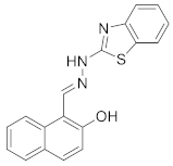
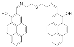
EntryProbeSolvent SystemLOD (μM)Linear Range (μM)Ref.
1Minerals 14 00649 i001CH3OH0.840–40[31]
2Minerals 14 00649 i002CH3CN/H2O (7/3)0.65-[32]
3Minerals 14 00649 i003DMSO/H2O (9/1)3.65-[33]
4Minerals 14 00649 i004CH3CN/HEPES (1/1)1.790–40[34]
5Minerals 14 00649 i005DMSO/H2O (1/1)1.24-[35]
This workMinerals 14 00649 i006EtOH/H2O (7/3)0.6360–220
Table 2. Study of the detection of Zn2+ in real water samples.
SamplespHAdded Amount
(μM)
Found Amount (μM)Std Dev
(μM)
RSD
(%, n = 3)
Recovery
(%, n = 3)
Tap water6.47100.00 95.720.350.9195.72
Ganjiang river water7.22100.00 87.770.541.0287.77
Rainwater5.84100.00 96.300.360.8896.30
Experimental conditions: Zn2+, 100 μM; concentration of P2, 100 μM; incubation time: 3 min. The pH values of water samples were determined by pH meter.
Disclaimer/Publisher’s Note: The statements, opinions and data contained in all publications are solely those of the individual author(s) and contributor(s) and not of MDPI and/or the editor(s). MDPI and/or the editor(s) disclaim responsibility for any injury to people or property resulting from any ideas, methods, instructions or products referred to in the content.