Article

Thermal Decomposition and Phase Transformation of Chrysotile in Asbestos-Containing Waste

Department of Geological and Environmental Sciences, Chonnam National University, Gwangju 61186, Republic of Korea
*
Author to whom correspondence should be addressed.
Minerals 2025, 15(4), 344; https://doi.org/10.3390/min15040344
Submission received: 17 February 2025 / Revised: 25 March 2025 / Accepted: 25 March 2025 / Published: 27 March 2025
(This article belongs to the Section Environmental Mineralogy and Biogeochemistry)

Abstract

:
In Korea, asbestos-containing waste (ACW) is disposed of in landfills. However, due to the limited landfill capacity and the potential health risks of asbestos contamination, alternative, safer disposal methods are needed. Heat treatment has been suggested as an alternative disposal method for ACW. Therefore, it is necessary to determine the optimal conditions for the thermal decomposition of chrysotile in ACW and reveal the mineralogical composition of heat-treated ACW. In this study, asbestos cement roof (ACR) and asbestos gypsum board (AGB) samples were heat-treated at 600, 700, 800, and 900 °C to identify the optimal heat treatment parameters to eliminate chrysotile fibers. The thermal, chemical, and mineralogical characteristics of the ACW were determined before and after heat treatment using multiple analytical methods. The ACR consisted of chrysotile, calcite, and ettringite, and the AGB consisted of chrysotile, gypsum, and calcite. After heat treatment at 900 °C, the ACR was mainly composed of cement component minerals and lime, while the AGB additionally contained anhydrite. SEM-EDS analysis confirmed the persistence of fibrous minerals in the ACW up to 800 °C. Furthermore, TEM-EDS analysis revealed hollow tubular morphology of chrysotile in the heat-treated ACR at up to 700 °C and in the heat-treated AGB at 600 °C. These results suggest that heat treatment at temperatures of at least 900 °C may be necessary for the complete thermal decomposition of chrysotile in ACW.

1. Introduction

Asbestos is a group of six naturally occurring hydrous silicate minerals characterized by a fibrous or needle-like habit with a fiber length of 5 μm or longer and an aspect ratio of at least 3:1 [1,2]. Asbestos has been widely used in construction materials due to its outstanding physical and chemical properties, including high extensile strength, heat insulation, and chemical resistance [3,4]. In particular, chrysotile, the most commonly used asbestos, was extensively used in construction materials such as asbestos cement roofs (ACRs) (Figure 1A–C) and asbestos gypsum boards (AGBs) (Figure 1D–F). However, between the late 1960s and early 1970s, it was found that the inhalation of asbestos fibers, which can become airborne when the material is disturbed or damaged, causes chronic diseases such as lung cancer and malignant mesothelioma [4,5], which led to asbestos being designated as a Group 1 carcinogen by the International Agency for Research on Cancer (IARC) in 1987. During Korea’s industrialization in the 1970s and 1980s, imported asbestos was also widely used in construction materials, accounting for 86%–96% of total asbestos consumption [6,7]. However, due to the severe health risks associated with asbestos exposure, in 2009, the Enforcement Decree of the Occupational Safety and Health Act was enacted, banning the import, production, and use of asbestos in Korea [8].
Since 2008, to address the health impacts associated with asbestos exposure, the Korean government has conducted an investigation of school buildings nationwide to identify buildings with asbestos-containing construction materials. This investigation found that, as of June 2018, over half of the schools in Korea (11,291 out of 20,986) contained such materials, primarily AGBs in ceilings (98%) [9]. In response, the Ministry of Education began the removal of asbestos-containing materials in schools, and by June 2022, a total area of 25,179,290 m2 had been removed. Moreover, in 2011, the government initiated a nationwide project to identify buildings containing ACRs and support their demolition. This effort identified approximately 950,000 ACR-containing buildings across the country; houses featuring ACRs were the most common, accounting for 67% of the total. By 2022, the government had demolished approximately 290,000 houses with ACRs, with a further 400,000 planned for demolition by 2033.
However, while these initiatives have reduced the number of asbestos-containing buildings, they have also generated a substantial quantity of asbestos-containing waste (ACW). In Korea, ACW is typically disposed of in landfills as “designated waste”, and the increased quantity of this waste has therefore led to a shortage of landfill capacity. The Ministry of Environment (2022) reported that, between 2017 and 2020, approximately 165,000 to 198,000 tons of ACW were disposed of annually, leaving only about 1,345,000 tons of landfill capacity as of 2022 [10]. To address this issue, the Ministry of Environment has established guidelines for expanding landfill capacity and managing the disposal of ACW, which came into effect in 2023.
The current disposal method for ACW involves packing the waste in polyethylene vinyl to prevent the release of chrysotile fibers before transporting it to landfills for burial. However, this approach poses health risks to site workers and local residents and may lead to land contamination [11,12,13]. Consequently, numerous studies have explored ways to decompose the chrysotile in ACW to avoid the need for landfilling and mitigate potential risks. Asbestos stabilizers have been applied to asbestos-containing ceiling tiles, demonstrating their ability to reduce asbestos fiber scattering and modify fiber lengths [14]. Mechanochemical treatment using a grinding mill has been used to process asbestos-containing waste (ACW), leading to the transformation of chrysotile into an amorphous phase or making asbestos inert at lower temperatures [15,16,17]. Acidic treatment has also been conducted, showing that asbestos decomposition occurs at lower temperatures compared to the traditional melting method [18,19]. However, despite its effectiveness, this method carries the risk of asbestos fiber dispersion and additional heat treatment is still required for complete decomposition. Heat treatment, however, is increasingly recognized as one of the most effective methods to decompose asbestos in ACW [13,20,21,22,23,24,25,26]. In addition to asbestos decomposition, this process also facilitates the recycling of treated ACW [24,26,27,28,29]. Notably, Durczak et al. demonstrated that clinkers produced from heat-treated asbestos–cement boards exhibit mechanical properties comparable to Portland cement, including high flexural and compressive strength [27].
Therefore, it is imperative to determine the optimal heat treatment conditions for chrysotile decomposition and examine the mineralogical characteristics of the heat-treated ACW to determine its suitability for recycling. Building on this, the present study aims to examine the mineralogical characteristics of ACW before and after heat treatment, focusing on the thermal decomposition and transformation of chrysotile fibers.

2. Materials and Methods

2.1. Materials and Heat Treatment

2.1.1. ACW and Reference Materials

ACR and AGB materials were selected as the ACW for this study. ACRs are usually made of cement and asbestos, while AGBs are made of gypsum, paper, cement, and asbestos (Table 1). The ACR samples (Figure 1B) were obtained from the roof of a warehouse in Gimje-si, Jeollabuk-do, built in 1990, whereas the AGB samples (Figure 1E) were obtained from an ACW disposal company in Jeonju-si, Jeollabuk-do.
Pure chrysotile, calcite, and gypsum were analyzed together with the ACW as references to identify the mineralogical composition of the ACW before and after heat treatment and to confirm the effect of the constituent minerals on the thermal transformation of chrysotile. Chrysotile fibers were obtained from the LAB chrysotile mine in Quebec, Canada, while calcite and gypsum were purchased from Ward’s Science (Rochester, New York, NY, USA) and Dongsung Chemicals (Seoul, Republic of Korea), respectively. Before the heat treatment, the ACR, AGB, calcite, and gypsum were pulverized to a particle size of ≤75 μm and placed in aluminum crucibles. The samples were then heated using an FX-63 electric furnace (Daihan Scientific, Wonju-si, Gangwon-do, Republic of Korea) for 2 h with a heating rate of 60 °C/min. Heat treatments were performed up to temperatures of 600, 700, 800, and 900 °C, respectively.

2.1.2. Analytical Methods

Various analytical methods were used to investigate the chemical, thermal, and mineralogical properties of the calcite, gypsum, chrysotile, and ACW samples before and after heat treatment.
The chemical composition of the ACW was determined through X-ray fluorescence (XRF) analysis using an Axios Mineral (Malvern Panalytical, Malvern, UK) instrument. The thermal properties of the ACW and the reference materials were determined using thermogravimetric and differential thermal analysis (TG-DTA) using a Thermo Plus TG-8120 (Rigaku, Tokyo, Japan) instrument. The samples were heated within the temperature range of 25 to 1200 °C in an air stream at a rate of 100 mL/min with a heating rate of 10 °C/min. The mineralogy of the calcite, gypsum, and ACW was determined before and after heat treatment through X-ray diffraction (XRD) analysis using an EMPYREAN (Malvern Panalytical, Malvern, UK) instrument with Ni-filtered Cu-Kα radiation. The measurements were performed at 40 kV and 30 mA with a step size of 0.01° in the 5 to 55° 2θ range. Additionally, Fourier-transform infrared spectroscopy (FT-IR) was employed to investigate the mineralogical changes in the ACW and reference materials. Infrared spectra were obtained at wavenumbers between 4000 and 550 cm−1 using a Frontier FT-IR/NIR Spectrum 3 (PerkinElmer, Shelton, WA, USA) instrument with KBr pellets. Additionally, polarized light microscopy (PLM) analysis was conducted before and after heat treatment using a DM750P (Leica, Wetzlar, Germany) instrument to identify the presence and type of asbestos by visually observing signs of elongation, dispersion staining color, and the extinction of the fibrous or columnar minerals in the ACW. A high-dispersion refractive index liquid (n = 1.550 HD) was used. Changes in the morphological properties of the chrysotile in the ACW following heat treatment were determined using scanning electron microscopy (SEM) equipped with energy dispersive spectrometry (EDS), which was performed using an FE-SEM S-4700 (Hitachi, Tokyo, Japan) instrument with an acceleration voltage of 15 kV, an emission current of 48 μA, and a working distance of 23.5 mm. Additionally, transmission electron microscopy (TEM) equipped with EDS was performed using a Tecnai F20 ST multifunctional TEM (FEI, Hillsboro, OR, USA) to examine changes in the structure and morphology of the chrysotile in the ACW before and after heat treatment; an acceleration voltage of 200 kV and a beam current of 1 nA were used.

3. Results and Discussion

3.1. Properties of the Reference Materials and the ACW

3.1.1. Chemical Composition of the ACW
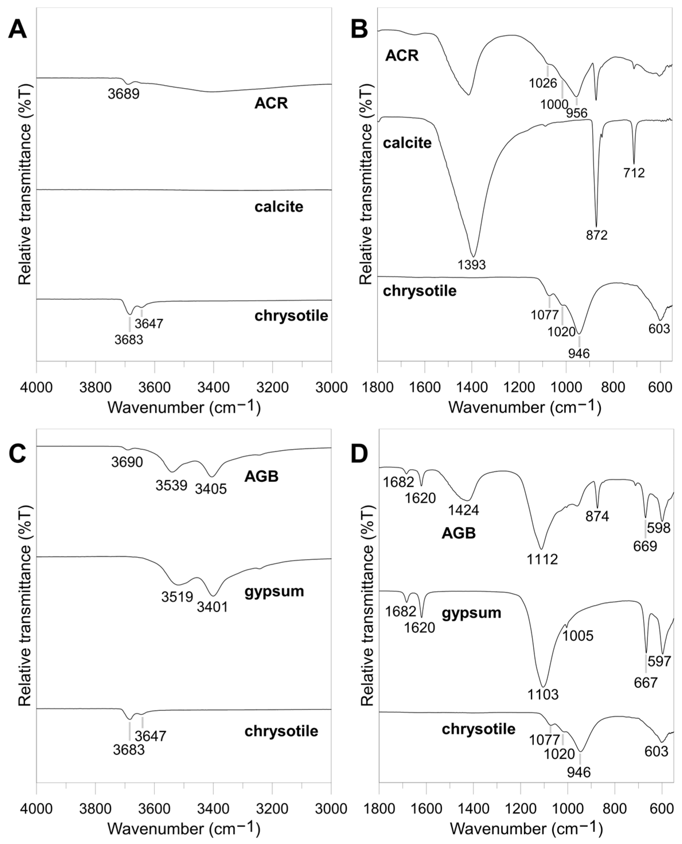
The chemical composition of the ACR and AGB samples is presented in Table 2. The ACR mainly consisted of CaO (57.9 wt.%), SiO2 (21.9 wt.%), and MgO (7.7 wt.%), with minor amounts of Al2O3 (4.5 wt.%), Fe2O3 (3.9 wt.%), etc. The AGB mainly consisted of CaO (49.3 wt.%), SO3 (33.5 wt.%), SiO2 (8.4 wt.%), and MgO (4.7 wt.%), with minor amounts of Al2O3 (1.1 wt.%), Fe2O3 (1.7 wt.%), etc. The loss on ignition is not analyzed.

3.1.2. Mineralogy

The XRD peaks of the untreated chrysotile were detected at 2θ = 12.1, 24.7, and 35.9° [30]. The FT-IR spectra of chrysotile exhibited doublet peaks at 3683 and 3647 cm−1, which are related to OH groups in the brucite layer (MgOH) of chrysotile [31] (Figure 2A,B). In the Si-O stretching region, triplet bands were observed at 1077, 1020, and 946 cm−1, which are related to SiO4. A band was also detected at 603 cm−1, which is associated with the Si-O bending vibration [32]. The XRD pattern of calcite (Figure 3A) showed the expected peaks at 2θ = 23.0, 29.4°, etc. In the FT-IR spectra of calcite (Figure 2A,B), three absorption bands were observed at ca. 1393, 872, and 712 cm−1, indicating the presence of CO32− [33]. The XRD pattern of the ACR (Figure 3A) exhibited peaks for ettringite (Ca6Al2(SO4)3(OH)12) [34] at 2θ = 9.1°, chrysotile at 2θ = 12.1°, brownmillerite (Al2Ca4Fe2O10) [21] at 2θ = 12.2, 32.8, and 34.7°, and calcite at 2θ = 29.5 and 23.0°. The FT-IR spectra of the ACR (Figure 2A,B) showed an absorption band at 3689 cm−1, indicating the presence of OH groups in chrysotile. Moreover, triplet bands were observed at 1026, 1000, and 956 cm−1, which closely match the bands for chrysotile (1077, 1020, and 946 cm−1). The absorption bands of calcite were also detected at 1393, 872, and 712 cm−1.
Figure 3B shows the XRD pattern of gypsum, exhibiting the main gypsum peaks at 2θ = 11.6, 20.7°, etc. The FT-IR spectra of gypsum (Figure 2C,D) showed absorption bands at 3519, 3401, 1682, and 1620 cm−1, which are attributed to water molecules within the crystal structure. Absorption bands were also detected at 1103, 1005, 667, and 597 cm−1, caused by SO4 tetrahedra [33,35]. The XRD pattern of the AGB (Figure 3B) showed the characteristic gypsum peaks at 2θ = 11.6, 20.7, 29.2°, etc. A chrysotile peak was also observed at 2θ = 12.1°. A quartz peak was observed at 2θ = 26.7° and a calcite peak at 2θ = 29.5°. The FT-IR spectra of the AGB (Figure 2C,D) exhibited three bands in the OH stretching vibration region, corresponding to chrysotile at 3690 cm−1 and to gypsum at 3539 and 3405 cm−1. Bands were also detected at 1682 and 1620 cm−1, associated with water molecules in the crystal structure of gypsum, along with bands at 1112, 669, and 598 cm−1, attributed to SO4 tetrahedra. Moreover, absorption bands attributed to calcite were observed at 1424 and 874 cm−1.

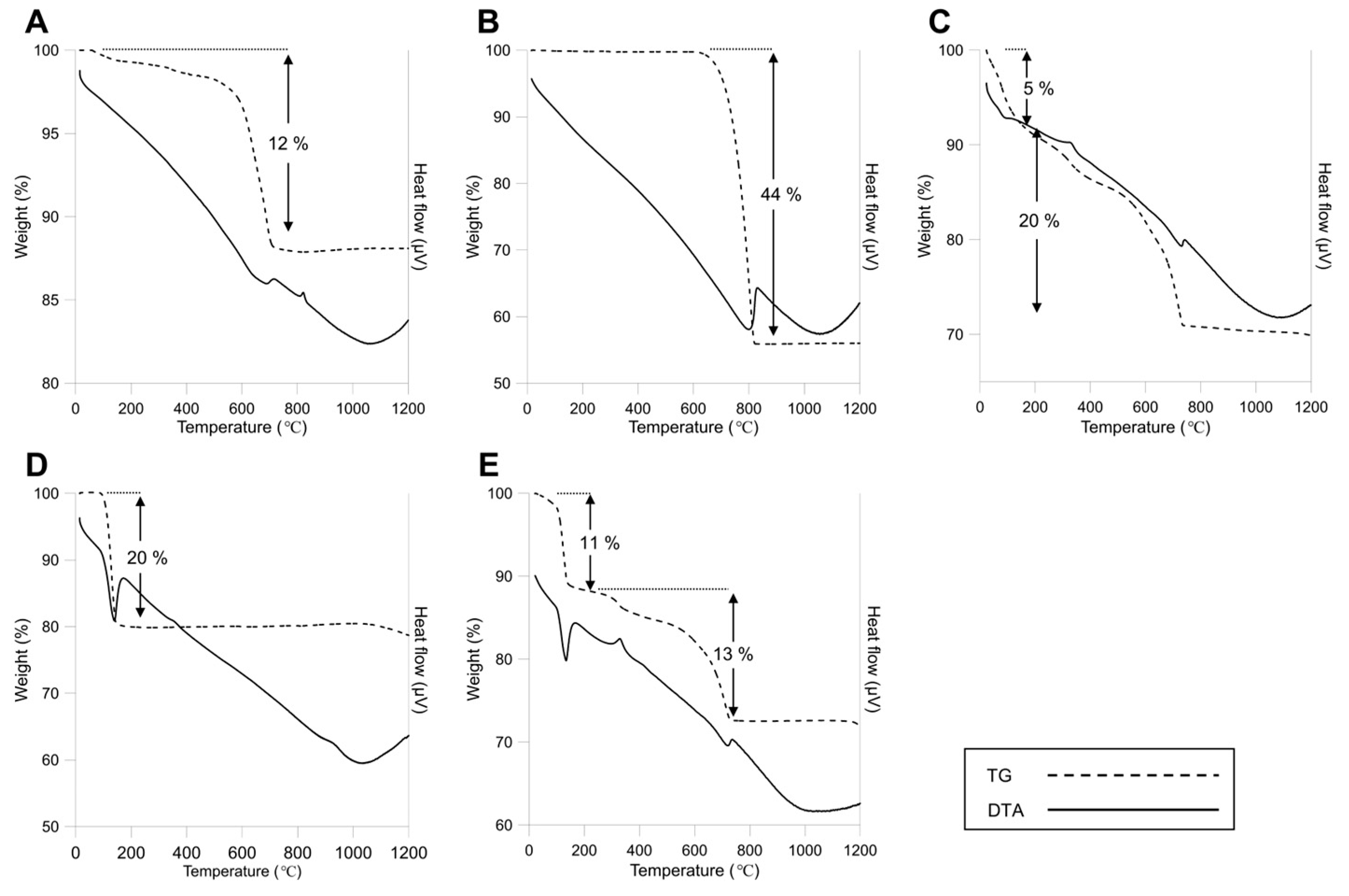
3.1.3. Thermal Characteristics of the Reference Materials and ACW

The TG-DTA curves of the chrysotile show that it underwent dehydroxylation up to a temperature of ca. 687 °C, and a weight loss of 12% was observed (Figure 4A). An exothermic peak was detected at ca. 820 °C, indicating the transformation of chrysotile into forsterite [30,36]. The TG-DTA curves of calcite (Figure 4B) show an endothermic peak at ca. 804 °C and a weight loss of 44%. This weight loss is attributed to the decomposition of calcite, which results in the emission of carbon dioxide [37]. In the TG-DTA curves of the ACR (Figure 4C), an endothermic peak was observed at ca. 97 °C, accompanied by a weight loss of 5%. This can be attributed to the loss of absorbed water in cement compounds [22,38], and possibly also to the loss of H2O in ettringite [33]. Between 200 and 700 °C, a weight loss of 24% was recorded. This value exceeds the theoretical weight loss of 13% expected from the dehydroxylation of chrysotile [31] but is lower than the weight loss expected from the decomposition of calcite (Figure 4B). The weight loss occurred in two distinct steps. The first stage, from 200 to 400 °C, resulted in a 5% weight loss, with an exothermic event at 337 °C, possibly due to the decomposition of organic reinforcement materials [23]. The second stage, from 400 to 700 °C, resulted in a 15% loss, primarily attributed to the decomposition of calcite [24,37,38], whose crystallinity is reduced by the manufacturing process or weathering [22,39], with additional contributions from the dehydration and dehydroxylation of chrysotile [36]. Furthermore, Portland cement also contributes to the weight loss through its decomposition [40].
The TG-DTA curves of gypsum (Figure 4D) revealed that dehydration occurred at approximately 139 °C, resulting in a weight loss of 20% that can be attributed to the dehydration of gypsum. Additionally, an exothermic peak was observed at around 330 °C, corresponding to the recrystallization of gypsum into β-anhydrite, with no associated weight loss. In the TG-DTA curves of the AGB, an endothermic peak was recorded at approximately 133 °C, resulting in a weight loss of 11% (Figure 4E). While this weight loss is smaller than that typically observed for pure gypsum, the temperature of the endothermic reaction was nearly identical to that observed for pure gypsum. The exothermic peak at 330 °C, which indicates recrystallization of gypsum into β-anhydrite. From 400 to 700 °C, a weight loss of 13% was observed, which can be attributed to the dehydration and dehydroxylation of chrysotile and the decomposition of calcite.

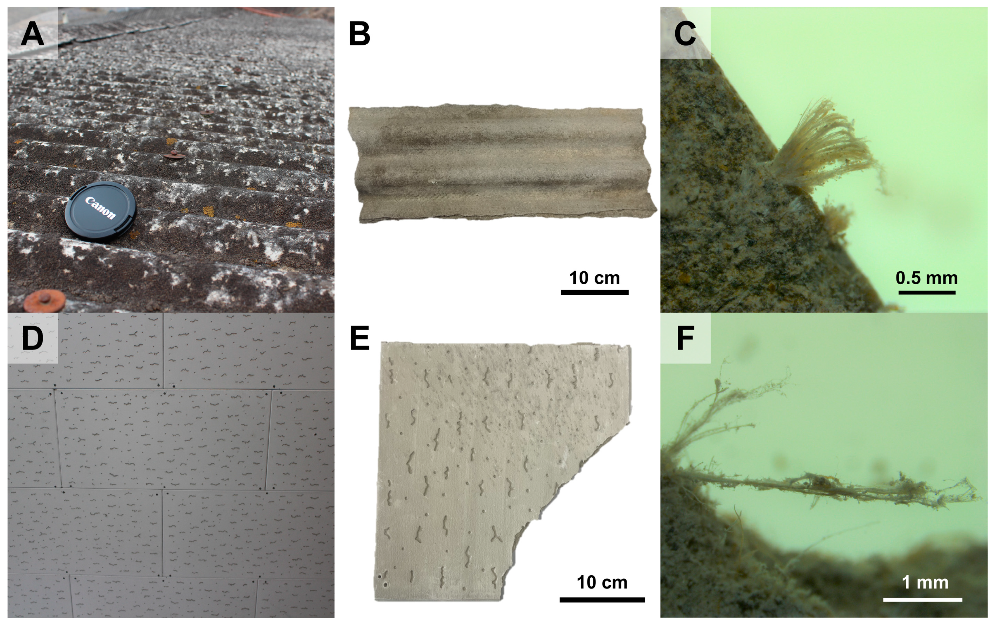
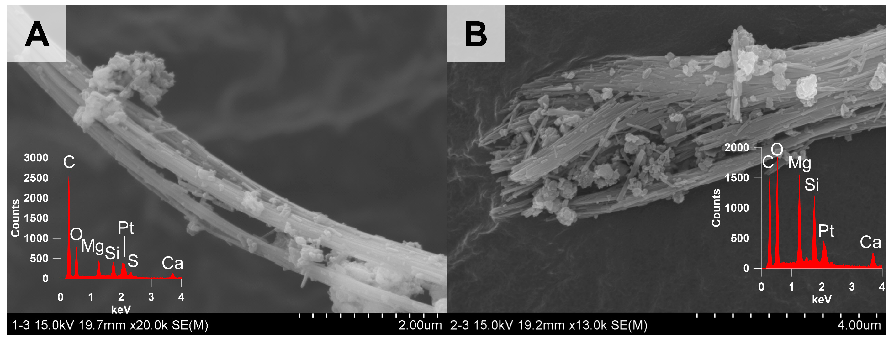
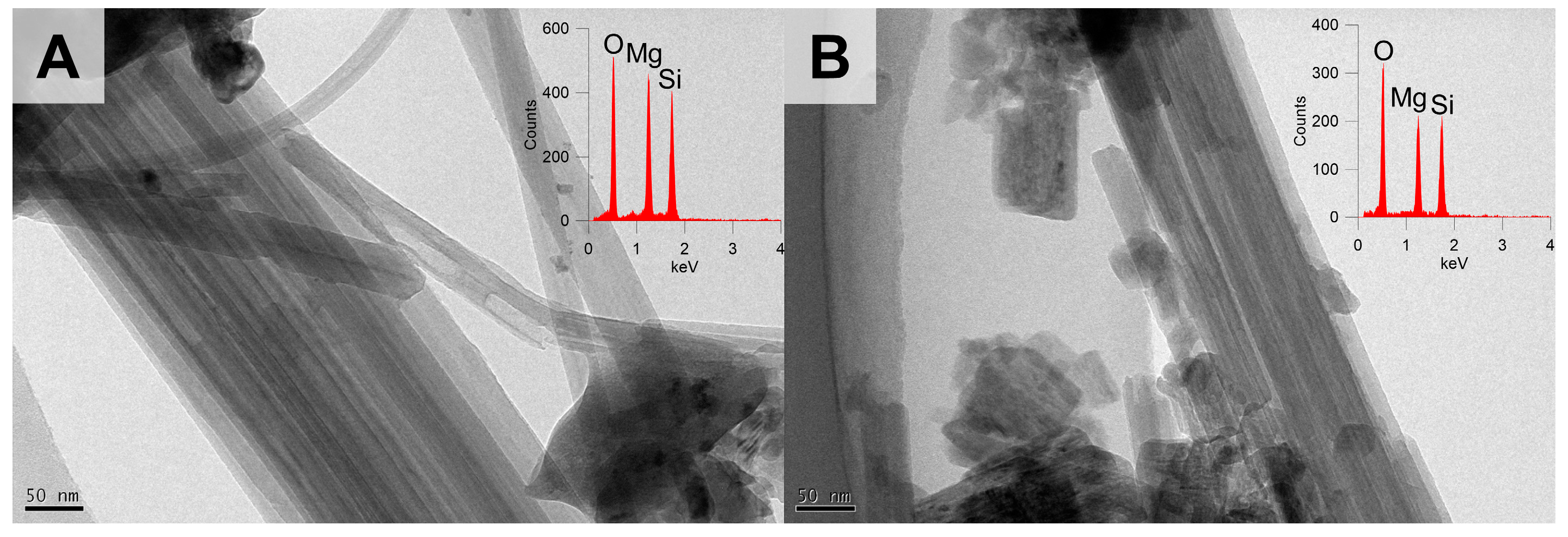
3.1.4. Optical, Morphological, and Structural Properties of the Chrysotile in ACW

The PLM images of the ACR (Figure S1) and AGB (Figure 5) confirmed the presence of chrysotile. Fibrous materials were widely observed in the ACR and AGB. These materials can be assumed to be chrysotile based on the following observations. When the retardation plate was inserted, this material appeared orange when placed in the northwestern orientation and blue when placed in the northeastern orientation (Figure S1A,D and Figure 5A,D), indicating a positive elongation sign [41,42], which is indicative of a higher refractive index along the longitudinal direction of chrysotile. Furthermore, the material displayed a dispersion staining color of magenta when placed parallel to the polarizer (n ‖) and blue when placed perpendicular to the polarizer (n ┴) (Figure S1B,E and Figure 5B,E). These colors correspond to their refractive indices and match chrysotile’s central stop dispersion staining colors [42,43]. The material also exhibited parallel extinction under crossed polarizers (Figure S1C,F and Figure 5C,F), further confirming its identification as chrysotile.
The SEM images of chrysotile in the ACR (Figure 6A) and AGB (Figure 6B) revealed a flexible fibrous morphology. Meanwhile, in the TEM images of the chrysotile in the ACR (Figure 7A) and AGB (Figure 7B), the chrysotile fibers exhibited a translucent appearance, and hollow tubular morphology was also observed. In the EDS spectra, the magnitudes of the peaks followed the order of oxygen, magnesium, and silicon, reflecting the elemental composition of chrysotile (Mg3Si2O5(OH)4).

3.2. Changes in the Characteristics of the ACW Following Heat Treatment

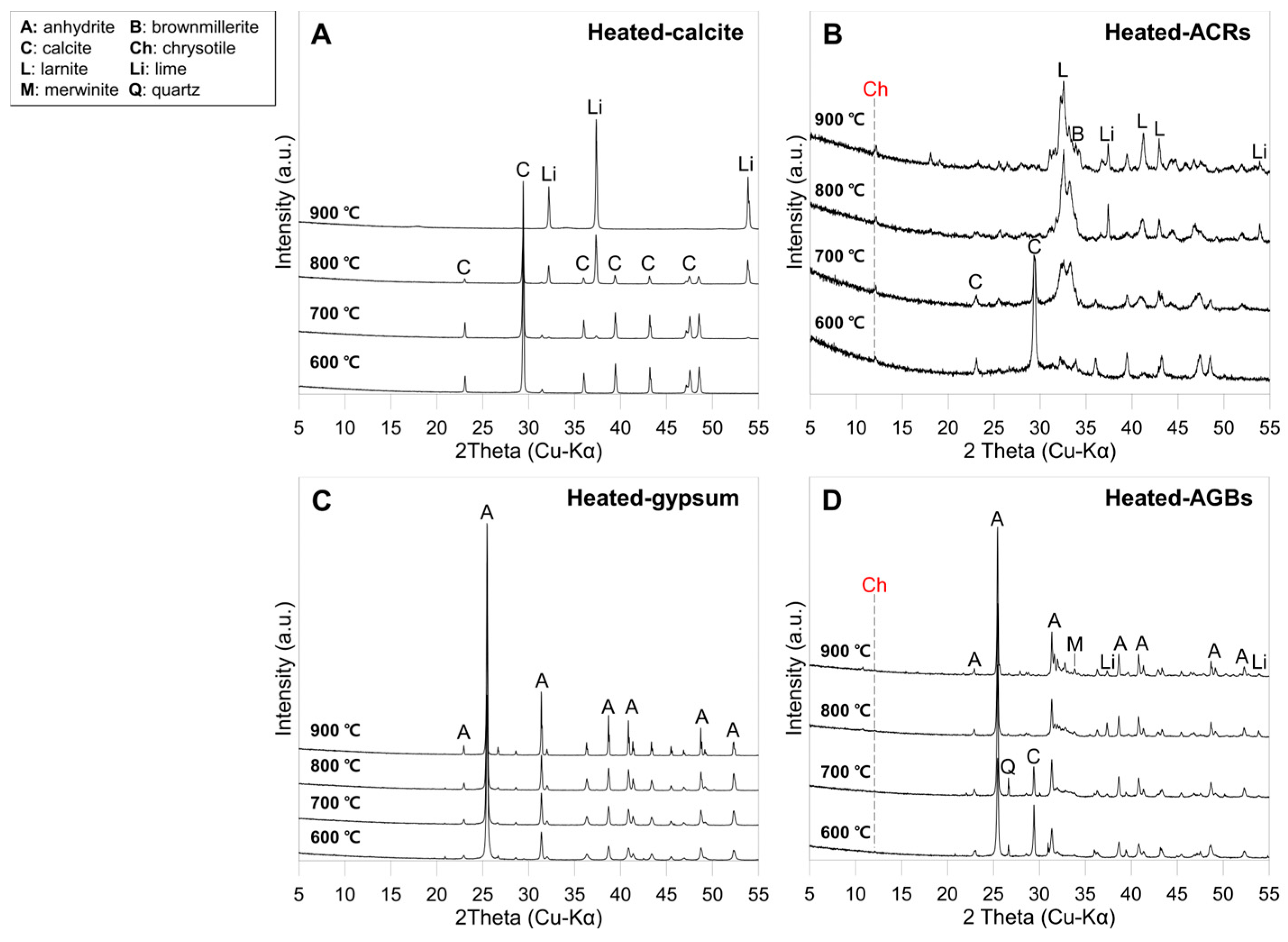
3.2.1. Mineralogical Changes in the ACW Following Heat Treatment

The XRD pattern of the chrysotile heat-treated at 600 °C exhibited a peak shift from 2θ = 12.2 to 2θ = 12.6°. After the heat treatments at 700, 800, and 900 °C, peaks were observed at 2θ = 22.9, 35.7, 36.5°, etc., corresponding to forsterite [30] (Figures S2 and S3). In the FT-IR spectra of the chrysotile heat-treated at 600 °C, an absorption band was observed at 3675 cm−1, in the OH stretching vibration region. Bands were also detected at 980, 956, 879, and 612 cm−1, associated with Si-O [30] (Figure S4). The chrysotile heat-treated at 700, 800, and 900 °C did not exhibit any bands in the OH stretching vibration region. However, in the Si-O stretching vibration region, bands were observed at 984, 873, and 840 cm−1, indicating the presence of forsterite [30] (Figure S5).
The XRD patterns of the calcite heat-treated at temperatures of 600, 700, 800, and 900 °C are shown in Figure 8A. The calcite peaks (2θ = 23.0, 29.4°, etc.) remained at temperatures of up to 800 °C but disappeared at 900 °C. At 800 °C, the peaks of lime at 2θ = 23.5, 37.4, and 53.9° were observed. The FT-IR spectra of all the heat-treated calcite samples showed three absorption bands at ca. 1393, 872, and 712 cm−1 (Figure 9A). The XRD patterns of the heat-treated ACR did not exhibit peaks corresponding to ettringite (Figure 8B), likely due to its decomposition during heat treatment as ettringite contains water in its crystal structure. In the heat-treated ACR at 600 °C, the chrysotile peak, originally observed at 2θ = 12.1° in the untreated sample, remained at a similar 2θ value. Nearby peaks, persisting up to 900 °C at 2θ = 12.2°, appear to correspond to brownmillerite [21]. The calcite peaks at 2θ = 29.5 and 23.0° were visible up to 700 °C but were replaced at 800 and 900 °C with the lime peaks at 2θ = 37.4 and 54.2°. The XRD patterns of the ACR heat-treated at 700, 800, and 900 °C exhibited the peaks of larnite at 2θ = 32.6°, etc., which is commonly found in cement [20,21,44]. The FT-IR spectra of the heat-treated ACR did not show the triplet bands of chrysotile at 1026, 1000, and 956 cm−1 (Figure 9B), whereas these bands were observed in the untreated ACR. This change indicates the absence of chrysotile or the collapse of its structure in the heat-treated ACR. In the ACR, the absorption bands of calcite at 1393, 872, and 712 cm−1 were detected up to 700 °C. Moreover, absorption bands at 993 and 845 cm−1 that first appeared at 800 °C and were more prominent at 900 °C are attributed to the presence of larnite [45].
The XRD patterns of the heat-treated gypsum (Figure 8C) show the peaks of anhydrite at 2θ = 25.5 and 31.4°, which were observed at all temperatures. In the FT-IR spectra of the heat-treated gypsum, no absorption bands were observed in the OH stretching area due to the dehydration of gypsum and the dehydroxylation of chrysotile (Figure 4A,D,E). However, the absorption bands at 1103, 1005, and 667 cm−1, caused by SO4 tetrahedra, persisted up to 900 °C (Figure 9C). The peak at 597 cm−1 that was observed in the untreated gypsum (Figure 2D) splits into a double band at 611 and 592 cm−1 in all heat treatments, indicating the presence of anhydrite [46]. The XRD patterns of the heat-treated AGB at 600 °C exhibited a weak chrysotile peak, which disappeared after heat treatment at 700, 800, and 900 °C (Figure 8D). Moreover, the XRD patterns of the heat-treated AGB did not exhibit gypsum peaks due to the dehydration of this mineral, which occurs at 139 °C. Instead, anhydrite peaks at 2θ = 25.5 and 23.0° were observed in the heat-treated AGB up to 900 °C (Figure 8D). The presence of anhydrite in all the heat-treated gypsum samples was also confirmed by XRD (Figure 8C) and FT-IR (Figure 9C). Additionally, in the AGB samples heat-treated at 800 °C, the calcite peaks disappeared due to the decomposition of this mineral and were replaced by lime peaks at 2θ = 37.4 and 53.9°. Furthermore, in the samples heat-treated at 800 and 900 °C, merwinite (Ca3Mg(SiO4)2) [21] peaks were detected at 2θ = 33.8 and 47.8°. This suggests the presence of magnesium, which may have originated from the decomposition of chrysotile. The FT-IR spectra of the heat-treated AGB did not exhibit absorption bands in the OH stretching vibration region, suggesting that chrysotile and gypsum (which were present in the untreated AGB) underwent dehydroxylation and dehydration, respectively (Figure 4A,D,E). The presence of calcite up to 700 °C was revealed by the absorption bands at 1424 and 874 cm−1. Additionally, all heat-treated AGB samples exhibited absorption bands attributed to SO4 tetrahedra at 1112 and 669 cm−1. In the heat-treated samples, the band at 597 cm−1 that was observed in the untreated gypsum (Figure 2D) splits into a double band at 612 and 594 cm−1 (Figure 9D), similar to the FT-IR spectra of heat-treated gypsum (Figure 9C), which suggests the presence of anhydrite in the heat-treated samples.
Chrysotile peaks were detected in the heat-treated ACR and AGB at 600 °C, suggesting that the dehydroxylation process remains incomplete at this temperature (Figure 4A,C,E). Chrysotile was not detected in the samples heat-treated at 700, 800, and 900 °C. Instead, its thermal decomposition resulted in the formation of minerals containing calcium, magnesium, or silicon such as larnite and merwinite. Notably, the heat-treated ACW was not found to contain forsterite—a mineral typically formed during the heat treatment of chrysotile at around 820 °C—in ACW. This absence suggests that constituent minerals such as calcite, gypsum, and quartz may prevent the transformation of chrysotile into forsterite [21,22,47]. In particular, Belardi and Piga stated that the presence of calcite leads to the formation of larnite [47]:
M g 3 S i 2 O 5 ( O H ) 4 + 4 C a C O 3 2 C a 2 S i O 4 + 3 M g O + 2 H 2 O + 4 C O 2

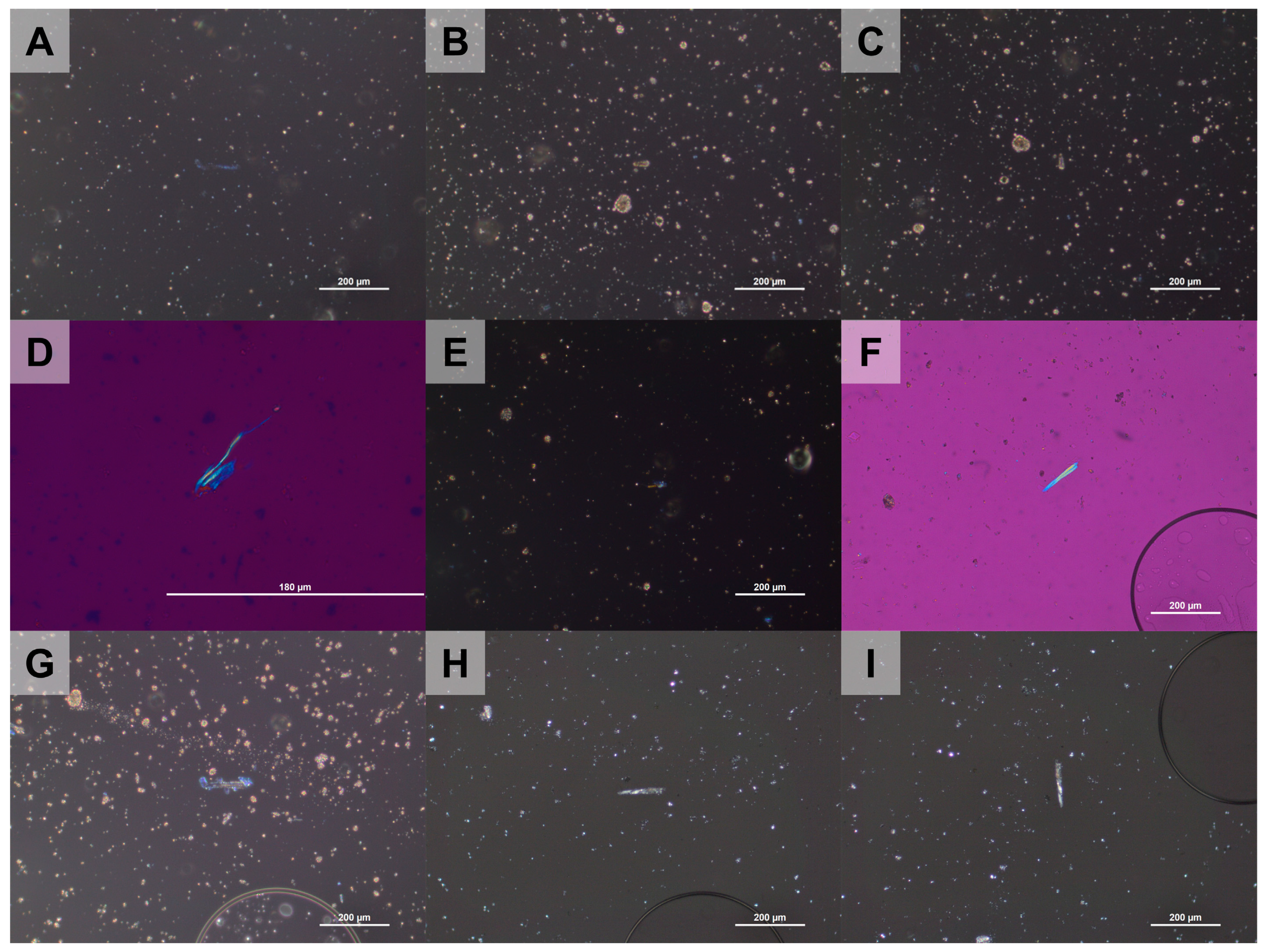
3.2.2. Changes in the Optical Properties of Chrysotile in the Heat-Treated ACW

In the ACR heat-treated at 600 °C, the fibrous minerals exhibited the same signs of elongation and extinction as pure chrysotile. However, the dispersion staining color was blue when placed parallel to the polarizer (n ‖) (Figure 10A), rather than the magenta that is expected for chrysotile [41,42,43], and the blue coloring persisted when placed perpendicular to the polarizer (n ┴). Therefore, chrysotile was not detected in the heat-treated ACR at 600 °C. In the ACR samples heat-treated at 700 and 800 °C, the elongation and extinction properties of the columnar mineral did not change and its dispersion staining color was yellow (n ‖ and n ┴) (Figure 10B,C). In the sample heat-treated at 900 °C, no fibrous or columnar minerals were observed, and amorphous minerals dominated. This was likely due to the detection limits of PLM, as minerals smaller than 0.2 µm are difficult to observe [48,49], along with the presence of a substantial cementitious amorphous phase [20]. In the AGB heat-treated at 600 and 700 °C, the elongation signs of the fibrous minerals were partially different from those of the untreated sample, with a yellow coloration in the interior of the minerals (Figure 10D) and a yellow dispersion staining (n ‖) (Figure 10E). In the AGB heat-treated at 800 and 900 °C, the elongation signs of the fibrous and columnar minerals were different from those of the untreated sample (Figure 10F); additionally, the dispersion staining color changed to yellow (n ‖) (Figure 10G) and the minerals no longer showed extinction upon rotation (Figure 10H,I), confirming the absence of chrysotile.
The PLM analyses revealed the presence of fibrous and columnar minerals in the heat-treated ACW samples. However, the optical properties of these minerals were not identical to those of chrysotile (Figure 10). Furthermore, the changes in the optical properties of the minerals caused by the heat treatment did not follow a distinct pattern. Additionally, it is unclear whether these minerals were originally chrysotile prior to the heat treatment, as ACW consists of various minerals (Section 3.1.2). Consequently, further electron microscope analyses were performed (Section 3.2.3 and Section 3.2.4) to determine the origins and characteristics of these minerals.

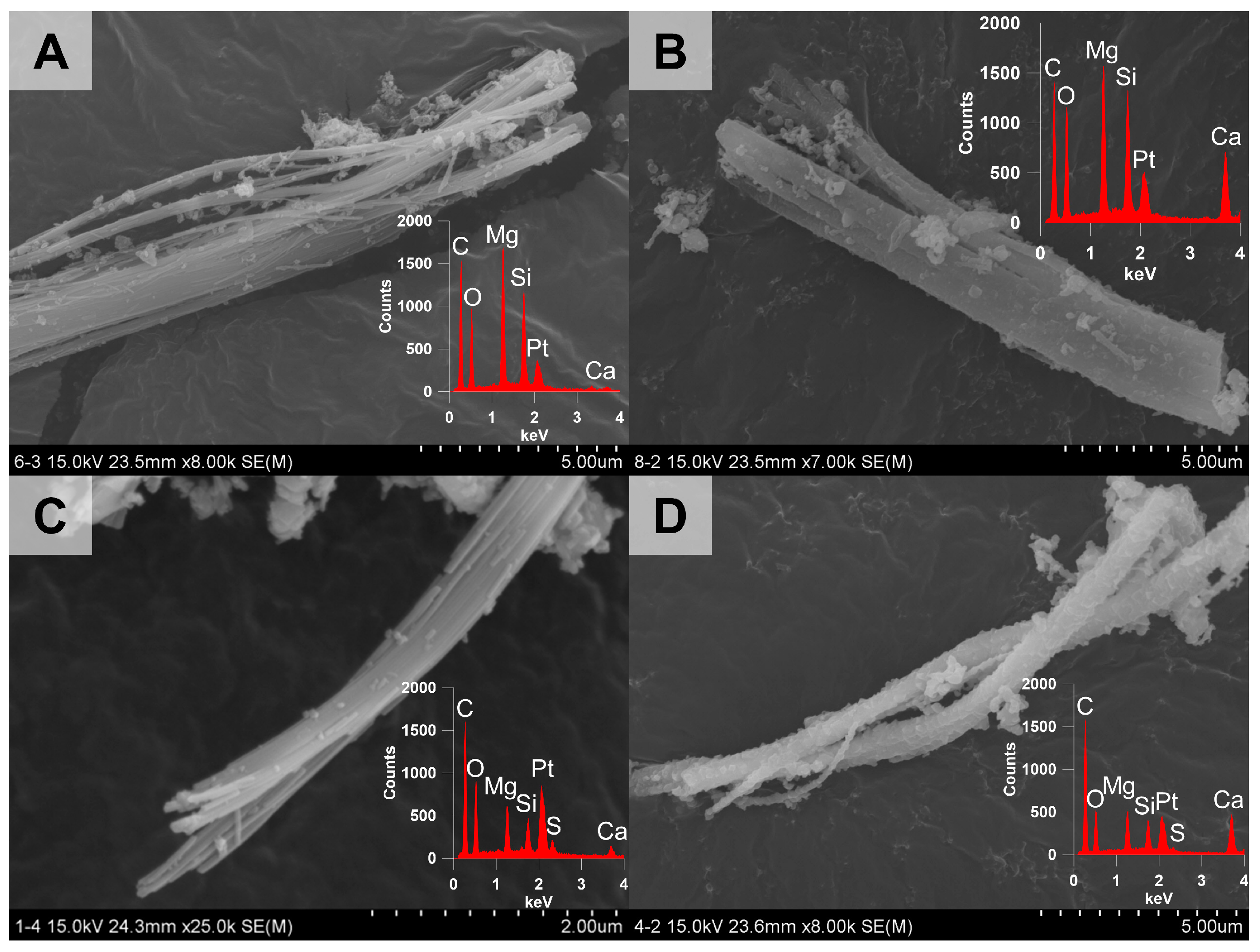
3.2.3. Morphological Changes of Chrysotile in the Heat-Treated ACW

The SEM images of the ACR heat-treated at 600 °C exhibited fibrous mineral bundles that resembled chrysotile. Furthermore, the EDS analysis confirmed that the elemental composition of these minerals was identical to that of chrysotile except for the presence of calcium and sulfur (Figure S6A), which may originate from the cement in the ACW. Fibrous minerals with hollow tabular morphology were also observed in the ACR heat-treated at 700 °C (Figure 11A and Figure 12A). At 800 °C, fibrous minerals were still present (Figure S6B), but the hollow tubular morphology was nearly absent (Figure S7B). Additionally, in the ACR heat-treated at 900 °C, columnar minerals with smooth surfaces were observed (Figure 11B). Similarly, the SEM images of the AGB heat-treated at 600, 700, and 800 °C also showed the presence of fibrous minerals (Figure 11C and Figure S6C,D, respectively), which were shown to have the same elemental composition as chrysotile except for the calcium and sulfur (Figure 6A,B). In the AGB sample heat-treated at 900 °C, minerals with collapsed cleavage were observed, showing a chemical composition similar to those at lower heat-treatment temperatures (Figure 11D).
The XRD and FT-IR analysis revealed the absence of chrysotile in the heat-treated ACW at 700, 800, and 900 °C. However, SEM-EDS revealed the presence of chrysotile in the ACW heat-treated at temperatures of 600, 700, and 800 °C. From these findings, it can be concluded that heat treatments at 600, 700, and 800 °C are insufficient to cause the complete thermal decomposition of chrysotile in ACW.

3.2.4. Structural Changes of Chrysotile in the Heat-Treated ACW

The TEM contrast images of the ACR heat-treated at 600 °C showed translucent fibrous minerals (Figure 12A). In the ACR heat-treated at 700 °C, these translucent fibrous minerals remained present (Figure S7A). In contrast, TEM images of the ACR heat-treated at 800 °C (Figure S7B) and 900 °C (Figure 12B) exhibited lumpy-shaped columnar minerals. These minerals were much more opaque than the chrysotile in the untreated ACR sample, indicating the destruction of the original structure of the chrysotile during heat treatment. Additionally, the EDS spectra of some columnar minerals in the ACR heat-treated at 900 °C exhibited a magnesium peak that was lower than the silicon peak (Figure 12B), which also indicates the collapse of the chrysotile structure. Meanwhile, the TEM contrast images of the AGB heat-treated at 600 °C showed fibrous minerals (Figure 12C), as well as a translucent appearance similar to the minerals observed in the ACR heat-treated at 600 °C. In the heat-treated AGB, lumpy-shaped columnar minerals were observed at 700, 800, and 900 °C (Figure 12D and Figure S7C,D). In the AGB heat-treated at 900 °C, the lumpy columnar minerals with rounded nodes appeared very dark in the TEM contrast images, indicating the destruction of the chrysotile structure. The minerals in all heated AGB samples consisted of magnesium, silicon, oxygen, etc., which are constituent elements of the AGB material (Table 2).
The TEM-EDS analyses demonstrated that the heat treatment at 900 °C almost completely decomposed the chrysotile in the ACW, as indicated by the absence of residual fibrous minerals. This suggests that the columnar minerals that remained after heat treatment at 900 °C were not chrysotile.

4. Conclusions

This study determined the mineralogical composition of ACW following heat treatment at various temperatures. SEM-EDS and TEM-EDS analyses demonstrated that a minimum temperature of 900 °C is required for the thermal decomposition of chrysotile in the ACW. Additionally, constituent minerals of the ACW prevent the transformation of chrysotile into forsterite and lead to the formation of cement minerals, anhydrite, and lime. These findings provide valuable information for the safe disposal and recycling of ACW to ensure human health and environmental safety.

Supplementary Materials

The following supporting information can be downloaded at: https://www.mdpi.com/article/10.3390/min15040344/s1. Figure S1. Polarized light microscopy (PLM) images of chrysotile in the asbestos cement roof (ACR) sample. (A,D) Chrysotile appears orange in the northwestern orientation (\) and blue in the northeastern orientation (/). (B,E) Chrysotile appears magenta when placed parallel (n ‖) to the polarizer and blue when placed perpendicular (n ┴). (C,F) Chrysotile shows parallel extinction; Figure S2. XRD patterns of untreated (25 °C) and heat-treated (600 and 650 °C) chrysotile [30]; Figure S3. XRD patterns of heat-treated chrysotile at 700, 800, 900, 1000, 1100, 1200, and 1300 °C [30]; Figure S4. Fourier-transform infrared spectroscopy (FT-IR) spectra of untreated (25 °C) and heat-treated (600 and 650 °C) chrysotile [30]; Figure S5. FT-IR spectra of heat-treated (700, 800, 900, 1000, 1100, 1200, and 1300 °C) chrysotile [30]; Figure S6. Scanning electron microscopy (SEM) images of minerals in heat-treated asbestos-containing waste (ACW). (A,B) Chrysotile in ACR samples heat-treated at 600 and 800 °C, respectively. (C,D) Chrysotile in AGB heat-treated samples at 700 and 800 °C, respectively. [C: carbon; Ca: calcium; Mg: magnesium; O: oxygen; Pt: platinum; S: sulfur; Si: silicon]; Figure S7. Transmission electron microscopy (TEM) images with EDS spectra of minerals in the heat-treated ACW. (A) Hollow tubular morphology of chrysotile that was commonly observed in the ACR heat-treated at 600 °C. (B–D) Lumpy columnar minerals observed in the ACR heat-treated at 800 °C, and in the AGB heat-treated at 800 and 900 °C, respectively. [C: carbon; Ca: calcium; Mg: magnesium; O: oxygen; S: sulfur; Si: silicon].

Author Contributions

Conceptualization, C.K. and Y.R.; methodology, C.K. and Y.R.; investigation, C.K. and Y.K.; writing—original draft preparation, C.K.; writing—review and editing, Y.K. and Y.R.; visualization, C.K. and Y.K.; supervision, Y.R. All authors have read and agreed to the published version of the manuscript.

Funding

This research received no external funding.

Data Availability Statement

All data generated or analyzed during this study are included in this published article and its Supplementary Information file.

Acknowledgments

We would like to thank the researchers at the CCRF of Chonnam National University in Gwangju (XRD and FT-IR analyses), Andong National University Central Research Facilities Laboratory (TG-DTA analysis), and the Korea Basic Science Institute—Gwangju Center (SEM-EDS and TEM-EDS analyses) for their analytical support.

Conflicts of Interest

The authors declare no conflicts of interest.

Abbreviations

The following abbreviations are used in this manuscript:
ACWAsbestos-containing waste
ACRAsbestos cement roof
AGBAsbestos gypsum board
N.D.Not detected
a.u.Arbitrary unit

References

  1. Enforcement Rules of the Asbestos Safety Management Act; Act No.00503; Ministry of Environment: Sejong, Republic of Korea, 2022; Available online: https://elaw.klri.re.kr/eng_mobile/viewer.do?hseq=29423&type=part&key=39 (accessed on 2 December 2024).
  2. Asbestos Safety Management Act; Act No.18907; Ministry of Environment: Sejong, Republic of Korea, 2022; Available online: https://elaw.klri.re.kr/eng_mobile/viewer.do?hseq=60952&type=part&key=39 (accessed on 2 December 2024).
  3. Park, J.I.; Yoon, C.S.; Paik, N.W. A Study on Exposure among Asbestos Textile Workers and Estimation of Their Historical Exposures. J. Korean Soc. Occup. Environ. Hyg. 1995, 5, 16–39, (In Korean with English abstract). [Google Scholar]
  4. Virta, R.L. Worldwide Asbestos Supply and Consumption Trends from 1900 Through 2003; US Geological Survey: Reston, VA, USA, 2006; ISBN 1-4113-1167-1.
  5. Becklake, M.R. Asbestos-Related Diseases of the Lung and Other Organs: Their Epidemiology and Implications for Clinical Practice. Am. Rev. Respir. Dis. 1976, 114, 187–227. [Google Scholar] [PubMed]
  6. Choi, J.K.; Paek, D.M.; Paik, N.W. The Production, the Use, the Number of Workers and Exposure Level of Asbestos in Korea. J. Korean Soc. Occup. Environ. Hyg. 1998, 8, 242–253, (In Korean with English abstract). [Google Scholar]
  7. Roh, Y.; Jeong, H.Y.; Park, B.N.; Kim, C.W.; Kim, Y.M.; Seo, M.A.; Shin, H.S.; Kim, H.W.; Sung, Y.J. Asbestos Trend in Korea from 1918 to 2027 Using Text Mining Techniques in a Big Data Environment. Econ. Environ. Geol. 2023, 56, 457–473, (In Korean with English abstract). [Google Scholar] [CrossRef]
  8. Enforcement Decree of the Occupational Safety and Health Act; Act No.34603; Ministry of Employment and Labor: Sejong, Republic of Korea, 2009. Available online: https://elaw.klri.re.kr/eng_service/lawView.do?hseq=42061&lang=kor (accessed on 2 December 2024).
  9. 5th School Asbestos Management Manual; Ministry of Education: Sejong, Republic of Korea, 2019. (In Korean)
  10. 3rd Basic Plan For Asbestos Safety Management (2023–2027); Ministry of Environment: Sejong, Republic of Korea, 2022. (In Korean)
  11. Advanced Management and Administration Standard of Designated Waste Materials; National Institute of Environmental Research: Incheon, Republic of Korea, 2007; Volume 144. (In Korean)
  12. Promentilla, M.A.B.; Peralta, G.L. An Evaluation of Landfill Disposal of Asbestos-Containing Waste and Geothermal Residues within a Risk-Assessment Framework. J. Mater. Cycles Waste Manag. 2003, 5, 0013–0021. [Google Scholar] [CrossRef]
  13. Yoon, S.; Jeong, H.; Park, B.; Kim, Y.; Kim, H.; Park, J.; Son, B.; Kim, T.; Mun, Y.; Lee, S. Inactivation of Asbestos-Containing Slate Using High-Temperature Plasma Reactor. Korean J. Mineral. Petrol. 2020, 33, 407–417, (In Korean with English abstract). [Google Scholar] [CrossRef]
  14. Jung, H.S.; Cha, J.S.; Kim, S.M.; Lee, W.S.; Lim, H.J.; Kim, H.W. Evaluating the Efficiency of an Asbestos Stabilizer on Ceiling Tiles and the Characteristics of the Released Asbestos Fibers. J. Hazard. Mater. 2015, 300, 378–386. [Google Scholar] [CrossRef]
  15. Plescia, P.; Gizzi, D.; Benedetti, S.; Camilucci, L.; Fanizza, C.; De Simone, P.; Paglietti, F. Mechanochemical Treatment to Recycling Asbestos-Containing Waste. Waste Manag. 2003, 23, 209–218. [Google Scholar] [CrossRef]
  16. Bloise, A.; Catalano, M.; Gualtieri, A.F. Effect of Grinding on Chrysotile, Amosite and Crocidolite and Implications for Thermal Treatment. Minerals 2018, 8, 135. [Google Scholar] [CrossRef]
  17. Colangelo, F.; Cioffi, R.; Lavorgna, M.; Verdolotti, L.; De Stefano, L. Treatment and Recycling of Asbestos-Cement Containing Waste. J. Hazard. Mater. 2011, 195, 391–397. [Google Scholar] [CrossRef]
  18. Yanagisawa, K.; Kozawa, T.; Onda, A.; Kanazawa, M.; Shinohara, J.; Takanami, T.; Shiraishi, M. A Novel Decomposition Technique of Friable Asbestos by CHClF2-Decomposed Acidic Gas. J. Hazard. Mater. 2009, 163, 593–599. [Google Scholar] [CrossRef]
  19. Nam, S.-N.; Jeong, S.K.; Lim, H.J. Thermochemical Destruction of Asbestos-Containing Roofing Slate and the Feasibility of Using Recycled Waste Sulfuric Acid. J. Hazard. Mater. 2014, 265, 151–157. [Google Scholar] [CrossRef]
  20. Viani, A.; Gualtieri, A.F.; Pollastri, S.; Rinaudo, C.; Croce, A.; Urso, G. Crystal Chemistry of the High Temperature Product of Transformation of Cement-Asbestos. J. Hazard. Mater. 2013, 248, 69–80. [Google Scholar] [CrossRef]
  21. Gualtieri, A.; Cavenati, C.; Zanatto, I.; Meloni, M.; Elmi, G.; Gualtieri, M.L. The Transformation Sequence of Cement–Asbestos Slates up to 1200 °C and Safe Recycling of the Reaction Product in Stoneware Tile Mixtures. J. Hazard. Mater. 2008, 152, 563–570. [Google Scholar] [CrossRef]
  22. Kusiorowski, R.; Zaremba, T.; Piotrowski, J.; Gerle, A. Thermal Decomposition of Asbestos-Containing Materials. J. Therm. Anal. Calorim. 2013, 113, 179–188. [Google Scholar] [CrossRef]
  23. Leonelli, C.; Veronesi, P.; Boccaccini, D.N.; Rivasi, M.R.; Barbieri, L.; Andreola, F.; Lancellotti, I.; Rabitti, D.; Pellacani, G.C. Microwave Thermal Inertisation of Asbestos Containing Waste and Its Recycling in Traditional Ceramics. J. Hazard. Mater. 2006, 135, 149–155. [Google Scholar] [CrossRef]
  24. Gualtieri, A.; Tartaglia, A. Thermal Decomposition of Asbestos and Recycling in Traditional Ceramics. J. Eur. Ceram. Soc. 2000, 20, 1409–1418. [Google Scholar] [CrossRef]
  25. Zaremba, T.; Peszko, M. Investigation of the Thermal Modification of Asbestos Wastes for Potential Use in Ceramic Formulation. J. Therm. Anal. Calorim. 2008, 92, 873–877. [Google Scholar] [CrossRef]
  26. Kusiorowski, R.; Zaremba, T.; Piotrowski, J.; Podwórny, J. Utilisation of Cement-Asbestos Wastes by Thermal Treatment and the Potential Possibility Use of Obtained Product for the Clinker Bricks Manufacture. J. Mater. Sci. 2015, 50, 6757–6767. [Google Scholar] [CrossRef]
  27. Durczak, K.; Pyzalski, M.; Brylewski, T.; Juszczyk, M.; Leśniak, A.; Libura, M.; Ustinovičius, L.; Vaišnoras, M. Modern Methods of Asbestos Waste Management as Innovative Solutions for Recycling and Sustainable Cement Production. Sustainability 2024, 16, 8798. [Google Scholar] [CrossRef]
  28. Kusiorowski, R.; Zaremba, T.; Piotrowski, J. The Potential Use of Cement–Asbestos Waste in the Ceramic Masses Destined for Sintered Wall Clay Brick Manufacture. Ceram. Int. 2014, 40, 11995–12002. [Google Scholar] [CrossRef]
  29. Gualtieri, A.; Boccaletti, M. Recycling of the Product of Thermal Inertization of Cement–Asbestos for the Production of Concrete. Constr. Build. Mater. 2011, 25, 3561–3569. [Google Scholar] [CrossRef]
  30. Jeong, H.; Moon, W.; Roh, Y. Characterization of Mineralogical Changes of Chrysotile and Its Thermal Decomposition by Heat Treatment. Econ. Environ. Geol. 2016, 49, 77–88, (In Korean with English abstract). [Google Scholar] [CrossRef]
  31. Martin, C. The Thermal Decomposition of Chrysotile. Mineral. Mag. 1977, 41, 453–459. [Google Scholar] [CrossRef]
  32. Jolicoeur, C.; Duchesne, D. Infrared and Thermogravimetric Studies of the Thermal Degradation of Chrysotile Asbestos Fibers: Evidence for Matrix Effects. Can. J. Chem. 1981, 59, 1521–1526. [Google Scholar] [CrossRef]
  33. Farmer, V.C. The Infrared Spectra of Minerals; Bartholomew Press: London, UK, 1974; Volume 4. [Google Scholar]
  34. Antao, S.M.; Duane, M.J.; Hassan, I. DTA, TG, and XRD Studies of Sturmanite and Ettringite. Can. Mineral. 2002, 40, 1403–1409. [Google Scholar] [CrossRef]
  35. Hass, M.; Sutherland, G.B.B.M. The Infra-Red Spectrum and Crystal Structure of Gypsum. Proc. R. Soc. Lond. Ser. Math. Phys. Sci. 1956, 236, 427–445. [Google Scholar] [CrossRef]
  36. Bloise, A.; Kusiorowski, R.; Gualtieri, M.L.; Gualtieri, A.F. Thermal Behaviour of Mineral Fibres. In Mineral Fibres: Crystal Chemistry, Chemical-Physical Properties, Biological Interaction and Toxicity; European Mineralogical Union: London, UK, 2017; ISBN 978-0-903056-65-6. [Google Scholar]
  37. Karunadasa, K.S.; Manoratne, C.; Pitawala, H.; Rajapakse, R. Thermal Decomposition of Calcium Carbonate (Calcite Polymorph) as Examined by in-Situ High-Temperature X-Ray Powder Diffraction. J. Phys. Chem. Solids 2013, 134, 21–28. [Google Scholar] [CrossRef]
  38. Dellisanti, F.; Minguzzi, V.; Morandi, N. Experimental Results from Thermal Treatment of Asbestos Containing Materials. GeoActa 2002, 1, 2001–2002. [Google Scholar]
  39. Dias, C.M.R.; Cincotto, M.A.; Savastano Jr, H.; John, V.M. Long-Term Aging of Fiber-Cement Corrugated Sheets–The Effect of Carbonation, Leaching and Acid Rain. Cem. Concr. Compos. 2008, 30, 255–265. [Google Scholar] [CrossRef]
  40. Miriello, D.; Bloise, A.; Crisci, G.M.; Barrese, E.; Apollaro, C. Effects of Milling: A Possible Factor Influencing the Durability of Historical Mortars. Archaeometry 2010, 52, 668–679. [Google Scholar] [CrossRef]
  41. Kwon, J. Asbestos Analysis of Bulk Samples Using a Polarized Light Microscope; Occupational Safety and Health Research Institute: Incheon, Republic of Korea, 2009; p. 80. (In Korean) [Google Scholar]
  42. U.S. Environmental Protection Agency. Interim Method for the Determination of Asbestos in Bulk Insulation Sample; EPA-600/M4-82-020; BiblioGov: Cincinanati, OH, USA, 1982. [Google Scholar]
  43. Perkins, R.L.; Harvey, B.W. Test Method: Method for the Determination of Asbestos in Bulk Building Materials. Available online: https://nepis.epa.gov/Exe/ZyPURL.cgi?Dockey=9100TKSO (accessed on 12 February 2025).
  44. Taylor, H. Cement Chemistry, 2nd ed.; Thomas Telford: London, UK, 1997. [Google Scholar]
  45. Vijayakumar, N.; Venkatraman, S.K.; Choudhary, R.; Indurkar, A.; Chatterjee, A.; Abraham, J.; Ostrovskiy, S.; Senatov, F.; Locs, J.; Swamiappan, S. Conversion of Biowaste into Larnite by Sol-gel Combustion Route for Biomedical Applications. ChemistrySelect 2022, 7, e202103783. [Google Scholar] [CrossRef]
  46. Liu, Y.; Wang, A.; Freemen, J. Raman, MIR, and NIR Spectroscopic Study of Calcium Sulfates: Gypsum, Bassanite, and Anhydrite. In Proceedings of the 40th Lunar and Planetary Science Conference, The Woodlands, TX, USA, 23–27 March 2009; p. 2128. [Google Scholar]
  47. Belardi, G.; Piga, L. Influence of Calcium Carbonate on the Decomposition of Asbestos Contained in End-of-Life Products. Thermochim. Acta 2013, 573, 220–228. [Google Scholar] [CrossRef]
  48. Ham, S.H.; Hwang, S.H.; Yoon, C.S.; Park, D.U. Review on asbestos analysis. J. Korean Soc. Occup. Environ. Hyg. 2009, 19, 213–232, (In Korean with English abstract). [Google Scholar]
  49. Dodson, R.F.; Hammar, S.P. Asbestos: Risk Assessment, Epidemiology, and Health Effects, 1st ed.; CRC press: Boca Raton, FL, USA, 2005; ISBN 0-429-12242-X. [Google Scholar]
Figure 1. Photographs showing (A) An asbestos cement roof (ACR). (B) A fragment of an ACR. (C) Asbestos fiber in an ACR. (D) Asbestos gypsum boards (AGBs). (E) A fragment of an AGB. (F) Asbestos fiber in an AGB.
Minerals 15 00344 g001
Figure 2. Fourier-transform infrared spectroscopy (FT-IR) spectra of untreated samples. (A,B) ACR, calcite, and chrysotile. (C,D) AGB, gypsum, and chrysotile.
Minerals 15 00344 g002
Figure 3. X-ray diffraction (XRD) spectra of untreated samples. (A) ACR and calcite; inset: magnification of the ACR spectrum focusing on 2θ = 11.0 to 14.0°. (B) AGB and gypsum; inset: magnification of the AGB spectrum focusing on 2θ = 11.0 to 14.0°. [B: brownmillerite; C: calcite; Ch: chrysotile;; E: ettringite; G: gypsum; Q: quartz].
Minerals 15 00344 g003
Figure 4. Thermogravimetric and differential thermal analysis (TG-DTA) curves of the untreated samples. (A) Chrysotile. (B) Calcite. (C) ACR. (D) Gypsum. (E) AGB.
Minerals 15 00344 g004
Figure 5. Polarized light microscopy (PLM) images of the untreated AGB sample indicating the presence of chrysotile. (A,D) With the retardation plate, the fibrous material appears orange in the northwestern orientation (\) and blue in the northeastern orientation (/), indicating a positive elongation sign. (B,E) The material appears magenta when placed parallel (n ‖) to the polarizer and blue when placed perpendicular (n ┴) to the polarizer. (C,F) The material shows parallel extinction.
Minerals 15 00344 g005
Figure 6. Scanning electron microscopy (SEM) images with energy dispersive spectrometry (EDS) spectra of chrysotile in the untreated ACW. (A) ACR. (B) AGB. [C: carbon; Ca: calcium; Mg: magnesium; O: oxygen; Pt: platinum; S: sulfur; Si: silicon].
Minerals 15 00344 g006
Figure 7. Transmission electron microscopy (TEM) images with EDS spectra of chrysotile in the untreated ACW. (A) ACR. (B) AGB. [Mg: magnesium; O: oxygen; Si: silicon].
Minerals 15 00344 g007
Figure 8. XRD patterns of heat-treated samples. (A) Calcite. (B) ACR. (C) Gypsum. (D) AGB. [A: anhydrite; B: brownmillerite; C: calcite; Ch: chrysotile; L: larnite; Li: lime; M: merwinite; Q: quartz].
Minerals 15 00344 g008
Figure 9. FT-IR spectra of the heat-treated samples. (A) Calcite. (B) ACR. (C) Gypsum. (D) AGB.
Minerals 15 00344 g009
Figure 10. PLM images of chrysotile in the heat-treated ACW. (A) Blue dispersion staining color of a columnar mineral in the ACR heat-treated at 600 °C (n ‖). (B,C) Yellow dispersion staining color of a columnar mineral in the ACR heat-treated at 700 °C (n ‖ and n ┴). (D) The elongation sign of a mineral in the AGB heat-treated at 600 °C (n ┴), which partially differed from that of the untreated sample. (E) Yellow dispersion staining color of a mineral in the AGB heat-treated at 700 °C. (F) The elongation sign of a mineral in the AGB heat-treated at 900 °C, which differed from that of the untreated sample. (G) The yellow dispersion staining color of a mineral in the AGB heat-treated at 900 °C (n ‖). (H,I) No extinction of a mineral in the AGB heat-treated at 900 °C.
Minerals 15 00344 g010
Figure 11. SEM images with EDS spectra of minerals in the heat-treated ACW. (A,C) Chrysotile in the ACR heat-treated at 700 °C and the AGB heat-treated at 600 °C, respectively. (B,D) Columnar minerals in the ACR and AGB heat-treated at 900 °C, respectively. [C: carbon; Ca: calcium; Mg: magnesium; O: oxygen; Pt: platinum; S: sulfur; Si: silicon].
Minerals 15 00344 g011
Figure 12. TEM images with EDS spectra of minerals in heat-treated ACW. (A,C) Chrysotile in the ACR heat-treated at 700 °C and AGB heat-treated at 600 °C, respectively. (B,D) Lumpy columnar minerals in the ACR heat-treated at 900 °C and the AGB heat-treated at 700 °C, respectively. [C: carbon; Ca: calcium; Mg: magnesium; O: oxygen; S: sulfur; Si: silicon].
Minerals 15 00344 g012
Table 1. The asbestos content, constituents, and uses of the asbestos-containing waste (ACW) used in this study (modified from [9]). [ACR: asbestos cement roof. AGB: asbestos gypsum board].
ACW TypeAsbestos Content (%)ConstituentsUse
ACR8–14Cement and asbestosRoofing
AGB4–6 Gypsum, asbestos, cement, and paperCeilings
Table 2. X-ray fluorescence (XRF) of the ACW (values in wt.%). [N.D.: not detected].
ACWCaOSiO2SO3MgOAl2O3Fe2O3TiO2K2OClP2O5Total
ACRs57.921.93.07.74.53.90.40.20.2N.D.99.7
AGBs49.38.433.54.71.11.70.40.2N.D.0.599.8
Disclaimer/Publisher’s Note: The statements, opinions and data contained in all publications are solely those of the individual author(s) and contributor(s) and not of MDPI and/or the editor(s). MDPI and/or the editor(s) disclaim responsibility for any injury to people or property resulting from any ideas, methods, instructions or products referred to in the content.

