Article

Formation Mechanism of Plagioclase–Amphibole and Amphibole–Spinel Symplectites in the Bijigou Layered Intrusion: Insights from Mineralogical and Crystallographic Constraints

School of Geosciences, Yangtze University, Wuhan 430100, China
*
Author to whom correspondence should be addressed.
Minerals 2025, 15(5), 433; https://doi.org/10.3390/min15050433
Submission received: 11 March 2025 / Revised: 18 April 2025 / Accepted: 18 April 2025 / Published: 22 April 2025

Abstract

:
The Bijigou layered intrusion is located in the northern margin of the Yangtze block. Based on cumulus mineral assemblages, the intrusion is divided into three major units from the base upwards: the lower zone (LZ), dominated by olivine gabbro; the middle zone (MZ), composed of gabbro and Fe-Ti oxide ore layers; and the upper zone (UZ), characterized by (quartz) diorite. Previous studies reported various vermicular symplectite textures in layered intrusions, which are thought to be related to the magmatic evolution of the layered intrusions and the mineralization of vanadium–titanium magnetite. However, detailed studies on the specific reaction mechanism of those symplectites are lacking. In this study, the characteristics, mineral compositions, and crystal orientation relationships of minerals in symplectites from Fe-Ti oxide Fe-Ti oxide-rich gabbro are in the Bijigou layered intrusion investigated by an Electron Probe Microanalyzer (EPMA) and Electron Backscattered Diffraction (EBSD) to reveal the formation process of symplectites in gabbros. In the Fe-Ti oxide-rich gabbro, abundant amphibole + spinel (Amp1 + Spl) symplectite and amphibole + plagioclase (Pl2 + Amp2) symplectite are developed between the primocryst plagioclase (Pl1) and Fe-Ti oxide; Pl2 had significantly higher An contents (An92–97) relative to Pl1. The Mg # for Amp1 and Amp2 was 0.78–1 and 0.6–0.84, respectively. Amphibole geothermometer calculations show Amp1 and Amp2 at 934–953 °C and 834–914 °C, suggesting that these symplectites crystallized at a late stage of magmatic evolution. The crystallographic orientation relationship between Amp1 and Spl varies in different areas, and Spl has a particular orientation relationship with the external Ilm. Pl2 and Amp2 inherit the crystallographic orientation of Amp1 and Pl1, respectively. We speculate that in the Bijigou layered intrusions, Amp1 + Spl and Pl2 + Amp2 were formed in two stages: Amp1 + Spl symplectite due to Ilm epitaxial growth as a result of supersaturation and rapid nucleation; and Pl2 + Amp2 symplectite due to dissolution–precipitation.

1. Introduction

Symplectite is a microstructure characterized by the fine-grained worm-like intergrowth of two or more minerals, which usually coexist in lamellae [1,2]. Symplectites and other microstructures in igneous rocks are vital records of magmatic transport, evolution, and solidification processes [3,4,5,6,7,8,9,10]. In recent years, significant progress has been made in studying the origin of symplectites. Baldwin proposed that changes in chemical potential gradient between minerals drive the formation of symplectites [11]; Keevil’s study of the Sept Iles intrusion revealed the influence of fluid activity on the microstructural evolution of symplectite through microzonation compositional analysis [7]. In terms of experimental simulation, Dharmapriya and Spruzeniece showed through experimental simulations that Na-K-rich fluids accelerate the formation of symplectite and microstructural adjustments through a dissolution–precipitation mechanism [12,13]; Yudovskaya showed through high-temperature and high-pressure experiments and numerical simulations that reactive melts led to the formation of spinel–clinopyroxene symplectite [14]; and Ashworth and Chambers proposed a new theory of symplectite formation in olivine based on the relationship between the spacing between crystals and the dynamics of the reactive interface [15]. However, there is still controversy about the formation mechanism of symplectite in layered intrusions. This study combined crystal orientation relationship analysis with mineralogical investigations on the Bijigou layered intrusion to unravel the formation mechanism of symplectites.
The Bijigou layered intrusion, hosting a large vanadium–titanium magnetite deposit, is located at the northern margin of the Yangtze block. It is the largest known basal-ultramafic layered intrusion in this area and the entire Qinling Orogenic Belt. Abundant symplectites are developed in the layered intrusions, including amphibole + plagioclase and amphibole + spinel symplectite developed between Fe-Ti oxides and plagioclase, and amphibole + plagioclase and clinopyroxene + magnetite symplectite developed between olivine and plagioclase. These symplectites are thought to be a result of reactions between Fe-rich melts and cumulus minerals formed by the immiscibility of the melts during magmatic evolution [8]. However, the mineral assemblages and developmental characteristics of these symplectites are distinctly different from those of typical structures formed by the reaction of immiscible Fe-rich melts with cumulus minerals [5,6,7,9]. On the other hand, the formation process and genesis of these symplectites have been studied mainly by analyzing the petrographic features and mineral microzonation compositions. However, there have been no mineralogical and crystallographic studies, so the detailed formation process and reaction mechanism of the symplectites are not yet clear. This study will address the information mechanism of symplectites in the Bijigou layered intrusion and provide insights into the magmatic evolution of layered intrusions and the genesis of vanadium–titanium magnetite deposits.

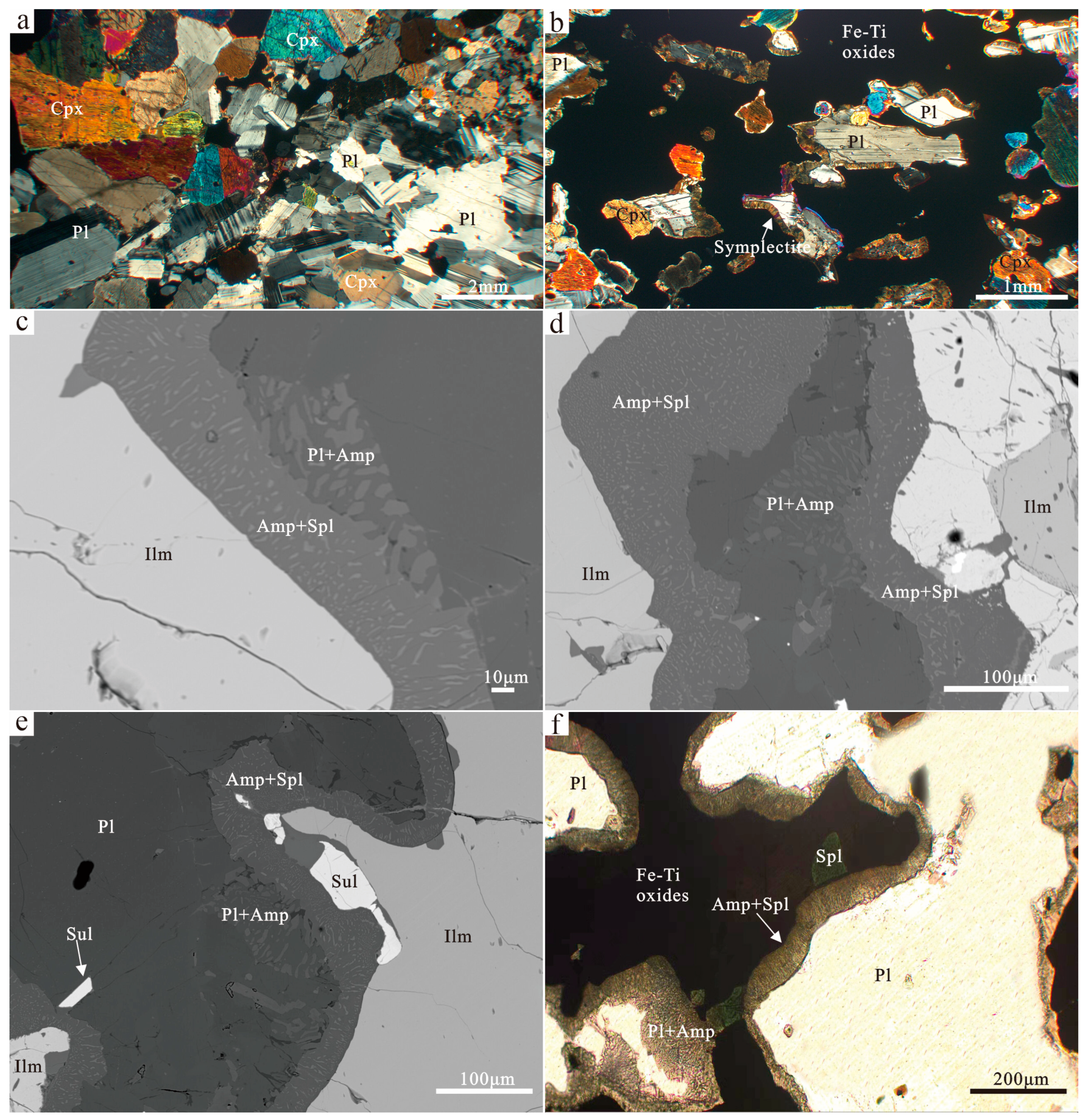
2. Geological Background and Petrography

The Yangtze block is adjacent to the Huaxia block in the southeast, the Songpan-Ganzi Orogenic Belt in the west, and the Qinling–Dabie Orogenic Belt and the southern margin of the North China Craton form the Central Orogenic Belt along the northern margin (Figure 1a). The Yangtze block has an expansive Archaean–Middle Proterozoic basement overlain by a Neoproterozoic–Cenozoic cover [16]. The Proterozoic–Mesoproterozoic basement mainly comprises metamorphic sedimentary strata and Granodioritic gneiss of the Kongling and Kangdian areas [17,18]. Late Sinian–Jurassic strata consist of carbonate rocks, terrestrial clastic rocks, and glacial deposits [16,19]. The long-term subduction of Neoarchean and the thickening of the crust along the northeastern Han-Nan Arc on the northern edge of the Yangtze block has resulted in the remelting of mafic–ultramafic layered intrusions and felsic intrusions [19,20,21,22]. Magnetite layered intrusions mainly include Bijigou, Wangjiangshan, Liushudian, and Luojiaba (Figure 1b). The Bijigou layered intrusion is the most considerable known basic–ultrabasic layered intrusion in the northern Yangtze block and even the entire Qinling Orogenic Belt, and it has an occurrence of sizeable vanadium–titanium magnetite deposits [20,23].
The Bijigou layered intrusion is about 50 km long, 6–12 km wide, and covers an area of about 500 km2. It is about 5 km thick (Figure 1b) [19,24]. The layered medium-sized and small-scale vanadium–titanium magnetite deposits form a curved mineralized zone with a total length of 5400 m. It can be divided into four mining sections from south to north: Bijigou, Zhoujiaban, Cuijiaping, and Zhantianliang (Figure 1c). The layered rock is divided into four lithofacies zones based on the mineral composition: from bottom to top, the bottom zone (BZ) is composed of brecciated gabbro; the lower zone (LZ) is mainly composed of olivine gabbro and troctolite; the middle zone (MZ) is mainly composed of gabbro and an Fe-Ti oxide ore layer; and the upper zone (UZ) is mainly composed of (quartz) diorite. The vanadium–titanium magnetite ore layer is mainly found in the central zone’s medium- to fine-grained gabbro (Figure 1d). The ore body is mainly found in a laminated, layered, or lens-like manner.
This paper mainly selects samples from the central part of the middle and upper parts of the medium-fine-grained gabbro and Fe-Ti oxide-rich gabbro for analysis. The gabbro is mainly composed of plagioclase (55–65%), clinopyroxene (15–35%), intergranular Fe-Ti oxides (<10%), and amphibole (<5%). Plagioclase and clinopyroxene are highly self-shaped, both in slabs, with a typical gabbroic texture. The size of the plagioclase grains is 0.2–3 mm (Figure 2a), and the self-shaped to semi-self-shaped clinopyroxene grains are 0.2–2.2 mm (Figure 2a). The Fe-Ti oxides are mainly distributed irregularly among the grains, and the amphiboles are irregularly distributed around the Fe-Ti oxides (Figure 2b). The Fe-Ti oxide-rich gabbro is mainly composed of plagioclase (10%~15%), clinopyroxene (10%~15%), Fe-Ti oxides (40%~55%), and a small amount of amphibole (<5%) (Figure 2b). Plagioclase is distributed in granular form, with sizes ranging from 0.2 to 2 mm. The size of clinopyroxene grains is 0.2 to 0.5 mm. Fe-Ti oxides are distributed irregularly among the cumulus plagioclase grains. Symplectite can be seen at the edge of the contact between plagioclase and Fe-Ti oxides (Figure 2b).
In the middle of the layered rock, the upper cumulus plagioclase can develop into symplectites at the contact edge with the intergranular mineral Fe-Ti oxides, especially in ore-rich areas. According to mineral composition, these symplectites can be divided into two categories: amphibole (Amp) + spinel (Spl) symplectite and plagioclase (Pl) + amphibole (Amp) symplectite (Figure 2c–f). In Amp + Spl, the spinel is distributed in the amphibole in the form of irregular, excellent particles, with the Spl measuring 1–20 μm; in Pl + Amp, on the other hand, the amphibole is 1–30 μm in size and is worm-like in shape, surrounded by plagioclase. These two types of symplectite can develop either independently or in symbiosis (Figure 2c,f). When they are adjacent to each other, Amp + Spl are close to ilmenite, and Pl + Amp are close to plagioclase (Figure 2c–e). Sometimes, the edges of Pl + Amp are connected to fishhook-like structures (Figure 2e).

3. Materials and Methods

3.1. Electron Probe Microanalyzer

The main components of amphibole, plagioclase, and spinel were tested using a JEOL JXA-8230 electron microprobe at the Key Laboratory of Mineralogy and Metallogeny of the Guangzhou Institute of Geochemistry, Chinese Academy of Sciences. The experimental conditions were an accelerating voltage of 15 kV, a test current of 20 nA, and a beam spot diameter of 1 μm. The reference materials used for amphibole analysis were Diopside (Si, Mg, and Ca), Magnetite (Fe), Potassium Feldspar (K), Sodium Feldspar (Na), Grossular (Al), Rutile (Ti), and Clinopyroxene (Mn). The plagioclase reference standards used for analysis are as follows: plagioclase (Si, Al, and Ca), Albite (Na), Potassium Feldspar (K), Rutile (Ti), Garnet (Fe and Mg), and Clinopyroxene (Mn). The reference materials used for the analysis of spinel are Diopside (Si and Ca), Olivine (Mg), Magnetite (Fe), Nickel metal (Ni), Garnet (Al), Rutile (Ti), Rose Clinopyroxene (Mn), Cr2O3 (Cr), and Vanadium metal (V). The analytical error is 2% for Si, Fe, and Mg and 5% for the other elements.

3.2. Electron Backscattered Diffraction

Electron Backscattered Diffraction (EBSD) analysis was performed using an FEI Quanta 450 FEG environmental scanning electron microscope (Thermo Fisher Scientific, Hillsboro, OR, USA) from the State Key Laboratory of Geological Processes and Mineral Resources at China University of Geosciences. The experimental conditions were as follows: low vacuum mode (30 pa), accelerating voltage 20 kV, spot size 6, working distance 23–25 mm, sample tilt 70°, and scanning step 50 μm. All samples were calibrated automatically and processed using the CHANNEL5.0 software from Oxford Instruments HKL Technology. All measurements were performed with the exclusion of erroneous, poorly oriented, and more than 5° misaligned data.

4. Results

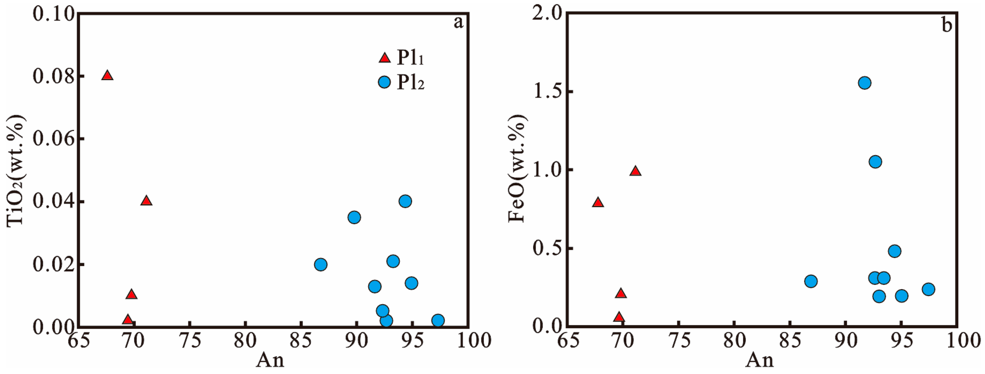
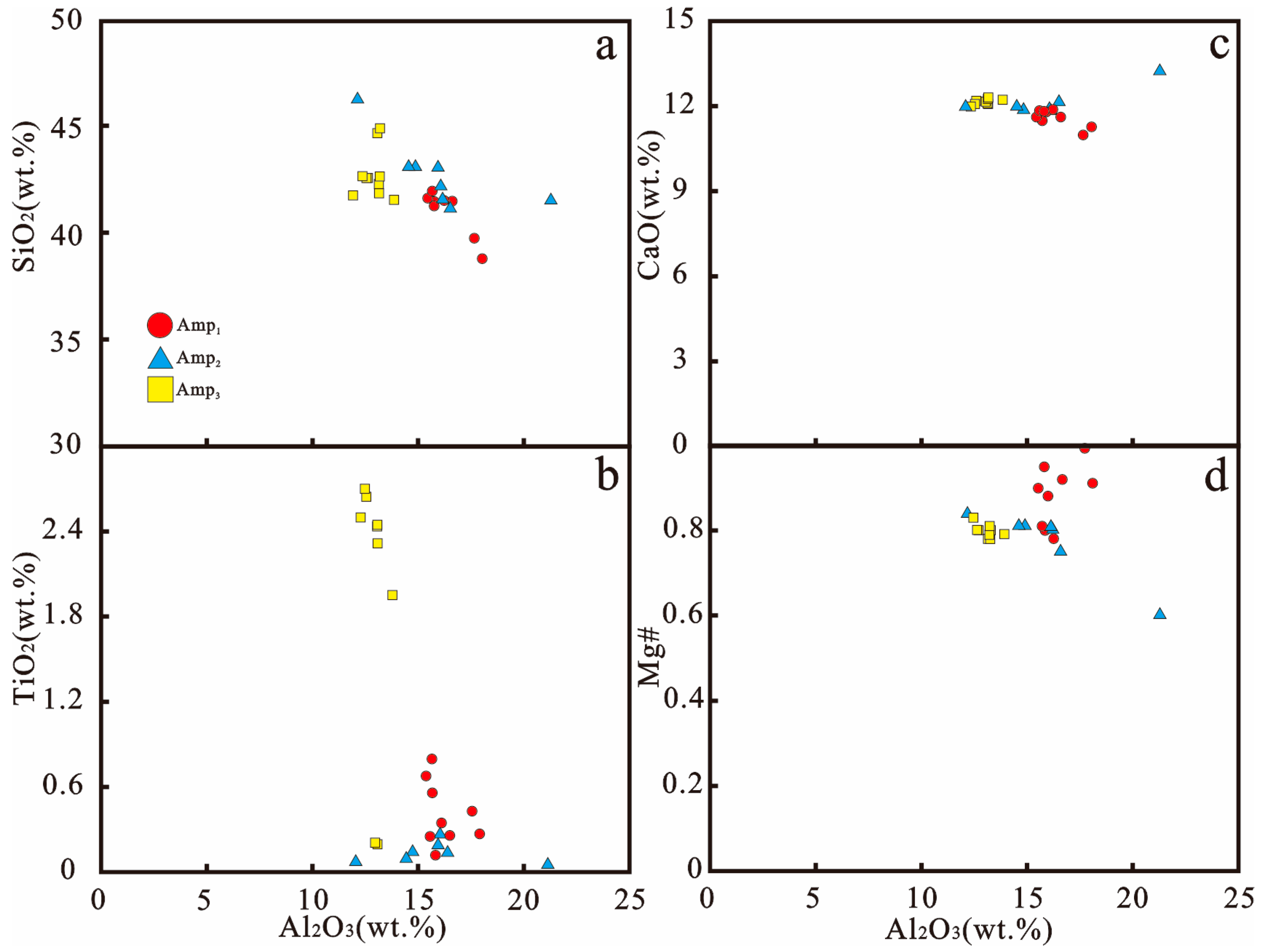
4.1. Mineral Chemistry

This paper analyzes the composition of Fe-Ti oxides, plagioclase (Pl1), and the symplectite minerals Amp (Amp1) + Spl-Pl (Pl2) + Amp (Amp2) that develop between them, as well as intergranular amphiboles (Amp3) in Fe-Ti oxide-rich gabbro. The composition of the plagioclase is shown in Table 1. Primocryst plagioclase (Pl1) has the following composition: 51.32–52.12 wt% SiO2, 30.20–30.98 wt% Al2O3, 0.20–0.98 wt% FeO, and 0–0.08 wt% TiO2. Its anorthite content (An = molar Ca/(Ca + Na)) ranges from 68 to 71. The Pl2 in the symplectite is different from primocryst plagioclase (Pl1). Pl2 has a higher An content (An92–97) and FeO (0.21–4.03 wt%) and a lower TiO2 content (Figure 3).
The amphibole components are shown in Table 2. Amp1 contains 38.83–41.99 wt% SiO2, 15.46–18.03 wt% Al2O3, 13.23–14.52 wt% FeO, 11.67–13.49 wt% MgO, 10.92–11.83 wt% CaO, 0.24–0.78 wt% TiO2, and Mg # ([Mg/(Mg + Fe2+)]) is 0.78–1. Compared with Amp1, Amp2 has a lower Mg # (Mg # 0.6–0.84) and TiO2 and higher SiO2 and CaO content (Figure 4). Amp3, compared with Amp1, has a lower Mg # (0.79–0.83) and Al2O3 (12.36–13.85) and higher TiO2 (0.18–2.7 wt%) (Figure 4).
The spinel in the Amp + Spl symplectite contains 0.05–9.58 wt% SiO2, 47.17–57.91 wt% Al2O3, 26.54–31.49 wt% FeO, and 8.59–9.34 wt% MgO (Table 3).

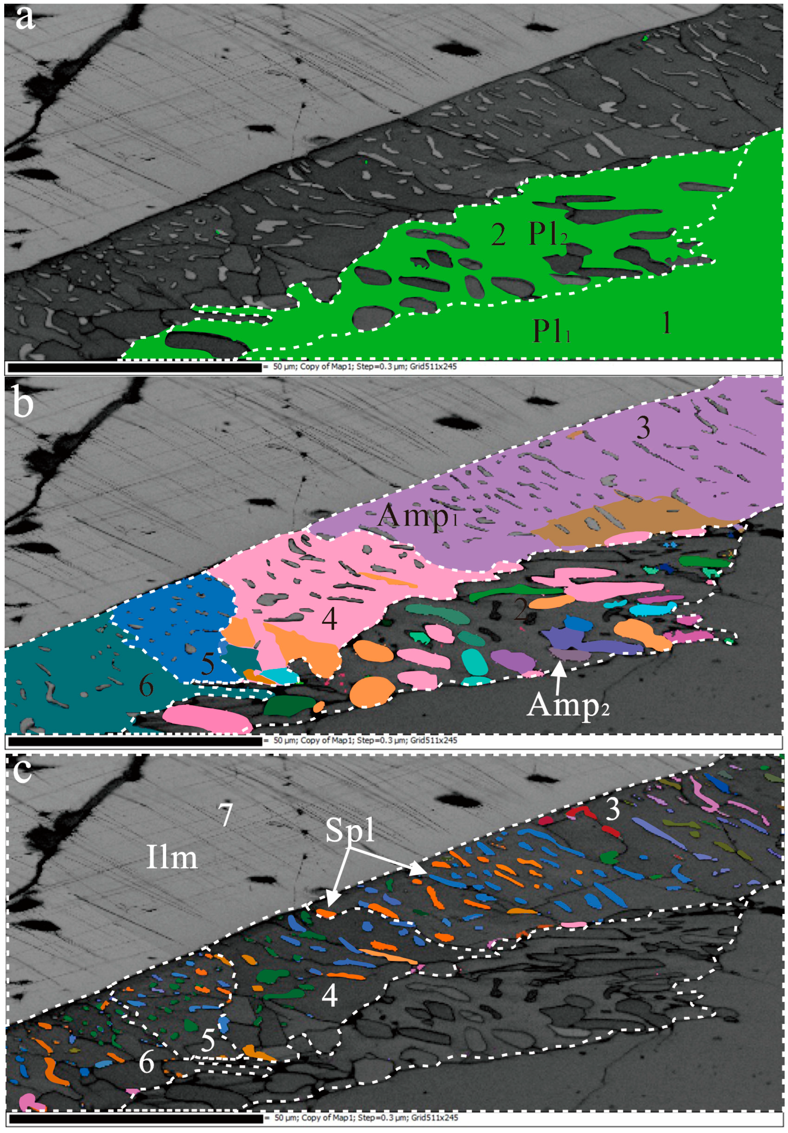
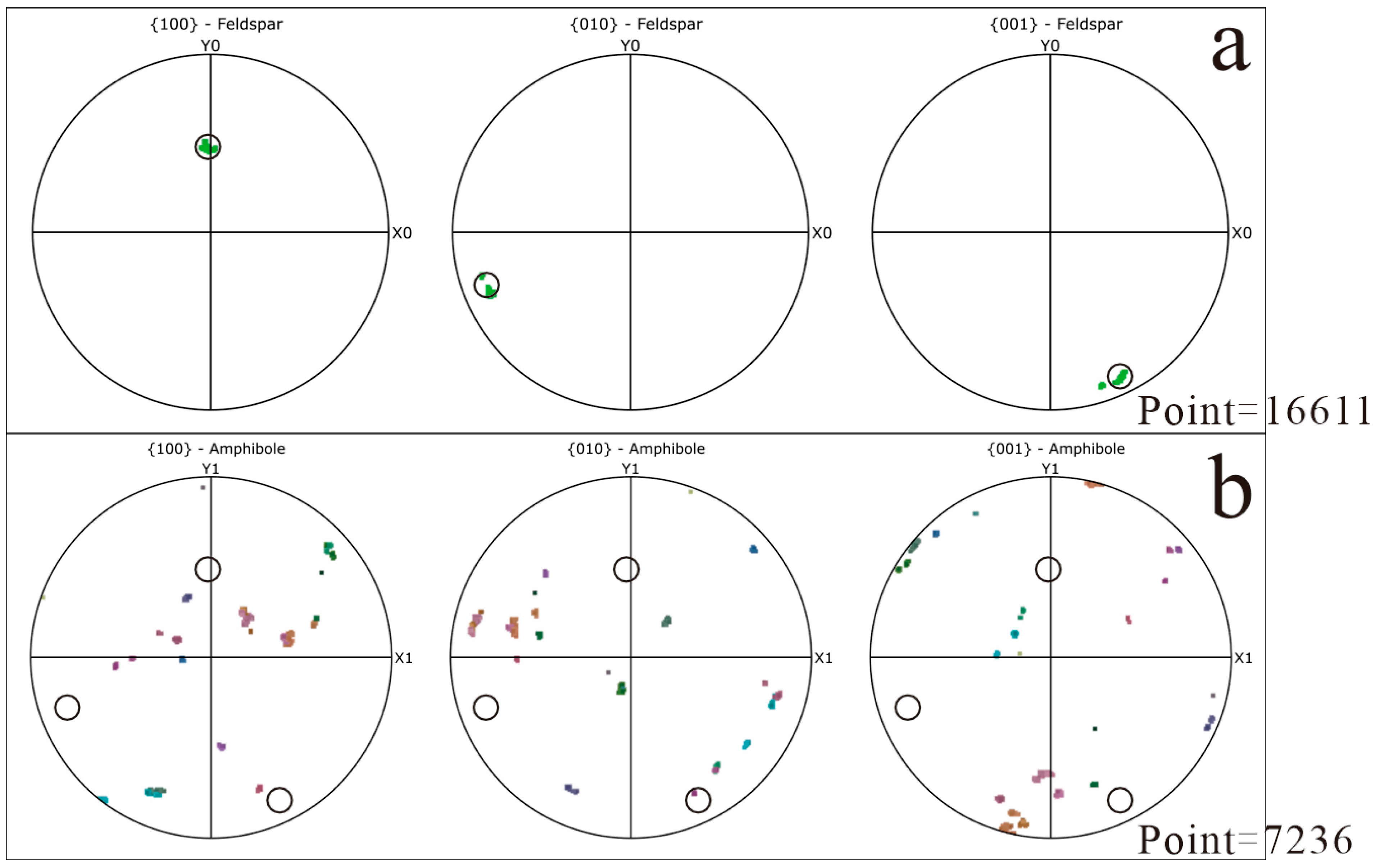
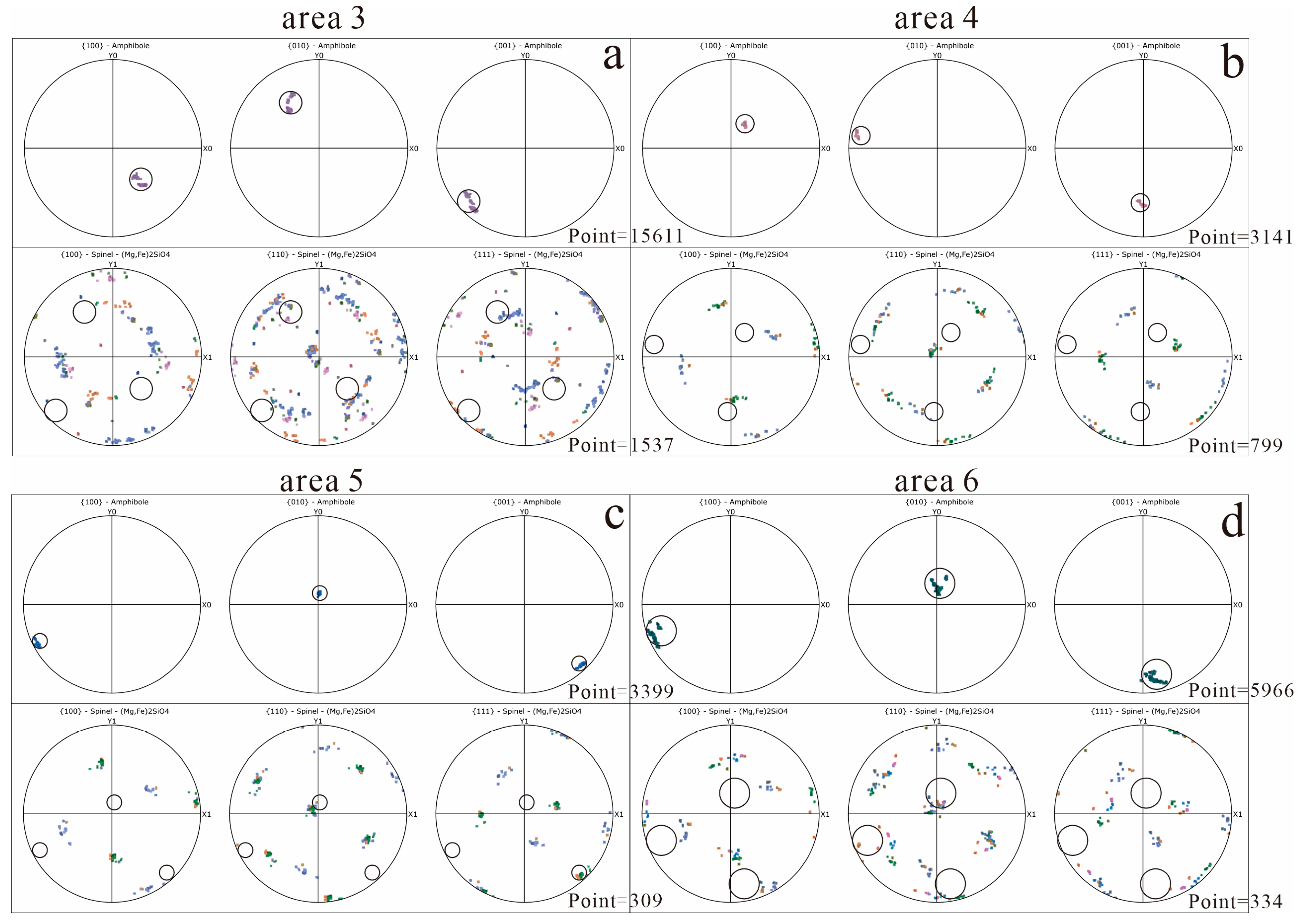
4.2. Crystal Orientation Relationship

This study uses AZtecCrystal EBSD data analysis software to analyze the orientation of the symplectite structure formed by Amp1 + Spl-Pl2 + Amp2 and the surrounding paramount crystals of Fe-Ti oxide and plagioclase (Pl1). The study area was divided into seven areas (Pl1 for area 1; Amp2 + Pl2 for area 2; Amp1 + Spl for areas 3–6; and Ilm for area 7) based on the difference in Euler angles of the EBSD results. In the sample, the plagioclase exhibits a consistent crystal orientation in a single area ((Figure 5a and Figure 6). The amphibole also shows a consistent crystal orientation in a single area except for area 2 (Figure 5a); however, the amphibole in area 2 shows significant differences in crystallographic orientation (Figure 5b). In addition, the orientation between Amp2 grains and Amp1 grains overlaps in some areas (Figure 6), indicating that Amp2 inherits the crystal orientation of Amp1 in some cases. The orientation of the spinel is more complicated than amphibole, with the spinel showing a variety of orientations within the same region (Figure 7). There are significant differences in the orientation distribution of spinel between different areas. However, in all pole figures, the spinel grains consistently rotate around a common axis that is sub-parallel to the normal of the Spl {110} plane (Figure 7), nearly perpendicular to the thin section surface. Ilmenite maintains the same orientation relationship in area 7 (Figure 8a).
Regarding crystallographic orientation, EBSD analysis shows that for the symplectites Pl2 + Amp2, there is no reasonable orientation relationship between Pl2 and Amp2 on the pole Figure (Figure 6). For the symplectite Amp1 + Spl, the orientation relationship in each area is more complex (as shown in Figure 5). Amphiboles show different orientation relationships with spinels colored according to their Euler angles. In area 3, spinel sections of different orientations show orientation relationships with host amphibole (i.e., Amp {100} overlaps with Spl {111} and {110}; Amp {010} overlaps with Spl {110}); however, area 4 shows only partial overlap of spinel {100} with host Amp {001} and no overlap with spinel {110} or {111} (Figure 7). In areas 5 and 6, Spl {110} also approximately overlaps with Amp {010}.
The crystallographic orientation analysis of spinel was conducted by coloring different Euler angles using external ilmenite as a reference phase. In Figure 8, it can be seen that almost all of the spinel and ilmenite have the following crystallographic orientation relationship: Ilm {1000} coincides with Spl {111}, Ilm {10-10} coincides with Spl {110}. This specific crystallographic relationship may be the cause of the generation of spinel. The origin of these differences will be analyzed in this study.

5. Discussion

5.1. Symplectite Formation Temperature

The microstructure of the layered intrusions in Bijigou was used to estimate the formation temperatures of the minerals using amphibole and plagioclase thermometers, respectively. The formation temperature of amphibole was estimated using the amphibole thermometer of Putirka, as follows:
T (℃) = 1781 − 132.74 × SiAmp + 116.6 × TiAmp + 69.41 × FetAmp + 101.62 × NaAmp
where SiAmp, TiAmp, and NaAmp are the calculated number of cations in 23 O atoms in amphibole; and FetAmp represents the total number of Fe cations, calculated as FeO [25]. The formation temperatures (T) of Amp1 and Amp2 are 934–953 °C and 834–914 °C, respectively.
The temperature data provide critical constraints on the physicochemical conditions governing symplectite formation. Temperature data provide key kinetic and thermodynamic constraints on the origin of the symplectites. We show that amphibole (Amp1 and Amp2) crystallized at temperatures in the 934–953 °C interval, respectively, revealing a gradual cooling process during the late stages of magma evolution [5]. This temperature constrains the cause of the stage formation of symplectites.

5.2. Amphibole + Spinel Symplectite Formation Mechanism

From the kinetic point of view, the amount of nucleation is closely related to the rate of nucleation, which is essentially a function of the degree of supersaturation. Under thermodynamic equilibrium conditions (∆G = 0), the nucleation rate is zero. As the pressure decreases or the supersaturation increases, the nucleation rate gradually increases. In the study by Obata, two different orientation relationships were observed between spinel and host clinopyroxene: in one area, the {100} of clinopyroxene perfectly coincided with the {111} of spinel, while in the other area, no clear crystallographic orientation relationship was observed [26]. During mantle peridotite decompression, low temperatures or rapid pressure drops drive the system far from thermodynamic equilibrium (i.e., the degree of supersaturation will increase). According to classical nucleation theory, an increase in supersaturation (∆C) directly enhances the driving force for phase transformation (∆G), resulting in an exponential increase in the nucleation rate (J) (J ∝ exp(–∆G2)). Under these conditions, new phases, such as spinel, form in large quantities due to rapid nucleation. However, due to the rapid nucleation rate, lattice orientation adjustment via interface diffusion to inherit the original arrangement of clinopyroxene is not possible, and a disordered topotaxic structure is ultimately formed.
The study analysis of the crystallographic orientation relationship between amphibole and spinel shows that no perfect orientation correlation feature is formed between them, but there is a specific crystallographic orientation relationship between spinel and external ilmenite. In contrast to Sept lles layered intrusions, the same Amp + Spl symplectite forms at similar temperatures but shows an ordered topology due to its slow diffusion in melts with high Si content [7]. This result can be attributed to delayed spinel nucleation during ilmenite epitaxial growth caused by a rapid pressure drop and sluggish diffusion kinetics that result in high supersaturation. This phenomenon may be attributed to a nucleation delay effect caused by a rapid decrease in phase transformation pressure and a slow diffusion kinetics rate, resulting in a rather large degree of supersaturation. At such a high degree of supersaturation, it is so rapid that the spinel in each area may not form the perfect orientation relationship with its host amphibole when nucleation occurs. Therefore, we speculate that the observed amphibole + spinel symplectites in the Bijigou layered intrusions are due to the epitaxial growth of spinel, the oversaturation of amphibole, and rapid nucleation.

5.3. Plagioclase + Amphibole (Pl2 + Amp2) Symplectite Formation Mechanism

The plagioclase + amphibole (Pl2 + Amp2) symplectite is adjacent to Pl1 (Figure 2c), where the crystal orientation of Pl2 inherits the crystal orientation of Pl1. The maximum orientation difference is 2° (Figure 6), indicating that the precipitation of Pl2 on Pl1 is the growth of pseudocrystals (maintaining the size and shape of pre-existing phases), which is often observed during the dissolution–precipitation process [27,28,29,30]. The slight (usually <2°) and local differences in the crystal orientation of Pl1 and Pl2 may be due to slightly different unit cell parameters [31]. The An content of Pl2 is significantly higher than that of Pl1, and the Ca content has increased significantly (Figure 9). The formation of high An in plagioclase is due to the reactive dissolution of plagioclase protoplasts by an evolved iron-rich unmixed melt [5,9], which was then precipitated. The fluid-rich melt penetrates along the grain boundary, causing the dissolution of the reaction interface along which Pl1 moves inward. The melt introduces Al ions, and the components in Pl1 release Na and Si, which combine with Ca2+ in the fluid to form Pl2 by reaction precipitation.
The orientation characteristics of amphibole indicate that its reaction history is similar to that of plagioclase. Although the Mg # of amphibole generally decreases in the direction indicated by the arrow in Figure 9a,b, the Mg content of Amp2 decreases significantly during the transition from Amp1 to Amp2, and in Amp1, the contact area between the two appears concave (Figure 8a). In the samples studied, the preferred orientation exhibited by Amp2 in area 2 is mainly inherited from Amp1 in area 4 (Figure 6b and Figure 7b). These features indicate that Amp1 has dissolved and Amp2 has precipitated [32,33,34,35].
If plagioclase is involved in forming amphibole, it will release Al during the reaction. Thus, the amphibole formed by reacting with plagioclase should be enriched in Al. As shown in Figure 9c, along the direction indicated in Figure 9a, the Al content of amphibole decreases with distance and does not increase. A comparison of the Al content of amphibole in symplectite with that of marginal amphibole (Figure 10) shows there is no regularity of increasing or decreasing Al content in amphibole and marginal amphibole in symplectite, and the Al content of plagioclase also does not decrease due to the increase in Al content in amphibole (Figure 9e). Plagioclase is not involved in amphibole growth, and the amphibole in the symplectite is formed by the epitaxial growth of marginal amphibole.
Our research has found that during magmatic evolution, as the temperature decreases, Pl1 is first formed, followed by the Amp1 boundary. As the temperature continues to decrease, Spl in Amp1, due to low saturation, quickly nucleates Amp1 + Spl to form symplectite; due to the dissolution of Pl1 by the melt, the melt replaces Pl1 and continues to precipitate to form Pl2. Amp2 is formed by the melt replacing Amp1 during the epitaxial growth of Amp1 and the melt replacing Pl2. From a temperature perspective, there is not much of a temperature difference between Pl2 and Amp2, and they have formed at the same time, thus forming the worm structure Pl2 + Amp2.

6. Conclusions

Based on our detailed investigation, we strongly conclude the following regarding the formation mechanisms of symplectites in the Bijigou layered intrusion:
We find that amphiboles crystallized during the late magmatic stage. Their formation temperatures range from 934 °C to 953 °C, revealing a gradual cooling process during the late stages of magma evolution.
In the amphibole–spinel symplectites, the crystallographic orientations vary significantly among different areas. Spinel often does not develop a perfect topotaxic relationship with its amphibole. Moreover, the epitaxial growth of spinel on ilmenite reinforces the notion that kinetic factors dominate the formation process. The combined topotaxic anomaly and the strong ilmenite–spinel relationship provide robust evidence for kinetic control during symplectite formation.
The plagioclase–amphibole symplectites are formed via a dissolution–precipitation mechanism. We observed that Pl2 inherits the crystallographic orientation of Pl1, with an angular deviation of less than 2°. Similarly, Amp2 partially retains the orientation of Amp1. These findings indicate that melt infiltration replaces Pl1 with Pl2 and replaces Amp1 with Amp2 through epitaxial growth.
Since symplectite is a common feature of layered igneous rocks, a study of the similar petrology and composition of symplectite in different bare igneous rocks can further refine the hypothesis proposed in this study.

Author Contributions

Conceptualization, B.S.; methodology, B.S.; software, B.S.; validation, B.S. and X.W.; formal analysis, B.S.; investigation, H.D.; data curation, H.D.; writing—original draft preparation, B.S.; writing—review and editing, H.D.; supervision, H.D.; project administration, H.D.; funding acquisition, H.D. All authors have read and agreed to the published version of the manuscript.

Funding

This study is supported by the National Natural Science Foundation of China (Grant No. 41802085) and the open fund of the Key Laboratory of Exploration Technologies for Oil and Gas Resources (Yangtze University), Ministry of Education (No. PI2023-04). Three reviewers are gratefully acknowledged for their constructive comments.

Data Availability Statement

All data and materials are available on request from the corresponding author. The data are not publicly available due to ongoing research using part of the data.

Conflicts of Interest

The authors declare no conflicts of interest.

References

  1. Mongkoltip, P.; Ashworth, J.R. Quantitative estimation of an open-system symplectite-forming reaction: Restricted diffusion of Al and Si in coronas around olivine. J. Pet. 1983, 24, 635–661. [Google Scholar] [CrossRef]
  2. Haselton, J.D.; Nash, W.P. Observations on Titanium in Lunar Oxides and Silicates. Abstr. Lunar Planet. Sci. Conf. 1975, 6, 343. [Google Scholar]
  3. Ambler, E.P.; Ashley, P.M.; McSween, H.Y. Mineralogy and petrology of the Dutchmans Creek gabbroic intrusion, South Carolina: Discussion and reply. Am. Mineral. 1980, 65, 1302–1306. [Google Scholar]
  4. Holness, M.B.; Stripp, G.; Humphreys, M.C.S. Silicate liquid Immiscibility within the crystal mush: Late-stage magmatic microstructures in the skaergaard Intrusion, east greenland. J. Pet. 2011, 52, 175–222. [Google Scholar] [CrossRef]
  5. Namur, O.; Charlier, B.; Holness, M.B. Dual origin of Fe-Ti-P gabbros by immiscibility and fractional crystallization of evolved tholeiitic basalts in the Sept Iles layered intrusion. Lithos 2012, 154, 100–114. [Google Scholar] [CrossRef]
  6. Keevil, H.A.; Namur, O.; Holness, M.B. Microstructures and late-stage magmatic processes in layered mafic intrusions: Symplectites from the sept iles intrusion, quebec, canada. J. Pet. 2020, 61, egaa071. [Google Scholar] [CrossRef]
  7. Wang, M.; Wang, C.Y.; Tan, W. Compositional evolution of interstitial liquid after onset of abundant Fe–Ti oxide crystallization in crystal mush: Insights from late-stage microstructures and mineral compositions of the bijigou layered intrusion, central china. J. Pet. 2022, 63, egac025. [Google Scholar] [CrossRef]
  8. Dong, H.; Wang, C.Y. Microstructures in the Baima layered intrusion, SW China: Constraints on the late-stage magmatic processes during solidification of a crystal mush. J. Asian Earth Sci. 2023, 255, 105774. [Google Scholar] [CrossRef]
  9. Xie, Q.; Zhang, Z.; Cheng, Z. Interstitial microstructures in Ji’nan mafic intrusion, North China Craton: Magmatic or hydrothermal origin? Eur. J. Mineral. 2017, 29, 839–850. [Google Scholar] [CrossRef]
  10. Baldwin, J.A.; Powell, R.; White, R.W.; Štípská, P. Ussing calculated chemical potential relationships to account for replacement of kyanite by symplectite in high pressure granulites. J. Metamorph. Geol. 2015, 33, 311–330. [Google Scholar] [CrossRef]
  11. Dharmapriya, P.L.; Malaviarachchi, S.P.K.; Kriegsman, L.M.; Galli, A.; Dyck, B.; Sajeev, K.; Su, B.-X.; Pitawala, A. Symplectite growth in the presence of alkaline fluids: Evidence from high-aluminous metasediments of the Highland Complex, Sri Lanka. Mineral. Petrol. 2020, 114, 515–538. [Google Scholar] [CrossRef]
  12. Spruzeniece, L.; Piazolo, S.; Daczko, N.R.; Kilburn, M.R.; Putnis, A. Symplectite formation in the presence of a reactive fluid: Insights from hydrothermal experiments. J. Metamorph. Geol. 2017, 35, 281–299. [Google Scholar] [CrossRef]
  13. Yudovskaya, M.A.; Kinnaird, J.A.; Costin, G.; McCreesh, M.; Shilovskikh, V.; Kovalchuk, E.; Kuzmin, D. Formation of spinel-orthopyroxene symplectites by reactive melt flow: Examples from the northern Bushveld Complex and implications for mineralization in layered intrusions. Econ. Geol. 2022, 117, 1935–1960. [Google Scholar] [CrossRef]
  14. Ashworth, J.R.; Chambers, A.D. Symplectic reaction in olivine and the controls of intergrowth spacing in symplectites. J. Pet. 2000, 41, 285–304. [Google Scholar] [CrossRef]
  15. Yan, D.P.; Zhou, M.F.; Song, H.L. Origin and tectonic significance of a Mesozoic multi-layer over-thrust system within the Yangtze Block (South China). Tectonophysics 2003, 361, 239–254. [Google Scholar] [CrossRef]
  16. Gao, S.; Ling, W.; Qiu, Y. Contrasting geochemical and Sm-Nd isotopic compositions of Archean metasediments from the Kongling high-grade terrain of the Yangtze craton: Evidence for cratonic evolution and redistribution of REE during crustal anatexis. Geochim. Cosmochim. Acta 1999, 63, 2071–2088. [Google Scholar] [CrossRef]
  17. Qiu, Y.M.; Gao, S.; McNaughton, N.J.; Groves, D.I.; Ling, W. First evidence of >3.2 Ga continental crust in the Yangtze craton of south China and its implications for Archean crustal evolution and Phanerozoic tectonics. Geology 2000, 28, 11–14. [Google Scholar] [CrossRef]
  18. Zhou, M.F.; Kennedy, A.K.; Sun, M. Neoproterozoic arc-related mafic Intrusions along the northern, margin of South China: Implications for the accretion of rodinia. J. Geol. 2002, 110, 611–618. [Google Scholar] [CrossRef]
  19. Zhao, J.H.; Zhou, M.F. Secular evolution of the Neoproterozoic lithospheric mantle underneath the northern margin of the Yangtze Block, South China. Lithos 2009, 107, 152–168. [Google Scholar] [CrossRef]
  20. Zhao, J.H.; Zhou, M.F. Neoproterozoic adakitic plutons in the northern margin the Yangtze Block, China: Partial melting of a thickened lower crust and implications for secular crustal evolution. Lithos 2008, 104, 231–248. [Google Scholar] [CrossRef]
  21. Ao, W.H.; Zhang, Y.K.; Zhang, R.Y.; Zhao, Y.; Sun, Y. Neoproterozoic Crustal Accretion of the Northern Margin of Yangtze Plate:Constrains from Geochemical Characteristics, LA-ICP-MS Zircon U-Pb Chronology and Hf Isotopic Compositions of Trondhjemite from Zushidian Area, Hannan Region. Geol. Rev. 2014, 60, 1393–1408. [Google Scholar]
  22. Wang, M.; Nebel, O.; Wang, C.Y. The flaw in the crustal ‘Zircon archive’: Mixed hf Isotope signatures record progressive contamination of late-stage liquid in mafic-ultramafic layered Intrusions. J. Pet. 2016, 57, 27–52. [Google Scholar] [CrossRef]
  23. Su, L. Studies of Neoproterozoie Mafie and Ultramafic Intrusionsin Western-Central China and Their Constraints on Breakupof Rodinia Supercontinent; Northwest University: Kirkland, WA, USA, 2004. [Google Scholar]
  24. Putirka, K. Special collection: Rates and depths of magma ascent on earth: Amphibole thermometers and barometers for igneous systems and some implications for eruption mechanisms of felsic magmas at arc volcanoes. Am. Mineral. 2016, 101, 841–858. [Google Scholar] [CrossRef]
  25. Zhang, R. Mineral Thermometers and Mineral Manometers; Geological Press: Beijing, China, 1983. [Google Scholar]
  26. Obata, M.; Ozawa, K. Topotaxic relationships between spinel and pyroxene in kelyphite after garnet in mantle-derived peridotites and their implications to reaction mechanism and kinetics. Mineral. Pet. 2011, 101, 217–224. [Google Scholar] [CrossRef]
  27. Engvik, A.K.; Putnis, A.; Fitz Gerald, J.D.; Austrheim, H. Albitization of granitic rocks: The mechanism of replacement of oligoclase by albite. Can. Mineral. 2008, 46, 1401–1415. [Google Scholar] [CrossRef]
  28. Hövelmann, J.; Putnis, A.; Geisler, T.; Schmidt, B.C.; Golla-Schindler, U. The replacement of plagioclase feldspars by albite: Observations from hydrothermal experiments. Contrib. Mineral. Pet. 2010, 159, 43–59. [Google Scholar] [CrossRef]
  29. Putnis, A.; Putnis, C.V. The mechanism of reequilibration of solids in the presence of a fluid phase. J. Solid State Chem. 2007, 180, 1783–1786. [Google Scholar] [CrossRef]
  30. Spruzeniece, L.; Piazolo, S.; Maynard-Casely, H.E. Deformation-resembling microstructure created by fluid-mediated dissolution-precipitation reactions. Nat. Commun. 2017, 8, 14032. [Google Scholar] [CrossRef]
  31. Giuntoli, F.; Menegon, L.; Warren, C.J. Replacement reactions and deformation by dissolution and precipitation processes in amphibolites. J. Metamorph. Geol. 2018, 36, 1263–1286. [Google Scholar] [CrossRef]
  32. Gratier, J.P.; Dysthe, D.K.; Renard, F. The role of pressure solution creep in the ductility of the Earth’s upper crust. Adv. Geophys. 2013, 54, 47–179. [Google Scholar]
  33. Hyppolito, T.; García-Casco, A.; Juliani, C.; Meira, V.T.; Hall, C. Late Paleozoic onset of subduction and exhumation at the western margin of Gondwana (Chilenia Terrane): Counterclockwise P-T paths and timing of metamorphism of deep-seated garnet-mica schist and amphibolite of Punta Sirena, Coastal Accretionary Complex, central Chile (34 S). Lithos 2014, 206, 409–434. [Google Scholar]
  34. Imon, R.; Okudaira, T.; Kanagawa, K. Development of shape-and lattice-preferred orientations of amphibole grains during initial cataclastic deformation and subsequent deformation by dissolution-precipitation creep in amphibolites from the Ryoke metamorphic belt, SW Japan. J. Struct. Geol. 2004, 26, 793–805. [Google Scholar] [CrossRef]
  35. Pearce, M.A.; Wheeler, J.; Prior, D.J. Relative strength of mafic and felsic rocks during amphibolite facies metamorphism and deformation. J. Struct. Geol. 2011, 33, 662–675. [Google Scholar] [CrossRef]
Figure 1. (a) Schematic diagram of the Neoproterozoic Hannan-Panxi Arc at the northern and western margins of the Yangtze block; (b) simplified geological map of the Neoproterozoic basic–ultrabasic and felsic rocks in the Hannan Arc at the northern margin of the Yangtze block; (c) plan view of the main iron-rich titan oxide section of the Bijigou layered intrusion. (d) Main rock composition of the Bijigou layered intrusion.
Minerals 15 00433 g001
Figure 2. Representative rocks and symplectites in the upper ore-bearing layer in the middle of the Bijigou layered complex. (a) Gabbro, orthopolarized, sample BJG-1515. (b) Fe-Ti oxide-rich gabbro, orthopolarized, sample BJG-1580. (c) Amp + Spl symplectite adjacent to Pl + Amp symplectite, BSE, Sample BJG-1579. (d) Visible Amp + Spl symplectite develops independently, partially symplastically with Pl + Amp symplectite, BSE, Sample BJG-1579. (e) Amp + Spl outer edge with fishhook-like structure connected to fishhook-like structure, BSE, Sample BJG-1579. (f) Uniaxial development in the plagioclase–amphibole and amphibole–spinel symplectites between Pl and Mt. Uniaxial, sample BJG-1579. Pl—plagioclase; Cpx—clinopyroxene; Amp—amphibole; Spl—spinel; Ilm—ilmenite; Sul—sulfide.
Minerals 15 00433 g002
Figure 3. Plagioclase composition of the Bijigou layered intrusion. A comparison between An and (a) TiO2 and (b) FeO is shown. Pl1 (blue circles); Pl2 (red triangles).
Minerals 15 00433 g003
Figure 4. Binaric diagram of amphibole components in the Bijigou layered intrusion. (a) Plots of Al2O3 versus SiO2, (b) plots of Al2O3 versus TiO2, (c) plots of Al2O3 versus CaO, (d) plots of Al2O3 versus Mg #. Amp1 (red circles); Amp2 (blue triangles); Amp₃ (yellow squares).
Minerals 15 00433 g004
Figure 5. Crystal orientation distribution maps of minerals in the symplectite area. (a) Plagioclase orientation distribution map, (b) amphibole orientation distribution map, and (c) spinel orientation distribution map. According to the distribution of positions and differences in orientation, the symplectite is divided into 7 areas: Pl—plagioclase, Amp—amphibole, Spl—spinel, Ilm—ilmenite.
Minerals 15 00433 g005
Figure 6. Orientations of plagioclase and amphibole in the Fe-Ti oxide-rich gabbro obtained by EBSD. (a) Pl1 and Pl2 pole figures and (b) Amp2 pole figures in area 2, divided according to the areas outlined in Figure 5. Each color in the pole figure corresponds to the Euler angle coloring in each area, as shown in Figure 5.
Minerals 15 00433 g006
Figure 7. Comparison of amphibole and spinel pole figures (a) in area 3, (b) in area 4, (c) in area 5, and (d) in area 6. The areas are defined according to Figure 5. Each color in the polar plot corresponds to a color in the Euler angle coloring of each area, as shown in Figure 5.
Minerals 15 00433 g007
Figure 8. (a) Ilmenite pole figure in area 7. Comparison of ilmenite and Spl pole figure (b) in area 3, (c) in area 4, (d) in area 5, and (e) in area 6. The areas are defined according to Figure 5. Each color in the polar plot corresponds to a color in the Euler angle coloring of each area, as shown in Figure 5.
Minerals 15 00433 g008
Figure 9. Sample BJG-1579: Changes in the composition of symplectites. (a) BSE images of amphibole and spinel symplectite and amphibole and plagioclase symplectite. The points in the images indicate electron microprobe analysis points. The solid circles are plagioclase, and the hollow circles are amphibole. (b) Graph of the variation of amphibole Mg # content with distance. (c) Graph of the variation of amphibole Al content with distance. (d) Graph of the variation of plagioclase An content with distance. (e) Graph of the variation of plagioclase Al content with distance. Al is the elemental content of Al.
Minerals 15 00433 g009
Figure 10. Comparison of the Mg #–Al content. Amp1 (red circles); Amp2 (blue triangles); Amp₃ (yellow squares).
Minerals 15 00433 g010
Table 1. Representative microanalyses of plagioclase in primocryst (Pl1) and plagioclase in symplectite (Pl2) (wt.%).
Sample1579-11579-21579-91579-101579-31579-41579-51579-61579-71579-81579-111579-121579-13
SiO251.3252.1251.6152.0246.2145.6046.1745.4345.2045.5444.8444.8645.86
TiO20.040.080.01b.db.d0.010.010.020.010.04b.d0.04b.d
Al2O330.2130.6130.9930.8434.7035.0134.3334.5933.2434.9635.4028.9833.17
FeO0.990.800.210.070.210.210.330.321.570.490.264.031.07
MnOb.d0.01b.d0.010.06b.d0.120.231.720.070.033.870.90
MgO0.440.010.03b.db.db.d0.020.010.03b.d0.010.020.01
CaO14.0713.5413.9214.0017.9618.9317.9218.1517.5418.3019.0916.2117.58
Na2O3.143.583.313.380.750.550.790.700.880.590.280.930.76
K2O0.020.010.020.030.010.010.030.020.010.010.010.140.03
total100.22100.75100.09100.3499.90100.3299.6999.47100.1910b.d99.9199.0699.39
Si2.342.362.352.362.142.102.142.112.082.112.082.092.13
Tib.db.db.db.db.db.db.db.db.db.db.db.db.d
Al1.621.631.661.651.891.901.881.891.801.911.931.591.82
Crb.db.db.db.db.db.db.db.db.db.db.db.db.d
Fe3+b.db.db.db.db.db.db.db.db.db.db.db.db.d
Fe2+0.040.030.01b.d0.010.010.010.010.060.020.010.160.04
Mnb.db.db.db.db.db.db.db.db.db.db.db.db.d
Mg0.03b.db.db.db.db.d0.010.020.12b.db.d0.270.06
Ca0.690.660.680.680.890.940.890.900.860.910.950.810.88
Bab.db.db.db.db.db.db.db.db.db.db.db.db.d
Na0.280.310.290.300.070.050.070.060.080.050.020.080.07
Kb.db.db.db.db.db.db.db.db.db.db.d0.01b.d
An71.1467.6469.8169.5192.9394.9792.5693.3291.6694.3897.4089.8192.55
Ab28.7732.3230.0630.337.024.977.286.548.295.542.579.307.25
Or0.090.040.140.160.050.060.150.140.050.070.040.890.20
Note: Sample 1579-1, 1579-2, 1579-9, 1579-10 for Pl1; Sample 1579-3~1579-13 for Pl2.
Table 2. Representative microanalyses of the Amp1 in Amp1 + Spl symplectite and Amp2 in the Amp2 + Pl2 symplectite in the Fe-Ti oxide-rich gabbro (wt.%).
SampleBJG1579-6BJG1579-7BJG1579-8BJG1579-9BJG1579-1BJG1579-2BJG1579-3BJG1579-4BJG1579-5
SiO241.5339.8141.6941.3546.2843.1842.2743.1743.14
TiO20.250.420.670.790.060.130.180.080.11
Al2O316.5817.6515.4615.7412.1214.8316.0514.5215.92
FeO13.0913.8513.2813.5911.9113.4913.0913.0412.24
MnO0.220.220.220.210.220.210.240.210.23
MgO13.1313.0413.3113.4714.0912.6712.3812.8513.36
CaO11.5410.9211.5611.4011.9111.8011.8211.9111.72
Na2O2.272.132.262.231.361.982.111.932.04
K2O0.160.160.200.180.100.170.130.150.12
Clb.db.db.db.d0.01b.d0.01b.d0.01
Total102.10101.87101.94102.43100.95101.47101.24100.80102.03
Si5.875.645.925.846.566.166.046.206.07
Aliv2.132.362.082.161.441.841.961.801.93
Alvi0.630.590.510.450.580.660.750.650.71
Ti0.030.040.070.080.010.010.020.010.01
Crb.db.db.db.db.db.db.db.db.d
Fe3+1.301.641.251.460.840.970.940.911.07
Fe2+0.25b.d0.320.150.570.640.620.660.37
Mn0.030.030.030.020.030.030.030.030.03
Mg2.772.752.822.832.982.692.642.752.80
Ca1.751.661.761.721.811.801.811.831.77
Na0.620.590.620.610.370.550.580.540.56
K0.030.030.040.030.020.030.020.030.02
Mg #0.921.000.900.950.840.810.810.810.88
Note: Sample 1579-6~1579-9 for Amp1; BJG1579-1~BJG1539-5 for Amp2.
Table 3. Representative microanalyses of spinel (Spl) in a symplectite (wt.%).
SampleTiO2FeOMnONiOCr2O3CaOV2O3SiO2Al2O3MgOTotal
Spl-BJG1579-10.1126.540.200.02b.d3.070.019.5847.179.3496.03
Spl-BJG1579-2b.d29.890.220.02b.d0.070.010.0557.918.8897.04
Spl-BJG1579-30.0731.490.210.01b.d0.05b.d0.1255.828.5996.36
Disclaimer/Publisher’s Note: The statements, opinions and data contained in all publications are solely those of the individual author(s) and contributor(s) and not of MDPI and/or the editor(s). MDPI and/or the editor(s) disclaim responsibility for any injury to people or property resulting from any ideas, methods, instructions or products referred to in the content.

