
MRI in Pregnancy and Precision Medicine: A Review from Literature


 ,
,  , ,
, ,  , , ,
, , ,  ,
,
Abstract
:1. Introduction
2. Methods
3. Body
3.1. Non Contrast MRI during Pregnancy
3.2. Gadolinium-Based Contrast Agents (GBCAs) MRI during Pregnancy
3.3. Risks Related to GBCA Administration
3.4. Pharmacokinetic Studies of GBCAs in Animal Models
3.5. GBCAs in Humans
3.6. Main Indications for Emergency MRI during Pregnancy
3.7. Main Maternal Indications for Emergency MRI during Pregnancy
3.8. Obstetric Causes
3.8.1. Abnormally Invasive Placenta
3.8.2. Placental Abruption
3.8.3. Uterine Rupture
3.8.4. Ovarian Cysts/Ovarian Torsion
3.8.5. Other Obstetric Causes
3.9. Non Obstetric Causes
3.9.1. Acute Abdominal Pain
3.9.2. Acute Appendicitis
3.9.3. Pancreatic and Biliary Pathology
3.9.4. Urolithiasis
3.9.5. Neurological Conditions
3.9.6. Cancer
3.9.7. Other Non-Obstetric Causes
3.10. Main Fetal Indications for MRI during Pregnancy
3.10.1. CNS Anomalies
3.10.2. Face and Neck
3.10.3. Chest
3.10.4. Abdomen
3.10.5. Limbs
3.11. GBCAs during Lactation
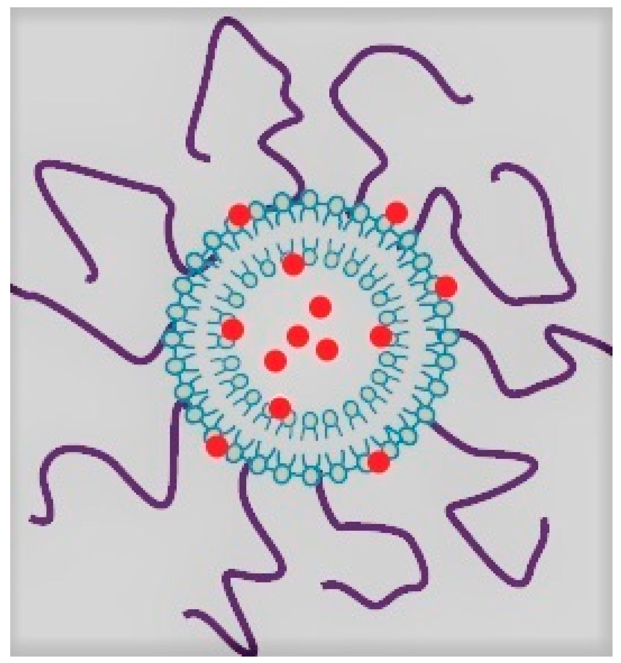
3.12. Liposomal Gadolinium Nanoparticle Contrast Agents
4. Conclusions
Author Contributions
Funding
Conflicts of Interest
References
- Mervak, B.M.; Altun, E.; McGinty, K.A.; Hyslop, W.B.; Semelka, R.C.; Burke, L.M. MRI in pregnancy: Indications and practical considerations. J. Magn. Reson. Imaging 2019, 49, 621–631. [Google Scholar] [CrossRef] [PubMed]
- Lum, M.; Tsiouris, A.J. MRI safety considerations during pregnancy. Clin. Imaging 2020, 62, 69–75. [Google Scholar] [CrossRef] [PubMed]
- Lazarus, E.; Debenedectis, C.; North, D.; Spencer, P.K.; Mayo-Smith, W.W. Utilization of imaging in pregnant patients: 10-year review of 5270 examinations in 3285 patients (1997–2006). Radiology 2009, 251, 517–524. [Google Scholar] [CrossRef]
- Coakley, F.V.; Cody, D.D.; Mahesh, M. The Pregnant Patient: Alternatives to CT and Dose-Saving Modifications to CT Technique. Image Wisely Web Site. 2010. Available online: https://www.imagewisely.org/Imaging-Modalities/ComputedTomography/Pregnant-Patient (accessed on 18 July 2020).
- ACOG. Guidelines for diagnostic imaging during pregnancy and lactation, Committee Opinion No. 723. Obstet. Gynecol. 2017, 130, e210–e216. [Google Scholar] [CrossRef] [PubMed]
- Tirada, N.; Dreizin, D.; Khati, N.J.; Akin, E.A.; Zeman, R.K. Imaging pregnant and lactating patients. RadioGraphics 2015, 35, 1751–1765. [Google Scholar] [CrossRef] [PubMed]
- McCarthy, S.M.; Stark, D.D.; Filly, R.A.; Callen, P.W.; Hricak, H.; Higgins, C.B. Obstetrical magnetic resonance imaging: Maternal anatomy. Radiology 1985, 154, 421–425. [Google Scholar] [CrossRef] [PubMed]
- Kazma, J.M.; van den Anker, J.; Allegaert, K.; Dallmann, A.; Ahmadzia, H.K. Anatomical and physiological alterations of pregnancy. J. Pharmacokinet. Pharmacodyn. 2020, 47, 271–285. [Google Scholar] [CrossRef]
- Murbach, M.; Neufeld, E.; Samaras, T.; Córcoles, J.; Robb, F.J.; Kainz, W.; Kuster, N. Pregnant women models analyzed for RF exposure and temperature increase in 3T RF shimmed birdcages. Magn. Reson. Med. 2017, 77, 2048–2056. [Google Scholar] [CrossRef] [PubMed]
- IEC. Medical Electrical Equipment-Part 2–33: Particular Requirements for the Basic Safety and Essential Performance of Magnetic Resonance Equipment for Medical Diagnosis; International Electrotechnical Commission: Ginevra, Switzerland, 2010. [Google Scholar]
- Ciet, P.; Litmanovich, D.E. MR safety issues particular to women. Magn. Reson. Imaging Clin. N. Am. 2015, 23, 59–67. [Google Scholar] [CrossRef] [PubMed]
- Marycz, K.; Kornicka, K.; Rocken, M. Static magnetic field (SMF) as a regulator of stem cell fate—New perspectives in regenerative medicine arising from an underestimated tool. Stem Cell Rev. Rep. 2018, 14, 785–792. [Google Scholar] [CrossRef]
- Zablotskii, V.; Polyakova, T.; Lunov, O.; Dejneka, A. How a high-gradient magnetic field could affect cell life. Sci. Rep. 2016, 6, 37407. [Google Scholar] [CrossRef]
- Heinrichs, W.L.; Fong, P.; Flannery, M.; Heinrichs, S.C.; Crooks, L.E.; Spindle, A.; Pedersen, R.A. Midgestational exposure of pregnant BALB/c mice to magnetic resonance imaging conditions. Magn. Reson. Imaging 1988, 6, 305–313. [Google Scholar] [CrossRef]
- Tyndall, D.A.; Sulik, K.K. Effects of magnetic resonance imaging on eye development in the C57BL/6J mouse. Teratology 1991, 43, 263–275. [Google Scholar] [CrossRef]
- Kok, R.D.; de Vries, M.M.; Heerschap, A.; van den Berg, P.P. Absence of harmful effects of magnetic resonance exposure at 1.5 T in utero during the third trimester of pregnancy: A follow-up study. Magn. Reson. Imaging 2004, 22, 851–854. [Google Scholar] [CrossRef] [PubMed]
- Baker, P.N.; Johnson, I.R.; Harvey, P.R.; Gowland, P.A.; Mansfield, P. A three-year follow-up of children imaged in utero with echo-planar magnetic resonance. Am. J. Obstet. Gynecol. 1994, 170 Pt 1, 32–33. [Google Scholar] [CrossRef]
- Myers, C.; Duncan, K.R.; Gowland, P.A.; Johnson, I.R.; Baker, P.N. Failure to detect intrauterine growth restriction following in utero exposure to MRI. Br. J. Radiol. 1998, 71, 549–551. [Google Scholar] [CrossRef]
- Chartier, A.L.; Bouvier, M.J.; McPherson, D.R.; Stepenosky, J.E.; Taysom, D.A.; Marks, R.M. The safety of maternal and fetal MRI at 3T. AJR Am. J. Roentgenol. 2019, 213, 1170–1173. [Google Scholar] [CrossRef]
- Ray, J.G.; Vermeulen, M.J.; Bharatha, A.; Montanera, W.J.; Park, A.L. Association between MRI exposure during pregnancy and fetal and childhood outcomes. JAMA 2016, 316, 952–961. [Google Scholar] [CrossRef]
- ACR Committee on MR Safety; Greenberg, T.D.; Hoff, M.N.; Gilk, T.B.; Jackson, E.F.; Kanal, E.; McKinney, A.M.; Och, J.G.; Pedrosa, I.; Rampulla, T.L.; et al. ACR guidance document on MR safe practices: Updates and critical information 2019. Magn. Reson. Imaging 2020, 51, 331–338. [Google Scholar]
- Sardu, C.; Gatta, G.; Pieretti, G.; Viola, L.; Sacra, C.; Di Grezia, G.; Musto, L.; Minelli, S.; La Forgia, D.; Capodieci, M.; et al. Pre-Menopausal Breast Fat Density Might Predict MACE During 10 Years of Follow-Up: The BRECARD Study. JACC Cardiovasc. Imaging 2021, 14, 426–438. [Google Scholar] [CrossRef] [PubMed]
- Sammet, S. Magnetic resonance safety. Abdom. Radiol. 2016, 41, 444–451. [Google Scholar] [CrossRef] [Green Version]
- Bird, S.T.; Gelperin, K.; Sahin, L.; Bleich, K.B.; Fazio-Eynullayeva, E.; Woods, C.; Radden, E.; Greene, P.; McCloskey, C.; Johnson, T.; et al. First-Trimester Exposure to Gadolinium-based Contrast Agents: A Utilization Study of 4.6 Million U.S. Pregnancies. Radiology 2019, 293, 193–200. [Google Scholar] [CrossRef] [PubMed]
- Cowling, T.; Frey, N. Macrocyclic and Linear Gadolinium Based Contrast Agents for Adults Undergoing Magnetic Resonance Imaging: A Review of Safety; Canadian Agency for Drugs and Technologies in Health: Ottawa, ON, Canada, 2019. [Google Scholar]
- Jost, G.; Frenzel, T.; Boyken, J.; Lohrke, J.; Nischwitz, V.; Pietsch, H. Long-term Excretion of Gadolinium-based Contrast Agents: Linear versus Macrocyclic Agents in an Experimental Rat Model. Radiology 2019, 290, 340–348. [Google Scholar] [CrossRef]
- Kanal, E. Gadolinium based contrast agents (GBCA): Safety overview after 3 decades of clinical experience. Magn. Reson. Imaging 2016, 34, 1341–1345. [Google Scholar] [CrossRef]
- Tsai, L.L.; Grant, A.K.; Mortele, K.J.; Kung, J.W.; Smith, M.P. A practical guide to MR imaging safety: What radiologists need to know. RadioGraphics 2015, 35, 1722–1737. [Google Scholar] [CrossRef] [PubMed]
- Fraum, T.J.; Ludwig, D.R.; Bashir, M.R.; Fowler, K.J. Gadolinium-based contrast agents: A comprehensive risk assessment. J. Magn. Reson. Imaging 2017, 6, 338–353. [Google Scholar] [CrossRef] [PubMed]
- Sikka, A.; Bisla, J.K.; Rajan, P.V.; Chalifoux, L.A.; Goodhartz, L.A.; Miller, F.H.; Yaghmai, V.; Horowitz, J.M. How to manage allergic reactions to contrast agent in pregnant patients. AJR Am. J. Roentgenol. 2016, 206, 247–252. [Google Scholar] [CrossRef] [PubMed]
- Simons, F.E.; Schatz, M. Anaphylaxis during pregnancy. J. Allergy Clin. Immunol. 2012, 130, 597–606. [Google Scholar] [CrossRef] [PubMed]
- Flanagan, E.; Bell, S. Abdominal Imaging in pregnancy (maternal and foetal risks). Best Pract. Res. Clin. Gastroenterol. 2020, 44–45, 101664. [Google Scholar] [CrossRef] [PubMed]
- Cowper, S.E.; Robin, H.S.; Steinberg, S.M.; Su, L.D.; Gupta, S.; Le Boit, P.E. Scleromyxoedema-like cutaneous diseases in renaldialysis patients. Lancet 2000, 356, 1000–1001. [Google Scholar] [CrossRef]
- Kaewlai, R.; Abujudeh, H. Nephrogenic systemic fibrosis. AJR Am. J. Roentgenol. 2012, 199, W17–W23. [Google Scholar] [CrossRef] [PubMed]
- Kanda, T.; Ishii, K.; Kawaguchi, H.; Kitajima, K.; Takenaka, D. High signal intensity in the dentate nucleus and globus pallidus on unenhanced T1-weighted MR images: Relationship with increasing cumulative dose of a gadoliniumbased contrast material. Radiology 2014, 270, 834–841. [Google Scholar] [CrossRef] [PubMed]
- McDonald, R.J.; McDonald, J.S.; Kallmes, D.; Jentoft, M.E.; Murray, D.L.; Thielen, K.R.; Williamson, E.E.; Eckel, L.J. Intracranial gadolinium deposition after contrastenhanced MR imaging. Radiology 2015, 275, 772–782. [Google Scholar] [CrossRef] [Green Version]
- Gibby, W.A.; Gibby, K.A.; Gibby, W.A. Comparison of Gd DTPA-BMA (Omniscan) versus Gd HP-DO3A (ProHance) retention in human bone tissue by inductively coupled plasma atomic emission spectroscopy. Investig. Radiol. 2004, 39, 138–142. [Google Scholar] [CrossRef]
- Blumfield, E.; Swenson, D.W.; Iyer, R.S.; Stanescu, A.L. Gadolinium-based contrast agents—Review of recent literature on magnetic resonance imaging signal intensity changes and tissue deposits, with emphasis on pediatric patients. Pediatr. Radiol. 2019, 49, 448–457. [Google Scholar] [CrossRef] [PubMed]
- Lyapustina, T.; Goldfine, C.; Rhyee, S.; Babu, K.M.; Griswold, M.K. Evaluating the patient with reported gadolinium-associated illness. J. Med. Toxicol. 2019, 15, 36–44. [Google Scholar] [CrossRef] [PubMed]
- Rozenfeld, M.N.; Podberesky, D.J. Gadolinium-based contrast agents in children. Pediatr. Radiol. 2018, 48, 1188–1196. [Google Scholar] [CrossRef]
- Elbeshlawi, I.; AbdelBaki, M.S. Safety of gadolinium administration in children. Pediatr. Neurol. 2018, 86, 27–32. [Google Scholar] [CrossRef] [PubMed]
- Khairinisa, M.A.; Takatsuru, Y.; Amano, I.; Erdene, K.; Nakajima, T.; Kameo, S.; Hiroshi, K.; Yoshito, T.; Noriyuki, K. The effect of perinatal gadolinium-based contrast agents on adult mice behavior. Investig. Radiol. 2018, 53, 110–118. [Google Scholar] [CrossRef]
- Prola-Netto, J.; Woods, M.; Roberts, V.H.J.; Sullivan, E.L.; Miller, C.A.; Frias, A.E.; Oh, K.Y. Gadolinium chelate safety in pregnancy: Barely detectable gadolinium levels in the juvenile nonhuman primate after in utero exposure. Radiology 2018, 286, 122–128. [Google Scholar] [CrossRef]
- Dean, P.B.; Niemi, P.; Kivisaari, L.; Kormano, M. Comparative pharmacokinetics of gadolinium DTPA and gadolinium chloride. Investig. Radiol. 1988, 23 (Suppl. S1), S258–S260. [Google Scholar] [CrossRef] [PubMed]
- Novak, Z.; Thurmond, A.S.; Ross, P.L.; Jones, M.K.; Thornburg, K.L.; Katzberg, R.W. Gadolinium-DTPA transplacental transfer and distribution in fetal tissue in rabbits. Investig. Radiol. 1993, 28, 828–830. [Google Scholar] [CrossRef]
- Mühler, M.R.; Clément, O.; Salomon, L.J.; Balvay, D.; Autret, G.; Vayssettes, C.; Cuénod, C.A.; Siauve, N. Maternofetal pharmacokinetics of a gadolinium chelate contrast agent in mice. Radiology 2011, 258, 455–460. [Google Scholar] [CrossRef]
- Oh, K.Y.; Roberts, V.H.; Schabel, M.C.; Grove, K.L.; Woods, M.; Frias, A.E. Gadolinium chelate contrast material in pregnancy: Fetal biodistribution in the nonhuman primate. Radiology 2015, 276, 110–118. [Google Scholar] [CrossRef] [Green Version]
- Rofsky, N.M.; Pizzarello, D.J.; Weinreb, J.C.; Ambrosino, M.M.; Rosenberg, C. Effect on fetal mouse development of exposure to MR imaging and gadopentetate dimeglumine. J. Magn. Reson. Imaging 1994, 4, 805–807. [Google Scholar] [CrossRef] [PubMed]
- Okuda, Y.; Sagami, F.; Tirone, P.; Morisetti, A.; Bussi, S.; Masters, R.E. Reproductive and developmental toxicity study of gadobenate dimeglumine formulation (E7155) (3)—Study of embryo-fetal toxicity in rabbits by intravenous administration. J. Toxicol. Sci. 1999, 24 (Suppl. S1), 79–87. [Google Scholar] [CrossRef] [Green Version]
- Soltys, R.A. Summary of preclinical safety evaluation of gadoteridol injection. Investig. Radiol. 1992, 27 (Suppl. S1), S7–S11. [Google Scholar] [CrossRef]
- De Santis, M.; Straface, G.; Cavaliere, A.F.; Carducci, B.; Caruso, A. Gadolinium periconceptional exposure: Pregnancy and neonatal outcome. Acta Obstet. Gynecol. Scand. 2007, 86, 99–101. [Google Scholar] [CrossRef] [PubMed]
- Morisetti, A.; Bussi, S.; Tirone, P.; de Haen, C. Toxicological safety evaluation of gadobenate dimeglumine 0.5 M solution for injection (MultiHance), a new magnetic resonance imaging contrast medium. J. Comput. Assist. Tomogr. 1999, 23 (Suppl. S1), S207–S217. [Google Scholar] [CrossRef] [PubMed]
- Tremblay, E.; Thérasse, E.; Thomassin-Naggara, I.; Trop, I. Quality initiatives: Guidelines for use of medical imaging during pregnancy and lactation. RadioGraphics 2012, 32, 897–911. [Google Scholar] [CrossRef] [Green Version]
- Members of Contrast Media Safety Committee of European Society of Urogenital Radiology (ESUR). ESUR Guidelines on Contrast Agents, V.10. ESUR Web Site. 2018. Available online: http://www.esur.org/fileadmin/content/2019/ESURGuidelines10.0FinalVersion.pdf (accessed on 25 August 2020).
- ACR Committee on Drugs and Contrast Media. ACR Manual on Contrast Media, Version 10.3. ACR Web Site. 2018. Available online: https://www.acr.org/-/media/ACR/Files/Clinical-Resources/ContrastMedia.pdf (accessed on 20 September 2020).
- American College of Radiology. Manual on Contrast Media Version 10.3. 2018. Available online: https://www.acr.org/-/media/ACR/Files/Clinical-Resources/Contrast_Media.pdf (accessed on 10 September 2021).
- Royal College of Radiologists. Guidance on Gadolinium-Based Contrast Agent Administration to Adult Patients. 2019. Available online: https://www.rcr.ac.uk/system/files/publication/field_publication_files/bfcr193-gadolinium-based-contrast-agent-adult-patients.pdf (accessed on 10 September 2021).
- European Society of Urogenital Radiology. Guidelines on Contrast Agents Version 10.0. 2018. Available online: http://www.esur-cm.org/ (accessed on 10 September 2021).
- Baughman, W.C.; Corteville, J.E.; Shah, R.R. Placenta accreta: Spectrum of US and MR imaging findings. RadioGraphics 2008, 28, 1905–1916. [Google Scholar] [CrossRef] [Green Version]
- Jaraquemada, J.M.P.; Bruno, C.H. Magnetic resonance imaging in 300 cases of placenta accreta: Surgical correlation of new findings. Acta Obstet. Gynecol. Scand. 2005, 84, 716–724. [Google Scholar] [CrossRef] [PubMed]
- Fadl, S.A.; Linnau, K.F.; Dighe, M.K. Placental abruption and hemorrhage-review of imaging appearance. Emerg. Radiol. 2019, 26, 87–97. [Google Scholar] [CrossRef]
- Jha, P.; Masselli, G.; Ohliger, M.A.; Pōder, L. Nonfetal Imaging during Pregnancy: Placental Disease. Radiol. Clin. N. Am. 2020, 58, 381–399. [Google Scholar] [CrossRef] [PubMed]
- Porreco, R.P.; Clark, S.L.; Belfort, M.A.; Dildy, G.A.; Meyers, J.A. The changing specter of uterine rupture. Am. J. Obstet. Gynecol. 2009, 200, 269.e1–269.e4. [Google Scholar] [CrossRef] [PubMed]
- Dow, M.; Wax, J.R.; Pinette, M.G.; Blackstone, J.; Cartin, A. Third-trimester uterine rupture without previous cesarean: A case series and review of the literature. Am. J. Perinatol. 2009, 26, 739–744. [Google Scholar] [CrossRef] [PubMed]
- Hoffmann, J.; Stumpp, P.; Exner, M.; Grothoff, M.; Stepan, H. Magnetic resonance imaging can be useful for advanced diagnostic of the lower uterine segment in patients after previous cesarean section. Ultrasound Obstet. Gynecol. 2018. Epub ahead of print. [Google Scholar] [CrossRef] [Green Version]
- Ribic-Pucelj, M.; Kobal, B.; Peternelj-Marinsek, S. Surgical treatment of adnexal masses in pregnancy: Indications, surgical approach and pregnancy outcome. J. Reprod. Med. 2007, 52, 273–279. [Google Scholar]
- Smorgick, N.; Pansky, M.; Feingold, M.; Herman, A.; Halperin, R.; Maymon, R. The clinical characteristics and sonographic findings of maternal ovarian torsion in pregnancy. Fertil. Steril. 2009, 92, 1983–1987. [Google Scholar] [CrossRef] [PubMed]
- Furey, E.A.; Bailey, A.A.; Pedrosa, I. Magnetic resonance imaging of acute abdominal and pelvic pain in pregnancy. Top. Magn. Reson. Imaging 2014, 23, 225–242. [Google Scholar] [CrossRef]
- Youssef, A.T. Uncommon obstetric and gynecologic emergencies associated with pregnancy: Ultrasound diagnosis. J. Ultrasound 2018, 21, 127–136. [Google Scholar] [CrossRef] [PubMed]
- McGahan, J.P.; Lamba, R.; Coakley, F.V. Imaging non-obstetrical causes of abdominal pain in the pregnant patient. Appl. Radiol. Scotch Plains 2010, 39, 10. [Google Scholar]
- Spalluto, L.B.; Woodfield, C.A.; De Benedectis, C.M.; Lazarus, E. MR imaging evaluation of abdominal pain during pregnancy: Appendicitis and other nonobstetric causes. RadioGraphics 2012, 32, 317–334. [Google Scholar] [CrossRef] [Green Version]
- Pastore, P.A.; Loomis, D.M.; Sauret, J. Appendicitis in pregnancy. J. Am. Board Fam. Med. 2006, 19, 621–626. [Google Scholar] [CrossRef]
- Dewhurst, C.; Beddy, P.; Pedrosa, I. MRI evaluation of acute appendicitis in pregnancy. J. Magn. Reson. Imaging 2013, 37, 566–575. [Google Scholar] [CrossRef]
- Burke, L.M.B.; Bashir, M.R.; Miller, F.H.; Siegelman, E.S.; Brown, M.; Alobaidy, M.; Jaffe, T.A.; Hussain, S.M.; Palmer, S.L.; Garon, B.L.; et al. Magnetic resonance imaging of acute appendicitis in pregnancy: A 5-year multiinstitutional study. Am. J. Obstet. Gynecol. 2015, 213, 693.e1–693.e6. [Google Scholar] [CrossRef]
- Fonseca, A.L.; Schuster, K.M.; Kaplan, L.J.; Maung, A.A.; Lui, F.Y.; Davis, K.A. The use of magnetic resonance imaging in the diagnosis of suspected appendicitis in pregnancy: Shortened length of stay without increase in hospital charges. JAMA Surg. 2014, 149, 687–693. [Google Scholar] [CrossRef] [PubMed] [Green Version]
- Håkansson, K.; Leander, P.; Ekberg, O.; Håkansson, H.-O. MR imaging in clinically suspected acute cholecystitis: A comparison with ultrasonography. Acta Radiol. 2000, 41, 322–328. [Google Scholar] [CrossRef] [PubMed]
- Mali, P. Pancreatitis in pregnancy: Etiology, diagnosis, treatment, and outcomes. Hepatobiliary Pancreat. Dis. Int. 2016, 15, 434–438. [Google Scholar] [CrossRef]
- Andreoiu, M.; MacMahon, R. Renal colic in pregnancy: Lithiasis or physiological hydronephrosis? Urology 2009, 74, 757–761. [Google Scholar] [CrossRef]
- Wing, D.A.; Fassett, M.J.; Getahun, D. Acute pyelonephritis in pregnancy: An 18-year retrospective analysis. Am. J. Obstet. Gynecol. 2014, 210, 219.e1–219.e6. [Google Scholar] [CrossRef]
- Maggioni, F.; Alessi, C.; Maggino, T.; Zanchin, G. Headache during pregnancy. Cephalalgia 1997, 17, 765–769. [Google Scholar] [CrossRef]
- Robbins, M.S.; Farmakidis, C.; Dayal, A.K.; Lipton, R.B. Acute headache diagnosis in pregnant women. Neurology 2015, 85, 1024–1030. [Google Scholar] [CrossRef] [PubMed] [Green Version]
- Raffaelli, B.; Neeb, L.; Israel-Willner, H.; Körner, J.; Liman, T.; Reuter, U.; Siebert, E. Brain imaging in pregnant women with acute headache. J. Neurol. 2018, 265, 1836–1843. [Google Scholar] [CrossRef] [PubMed]
- Kanekar, S.; Bennett, S. Imaging of neurologic conditions in pregnant patients. RadioGraphics 2016, 36, 2102–2122. [Google Scholar] [CrossRef] [PubMed]
- Edlow, J.A.; Caplan, L.R.; O’Brien, K.; Tibbles, C.D. Diagnosis of acute neurological emergencies in pregnant and post-partum women. Lancet Neurol. 2013, 12, 175–185. [Google Scholar] [CrossRef]
- Gutke, A.; Ostgaard, H.C.; Oberg, B. Predicting persistent pregnancy-related low back pain. Spine 2008, 33, E386–E393. [Google Scholar] [CrossRef]
- Mousavi, S.J.; Parnianpour, M.; Vleeming, A. Pregnancy related pelvic girdle pain and low back pain in an Iranian population. Spine 2007, 32, E100–E104. [Google Scholar] [CrossRef]
- Han, I.H. Pregnancy and spinal problems. Curr. Opin. Obstet. Gynecol. 2010, 22, 477–481. [Google Scholar] [CrossRef] [PubMed]
- Bonfield, C.M.; Engh, J.A. Pregnancy and brain tumors. Neurol. Clin. 2012, 30, 937–946. [Google Scholar] [CrossRef] [PubMed]
- Simon, R. Brain tumors in pregnancy. Semin. Neurol. 1988, 8, 214–221. [Google Scholar] [CrossRef]
- Canibaño, B.; Deleu, D.; Mesraoua, B.; Melikyan, G.; Ibrahim, F.; Hanssens, Y. Pregnancy-related issues in women with multiple sclerosis: An evidence-based review with practical recommendations. J. Drug Assess. 2020, 9, 20–36. [Google Scholar] [CrossRef] [Green Version]
- Simon, J.; Li, D.; Traboulsee, A.; Coyle, P.; Arnold, D.; Barkhof, F.; Frank, J.; Grossman, R.; Paty, D.; Radue, E.; et al. Standardized MR imaging protocol for multiple sclerosis: Consortium of MS Centers consensus guidelines. AJNR Am. J. Neuroradiol. 2006, 27, 455–461. [Google Scholar]
- Smith, L.H.; Danielsen, B.; Allen, M.E.; Cress, R. Cancer associated with obstetric delivery: Results of linkage with the California cancer registry. Am. J. Obstet. Gynecol. 2003, 189, 1128–1135. [Google Scholar] [CrossRef]
- Parazzini, F.; Franchi, M.; Tavani, A.; Negri, E.; Peccatori, F.A. Frequency of pregnancy related cancer: A population based linkage study in Lombardy, Italy. Int. J. Gynecol. Cancer 2017, 27, 613–619. [Google Scholar] [CrossRef] [PubMed]
- Stensheim, H.; Moller, B.; van Dijk, T.; Fossa, S.D. Cause-specific survival for women diagnosed with cancer during pregnancy or lactation: A registry-based cohort study. J. Clin. Oncol. 2009, 27, 45–51. [Google Scholar] [CrossRef] [PubMed]
- Vashi, R.; Hooley, R.; Butler, R. Breast imaging of the pregnant and lactating patient: Imaging modalities and pregnancy-associated breast cancer. AJR Am. J. Roentgenol. 2013, 200, 321–328. [Google Scholar] [CrossRef] [PubMed]
- Nissan, N.; Furman-Haran, E.; Allweis, T.; Menes, T.; Golan, O.; Kent, V.; Barsuk, D.; Paluch-Shimon, S.; Haas, I.; Brodsky, M.; et al. Non-contrast Breast MRI During Pregnancy Using Diffusion Tensor Imaging: A Feasibility Study. J. Magn. Reson. Imaging 2019, 49, 508–517. [Google Scholar] [CrossRef] [PubMed]
- Nissan, N.; Anaby, D.; Sklair-Levy, M. Breast MRI without Contrast Is Feasible and Appropriate During Pregnancy. J. Am. Coll. Radiol. 2019, 16 Pt A, 408–409. [Google Scholar] [CrossRef]
- Han, S.; Amant, F.; Michielsen, K.; De Keyzer, F.; Fieuws, S.; Van Calsteren, K.; Dresen, R.C.; Gziri, M.M.; Vandecaveye, V. Feasibility of whole-body diffusion-weighted MRI for detection of primary tumour, nodal and distant metastases in women with cancer during pregnancy: A pilot study. Eur. Radiol. 2018, 28, 1862–1874. [Google Scholar] [CrossRef] [PubMed]
- Peccatori, F.A.; Codacci-Pisanelli, G.; Del Grande, M.; Scarfone, G.; Zugni, F.; Petralia, G. Whole body MRI for systemic staging of breast cancer in pregnant women. Breast 2017, 35, 177–181. [Google Scholar] [CrossRef]
- Bourgioti, C.; Konidari, M.; Moulopoulos, L.A. Imaging of gynecologic malignancy in a reproductive age female: Cancer during pregnancy. Radiol. Clin. N. Am. 2020, 58, 413–430. [Google Scholar] [CrossRef]
- Boregowda, G.; Shehata, H.A. Gastrointestinal and liver disease in pregnancy. Best Pract. Res. Clin. Obstet. Gynaecol. 2013, 27, 835–853. [Google Scholar] [CrossRef] [PubMed]
- Barton, J.R.; Sibai, B.M. Gastrointestinal complications of pre-eclampsia. Semin. Perinatol. 2009, 33, 179–188. [Google Scholar] [CrossRef] [PubMed]
- Girard, N.; Raybaud, C.; Dercole, C.; Boubli, L.; Chau, C.; Cahen, S.; Potier, A.; Gamerre, M. In vivo MRI of the fetal brain. Neuroradiology 1993, 35, 431–436. [Google Scholar] [CrossRef] [PubMed]
- Guo, W.-Y.; Chang, C.-Y.; Ho, D.M.; Wong, T.-T.; Sheu, M.-H.; Cheng, H.-C.; Chen, S.-J.; Hung, J.-H. A comparative MR and pathological study on fetal CNS disorders. Child’s Nerv. Syst. 2001, 17, 512–518. [Google Scholar] [CrossRef]
- Merzoug, V.; Ferey, S.; Andre, C.; Gelot, A.; Adamsbaum, C. Magnetic resonance imaging of the fetal brain. J. Neuroradiol. 2002, 29, 76–90. [Google Scholar] [PubMed]
- Ghi, T.; Tani, G.; Savelli, L.; Colleoni, G.G.; Pilu, G.; Bovicelli, L. Prenatal imaging of facial clefts by magnetic resonance imaging with emphasis on the posterior palate. Prenat. Diagn. 2003, 23, 970–975. [Google Scholar] [CrossRef]
- Shinmoto, H.; Kashima, K.; Yuasa, Y.; Tanimoto, A.; Morikawa, Y.; Ishimoto, H.; Yoshimura, Y.; Hiramatsu, K. MR imaging of non-CNS fetal abnormalities: A pictorial essay. RadioGraphics 2000, 20, 1227–1243. [Google Scholar] [CrossRef]
- Quinn, T.M.; Hubbard, A.M.; Adzick, N.S. Prenatal magnetic resonance imaging enhances fetal diagnosis. J. Pediatr. Surg. 1998, 33, 553–558. [Google Scholar] [CrossRef]
- Cha, I.; Adzick, N.S.; Harrison, M.R.; Finkbeiner, W.E. Fetal congenital cystic adenomatoid malformations of the lung: A clinicopathologic study of eleven cases. Am. J. Surg. Pathol. 1997, 21, 537–544. [Google Scholar] [CrossRef] [PubMed]
- Cass, D.L.; Crombleholme, T.M.; Howell, L.J.; Stafford, P.W.; Ruchelli, E.D.; Adzick, N.S. Cystic lung lesions with systemic arterial blood supply: A hybrid of congenital cystic adenomatoid malformation and bronchopulmonary sequestration. J. Pediatr. Surg. 1997, 32, 986–990. [Google Scholar] [CrossRef]
- Levine, D.; Barnewolt, C.E.; Mehta, T.S.; Trop, I.; Estroff, J.; Wong, G. Fetal thoracic abnormalities: MR imaging. Radiology 2003, 228, 379–388. [Google Scholar] [CrossRef] [PubMed]
- Kasprian, G.; Balassy, C.; Brugger, P.C.; Prayer, D. MRI of normal and pathological fetal lung development. Eur. J. Radiol. 2006, 57, 261–270. [Google Scholar] [CrossRef]
- Datin-Dorriere, V.; Rouzies, S.; Taupin, P.; Walter-Nicolet, E.; Benachi, A.; Sonigo, P.; Mitanchez, D. Prenatal prognosis in isolated congenital diaphragmatic hernia. Am. J. Obstet. Gynecol. 2008, 198, 80.e1–80.e5. [Google Scholar] [CrossRef] [PubMed]
- Achiron, R.; Orvieto, R. Assessment of fetal cardiovascular function: Ultrasound study of the fetal circulatory compartments. Curr. Opin. Obstet. Gynecol. 1999, 11, 119–123. [Google Scholar] [CrossRef] [PubMed]
- Chang, C.H.; Chang, F.M.; Yu, C.H.; Liang, R.I.; Ko, H.C.; Chen, H.Y. Systemic assessment of fetal hemodynamics by Doppler ultrasound. Ultrasound Med. Biol. 2000, 26, 777–785. [Google Scholar] [CrossRef]
- Manganaro, L.; Savelli, S.; Di Maurizio, M.; Perrone, A.; Tesei, J.; Francioso, A.; Angeletti, M.; Coratella, F.; Irimia, D.; Fierro, F.; et al. Potential role of fetal cardiac evaluation with magnetic resonance imaging: Preliminary experience. Prenat. Diagn. 2008, 28, 148–156. [Google Scholar] [CrossRef]
- Garel, C.; Dreux, S.; Philippe-Chomette, P.; Vuillard, E.; Oury, J.F.; Muller, F. Contribution of fetalmagnetic resonance imaging and amniotic fluid digestive enzyme assays to the evaluation of gastrointestinal tract abnormalities. Ultrasound Obstet. Gynecol. 2006, 28, 282–291. [Google Scholar] [CrossRef] [PubMed]
- Saguintaah, M.; Couture, A.; Veyrac, C.; Baud, C.; Quere, M.P. MRI of the fetal gastrointestinal tract. Pediatr. Radiol. 2002, 32, 395–404. [Google Scholar] [CrossRef]
- Brugger, P.C.; Prayer, D. Fetal abdominal magnetic resonance imaging. Eur. J. Radiol. 2006, 57, 278–293. [Google Scholar] [CrossRef]
- Witzani, L.; Brugger, P.C.; Hormann, M.; Kasprian, G.; Csapone-Balassy, C.; Prayer, D. Normal renal development investigated with fetal MRI. Eur. J. Radiol. 2006, 57, 294–302. [Google Scholar] [CrossRef]
- Malinger, G.; Brugger, P.C.; Prayer, D. Fetal MRI of the femur—Preliminary results. Ultrasound Obstet. Gynecol. 2006, 27, 593. [Google Scholar]
- Kubik-Huch, R.A.; Gottstein-Aalame, N.M.; Frenzel, T.; Seifert, B.; Puchert, E.; Wittek, S.; Debatin, J.F. Gadopentetate dimeglumine excretion into human breast milk during lactation. Radiology 2000, 216, 555–558. [Google Scholar] [CrossRef] [PubMed]
- Schmiedl, U.; Maravilla, K.R.; Gerlach, R.; Dowling, C.A. Excretion of gadopentetate dimeglumine in human breast milk. AJR Am. J. Roentgenol. 1990, 154, 1305–1306. [Google Scholar] [CrossRef] [PubMed] [Green Version]
- Webb, J.A.; Thomsen, H.S.; Morcos, S.K.; Members of Contrast Media Safety Committee of European Society of Urogenital Radiology. The use of iodinated and gadolinium contrast media during pregnancy and lactation. Eur. Radiol. 2005, 15, 1234–1240. [Google Scholar] [CrossRef] [PubMed]
- Shetty, A.N.; Pautler, R.; Ghagahda, K.; Rendon, D.; Gao, H.; Starosolski, Z.; Bhavane, R.; Patel, C.; Annapragada, A.; Yallampalli, C.; et al. A liposomal Gd contrast agent does not cross the mouse placental barrier. Sci. Rep. 2016, 6, 27863. [Google Scholar] [CrossRef]
- Mulder WJ, M.; Strijkers, G.J.; van Tilborg GA, F.; Griffioen, A.W.; Nicolay, K. Lipid-based nanoparticles for contrast-enhanced MRI and molecular imaging. NMR Biomed. 2006, 19, 142–164. [Google Scholar] [CrossRef]
- Ghaghada, K.; Hawley, C.; Kawaji, K.; Annapragada, A.; Mukundan, S. T1 relaxivity of core-encapsulated gadolinium liposomal contrast agents–effect of liposome size and internal gadolinium concentration. Acad. Radiol. 2008, 15, 1259–1263. [Google Scholar] [CrossRef] [Green Version]
- Ghaghada, K.B.; Ravoori, M.; Sabapathy, D.; Bankson, J.; Kundra, V.; Annapragada, A. New dual mode gadolinium nanoparticle contrast agent for magnetic resonance imaging. PLoS ONE 2009, 4, e7628. [Google Scholar] [CrossRef] [Green Version]
- Ghaghada, K.B.; Starosolski, Z.A.; Bhayana, S.; Stupin, I.; Patel, C.V.; Bhavane, R.C.; Gao, H.; Bednov, A.; Yallampalli, C.; Belfort, M.; et al. Pre-clinical evaluation of a nanoparticle-based blood-pool contrast agent for MR imaging of the placenta. Placenta 2017, 57, 60–70. [Google Scholar] [CrossRef]
- Badachhape, A.A.; Kumar, A.; Ghaghada, K.B.; Stupin, I.V.; Srivastava, M.; Devkota, L.; Starosolski, Z.; Tanifum, E.A.; George, V.; Fox, K.A.; et al. Pre-clinical magnetic resonance imaging of retroplacental clear space throughout gestation. Placenta 2019, 77, 1–7. [Google Scholar] [CrossRef] [PubMed]
- Badachhape, A.A.; Devkota, L.; Stupin, I.V.; Sarkar, P.; Srivastava, M.; Tanifum, E.A.; Fox, K.A.; Yallampalli, C.; Annapragada, A.V.; Ghaghada, K.B. Nanoparticle Contrast-enhanced T1-Mapping Enables Estimation of Placental Fractional Blood Volume in a Pregnant Mouse Model. Sci. Rep. 2019, 9, 18707. [Google Scholar] [CrossRef] [PubMed] [Green Version]
- Costelloe, C.M.; Amini, B.; Madewell, J.E. Risks and Benefits of Gadolinium-Based Contrast-Enhanced MRI. Semin. Ultrasound CT MRI 2020, 41, 170–182. [Google Scholar] [CrossRef]
- Colosimo, C.; Ruscalleda, J.; Korves, M.; La Ferla, R.; Wool, C.; Pianezzola, P.; Kirchin, M.A. Detection of intracranial metastases: A multicenter, intrapatient comparison of gadobenate dimeglumine-enhanced MRI with routinely used contrast agents at equal dosage. Investig. Radiol. 2001, 36, 72–81. [Google Scholar] [CrossRef] [PubMed]


| Trade Name | Marketing Authorisation Holder | Compound | Chemical Structure | Use |
|---|---|---|---|---|
| Dotarem® | Guerbet Diagnostic Imaging | Gadoterate meglumine | Macrocyclic | Intraarticular/Intravenous |
| Gadovist® | Bayer Pharmaceuticals | Gadobutrolo | Macrocyclic | Intravenous |
| Magnevist® | Bayer Pharmaceuticals | Gadopentetate dimeglumine | Linear | Intraarticular |
| Multihance® | Bracco Imaging | Gadobenate dimeglumine | Linear | Intravenous |
| Primovist® | Bayer Pharmaceuticals | Gadoxetate disodium | Linear | Intravenous |
| Prohance® | Bracco Imaging | Gadoteridol | Macrocyclic | Intravenous |
| Site | Indications |
|---|---|
| CNS anomalies | Ventriculomegaly, hemorrhages, lissencephaly, polymicrogyria/pachygyria, gray matter heterotopias, cortical dysplasias, and neural tube defects (e.g., spina bifida/diastematomyelia). |
| Face and palate | In cases in which there is a significant risk of associated brain abnormalities. |
| Neck masses | Neck masses could impair the airway leading to asphyxia at birth. |
| Chest | Congenital diaphragmatic hernias. |
| Abdomen | Abdominal masses or bowel pathologies, including obstruction and atresia. |
Publisher’s Note: MDPI stays neutral with regard to jurisdictional claims in published maps and institutional affiliations. |
© 2021 by the authors. Licensee MDPI, Basel, Switzerland. This article is an open access article distributed under the terms and conditions of the Creative Commons Attribution (CC BY) license (https://creativecommons.org/licenses/by/4.0/).
Share and Cite
Gatta, G.; Di Grezia, G.; Cuccurullo, V.; Sardu, C.; Iovino, F.; Comune, R.; Ruggiero, A.; Chirico, M.; La Forgia, D.; Fanizzi, A.; et al. MRI in Pregnancy and Precision Medicine: A Review from Literature. J. Pers. Med. 2022, 12, 9. https://doi.org/10.3390/jpm12010009
Gatta G, Di Grezia G, Cuccurullo V, Sardu C, Iovino F, Comune R, Ruggiero A, Chirico M, La Forgia D, Fanizzi A, et al. MRI in Pregnancy and Precision Medicine: A Review from Literature. Journal of Personalized Medicine. 2022; 12(1):9. https://doi.org/10.3390/jpm12010009
Chicago/Turabian StyleGatta, Gianluca, Graziella Di Grezia, Vincenzo Cuccurullo, Celestino Sardu, Francesco Iovino, Rosita Comune, Angelo Ruggiero, Marilena Chirico, Daniele La Forgia, Annarita Fanizzi, and et al. 2022. "MRI in Pregnancy and Precision Medicine: A Review from Literature" Journal of Personalized Medicine 12, no. 1: 9. https://doi.org/10.3390/jpm12010009
APA StyleGatta, G., Di Grezia, G., Cuccurullo, V., Sardu, C., Iovino, F., Comune, R., Ruggiero, A., Chirico, M., La Forgia, D., Fanizzi, A., Massafra, R., Belfiore, M. P., Falco, G., Reginelli, A., Brunese, L., Grassi, R., Cappabianca, S., & Viola, L. (2022). MRI in Pregnancy and Precision Medicine: A Review from Literature. Journal of Personalized Medicine, 12(1), 9. https://doi.org/10.3390/jpm12010009