
The Role of Endometrial Stem/Progenitor Cells in Recurrent Reproductive Failure


 , and
, and 
Abstract
:1. Introduction
2. Scope of This Review
3. Materials and Methods
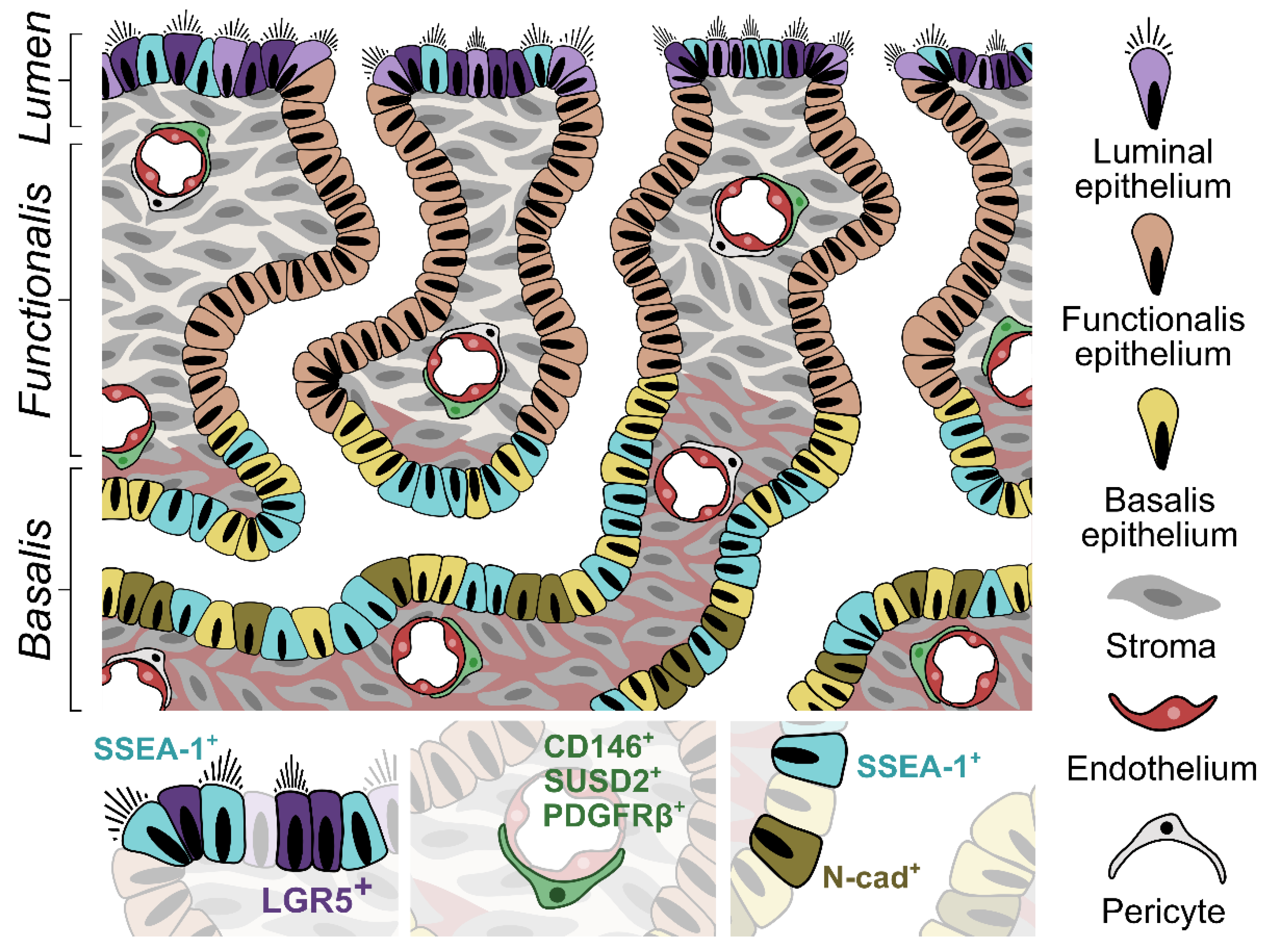
4. Endometrial Stem/Progenitor Cells
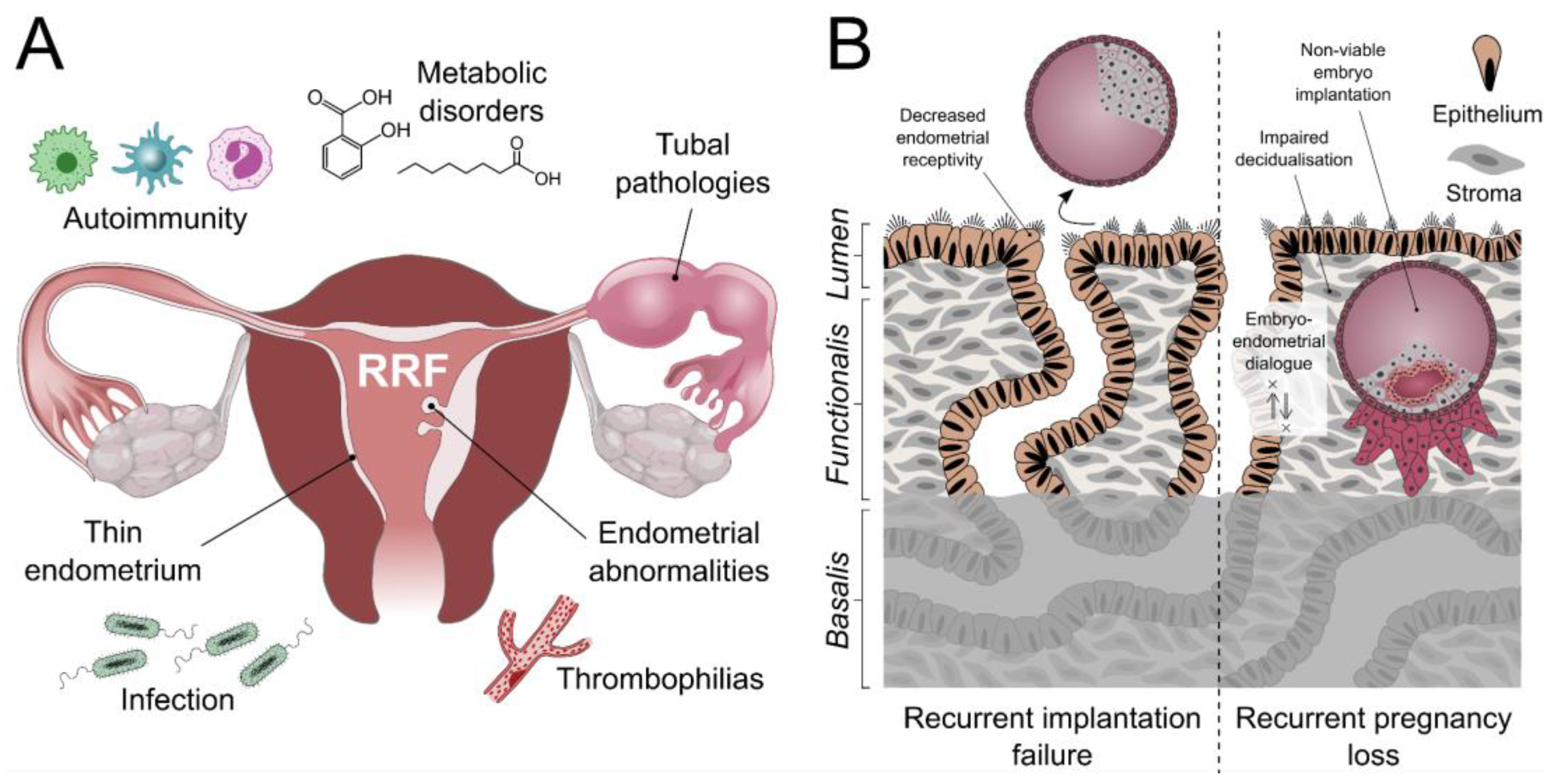
5. Endometrial Stem/Progenitor Cells and Recurrent Reproductive Failure (RRF)
5.1. Recurrent Implantation Failure (RIF)
5.2. Recurrent Pregnancy Loss (RPL)
6. Applications in Clinical Management of RRF
7. Avenues for Future Research
8. Discussion
Author Contributions
Funding
Institutional Review Board Statement
Informed Consent Statement
Acknowledgments
Conflicts of Interest
References
- Okada, H.; Tsuzuki, T.; Murata, H. Decidualization of the Human Endometrium. Reprod. Med. Biol. 2018, 17, 220–227. [Google Scholar] [CrossRef] [Green Version]
- Diedrich, K.; Fauser, B.C.J.M.; Devroey, P.; Griesinger, G.; on behalf of the Evian Annual Reproduction (EVAR) Workshop Group. The Role of the Endometrium and Embryo in Human Implantation. Hum. Reprod. Update 2007, 13, 365–377. [Google Scholar] [CrossRef]
- Hapangama, D.K.; Turner, M.A.; Drury, J.A.; Martin-Ruiz, C.; Von Zglinicki, T.; Farquharson, R.G.; Quenby, S. Endometrial Telomerase Shows Specific Expression Patterns in Different Types of Reproductive Failure. Reprod. Biomed. Online 2008, 17, 416–424. [Google Scholar] [CrossRef]
- Kim, S.-M.; Kim, J.-S. A Review of Mechanisms of Implantation. Dev. Reprod. 2017, 21, 351–359. [Google Scholar] [CrossRef] [Green Version]
- Quenby, S.; Nik, H.; Innes, B.; Lash, G.; Turner, M.; Drury, J.; Bulmer, J. Uterine Natural Killer Cells and Angiogenesis in Recurrent Reproductive Failure. Hum. Reprod. 2009, 24, 45–54. [Google Scholar] [CrossRef] [Green Version]
- Tempest, N.; Batchelor, E.; Hill, C.J.; Al-Lamee, H.; Drury, J.; Drakeley, A.J.; Hapangama, D.K. Anterior Gradient Protein 3 and S100 Calcium-Binding Protein P Levels in Different Endometrial Epithelial Compartments May Play an Important Role in Recurrent Pregnancy Failure. Int. J. Mol. Sci. 2021, 22, 3835. [Google Scholar] [CrossRef]
- Al-Lamee, H.; Ellison, A.; Drury, J.; Hill, C.J.; Drakeley, A.J.; Hapangama, D.K.; Tempest, N. Altered Endometrial Oestrogen-Responsiveness and Recurrent Reproductive Failure. Reprod. Fertil. 2022, 3, 30–38. [Google Scholar] [CrossRef]
- Busnelli, A.; Reschini, M.; Cardellicchio, L.; Vegetti, W.; Somigliana, E.; Vercellini, P. How Common Is Real Repeated Implantation Failure? An Indirect Estimate of the Prevalence. Reprod. Biomed. Online 2020, 40, 91–97. [Google Scholar] [CrossRef] [Green Version]
- Busnelli, A.; Somigliana, E.; Cirillo, F.; Baggiani, A.; Levi-Setti, P.E. Efficacy of Therapies and Interventions for Repeated Embryo Implantation Failure: A Systematic Review and Meta-Analysis. Sci. Rep. 2021, 11, 1747. [Google Scholar] [CrossRef]
- Polanski, L.T.; Baumgarten, M.N.; Quenby, S.; Brosens, J.; Campbell, B.K.; Raine-Fenning, N.J. What Exactly Do We Mean by “Recurrent Implantation Failure”? A Systematic Review and Opinion. Reprod. Biomed. Online 2014, 28, 409–423. [Google Scholar] [CrossRef] [Green Version]
- Practice Committee of the American Society for Reproductive Medicine. Definitions of Infertility and Recurrent Pregnancy Loss: A Committee Opinion. Fertil. Steril. 2013, 99, 63. [Google Scholar] [CrossRef]
- ESHRE Guideline Group on RPL; Bender Atik, R.; Christiansen, O.B.; Elson, J.; Kolte, A.M.; Lewis, S.; Middeldorp, S.; Nelen, W.; Peramo, B.; Quenby, S.; et al. ESHRE Guideline: Recurrent Pregnancy Loss. Hum. Reprod. Open 2018, 2018, hoy004. [Google Scholar] [CrossRef]
- El Hachem, H.; Crepaux, V.; May-Panloup, P.; Descamps, P.; Legendre, G.; Bouet, P.-E. Recurrent Pregnancy Loss: Current Perspectives. Int. J. Women’s Health 2017, 9, 331–345. [Google Scholar] [CrossRef] [Green Version]
- Royal College of Obstetritians and Gynaecologists (RCOG). The Investigation and Treatment of Couples with Recurrent First-Trimester and Second-Trimester Miscarriage; RCOG: London, UK, 2011. [Google Scholar]
- Farren, J.; Jalmbrant, M.; Falconieri, N.; Mitchell-Jones, N.; Bobdiwala, S.; Al-Memar, M.; Tapp, S.; Van Calster, B.; Wynants, L.; Timmerman, D.; et al. Posttraumatic Stress, Anxiety and Depression Following Miscarriage and Ectopic Pregnancy: A Multicenter, Prospective, Cohort Study. Am. J. Obstet. Gynecol. 2020, 222, 367.e1–367.e22. [Google Scholar] [CrossRef]
- Margalioth, E.J.; Ben-Chetrit, A.; Gal, M.; Eldar-Geva, T. Investigation and Treatment of Repeated Implantation Failure Following IVF-ET. Hum. Reprod. 2006, 21, 3036–3043. [Google Scholar] [CrossRef] [Green Version]
- Dimitriadis, E.; Menkhorst, E.; Saito, S.; Kutteh, W.H.; Brosens, J.J. Recurrent Pregnancy Loss. Nat. Rev. Dis. Primers 2020, 6, 98. [Google Scholar] [CrossRef]
- Bashiri, A.; Halper, K.I.; Orvieto, R. Recurrent Implantation Failure-Update Overview on Etiology, Diagnosis, Treatment and Future Directions. Reprod. Biol. Endocrinol. 2018, 16, 121. [Google Scholar] [CrossRef] [Green Version]
- Demirol, A.; Gurgan, T. Effect of Treatment of Intrauterine Pathologies with Office Hysteroscopy in Patients with Recurrent IVF Failure. Reprod. Biomed. Online 2004, 8, 590–594. [Google Scholar] [CrossRef]
- Turocy, J.M.; Rackow, B.W. Uterine Factor in Recurrent Pregnancy Loss. Semin. Perinatol. 2019, 43, 74–79. [Google Scholar] [CrossRef]
- Meyer, W.R.; Castelbaum, A.J.; Somkuti, S.; Sagoskin, A.W.; Doyle, M.; Harris, J.E.; Lessey, B.A. Hydrosalpinges Adversely Affect Markers of Endometrial Receptivity. Hum. Reprod. 1997, 12, 1393–1398. [Google Scholar] [CrossRef] [Green Version]
- Backos, M.; Rai, R.; Regan, L. Antiphospholipid Antibodies and Infertility. Hum. Fertil. 2002, 5, 30–34. [Google Scholar] [CrossRef]
- Pirtea, P.; Cicinelli, E.; De Nola, R.; de Ziegler, D.; Ayoubi, J.M. Endometrial Causes of Recurrent Pregnancy Losses: Endometriosis, Adenomyosis, and Chronic Endometritis. Fertil. Steril. 2021, 115, 546–560. [Google Scholar] [CrossRef]
- Mahajan, N.; Sharma, S. The Endometrium in Assisted Reproductive Technology: How Thin Is Thin? J. Hum. Reprod. Sci. 2016, 9, 3–8. [Google Scholar] [CrossRef]
- Vartanyan, E.; Tsaturova, K.; Devyatova, E. Thin Endometrium Problem in IVF Programs. Gynecol. Endocrinol. 2020, 36, 24–27. [Google Scholar] [CrossRef]
- Bellver, J.; Soares, S.R.; Alvarez, C.; Muñoz, E.; Ramírez, A.; Rubio, C.; Serra, V.; Remohí, J.; Pellicer, A. The Role of Thrombophilia and Thyroid Autoimmunity in Unexplained Infertility, Implantation Failure and Recurrent Spontaneous Abortion. Hum. Reprod. 2008, 23, 278–284. [Google Scholar] [CrossRef] [Green Version]
- Alecsandru, D.; Barrio, A.; Andia, V.; Cruz, E.; Aparicio, P.; Serna, J.; Cruz, M.; Pellicer, A.; Garcia-Velasco, J.A. Pancreatic Autoimmunity: An Unknown Etiology on Patients with Assisted Reproductive Techniques (ART)-Recurrent Reproductive Failure. PLoS ONE 2018, 13, e0203446. [Google Scholar] [CrossRef]
- Sheikhansari, G.; Soltani-Zangbar, M.S.; Pourmoghadam, Z.; Kamrani, A.; Azizi, R.; Aghebati-Maleki, L.; Danaii, S.; Koushaeian, L.; Hojat-Farsangi, M.; Yousefi, M. Oxidative Stress, Inflammatory Settings, and MicroRNA Regulation in the Recurrent Implantation Failure Patients with Metabolic Syndrome. Am. J. Reprod. Immunol. 2019, 82, e13170. [Google Scholar] [CrossRef]
- Broughton, D.E.; Moley, K.H. Obesity and Female Infertility: Potential Mediators of Obesity’s Impact. Fertil. Steril. 2017, 107, 840–847. [Google Scholar] [CrossRef] [Green Version]
- Vitagliano, A.; Saccardi, C.; Noventa, M.; Di Spiezio Sardo, A.; Saccone, G.; Cicinelli, E.; Pizzi, S.; Andrisani, A.; Litta, P.S. Effects of Chronic Endometritis Therapy on In Vitro Fertilization Outcome in Women with Repeated Implantation Failure: A Systematic Review and Meta-Analysis. Fertil. Steril. 2018, 110, 103–112.e1. [Google Scholar] [CrossRef]
- Odendaal, J.; Quenby, S.; Sammaritano, L.; Macklon, N.; Branch, D.W.; Rosenwaks, Z. Immunologic and Rheumatologic Causes and Treatment of Recurrent Pregnancy Loss: What Is the Evidence? Fertil. Steril. 2019, 112, 1002–1012. [Google Scholar] [CrossRef] [Green Version]
- Ticconi, C.; Pietropolli, A.; Di Simone, N.; Piccione, E.; Fazleabas, A. Endometrial Immune Dysfunction in Recurrent Pregnancy Loss. Int. J. Mol. Sci. 2019, 20, 5332. [Google Scholar] [CrossRef] [Green Version]
- Vomstein, K.; Voss, P.; Molnar, K.; Ainsworth, A.; Daniel, V.; Strowitzki, T.; Toth, B.; Kuon, R.-J. Two of a Kind? Immunological and Clinical Risk Factors Differ between Recurrent Implantation Failure and Recurrent Miscarriage. J. Reprod. Immunol. 2020, 141, 103166. [Google Scholar] [CrossRef] [PubMed]
- Valdes, C.T.; Schutt, A.; Simon, C. Implantation Failure of Endometrial Origin: It Is Not Pathology, but Our Failure to Synchronize the Developing Embryo with a Receptive Endometrium. Fertil. Steril. 2017, 108, 15–18. [Google Scholar] [CrossRef] [Green Version]
- Santamaria, X.; Simón, C. Endometrial Factor in Unexplained Infertility and Recurrent Implantation Failure. Semin. Reprod. Med. 2021, 39, 227–232. [Google Scholar] [CrossRef] [PubMed]
- Salker, M.; Teklenburg, G.; Molokhia, M.; Lavery, S.; Trew, G.; Aojanepong, T.; Mardon, H.J.; Lokugamage, A.U.; Rai, R.; Landles, C.; et al. Natural Selection of Human Embryos: Impaired Decidualization of Endometrium Disables Embryo-Maternal Interactions and Causes Recurrent Pregnancy Loss. PLoS ONE 2010, 5, e10287. [Google Scholar] [CrossRef] [PubMed] [Green Version]
- Teklenburg, G.; Salker, M.; Heijnen, C.; Macklon, N.S.; Brosens, J.J. The Molecular Basis of Recurrent Pregnancy Loss: Impaired Natural Embryo Selection. Mol. Hum. Reprod. 2010, 16, 886–895. [Google Scholar] [CrossRef] [Green Version]
- Simon, A.; Laufer, N. Repeated Implantation Failure: Clinical Approach. Fertil. Steril. 2012, 97, 1039–1043. [Google Scholar] [CrossRef]
- Quenby, S.; Anim-Somuah, M.; Kalumbi, C.; Farquharson, R.; Aplin, J.D. Different Types of Recurrent Miscarriage Are Associated with Varying Patterns of Adhesion Molecule Expression in Endometrium. Reprod. Biomed. Online 2007, 14, 224–234. [Google Scholar] [CrossRef]
- Yuan, J.; Cha, J.; Deng, W.; Bartos, A.; Sun, X.; Ho, H.-Y.H.; Borg, J.-P.; Yamaguchi, T.P.; Yang, Y.; Dey, S.K. Planar Cell Polarity Signaling in the Uterus Directs Appropriate Positioning of the Crypt for Embryo Implantation. Proc. Natl. Acad. Sci. USA 2016, 113, E8079–E8088. [Google Scholar] [CrossRef] [Green Version]
- Yuan, J.; Deng, W.; Cha, J.; Sun, X.; Borg, J.-P.; Dey, S.K. Tridimensional Visualization Reveals Direct Communication between the Embryo and Glands Critical for Implantation. Nat. Commun. 2018, 9, 603. [Google Scholar] [CrossRef]
- Salamonsen, L.A.; Evans, J.; Nguyen, H.P.T.; Edgell, T.A. The Microenvironment of Human Implantation: Determinant of Reproductive Success. Am. J. Reprod. Immunol. 2016, 75, 218–225. [Google Scholar] [CrossRef] [PubMed]
- Filant, J.; Spencer, T.E. Uterine Glands: Biological Roles in Conceptus Implantation, Uterine Receptivity and Decidualization. Int. J. Dev. Biol. 2014, 58, 107–116. [Google Scholar] [CrossRef] [PubMed]
- Kelleher, A.M.; Milano-Foster, J.; Behura, S.K.; Spencer, T.E. Uterine Glands Coordinate On-Time Embryo Implantation and Impact Endometrial Decidualization for Pregnancy Success. Nat. Commun. 2018, 9, 2435. [Google Scholar] [CrossRef]
- Nogales, F.; Martinez, H.; Parache, J. Shedding and reconstruction of the human endometrium. Gynakol. Rundsch. 1969, 7, 292–312. [Google Scholar] [CrossRef] [PubMed]
- Nogales-Ortiz, F.; Puerta, J.; Nogales, F.F. The Normal Menstrual Cycle. Chronology and Mechanism of Endometrial Desquamation. Obstet. Gynecol. 1978, 51, 259–264. [Google Scholar] [CrossRef] [PubMed]
- Ferenczy, A. Studies on the Cytodynamics of Human Endometrial Regeneration. I. Scanning Electron Microscopy. Am. J. Obstet. Gynecol. 1976, 124, 64–74. [Google Scholar] [CrossRef]
- Tempest, N.; Hill, C.J.; Maclean, A.; Marston, K.; Powell, S.G.; Al-Lamee, H.; Hapangama, D.K. Novel Microarchitecture of Human Endometrial Glands: Implications in Endometrial Regeneration and Pathologies. Hum. Reprod. Update 2022, 28, 153–171. [Google Scholar] [CrossRef]
- Kurman, R.J.; Ellenson, L.H.; Ronnett, B.M. Blaustein’s Pathology of the Female Genital Tract; Springer: Cham, Switzerland, 2019; ISBN 978-3-319-46333-9. [Google Scholar]
- Tempest, N.; Jansen, M.; Baker, A.-M.; Hill, C.J.; Hale, M.; Magee, D.; Treanor, D.; Wright, N.A.; Hapangama, D.K. Histological 3D Reconstruction and in Vivo Lineage Tracing of the Human Endometrium. J. Pathol. 2020, 251, 440–451. [Google Scholar] [CrossRef]
- Yamaguchi, M.; Yoshihara, K.; Suda, K.; Nakaoka, H.; Yachida, N.; Ueda, H.; Sugino, K.; Mori, Y.; Yamawaki, K.; Tamura, R.; et al. Three-Dimensional Understanding of the Morphological Complexity of the Human Uterine Endometrium. iScience 2021, 24, 102258. [Google Scholar] [CrossRef]
- Yamaguchi, M.; Yoshihara, K.; Yachida, N.; Suda, K.; Tamura, R.; Ishiguro, T.; Enomoto, T. The New Era of Three-Dimensional Histoarchitecture of the Human Endometrium. J. Pers. Med. 2021, 11, 713. [Google Scholar] [CrossRef]
- Chan, R.W.S.; Schwab, K.E.; Gargett, C.E. Clonogenicity of Human Endometrial Epithelial and Stromal Cells. Biol. Reprod. 2004, 70, 1738–1750. [Google Scholar] [CrossRef] [PubMed]
- Valentijn, A.J.; Palial, K.; Al-Lamee, H.; Tempest, N.; Drury, J.; Von Zglinicki, T.; Saretzki, G.; Murray, P.; Gargett, C.E.; Hapangama, D.K. SSEA-1 Isolates Human Endometrial Basal Glandular Epithelial Cells: Phenotypic and Functional Characterization and Implications in the Pathogenesis of Endometriosis. Hum. Reprod. 2013, 28, 2695–2708. [Google Scholar] [CrossRef] [PubMed] [Green Version]
- Fayazi, M.; Salehnia, M.; Ziaei, S. Characteristics of Human Endometrial Stem Cells in Tissue and Isolated Cultured Cells: An Immunohistochemical Aspect. Iran. Biomed. J. 2016, 20, 109–116. [Google Scholar] [CrossRef] [PubMed]
- Gargett, C.E.; Schwab, K.E.; Deane, J.A. Endometrial Stem/Progenitor Cells: The First 10 Years. Hum. Reprod. Update 2016, 22, 137–163. [Google Scholar] [CrossRef] [Green Version]
- Tempest, N.; Maclean, A.; Hapangama, D.K. Endometrial Stem Cell Markers: Current Concepts and Unresolved Questions. Int. J. Mol. Sci. 2018, 19, 3240. [Google Scholar] [CrossRef] [Green Version]
- Hapangama, D.K.; Turner, M.A.; Drury, J.A.; Quenby, S.; Saretzki, G.; Martin-Ruiz, C.; Von Zglinicki, T. Endometriosis Is Associated with Aberrant Endometrial Expression of Telomerase and Increased Telomere Length. Hum. Reprod. 2008, 23, 1511–1519. [Google Scholar] [CrossRef] [Green Version]
- Kyo, S.; Maida, Y.; Inoue, M. Stem Cells in Endometrium and Endometrial Cancer: Accumulating Evidence and Unresolved Questions. Cancer Lett. 2011, 308, 123–133. [Google Scholar] [CrossRef]
- Cousins, F.L.; Dorien, F.O.; Gargett, C.E. Endometrial Stem/Progenitor Cells and Their Role in the Pathogenesis of Endometriosis. Best Pract. Res. Clin. Obstet. Gynaecol. 2018, 50, 27–38. [Google Scholar] [CrossRef]
- Hapangama, D.K.; Drury, J.; Da Silva, L.; Al-Lamee, H.; Earp, A.; Valentijn, A.J.; Edirisinghe, D.P.; Murray, P.A.; Fazleabas, A.T.; Gargett, C.E. Abnormally Located SSEA1+/SOX9+ Endometrial Epithelial Cells with a Basalis-like Phenotype in the Eutopic Functionalis Layer May Play a Role in the Pathogenesis of Endometriosis. Hum. Reprod. 2019, 34, 56–68. [Google Scholar] [CrossRef]
- Strug, M.; Aghajanova, L. Making More Womb: Clinical Perspectives Supporting the Development and Utilization of Mesenchymal Stem Cell Therapy for Endometrial Regeneration and Infertility. J. Pers. Med. 2021, 11, 1364. [Google Scholar] [CrossRef]
- Dreisler, E.; Kjer, J.J. Asherman’s Syndrome: Current Perspectives on Diagnosis and Management. Int. J. Women’s Health 2019, 11, 191–198. [Google Scholar] [CrossRef] [PubMed]
- Diniz-da-Costa, M.; Kong, C.-S.; Fishwick, K.J.; Rawlings, T.; Brighton, P.J.; Hawkes, A.; Odendaal, J.; Quenby, S.; Ott, S.; Lucas, E.S.; et al. Characterization of Highly Proliferative Decidual Precursor Cells during the Window of Implantation in Human Endometrium. Stem Cells 2021, 39, 1067–1080. [Google Scholar] [CrossRef] [PubMed]
- De Miguel-Gómez, L.; Ferrero, H.; López-Martínez, S.; Campo, H.; López-Pérez, N.; Faus, A.; Hervás, D.; Santamaría, X.; Pellicer, A.; Cervelló, I. Stem Cell Paracrine Actions in Tissue Regeneration and Potential Therapeutic Effect in Human Endometrium: A Retrospective Study. BJOG Int. J. Obstet. Gynaecol. 2020, 127, 551–560. [Google Scholar] [CrossRef] [PubMed]
- Alawadhi, F.; Du, H.; Cakmak, H.; Taylor, H.S. Bone Marrow-Derived Stem Cell (BMDSC) Transplantation Improves Fertility in a Murine Model of Asherman’s Syndrome. PLoS ONE 2014, 9, e96662. [Google Scholar] [CrossRef] [PubMed]
- Singh, N.; Mohanty, S.; Seth, T.; Shankar, M.; Bhaskaran, S.; Dharmendra, S. Autologous Stem Cell Transplantation in Refractory Asherman’s Syndrome: A Novel Cell Based Therapy. J. Hum. Reprod. Sci. 2014, 7, 93–98. [Google Scholar] [CrossRef]
- Singh, N.; Shekhar, B.; Mohanty, S.; Kumar, S.; Seth, T.; Girish, B. Autologous Bone Marrow-Derived Stem Cell Therapy for Asherman’s Syndrome and Endometrial Atrophy: A 5-Year Follow-up Study. J. Hum. Reprod. Sci. 2020, 13, 31–37. [Google Scholar] [CrossRef]
- Yi, K.W.; Mamillapalli, R.; Sahin, C.; Song, J.; Tal, R.; Taylor, H.S. Bone Marrow-Derived Cells or C-X-C Motif Chemokine 12 (CXCL12) Treatment Improve Thin Endometrium in a Mouse Model. Biol. Reprod. 2019, 100, 61–70. [Google Scholar] [CrossRef]
- Lucas, E.S.; Dyer, N.P.; Fishwick, K.; Ott, S.; Brosens, J.J. Success after Failure: The Role of Endometrial Stem Cells in Recurrent Miscarriage. Reproduction 2016, 152, R159–R166. [Google Scholar] [CrossRef] [Green Version]
- Tempest, N.; Baker, A.M.; Wright, N.A.; Hapangama, D.K. Does Human Endometrial LGR5 Gene Expression Suggest the Existence of Another Hormonally Regulated Epithelial Stem Cell Niche? Hum. Reprod. 2018, 33, 1052–1062. [Google Scholar] [CrossRef] [Green Version]
- Valatkaitė, E.; Baušytė, R.; Vitkevičienė, A.; Ramašauskaitė, D.; Navakauskienė, R. Decidualization Potency and Epigenetic Changes in Human Endometrial Origin Stem Cells During Propagation. Front. Cell Dev. Biol. 2021, 9, 765265. [Google Scholar] [CrossRef]
- Gorsek Sparovec, T.; Markert, U.R.; Reif, P.; Schoell, W.; Moser, G.; Feichtinger, J.; Mihalic, Z.N.; Kargl, J.; Gargett, C.E.; Gold, D. The Fate of Human SUSD2+ Endometrial Mesenchymal Stem Cells during Decidualization. Stem Cell Res. 2022, 60, 102671. [Google Scholar] [CrossRef] [PubMed]
- Papanicolaou, G.N. Epithelial Regeneration in the Uterine Glands and on the Surface of the Uterus. Am. J. Obstet. Gynecol. 1933, 25, 30–37. [Google Scholar] [CrossRef]
- Prianishnikov, V.A. A Functional Model of the Structure of the Epithelium of Normal, Hyperplastic and Malignant Human Endometrium: A Review. Gynecol. Oncol. 1978, 6, 420–428. [Google Scholar] [CrossRef]
- Padykula, H.A.; Coles, L.G.; Okulicz, W.C.; Rapaport, S.I.; McCracken, J.A.; King, N.W.; Longcope, C.; Kaiserman-Abramof, I.R. The Basalis of the Primate Endometrium: A Bifunctional Germinal Compartment. Biol. Reprod. 1989, 40, 681–690. [Google Scholar] [CrossRef] [Green Version]
- Gargett, C.E. Uterine Stem Cells: What Is the Evidence? Hum. Reprod. Update 2007, 13, 87–101. [Google Scholar] [CrossRef]
- Cousins, F.L.; Pandoy, R.; Jin, S.; Gargett, C.E. The Elusive Endometrial Epithelial Stem/Progenitor Cells. Front. Cell Dev. Biol. 2021, 9, 868. [Google Scholar] [CrossRef]
- Fleischer, A.C. Sonographic Assessment of Endometrial Disorders. Semin. Ultrasound CT MRI 1999, 20, 259–266. [Google Scholar] [CrossRef]
- Tresserra, F.; Grases, P.; Ubeda, A.; Pascual, M.A.; Grases, P.J.; Labastida, R. Morphological Changes in Hysterectomies after Endometrial Ablation. Hum. Reprod. 1999, 14, 1473–1477. [Google Scholar] [CrossRef] [Green Version]
- Gargett, C.E. Identification and Characterisation of Human Endometrial Stem/Progenitor Cells. Aust. N. Z. J. Obstet. Gynaecol. 2006, 46, 250–253. [Google Scholar] [CrossRef]
- Hapangama, D.K.; Kamal, A.M.; Bulmer, J.N. Estrogen Receptor β: The Guardian of the Endometrium. Hum. Reprod. Update 2015, 21, 174–193. [Google Scholar] [CrossRef] [Green Version]
- Gargett, C.E.; Chan, R.W.S.; Schwab, K.E. Endometrial Stem Cells. Curr. Opin. Obstet. Gynecol. 2007, 19, 377–383. [Google Scholar] [CrossRef] [PubMed]
- Sourial, S.; Tempest, N.; Hapangama, D.K. Theories on the Pathogenesis of Endometriosis. Int. J. Reprod. Med. 2014, 2014, 179515. [Google Scholar] [CrossRef] [PubMed] [Green Version]
- Taylor, H.S. Endometrial Cells Derived from Donor Stem Cells in Bone Marrow Transplant Recipients. JAMA 2004, 292, 81–85. [Google Scholar] [CrossRef] [PubMed] [Green Version]
- Schwab, K.E.; Chan, R.W.S.; Gargett, C.E. Putative Stem Cell Activity of Human Endometrial Epithelial and Stromal Cells during the Menstrual Cycle. Fertil. Steril. 2005, 84 (Suppl. S2), 1124–1130. [Google Scholar] [CrossRef] [PubMed]
- Chan, R.W.S.; Gargett, C.E. Identification of Label-Retaining Cells in Mouse Endometrium. Stem Cells 2006, 24, 1529–1538. [Google Scholar] [CrossRef]
- Cervelló, I.; Martínez-Conejero, J.A.; Horcajadas, J.A.; Pellicer, A.; Simón, C. Identification, Characterization and Co-Localization of Label-Retaining Cell Population in Mouse Endometrium with Typical Undifferentiated Markers. Hum. Reprod. 2007, 22, 45–51. [Google Scholar] [CrossRef] [Green Version]
- Cervelló, I.; Gil-Sanchis, C.; Mas, A.; Delgado-Rosas, F.; Martínez-Conejero, J.A.; Galán, A.; Martínez-Romero, A.; Martínez, S.; Navarro, I.; Ferro, J.; et al. Human Endometrial Side Population Cells Exhibit Genotypic, Phenotypic and Functional Features of Somatic Stem Cells. PLoS ONE 2010, 5, e10964. [Google Scholar] [CrossRef] [Green Version]
- Cervelló, I.; Mas, A.; Gil-Sanchis, C.; Peris, L.; Faus, A.; Saunders, P.T.K.; Critchley, H.O.D.; Simón, C. Reconstruction of Endometrium from Human Endometrial Side Population Cell Lines. PLoS ONE 2011, 6, e21221. [Google Scholar] [CrossRef]
- Du, H.; Taylor, H.S. Contribution of Bone Marrow-Derived Stem Cells to Endometrium and Endometriosis. Stem Cells 2007, 25, 2082–2086. [Google Scholar] [CrossRef]
- Kato, K.; Yoshimoto, M.; Kato, K.; Adachi, S.; Yamayoshi, A.; Arima, T.; Asanoma, K.; Kyo, S.; Nakahata, T.; Wake, N. Characterization of Side-Population Cells in Human Normal Endometrium. Hum. Reprod. 2007, 22, 1214–1223. [Google Scholar] [CrossRef] [Green Version]
- Schwab, K.E.; Gargett, C.E. Co-Expression of Two Perivascular Cell Markers Isolates Mesenchymal Stem-like Cells from Human Endometrium. Hum. Reprod. 2007, 22, 2903–2911. [Google Scholar] [CrossRef] [PubMed] [Green Version]
- Götte, M.; Wolf, M.; Staebler, A.; Buchweitz, O.; Kelsch, R.; Schüring, A.N.; Kiesel, L. Increased Expression of the Adult Stem Cell Marker Musashi-1 in Endometriosis and Endometrial Carcinoma. J. Pathol. 2008, 215, 317–329. [Google Scholar] [CrossRef] [PubMed]
- Du, H.; Naqvi, H.; Taylor, H.S. Ischemia/Reperfusion Injury Promotes and Granulocyte-Colony Stimulating Factor Inhibits Migration of Bone Marrow-Derived Stem Cells to Endometrium. Stem Cells Dev. 2012, 21, 3324–3331. [Google Scholar] [CrossRef] [PubMed] [Green Version]
- Masuda, H.; Anwar, S.S.; Bühring, H.-J.; Rao, J.R.; Gargett, C.E. A Novel Marker of Human Endometrial Mesenchymal Stem-like Cells. Cell Transplant. 2012, 21, 2201–2214. [Google Scholar] [CrossRef]
- Kim, J.Y.; Tavaré, S.; Shibata, D. Counting Human Somatic Cell Replications: Methylation Mirrors Endometrial Stem Cell Divisions. Proc. Natl. Acad. Sci. USA 2005, 102, 17739–17744. [Google Scholar] [CrossRef] [PubMed] [Green Version]
- Potten, C.S.; Loeffler, M. Stem Cells: Attributes, Cycles, Spirals, Pitfalls and Uncertainties. Lessons for and from the Crypt. Development 1990, 110, 1001–1020. [Google Scholar] [CrossRef]
- Gargett, C.E. Review Article: Stem Cells in Human Reproduction. Reprod. Sci. 2007, 14, 405–424. [Google Scholar] [CrossRef]
- Gargett, C.E.; Schwab, K.E.; Zillwood, R.M.; Nguyen, H.P.T.; Wu, D. Isolation and Culture of Epithelial Progenitors and Mesenchymal Stem Cells from Human Endometrium. Biol. Reprod. 2009, 80, 1136–1145. [Google Scholar] [CrossRef] [Green Version]
- Fayazi, M.; Salehnia, M.; Ziaei, S. Differentiation of Human CD146-Positive Endometrial Stem Cells to Adipogenic-, Osteogenic-, Neural Progenitor-, and Glial-like Cells. In Vitro Cell. Dev. Biol.-Anim. 2015, 51, 408–414. [Google Scholar] [CrossRef]
- Azami, M.; Ai, J.; Ebrahimi-Barough, S.; Farokhi, M.; Fard, S.E. In Vitro Evaluation of Biomimetic Nanocomposite Scaffold Using Endometrial Stem Cell Derived Osteoblast-like Cells. Tissue Cell 2013, 45, 328–337. [Google Scholar] [CrossRef]
- Shoae-Hassani, A.; Sharif, S.; Seifalian, A.M.; Mortazavi-Tabatabaei, S.A.; Rezaie, S.; Verdi, J. Endometrial Stem Cell Differentiation into Smooth Muscle Cell: A Novel Approach for Bladder Tissue Engineering in Women. BJU Int. 2013, 112, 854–863. [Google Scholar] [CrossRef] [PubMed]
- Trapero, C.; Vidal, A.; Rodríguez-Martínez, A.; Sévigny, J.; Ponce, J.; Coroleu, B.; Matias-Guiu, X.; Martín-Satué, M. The Ectonucleoside Triphosphate Diphosphohydrolase-2 (NTPDase2) in Human Endometrium: A Novel Marker of Basal Stroma and Mesenchymal Stem Cells. Purinergic Signal. 2019, 15, 225–236. [Google Scholar] [CrossRef] [PubMed]
- Nguyen, H.P.T.; Xiao, L.; Deane, J.A.; Tan, K.-S.; Cousins, F.L.; Masuda, H.; Sprung, C.N.; Rosamilia, A.; Gargett, C.E. N-Cadherin Identifies Human Endometrial Epithelial Progenitor Cells by In Vitro Stem Cell Assays. Hum. Reprod. 2017, 32, 2254–2268. [Google Scholar] [CrossRef] [Green Version]
- Nguyen, H.P.T.; Sprung, C.N.; Gargett, C.E. Differential Expression of Wnt Signaling Molecules between Pre- and Postmenopausal Endometrial Epithelial Cells Suggests a Population of Putative Epithelial Stem/Progenitor Cells Reside in the Basalis Layer. Endocrinology 2012, 153, 2870–2883. [Google Scholar] [CrossRef]
- Syed, S.M.; Kumar, M.; Ghosh, A.; Tomasetig, F.; Ali, A.; Whan, R.M.; Alterman, D.; Tanwar, P.S. Endometrial Axin2+ Cells Drive Epithelial Homeostasis, Regeneration, and Cancer Following Oncogenic Transformation. Cell Stem Cell 2020, 26, 64–80.e13. [Google Scholar] [CrossRef]
- Sampson, J.A. Metastatic or Embolic Endometriosis, Due to the Menstrual Dissemination of Endometrial Tissue into the Venous Circulation. Am. J. Pathol. 1927, 3, 93–110.43. [Google Scholar]
- Leyendecker, G.; Herbertz, M.; Kunz, G.; Mall, G. Endometriosis Results from the Dislocation of Basal Endometrium. Hum. Reprod. 2002, 17, 2725–2736. [Google Scholar] [CrossRef] [Green Version]
- Macer, M.L.; Taylor, H.S. Endometriosis and Infertility: A Review of the Pathogenesis and Treatment of Endometriosis-Associated Infertility. Obstet. Gynecol. Clin. N. Am. 2012, 39, 535–549. [Google Scholar] [CrossRef] [Green Version]
- Wang, H.; Dey, S.K. Roadmap to Embryo Implantation: Clues from Mouse Models. Nat. Rev. Genet. 2006, 7, 185–199. [Google Scholar] [CrossRef]
- Achache, H.; Revel, A. Endometrial Receptivity Markers, the Journey to Successful Embryo Implantation. Hum. Reprod. Update 2006, 12, 731–746. [Google Scholar] [CrossRef] [Green Version]
- Grewal, S.; Carver, J.G.; Ridley, A.J.; Mardon, H.J. Implantation of the Human Embryo Requires Rac1-Dependent Endometrial Stromal Cell Migration. Proc. Natl. Acad. Sci. USA 2008, 105, 16189–16194. [Google Scholar] [CrossRef] [PubMed] [Green Version]
- Lessey, B.A.; Castelbaum, A.J.; Sawin, S.W.; Sun, J. Integrins as Markers of Uterine Receptivity in Women with Primary Unexplained Infertility. Fertil. Steril. 1995, 63, 535–542. [Google Scholar] [CrossRef]
- Esmaeilzadeh, S.; Mohammadi, A.; Mahdinejad, N.; Ghofrani, F.; Ghasemzadeh-Hasankolaei, M. Receptivity Markers in Endometrial Mesenchymal Stem Cells of Recurrent Implantation Failure and Non-Recurrent Implantation Failure Women: A Pilot Study. J. Obstet. Gynaecol. Res. 2020, 46, 1393–1402. [Google Scholar] [CrossRef]
- Dominici, M.; Le Blanc, K.; Mueller, I.; Slaper-Cortenbach, I.; Marini, F.; Krause, D.; Deans, R.; Keating, A.; Prockop, D.; Horwitz, E. Minimal Criteria for Defining Multipotent Mesenchymal Stromal Cells. The International Society for Cellular Therapy Position Statement. Cytotherapy 2006, 8, 315–317. [Google Scholar] [CrossRef] [PubMed]
- Chau, Y.M.; Pando, S.; Taylor, H.S. HOXA11 Silencing and Endogenous HOXA11 Antisense Ribonucleic Acid in the Uterine Endometrium. J. Clin. Endocrinol. Metab. 2002, 87, 2674–2680. [Google Scholar] [CrossRef] [PubMed]
- He, B.; Ni, Z.-L.; Kong, S.-B.; Lu, J.-H.; Wang, H.-B. Homeobox Genes for Embryo Implantation: From Mouse to Human. Anim. Models Exp. Med. 2018, 1, 14–22. [Google Scholar] [CrossRef] [Green Version]
- Docheva, D.; Popov, C.; Mutschler, W.; Schieker, M. Human Mesenchymal Stem Cells in Contact with Their Environment: Surface Characteristics and the Integrin System. J. Cell. Mol. Med. 2007, 11, 21–38. [Google Scholar] [CrossRef]
- Merviel, P.; Challier, J.C.; Carbillon, L.; Foidart, J.M.; Uzan, S. The Role of Integrins in Human Embryo Implantation. Fetal Diagn. Ther. 2001, 16, 364–371. [Google Scholar] [CrossRef]
- Zhao, H.; Hu, S.; Qi, J.; Wang, Y.; Ding, Y.; Zhu, Q.; He, Y.; Lu, Y.; Yao, Y.; Wang, S.; et al. Increased Expression of HOXA11-AS Attenuates Endometrial Decidualization in Recurrent Implantation Failure Patients. Mol. Ther. 2022, 30, 1706–1720. [Google Scholar] [CrossRef]
- Revel, A.; Koler, M.; Prus, D.; Tsafrir, A.; Laufer, N.; Reich, R. Implementation of Integrin Β3 Level as Predictor of Implantation in an IVF Program. Fertil. Steril. 2005, 84, S144. [Google Scholar] [CrossRef]
- Gendron, R.L.; Paradis, H.; Hsieh-Li, H.M.; Lee, D.W.; Potter, S.S.; Markoff, E. Abnormal Uterine Stromal and Glandular Function Associated with Maternal Reproductive Defects in Hoxa-11 Null Mice. Biol. Reprod. 1997, 56, 1097–1105. [Google Scholar] [CrossRef] [PubMed]
- Quenby, S.; Vince, G.; Farquharson, R.; Aplin, J. Recurrent Miscarriage: A Defect in Nature’s Quality Control? Hum. Reprod. 2002, 17, 1959–1963. [Google Scholar] [CrossRef] [PubMed] [Green Version]
- Lucas, E.S.; Dyer, N.P.; Murakami, K.; Lee, Y.H.; Chan, Y.-W.; Grimaldi, G.; Muter, J.; Brighton, P.J.; Moore, J.D.; Patel, G.; et al. Loss of Endometrial Plasticity in Recurrent Pregnancy Loss. Stem Cells 2016, 34, 346–356. [Google Scholar] [CrossRef] [PubMed] [Green Version]
- Gargett, C.E.; Nguyen, H.P.T.; Ye, L. Endometrial Regeneration and Endometrial Stem/Progenitor Cells. Rev. Endocr. Metab. Disord. 2012, 13, 235–251. [Google Scholar] [CrossRef]
- He, Y.; Han, Y.; Ye, Y. Therapeutic Potential of Menstrual Blood-Derived Stem Cell Transplantation for Intrauterine Adhesions. Front. Surg. 2022, 9, 847213. [Google Scholar] [CrossRef]
- Lebovitz, O.; Orvieto, R. Treating Patients with “Thin” Endometrium—An Ongoing Challenge. Gynecol. Endocrinol. 2014, 30, 409–414. [Google Scholar] [CrossRef]
- Timeva, T.; Shterev, A.; Kyurkchiev, S. Recurrent Implantation Failure: The Role of the Endometrium. J. Reprod. Infertil. 2014, 15, 173–183. [Google Scholar]
- Liu, S.-M.; Zhou, Y.-Z.; Wang, H.-B.; Sun, Z.-Y.; Zhen, J.-R.; Shen, K.; Deng, C.-Y.; Lang, J.-H. Factors Associated with Effectiveness of Treatment and Reproductive Outcomes in Patients with Thin Endometrium Undergoing Estrogen Treatment. Chin. Med. J. 2015, 128, 3173–3177. [Google Scholar] [CrossRef]
- Liu, K.E.; Hartman, M.; Hartman, A. Management of Thin Endometrium in Assisted Reproduction: A Clinical Practice Guideline from the Canadian Fertility and Andrology Society. Reprod. Biomed. Online 2019, 39, 49–62. [Google Scholar] [CrossRef] [Green Version]
- Vaegter, K.K.; Lakic, T.G.; Olovsson, M.; Berglund, L.; Brodin, T.; Holte, J. Which Factors Are Most Predictive for Live Birth after In Vitro Fertilization and Intracytoplasmic Sperm Injection (IVF/ICSI) Treatments? Analysis of 100 Prospectively Recorded Variables in 8400 IVF/ICSI Single-Embryo Transfers. Fertil. Steril. 2017, 107, 641–648.e2. [Google Scholar] [CrossRef] [Green Version]
- Gallos, I.D.; Khairy, M.; Chu, J.; Rajkhowa, M.; Tobias, A.; Campbell, A.; Dowell, K.; Fishel, S.; Coomarasamy, A. Optimal Endometrial Thickness to Maximize Live Births and Minimize Pregnancy Losses: Analysis of 25,767 Fresh Embryo Transfers. Reprod. Biomed. Online 2018, 37, 542–548. [Google Scholar] [CrossRef] [PubMed]
- Liu, K.E.; Hartman, M.; Hartman, A.; Luo, Z.-C.; Mahutte, N. The Impact of a Thin Endometrial Lining on Fresh and Frozen-Thaw IVF Outcomes: An Analysis of over 40,000 Embryo Transfers. Hum. Reprod. 2018, 33, 1883–1888. [Google Scholar] [CrossRef] [PubMed] [Green Version]
- Gharibeh, N.; Aghebati-Maleki, L.; Madani, J.; Pourakbari, R.; Yousefi, M.; Ahmadian Heris, J. Cell-Based Therapy in Thin Endometrium and Asherman Syndrome. Stem Cell Res. Ther. 2022, 13, 33. [Google Scholar] [CrossRef] [PubMed]
- Deane, J.A.; Gualano, R.C.; Gargett, C.E. Regenerating Endometrium from Stem/Progenitor Cells: Is It Abnormal in Endometriosis, Asherman’s Syndrome and Infertility? Curr. Opin. Obstet. Gynecol. 2013, 25, 193–200. [Google Scholar] [CrossRef] [PubMed]
- Jing, Z.; Qiong, Z.; Yonggang, W.; Yanping, L. Rat Bone Marrow Mesenchymal Stem Cells Improve Regeneration of Thin Endometrium in Rat. Fertil. Steril. 2014, 101, 587–594. [Google Scholar] [CrossRef] [PubMed]
- Azizi, R.; Aghebati-Maleki, L.; Nouri, M.; Marofi, F.; Negargar, S.; Yousefi, M. Stem Cell Therapy in Asherman Syndrome and Thin Endometrium: Stem Cell- Based Therapy. Biomed. Pharmacother. 2018, 102, 333–343. [Google Scholar] [CrossRef]
- Abuwala, N.; Tal, R. Endometrial Stem Cells: Origin, Biological Function, and Therapeutic Applications for Reproductive Disorders. Curr. Opin. Obstet. Gynecol. 2021, 33, 232–240. [Google Scholar] [CrossRef]
- Zhang, Y.; Shi, L.; Lin, X.; Zhou, F.; Xin, L.; Xu, W.; Yu, H.; Li, J.; Pan, M.; Pan, Y.; et al. Unresponsive Thin Endometrium Caused by Asherman Syndrome Treated with Umbilical Cord Mesenchymal Stem Cells on Collagen Scaffolds: A Pilot Study. Stem Cell Res. Ther. 2021, 12, 420. [Google Scholar] [CrossRef]
- Mouanness, M.; Ali-Bynom, S.; Jackman, J.; Seckin, S.; Merhi, Z. Use of Intra-Uterine Injection of Platelet-Rich Plasma (PRP) for Endometrial Receptivity and Thickness: A Literature Review of the Mechanisms of Action. Reprod. Sci. 2021, 28, 1659–1670. [Google Scholar] [CrossRef]
- Strohmer, H.; Obruca, A.; Radner, K.M.; Feichtinger, W. Relationship of the Individual Uterine Size and the Endometrial Thickness in Stimulated Cycles. Fertil. Steril. 1994, 61, 972–975. [Google Scholar] [CrossRef]
- Scioscia, M.; Lamanna, G.; Lorusso, F.; Serrati, G.; Selvaggi, L.E.; Depalo, R. Characterization of Endometrial Growth in Proliferative and Early Luteal Phase in IVF Cycles. Reprod. Biomed. Online 2009, 18, 73–78. [Google Scholar] [CrossRef]
- Hu, J.; Song, K.; Zhang, J.; Zhang, Y.; Tan, B.-Z. Effects of Menstrual Blood-Derived Stem Cells on Endometrial Injury Repair. Mol. Med. Rep. 2019, 19, 813–820. [Google Scholar] [CrossRef] [PubMed] [Green Version]
- Zhao, J.; Zhang, Q.; Wang, Y.; Li, Y. Uterine Infusion with Bone Marrow Mesenchymal Stem Cells Improves Endometrium Thickness in a Rat Model of Thin Endometrium. Reprod. Sci. 2015, 22, 181–188. [Google Scholar] [CrossRef] [PubMed] [Green Version]
- Conforti, A.; Alviggi, C.; Mollo, A.; De Placido, G.; Magos, A. The Management of Asherman Syndrome: A Review of Literature. Reprod. Biol. Endocrinol. 2013, 11, 118. [Google Scholar] [CrossRef] [Green Version]
- Khan, Z.; Goldberg, J.M. Hysteroscopic Management of Asherman’s Syndrome. J. Minim. Invasive Gynecol. 2018, 25, 218–228. [Google Scholar] [CrossRef]
- Hanstede, M.M.F.; van der Meij, E.; Goedemans, L.; Emanuel, M.H. Results of Centralized Asherman Surgery, 2003–2013. Fertil. Steril. 2015, 104, 1561–1568.e1. [Google Scholar] [CrossRef] [Green Version]
- Chen, L.; Zhang, H.; Wang, Q.; Xie, F.; Gao, S.; Song, Y.; Dong, J.; Feng, H.; Xie, K.; Sui, L. Reproductive Outcomes in Patients With Intrauterine Adhesions Following Hysteroscopic Adhesiolysis: Experience From the Largest Women’s Hospital in China. J. Minim. Invasive Gynecol. 2017, 24, 299–304. [Google Scholar] [CrossRef]
- Min, J.; Lu, N.; Huang, S.; Chai, X.; Wang, S.; Peng, L.; Wang, J. Phenotype and Biological Characteristics of Endometrial Mesenchymal Stem/Stromal Cells: A Comparison between Intrauterine Adhesion Patients and Healthy Women. Am. J. Reprod. Immunol. 2021, 85, e13379. [Google Scholar] [CrossRef]
- Zhang, Y.; Lin, X.; Dai, Y.; Hu, X.; Zhu, H.; Jiang, Y.; Zhang, S. Endometrial Stem Cells Repair Injured Endometrium and Induce Angiogenesis via AKT and ERK Pathways. Reproduction 2016, 152, 389–402. [Google Scholar] [CrossRef] [Green Version]
- Tewary, S.; Lucas, E.S.; Fujihara, R.; Kimani, P.K.; Polanco, A.; Brighton, P.J.; Muter, J.; Fishwick, K.J.; Da Costa, M.J.M.D.; Ewington, L.J.; et al. Impact of Sitagliptin on Endometrial Mesenchymal Stem-like Progenitor Cells: A Randomised, Double-Blind Placebo-Controlled Feasibility Trial. EBioMedicine 2020, 51, 102597. [Google Scholar] [CrossRef] [Green Version]
- Vella, A. Mechanism of Action of DPP-4 Inhibitors—New Insights. J. Clin. Endocrinol. Metab. 2012, 97, 2626–2628. [Google Scholar] [CrossRef] [PubMed] [Green Version]
- Kieffer, T.J.; Mc Intosh, C.H.S.; Pederson, R.A. Degradation of Glucose-Dependent Insulinotropic Polypeptide and Truncated Glucagon-like Peptide 1 In Vitro and In Vivo by Dipeptidyl Peptidase Iv. Endocrinology 1995, 136, 3585–3596. [Google Scholar] [CrossRef] [PubMed]
- Zhong, J.; Rajagopalan, S. Dipeptidyl Peptidase-4 Regulation of SDF-1/CXCR4 Axis: Implications for Cardiovascular Disease. Front. Immunol. 2015, 6, 477. [Google Scholar] [CrossRef] [PubMed] [Green Version]
- Munné, S.; Kaplan, B.; Frattarelli, J.L.; Child, T.; Nakhuda, G.; Shamma, F.N.; Silverberg, K.; Kalista, T.; Handyside, A.H.; Katz-Jaffe, M.; et al. Preimplantation Genetic Testing for Aneuploidy versus Morphology as Selection Criteria for Single Frozen-Thawed Embryo Transfer in Good-Prognosis Patients: A Multicenter Randomized Clinical Trial. Fertil. Steril. 2019, 112, 1071–1079.e7. [Google Scholar] [CrossRef]
- Nagori, C.B.; Panchal, S.Y.; Patel, H. Endometrial Regeneration Using Autologous Adult Stem Cells Followed by Conception by In Vitro Fertilization in a Patient of Severe Asherman’s Syndrome. J. Hum. Reprod. Sci. 2011, 4, 43–48. [Google Scholar] [CrossRef]
- Santamaria, X.; Cabanillas, S.; Cervelló, I.; Arbona, C.; Raga, F.; Ferro, J.; Palmero, J.; Remohí, J.; Pellicer, A.; Simón, C. Autologous Cell Therapy with CD133+ Bone Marrow-Derived Stem Cells for Refractory Asherman’s Syndrome and Endometrial Atrophy: A Pilot Cohort Study. Hum. Reprod. 2016, 31, 1087–1096. [Google Scholar] [CrossRef] [Green Version]
- Zhang, L.; Li, Y.; Guan, C.-Y.; Tian, S.; Lv, X.-D.; Li, J.-H.; Ma, X.; Xia, H.-F. Therapeutic Effect of Human Umbilical Cord-Derived Mesenchymal Stem Cells on Injured Rat Endometrium during Its Chronic Phase. Stem Cell Res. Ther. 2018, 9, 36. [Google Scholar] [CrossRef] [Green Version]
- Xin, L.; Lin, X.; Pan, Y.; Zheng, X.; Shi, L.; Zhang, Y.; Ma, L.; Gao, C.; Zhang, S. A Collagen Scaffold Loaded with Human Umbilical Cord-Derived Mesenchymal Stem Cells Facilitates Endometrial Regeneration and Restores Fertility. Acta Biomater. 2019, 92, 160–171. [Google Scholar] [CrossRef]
- Cao, Y.; Sun, H.; Zhu, H.; Zhu, X.; Tang, X.; Yan, G.; Wang, J.; Bai, D.; Wang, J.; Wang, L.; et al. Allogeneic Cell Therapy Using Umbilical Cord MSCs on Collagen Scaffolds for Patients with Recurrent Uterine Adhesion: A Phase I Clinical Trial. Stem Cell Res. Ther. 2018, 9, 192. [Google Scholar] [CrossRef] [Green Version]
- Gan, L.; Duan, H.; Xu, Q.; Tang, Y.-Q.; Li, J.-J.; Sun, F.-Q.; Wang, S. Human Amniotic Mesenchymal Stromal Cell Transplantation Improves Endometrial Regeneration in Rodent Models of Intrauterine Adhesions. Cytotherapy 2017, 19, 603–616. [Google Scholar] [CrossRef]
- Li, B.; Zhang, Q.; Sun, J.; Lai, D. Human Amniotic Epithelial Cells Improve Fertility in an Intrauterine Adhesion Mouse Model. Stem Cell Res. Ther. 2019, 10, 257. [Google Scholar] [CrossRef] [PubMed] [Green Version]
- Ouyang, X.; You, S.; Zhang, Y.; Zhang, C.; Zhang, G.; Shao, X.; He, F.; Hu, L. Transplantation of Human Amnion Epithelial Cells Improves Endometrial Regeneration in Rat Model of Intrauterine Adhesions. Stem Cells Dev. 2020, 29, 1346–1362. [Google Scholar] [CrossRef] [PubMed]
- Bai, X.; Liu, J.; Yuan, W.; Liu, Y.; Li, W.; Cao, S.; Yu, L.; Wang, L. Therapeutic Effect of Human Amniotic Epithelial Cells in Rat Models of Intrauterine Adhesions. Cell Transplant. 2020, 29, 963689720908495. [Google Scholar] [CrossRef] [PubMed] [Green Version]
- Choi, J.; Jo, M.; Lee, E.; Oh, Y.K.; Choi, D. The Role of Autophagy in Human Endometrium. Biol. Reprod. 2012, 86, 70. [Google Scholar] [CrossRef]
- Kilic, S.; Yuksel, B.; Pinarli, F.; Albayrak, A.; Boztok, B.; Delibasi, T. Effect of Stem Cell Application on Asherman Syndrome, an Experimental Rat Model. J. Assist. Reprod. Genet. 2014, 31, 975–982. [Google Scholar] [CrossRef] [Green Version]
- Shao, X.; Ai, G.; Wang, L.; Qin, J.; Li, Y.; Jiang, H.; Zhang, T.; Zhou, L.; Gao, Z.; Cheng, J.; et al. Adipose-Derived Stem Cells Transplantation Improves Endometrial Injury Repair. Zygote 2019, 27, 367–374. [Google Scholar] [CrossRef]
- Monsef, F.; Artimani, T.; Alizadeh, Z.; Ramazani, M.; Solgi, G.; Yavangi, M.; Asl, S.S. Comparison of the Regenerative Effects of Bone Marrow/Adipose-Derived Stem Cells in the Asherman Model Following Local or Systemic Administration. J. Assist. Reprod. Genet. 2020, 37, 1861–1868. [Google Scholar] [CrossRef]
- Lee, S.Y.; Shin, J.E.; Kwon, H.; Choi, D.H.; Kim, J.H. Effect of Autologous Adipose-Derived Stromal Vascular Fraction Transplantation on Endometrial Regeneration in Patients of Asherman’s Syndrome: A Pilot Study. Reprod. Sci. 2020, 27, 561–568. [Google Scholar] [CrossRef]
- Rawlings, T.M.; Makwana, K.; Tryfonos, M.; Lucas, E.S. Organoids to Model the Endometrium: Implantation and Beyond. Reprod. Fertil. 2021, 2, R85–R101. [Google Scholar] [CrossRef]
- Turco, M.Y.; Gardner, L.; Hughes, J.; Cindrova-Davies, T.; Gomez, M.J.; Farrell, L.; Hollinshead, M.; Marsh, S.G.E.; Brosens, J.J.; Critchley, H.O.; et al. Long-Term, Hormone-Responsive Organoid Cultures of Human Endometrium in a Chemically Defined Medium. Nat. Cell Biol. 2017, 19, 568–577. [Google Scholar] [CrossRef]
- Luddi, A.; Pavone, V.; Semplici, B.; Governini, L.; Criscuoli, M.; Paccagnini, E.; Gentile, M.; Morgante, G.; Leo, V.D.; Belmonte, G.; et al. Organoids of Human Endometrium: A Powerful In Vitro Model for the Endometrium-Embryo Cross-Talk at the Implantation Site. Cells 2020, 9, 1121. [Google Scholar] [CrossRef] [PubMed]
- Kim, J.; Koo, B.-K.; Knoblich, J.A. Human Organoids: Model Systems for Human Biology and Medicine. Nat. Rev. Mol. Cell Biol. 2020, 21, 571–584. [Google Scholar] [CrossRef] [PubMed]
- Zheng, Y.; Xue, X.; Shao, Y.; Wang, S.; Esfahani, S.N.; Li, Z.; Muncie, J.M.; Lakins, J.N.; Weaver, V.M.; Gumucio, D.L.; et al. Controlled Modelling of Human Epiblast and Amnion Development Using Stem Cells. Nature 2019, 573, 421–425. [Google Scholar] [CrossRef] [PubMed]
- Liu, X.; Tan, J.P.; Schröder, J.; Aberkane, A.; Ouyang, J.F.; Mohenska, M.; Lim, S.M.; Sun, Y.B.Y.; Chen, J.; Sun, G.; et al. Modelling Human Blastocysts by Reprogramming Fibroblasts into IBlastoids. Nature 2021, 591, 627–632. [Google Scholar] [CrossRef] [PubMed]
- Yu, L.; Wei, Y.; Duan, J.; Schmitz, D.A.; Sakurai, M.; Wang, L.; Wang, K.; Zhao, S.; Hon, G.C.; Wu, J. Blastocyst-like Structures Generated from Human Pluripotent Stem Cells. Nature 2021, 591, 620–626. [Google Scholar] [CrossRef]
- Haider, S.; Meinhardt, G.; Saleh, L.; Kunihs, V.; Gamperl, M.; Kaindl, U.; Ellinger, A.; Burkard, T.R.; Fiala, C.; Pollheimer, J.; et al. Self-Renewing Trophoblast Organoids Recapitulate the Developmental Program of the Early Human Placenta. Stem Cell Rep. 2018, 11, 537–551. [Google Scholar] [CrossRef] [Green Version]
- Turco, M.Y.; Gardner, L.; Kay, R.G.; Hamilton, R.S.; Prater, M.; Hollinshead, M.S.; McWhinnie, A.; Esposito, L.; Fernando, R.; Skelton, H.; et al. Trophoblast Organoids as a Model for Maternal-Fetal Interactions during Human Placentation. Nature 2018, 564, 263–267. [Google Scholar] [CrossRef] [Green Version]
- Rinehart, C.A.; Lyn-Cook, B.D.; Kaufman, D.G. Gland Formation from Human Endometrial Epithelial Cells In Vitro. In Vitro Cell. Dev. Biol. 1988, 24, 1037–1041. [Google Scholar] [CrossRef]
- Bläuer, M.; Heinonen, P.K.; Martikainen, P.M.; Tomás, E.; Ylikomi, T. A Novel Organotypic Culture Model for Normal Human Endometrium: Regulation of Epithelial Cell Proliferation by Estradiol and Medroxyprogesterone Acetate. Hum. Reprod. 2005, 20, 864–871. [Google Scholar] [CrossRef] [Green Version]
- Boretto, M.; Maenhoudt, N.; Luo, X.; Hennes, A.; Boeckx, B.; Bui, B.; Heremans, R.; Perneel, L.; Kobayashi, H.; Van Zundert, I.; et al. Patient-Derived Organoids from Endometrial Disease Capture Clinical Heterogeneity and Are Amenable to Drug Screening. Nat. Cell Biol. 2019, 21, 1041–1051. [Google Scholar] [CrossRef]
- Hill, C.J.; Alijassim, F.; Phelan, M.M.; Tempest, N.; Hapangama, D.K. Bipartite Collagen-Agarose Scaffolds to Model Eutopic Embryo Implantation. In Proceedings of the Reproductive Sciences, Denver, CO, USA, 1 March 2022; Volume 29, p. 185. [Google Scholar]
Publisher’s Note: MDPI stays neutral with regard to jurisdictional claims in published maps and institutional affiliations. |
© 2022 by the authors. Licensee MDPI, Basel, Switzerland. This article is an open access article distributed under the terms and conditions of the Creative Commons Attribution (CC BY) license (https://creativecommons.org/licenses/by/4.0/).
Share and Cite
Al-Lamee, H.; Hill, C.J.; Turner, F.; Phan, T.; Drakeley, A.J.; Hapangama, D.K.; Tempest, N. The Role of Endometrial Stem/Progenitor Cells in Recurrent Reproductive Failure. J. Pers. Med. 2022, 12, 775. https://doi.org/10.3390/jpm12050775
Al-Lamee H, Hill CJ, Turner F, Phan T, Drakeley AJ, Hapangama DK, Tempest N. The Role of Endometrial Stem/Progenitor Cells in Recurrent Reproductive Failure. Journal of Personalized Medicine. 2022; 12(5):775. https://doi.org/10.3390/jpm12050775
Chicago/Turabian StyleAl-Lamee, Hannan, Christopher J. Hill, Florence Turner, Thuan Phan, Andrew J. Drakeley, Dharani K. Hapangama, and Nicola Tempest. 2022. "The Role of Endometrial Stem/Progenitor Cells in Recurrent Reproductive Failure" Journal of Personalized Medicine 12, no. 5: 775. https://doi.org/10.3390/jpm12050775
APA StyleAl-Lamee, H., Hill, C. J., Turner, F., Phan, T., Drakeley, A. J., Hapangama, D. K., & Tempest, N. (2022). The Role of Endometrial Stem/Progenitor Cells in Recurrent Reproductive Failure. Journal of Personalized Medicine, 12(5), 775. https://doi.org/10.3390/jpm12050775