
Retinal Microvascular Changes in Familial Hypercholesterolemia: Analysis with Swept-Source Optical Coherence Tomography Angiography

 ,
,  , ,
, ,
Abstract
:1. Introduction
2. Materials and Methods
2.1. Participants
2.2. Imaging
2.3. Coronary Artery Calcium Score
2.4. Statistical Analysis
3. Results
3.1. Characteristics of the Study Population
3.2. Retinal Microvascularization and CAC Score in the FH Group
3.3. Retinal Microvascularization in the FH and CT Groups
4. Discussion
5. Conclusions
Author Contributions
Funding
Institutional Review Board Statement
Informed Consent Statement
Data Availability Statement
Acknowledgments
Conflicts of Interest
References
- Bérard, E.; Bongard, V.; Haas, B.; Dallongeville, J.; Moitry, M.; Cottel, D.; Ruidavets, J.B.; Ferrières, J. Prevalence and Treatment of Familial Hypercholesterolemia in France. Can. J. Cardiol. 2019, 35, 744–752. [Google Scholar] [CrossRef] [PubMed]
- Bouhairie, V.E.; Goldberg, A.C. Familial hypercholesterolemia. Cardiol. Clin. 2015, 33, 169–179. [Google Scholar] [CrossRef] [PubMed] [Green Version]
- Turgeon, R.D.; Barry, A.R.; Pearson, G.J. Familial hypercholesterolemia: Review of diagnosis, screening, and treatment. Can. Fam. Physician 2016, 62, 32–37. [Google Scholar] [PubMed]
- Ferrières, J.; Bruckert, É.; Béliard, S.; Rabès, J.P.; Farnier, M.; Krempf, M.; Cariou, B.; Danchin, N. Familial hypercholesterolemia: A largely underestimated cardiovascular risk. Annales Cardiologie d’angeiologie 2018, 67, 1–8. [Google Scholar] [CrossRef]
- Degoma, E.M.; Ahmad, Z.S.; O’Brien, E.C.; Kindt, I.; Shrader, P.; Newman, C.B.; Pokharel, Y.; Baum, S.J.; Hemphill, L.C.; Hudgins, L.C.; et al. Treatment Gaps in Adults with Heterozygous Familial Hypercholesterolemia in the United States: Data From the CASCADE-FH Registry. Circ. Cardiovasc. Genet. 2016, 9, 240–249. [Google Scholar] [CrossRef] [Green Version]
- Alonso, R.; de Isla, L.P.; Muñiz-Grijalvo, O.; Diaz-Diaz, J.L.; Mata, P. Familial Hypercholesterolaemia Diagnosis and Management. Eur. Cardiol. 2018, 13, 14–20. [Google Scholar] [CrossRef]
- Sharifi, M.; Rakhit, R.D.; Humphries, S.E.; Nair, D. Cardiovascular risk stratification in familial hypercholesterolaemia. Heart 2016, 102, 1003–1008. [Google Scholar] [CrossRef]
- Agatston, A.S.; Janowitz, W.R.; Hildner, F.J.; Zusmer, N.R.; Viamonte, M., Jr.; Detrano, R. Quantification of coronary artery calcium using ultrafast computed tomography. J. Am. Coll. Cardiol. 1990, 15, 827–832. [Google Scholar] [CrossRef] [Green Version]
- Gallo, A.; de Isla, L.P.; Charrière, S.; Vimont, A.; Alonso, R.; Muñiz-Grijalvo, O.; Díaz-Díaz, J.L.; Zambón, D.; Moulin, P.; Bruckert, E.; et al. The Added Value of Coronary Calcium Score in Predicting Cardiovascular Events in Familial Hypercholesterolemia. JACC Cardiovasc. Imaging 2021, 14, 2414–2424. [Google Scholar] [CrossRef]
- Shapiro, M.D.; Blankstein, R. Reclassifying Risk in Familial Hypercholesterolemia: The Power of a Coronary Artery Calcium Score of Zero. JACC Cardiovasc. Imaging 2019, 12, 1805–1807. [Google Scholar] [CrossRef]
- Singh, S.; Bittner, V. Familial hypercholesterolemia—Epidemiology, diagnosis, and screening. Curr. Atheroscler. Rep. 2015, 17, 482–485. [Google Scholar] [CrossRef]
- Spaide, R.F.; Fujimoto, J.G.; Waheed, N.K.; Sadda, S.R.; Staurenghi, G. Optical coherence tomography angiography. Prog. Retin. Eye Res. 2018, 64, 1–55. [Google Scholar] [CrossRef]
- Kushner-Lenhoff, S.; Li, Y.; Zhang, Q.; Wang, R.K.; Jiang, X.; Kashani, A.H. OCTA Derived Vessel Skeleton Density Versus Flux and Their Associations with Systemic Determinants of Health. Investig. Ophthalmol. Vis. Sci. 2022, 63, 19. [Google Scholar] [CrossRef]
- Arnould, L.; Guenancia, C.; Azemar, A.; Alan, G.; Pitois, S.; Bichat, F.; Zeller, M.; Gabrielle, P.H.; Bron, A.M.; Creuzot-Garcher, C.; et al. The EYE-MI Pilot Study: A Prospective Acute Coronary Syndrome Cohort Evaluated with Retinal Optical Coherence Tomography Angiography. Investig. Ophthalmol. Vis. Sci. 2018, 59, 4299–4306. [Google Scholar] [CrossRef] [Green Version]
- Hannappe, M.A.; Arnould, L.; Méloux, A.; Mouhat, B.; Bichat, F.; Zeller, M.; Cottin, Y.; Binquet, C.; Vergely, C.; Creuzot-Garcher, C.; et al. Vascular density with optical coherence tomography angiography and systemic biomarkers in low and high cardiovascular risk patients. Sci. Rep. 2020, 10, 16718–16728. [Google Scholar] [CrossRef]
- O’Bryhim, B.E.; Lin, J.B.; Van Stavern, G.P.; Apte, R.S. OCT Angiography Findings in Preclinical Alzheimer’s Disease: 3-Year Follow-Up. Ophthalmology 2021, 128, 1489–1491. [Google Scholar] [CrossRef]
- Chua, J.; Sim, R.; Tan, B.; Wong, D.; Yao, X.; Liu, X.; Ting, D.S.W.; Schmidl, D.; Ang, M.; Garhöfer, G.; et al. Optical Coherence Tomography Angiography in Diabetes and Diabetic Retinopathy. J. Clin. Med. 2020, 9, 1723. [Google Scholar] [CrossRef]
- von Elm, E.; Altman, D.G.; Egger, M.; Pocock, S.J.; Gøtzsche, P.C.; Vandenbroucke, J.P. The Strengthening the Reporting of Observational Studies in Epidemiology (STROBE) statement: Guidelines for reporting observational studies. Lancet 2007, 370, 1453–1457. [Google Scholar] [CrossRef]
- Neves, P.O.; Andrade, J.; Monção, H. Coronary artery calcium score: Current status. Radiol. Bras. 2017, 50, 182–189. [Google Scholar] [CrossRef] [Green Version]
- Ying, G.S.; Maguire, M.G.; Glynn, R.; Rosner, B. Tutorial on Biostatistics: Statistical Analysis for Correlated Binary Eye Data. Ophthalmic Epidemiol. 2018, 25, 1–12. [Google Scholar] [CrossRef]
- Stapleton, P.A.; Goodwill, A.G.; James, M.E.; Brock, R.W.; Frisbee, J.C. Hypercholesterolemia and microvascular dysfunction: Interventional strategies. J. Inflamm. 2010, 7, 54–64. [Google Scholar] [CrossRef] [PubMed] [Green Version]
- Nägele, M.P.; Barthelmes, J.; Ludovici, V.; Cantatore, S.; Frank, M.; Ruschitzka, F.; Flammer, A.J.; Sudano, I. Retinal microvascular dysfunction in hypercholesterolemia. J. Clin. Lipidol. 2018, 12, 1523–1531. [Google Scholar] [CrossRef] [PubMed]
- Flammer, A.J.; Anderson, T.; Celermajer, D.S.; Creager, M.A.; Deanfield, J.; Ganz, P.; Hamburg, N.M.; Lüscher, T.F.; Shechter, M.; Taddei, S.; et al. The assessment of endothelial function: From research into clinical practice. Circulation 2012, 126, 753–767. [Google Scholar] [CrossRef] [PubMed]
- Al Rifai, M.; Cainzos-Achirica, M.; Kianoush, S.; Mirbolouk, M.; Peng, A.; Comin-Colet, J.; Blaha, M.J. Coronary Artery Calcium: Recommendations for Risk Assessment in Cardiovascular Prevention Guidelines. Curr. Treat. Options Cardiovasc. Med. 2018, 20, 11–24. [Google Scholar] [CrossRef]
- Chua, A.; Blankstein, R.; Ko, B. Coronary artery calcium in primary prevention. Aust. J. Gen. Pract. 2020, 49, 464–469. [Google Scholar] [CrossRef] [PubMed]
- Wang, J.J.; Liew, G.; Wong, T.Y.; Smith, W.; Klein, R.; Leeder, S.R.; Mitchell, P. Retinal vascular calibre and the risk of coronary heart disease-related death. Heart 2006, 92, 1583–1587. [Google Scholar] [CrossRef] [PubMed]
- McGeechan, K.; Liew, G.; Macaskill, P.; Irwig, L.; Klein, R.; Sharrett, A.R.; Klein, B.E.; Wang, J.J.; Chambless, L.E.; Wong, T.Y. Risk prediction of coronary heart disease based on retinal vascular caliber (from the Atherosclerosis Risk In Communities [ARIC] Study). Am. J. Cardiol. 2008, 102, 58–63. [Google Scholar] [CrossRef] [Green Version]
- Furino, C.; Montrone, G.; Cicinelli, M.V.; Balestra, S.; Grassi, M.O.; Reibaldi, M.; Boscia, F.; Alessio, G. Optical coherence tomography angiography in diabetic patients without diabetic retinopathy. Eur. J. Ophthalmol. 2020, 30, 1418–1423. [Google Scholar] [CrossRef]
- Vujosevic, S.; Toma, C.; Villani, E.; Gatti, V.; Brambilla, M.; Muraca, A.; Ponziani, M.C.; Aimaretti, G.; Nuzzo, A.; Nucci, P.; et al. Early Detection of Microvascular Changes in Patients with Diabetes Mellitus without and with Diabetic Retinopathy: Comparison between Different Swept-Source OCT-A Instruments. J. Diabetes Res. 2019, 2019, 2547216. [Google Scholar] [CrossRef] [Green Version]
- Donati, S.; Maresca, A.M.; Cattaneo, J.; Grossi, A.; Mazzola, M.; Caprani, S.M.; Premoli, L.; Docchio, F.; Rizzoni, D.; Guasti, L.; et al. Optical coherence tomography angiography and arterial hypertension: A role in identifying subclinical microvascular damage? Eur. J. Ophthalmol. 2021, 31, 158–165. [Google Scholar] [CrossRef]
- Yilmaz, H.; Gultekin, M.H.; Yalcin, A. Erectile dysfunction and retinal microvascular network: An optical coherence tomography angiography study. Int. J. Impot. Res. 2021, 33, 318–324. [Google Scholar] [CrossRef]
- Alan, G.; Guenancia, C.; Arnould, L.; Azemar, A.; Pitois, S.; Maza, M.; Bichat, F.; Zeller, M.; Gabrielle, P.H.; Bron, A.M.; et al. Retinal Vascular Density as A Novel Biomarker of Acute Renal Injury after Acute Coronary Syndrome. Sci. Rep. 2019, 9, 8060–8069. [Google Scholar] [CrossRef] [Green Version]
- Lavia, C.; Bonnin, S.; Maule, M.; Erginay, A.; Tadayoni, R.; Gaudric, A. Vessel density of superficial, intermediate and deep capillary plexuses using optical coherence tomography angiography. Retina 2019, 39, 247–258. [Google Scholar] [CrossRef]
- Brücher, V.C.; Storp, J.J.; Eter, N.; Alnawaiseh, M. Optical coherence tomography angiography-derived flow density: A review of the influencing factors. Graefe’s Arch. Clin. Exp. Ophthalmol. 2020, 258, 701–710. [Google Scholar] [CrossRef]
- Yeung, L.; Wu, I.W.; Sun, C.C.; Liu, C.F.; Chen, S.Y.; Tseng, C.H.; Lee, H.C.; Lee, C.C. Early retinal microvascular abnormalities in patients with chronic kidney disease. Microcirculation 2019, 26, e12555–e12565. [Google Scholar] [CrossRef]
- Dastiridou, A.; Kassos, I.; Samouilidou, M.; Koutali, D.; Mataftsi, A.; Androudi, S.; Ziakas, N. Age and signal strength-related changes in vessel density in the choroid and the retina: An OCT angiography study of the macula and optic disc. Acta Ophthalmol. 2021. [Google Scholar] [CrossRef]
- Rim, T.H.; Lee, C.J.; Tham, Y.C.; Cheung, N.; Yu, M.; Lee, G.; Kim, Y.; Ting, D.S.W.; Chong, C.C.Y.; Choi, Y.S.; et al. Deep-learning-based cardiovascular risk stratification using coronary artery calcium scores predicted from retinal photographs. Lancet Digit. Health 2021, 3, e306–e316. [Google Scholar] [CrossRef]
- Son, J.; Shin, J.Y.; Chun, E.J.; Jung, K.H.; Park, K.H.; Park, S.J. Predicting High Coronary Artery Calcium Score from Retinal Fundus Images with Deep Learning Algorithms. Transl. Vis. Sci. Technol. 2020, 9, 28–36. [Google Scholar] [CrossRef]
- Arnould, L.; Guenancia, C.; Bourredjem, A.; Binquet, C.; Gabrielle, P.H.; Eid, P.; Baudin, F.; Kawasaki, R.; Cottin, Y.; Creuzot-Garcher, C.; et al. Prediction of Cardiovascular Parameters with Supervised Machine Learning from Singapore “I” Vessel Assessment and OCT-Angiography: A Pilot Study. Transl. Vis. Sci. Technol. 2021, 10, 20:1–20:12. [Google Scholar] [CrossRef]
- Augustin, A.J.; Atorf, J. The Value of Optical Coherence Tomography Angiography (OCT-A) in Neurological Diseases. Diagnostics 2022, 12, 468. [Google Scholar] [CrossRef]
- Anvari, P.; Ashrafkhorasani, M.; Habibi, A.; Falavarjani, K.G. Artifacts in Optical Coherence Tomography Angiography. J. Ophthalmic Vis. Res. 2021, 16, 271–286. [Google Scholar] [CrossRef] [PubMed]

| FH Group | CT Group | p | |
|---|---|---|---|
| Patients (eyes) | 83 (162) | 78 (121) | |
| Age (years) | 56.0 (41.0; 69.0) | 49.1 (30.0; 66.0) | 0.342 |
| Gender (male) | 44 (53.0) | 31 (39.7) | 0.092 |
| Cardiovascular history | |||
| Hypertension | 30 (36.1) | 11 (14.5) | 0.002 |
| Diabetes | 4 (4.8) | 3 (4.2) | 0.860 |
| Smoking status (current of former smoker) | 36 (43.4) | 14 (18.7) | <0.001 |
| Ocular characteristics | |||
| Intraocular pressure (mmHg) | 15.0 (13.0; 18.0) | 15.0 (13.0; 18.0) | 0.524 |
| Axial length (mm) | 23.5 (22.9; 24.0) | 23.4 (23.0; 24.3) | 0.659 |
| Lens status (phakic) | 158 (97.5) | 107 (88.4) | 0.002 |
| Multivariate Analysis (n = 109) | ||
|---|---|---|
| β [95% CI] | p | |
| Superficial capillary plexus | ||
| Vessel density (mm−1) | −0.0009 [−0.002; 0.0003] | 0.162 |
| Vessel length | −0.00002 [−0.0003; 0.0000008] | 0.067 * |
| Deep capillary plexus | ||
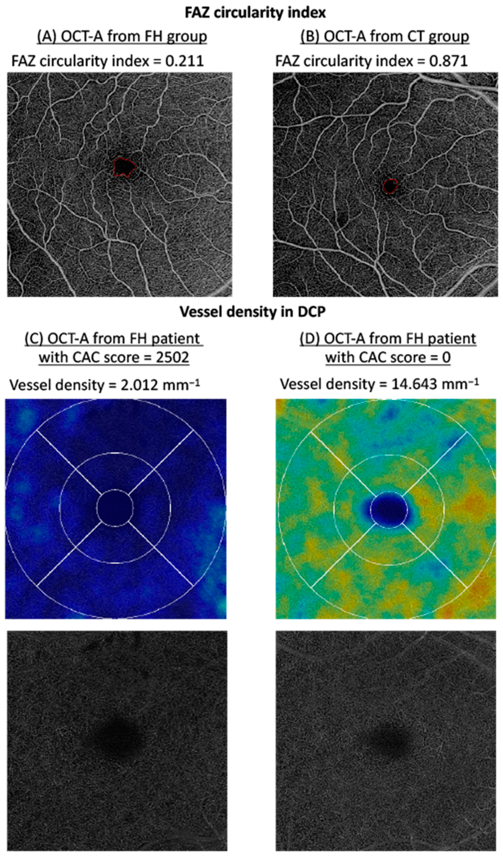
| Vessel density (mm−1) | −0.002 [−0.004; −0.0005] | 0.010 * |
| Vessel length | −0.00005 [−0.00008; −0.00001] | 0.010 |
| FAZ size (mm2) | 0.0001 [−0.00002; 0.0003] | 0.101 |
| FAZ length (mm) | −0.0003 [−0.0001; 0.001] | 0.117 |
| FAZ circularity index | −0.00002 [−0.0008; −0.00005] | 0.573 |
| Multivariate Analysis | ||||
|---|---|---|---|---|
| FH Group (n = 162) | CT Group (n = 121) | β [95% CI] | p | |
| Superficial capillary plexus | ||||
| Vessel density (mm−1) | 18.73 ± 2.41 | 19.34 ± 1.51 | 0.38 [−0.22; 0.07] | 0.211 |
| Vessel length | 0.41 ± 0.05 | 0.42 ± 0.03 | 0.007 [−0.006; 0.02] | 0.297 *† |
| Deep capillary plexus | ||||
| Vessel density (mm−1) | 11.26 ± 4.69 | 12.14 ± 4.04 | 0.46 [−0.66; 1.58] | 0.419 *† |
| Vessel length | 0.26 ± 0.47 | 0.24 ± 0.08 | −0.003 [−0.09; 0.09] | 0.944 |
| FAZ size (mm2) | 0.32 ± 0.47 | 0.27 ± 0.16 | −0.02 [−1.11; 0.08] | 0.701 * |
| FAZ length (mm) | 2.45 ± 2.63 | 2.13 ± 0.77 | −0.13 [0.65; 0.39] | 0.627 * |
| FAZ circularity index | 0.67 ± 0.16 | 0.72 ± 0.10 | 0.04 [0.002; 0.07] | 0.037 * |
Publisher’s Note: MDPI stays neutral with regard to jurisdictional claims in published maps and institutional affiliations. |
© 2022 by the authors. Licensee MDPI, Basel, Switzerland. This article is an open access article distributed under the terms and conditions of the Creative Commons Attribution (CC BY) license (https://creativecommons.org/licenses/by/4.0/).
Share and Cite
Eid, P.; Arnould, L.; Gabrielle, P.-H.; Aho, L.S.; Farnier, M.; Creuzot-Garcher, C.; Cottin, Y. Retinal Microvascular Changes in Familial Hypercholesterolemia: Analysis with Swept-Source Optical Coherence Tomography Angiography. J. Pers. Med. 2022, 12, 871. https://doi.org/10.3390/jpm12060871
Eid P, Arnould L, Gabrielle P-H, Aho LS, Farnier M, Creuzot-Garcher C, Cottin Y. Retinal Microvascular Changes in Familial Hypercholesterolemia: Analysis with Swept-Source Optical Coherence Tomography Angiography. Journal of Personalized Medicine. 2022; 12(6):871. https://doi.org/10.3390/jpm12060871
Chicago/Turabian StyleEid, Pétra, Louis Arnould, Pierre-Henry Gabrielle, Ludwig S. Aho, Michel Farnier, Catherine Creuzot-Garcher, and Yves Cottin. 2022. "Retinal Microvascular Changes in Familial Hypercholesterolemia: Analysis with Swept-Source Optical Coherence Tomography Angiography" Journal of Personalized Medicine 12, no. 6: 871. https://doi.org/10.3390/jpm12060871
APA StyleEid, P., Arnould, L., Gabrielle, P.-H., Aho, L. S., Farnier, M., Creuzot-Garcher, C., & Cottin, Y. (2022). Retinal Microvascular Changes in Familial Hypercholesterolemia: Analysis with Swept-Source Optical Coherence Tomography Angiography. Journal of Personalized Medicine, 12(6), 871. https://doi.org/10.3390/jpm12060871