Review

Primary Synovial Sarcoma of the Kidney: Diagnostic Approach and Therapeutic Modalities for a Rare Nosological Entity

by
Aikaterini Mastoraki
1,
Dimitrios Schizas
1,
Despoina Maria Karavolia
1,
Antonios Smailis
1,
Nikolaos Machairas
2,*,
Michail Vailas
1,
Adamantios Michalinos
3,
Dimitrios Tsapralis
4,
Ioannis Anastasiou
5 and
George Agrogiannis
6
1
First Department of Surgery, National and Kapodistrian University of Athens, Laikon General Hospital, 11527 Athens, Greece
2
Second Department of Propaedeutic Surgery, National and Kapodistrian University of Athens, Laikon General Hospital, 11527 Athens, Greece
3
Department of Anatomy, European University of Cyprus, 1516 Nicosia, Cyprus
4
Department of Surgery, General Hospital of Ierapetra, 72200 Ierapetra, Greece
5
First University Urology Clinic, National and Kapodistrian University of Athens, Laikon General Hospital, 11527 Athens, Greece
6
First Department of Pathology, Medical School, National and Kapodistrian University of Athens, 11527 Athens, Greece
*
Author to whom correspondence should be addressed.
J. Pers. Med. 2022, 12(9), 1450; https://doi.org/10.3390/jpm12091450
Submission received: 10 August 2022 / Revised: 27 August 2022 / Accepted: 31 August 2022 / Published: 2 September 2022
(This article belongs to the Section Clinical Medicine, Cell, and Organism Physiology)

Abstract

:
Synovial sarcoma (SS) is a rare mesenchymal entity that represents 5–10% among soft tissue sarcomas (STS). Primary renal synovial sarcoma (PRSS) is an uncommon, rapidly growing tumor, with potential metastatic dissemination. The main prognostic factors of PRSS include tumor size and histologic grade, while translocation t (X; 18) (p11.2; q11.2) (fusion of SYT gene -chromosome 18- with SSX genes (1, 2 or 4)-chromosome X) is the most common pathognomonic sign. Aggressive surgical resection of the tumor along with concomitant regional lymphadenectomy is the treatment of choice for PRSS, while additional en bloc resection of the adjacent affected organs is often performed. To date, the role of preoperative or postoperative chemotherapy remains equivocal. The prognosis of patients with PRSS is poor, as the 5-year survival rate is only 20–30% and further deteriorates when a high mitotic activity is detected. Local recurrence even after complete R0 surgical excision remains the most frequent cause of death. The aim of this review was to meticulously discuss clinical features, histogenesis, and morphological and immunochemical findings of PRSS, while the role of current diagnostic and therapeutic management of this aggressive neoplasm was emphasized.

Graphical Abstract

1. Introduction

Synovial sarcoma (SS) is a rare mesenchymal entity that represents 5–10% among soft tissue sarcomas (STS) [1]. Lejars and Rubens-Duval reported and initially named it synovial endothelioma in 1919. The most predominant theory for its origin is the retrograde differentiation of an undefined mesenchymal cell [2,3]. This type of tumor can be encountered either in the extremities close to articulations (85–95%) or bursas, sinews and the head and neck region (10%) [4]. It also can be detected in unusual parts of the body without correlation to the joints, including the nervous system, thoracic and abdominal wall cavity, prostate, fallopian tubes, retroperitoneum, bones and kidneys [4,5,6,7,8].
Primary renal synovial sarcoma (PRSS) presents an incidence of 1–3% among all renal tumors [9]. The first description was in 1999 by Faria, while it was previously categorized as an embryonal sarcoma of the kidney [10,11]. Clinical features of PRSS typically range from presence of an enlarged abdominal mass, vague pain and hematuria to local invasion as well as liver and lung metastatic disease [12,13,14,15,16]. Due to the limited number of sporadic cases of PRSS, standard protocols for the diagnosis and treatment of this rare neoplasm are strongly required [17,18,19,20]. The aim of this review was to meticulously discuss clinical features, histogenesis, and morphological and immunochemical findings of PRSS, while the role of current diagnostic and therapeutic management of this aggressive neoplasm was emphasized.

2. Epidemiology and Classification

STS is a rare mesenchymal malignancy that accounts for less than 1% of all adult tumors and affects 2–3 per 100,000 people. In addition, 5–10% of all STSs are represented by SS, which can rarely be encountered in the kidney [20,21]. PRSS is the fourth most common subtype of primary sarcoma of the kidney following undifferentiated pleomorphic sarcoma, liposarcoma and rhabdomyosarcoma [9]. To date, only 185 cases have been reported in the literature, since the first description of PRSS in 1999 [20,22,23,24,25]. Notably, the translocation t(X; 18) (p11.2; q11.2) is present in around 95% of the affected patients [26,27].
The above-mentioned cases were recently systematically reviewed, and more comprehensive epidemiological data were revealed [25]. In that study the median age of PRSS patients was 36.2 years, and the male to female ratio was 1.14:1. The main localization of tumor was in the right kidney with a right to left ratio of 1.53:1. At the time of diagnosis, 65% of affected patients were symptomatic, 46.3% appeared with hematuria, and 43% with pain. Moreover, 8% of the patients presented with metastatic disease, whereas there was caval thrombus formation in 48.2% of the cases [12,25,28,29]. As reported, the lungs are the most common sites for distant metastasis, while the median survival time is 34 months [25,29].
PRSS develops in three possible histological patterns: the monophasic (MSS), the biphasic (BSS), and the poorly differentiated type (PDSS) [8,17]. Relevant prevalence of the above-mentioned PRSS classifications has been identified in 76%, 14% and 10% of cases, respectively [23,25]. Firstly, MSS reveals, microscopically, ovoid or spindled cells with moderate nuclear pleomorphism, arranged in solid compact collagenous sheets without epithelial cell component, and abundant solitary fibrous tumor-like vessels in the background. The monophasic type may be misinterpreted with spindle cell tumors, such as fibrosarcoma, malignant peripheral nerve sheath tumors, adult Wilms’ tumor and vessel-derived tumors such as solitary fibrous tumor. BSS combines MSS’ microscopic features and spindle cells with glandular elements arrayed with mucosal epithelium [14,20,22,27]. It is the pattern most easily distinguishable from other tumors and expresses the SYT-SSX1 fusion, while the monophasic form shows the SYT-SSX2 fusion. Other than that, both MSS and BSS have the same clinical, ultrastructural, and molecular features [30,31]. On the other hand, PDSS includes undifferentiated round cells and is characterized by increased mitotic rate with the poorest prognosis [12,22].
Three PDSS variants have been described, including a subtype of large cells (with fusiform, epithelioid and rhabdoid characteristics), a type of small cells (comparable to Ewing sarcoma and NETs) and a fusocellular category (similar to fibrosarcoma and malignant peripheral nerve tumors). However, PDSS can be more easily distinguished because of the presence of cellular areas with hypocellular zones with calcification or hyalinization and mast cells near fusiform cells [3]. Lastly, there is a TNM classification by the American Joint Committee on Cancer for all the primary renal sarcomas, but its use for staging sarcomas does not sufficiently predict patient prognosis. Furthermore, the histological grade can be adequately determined based upon the scoring system of the French Federation, although SS is considered as grade 3 by definition [32].

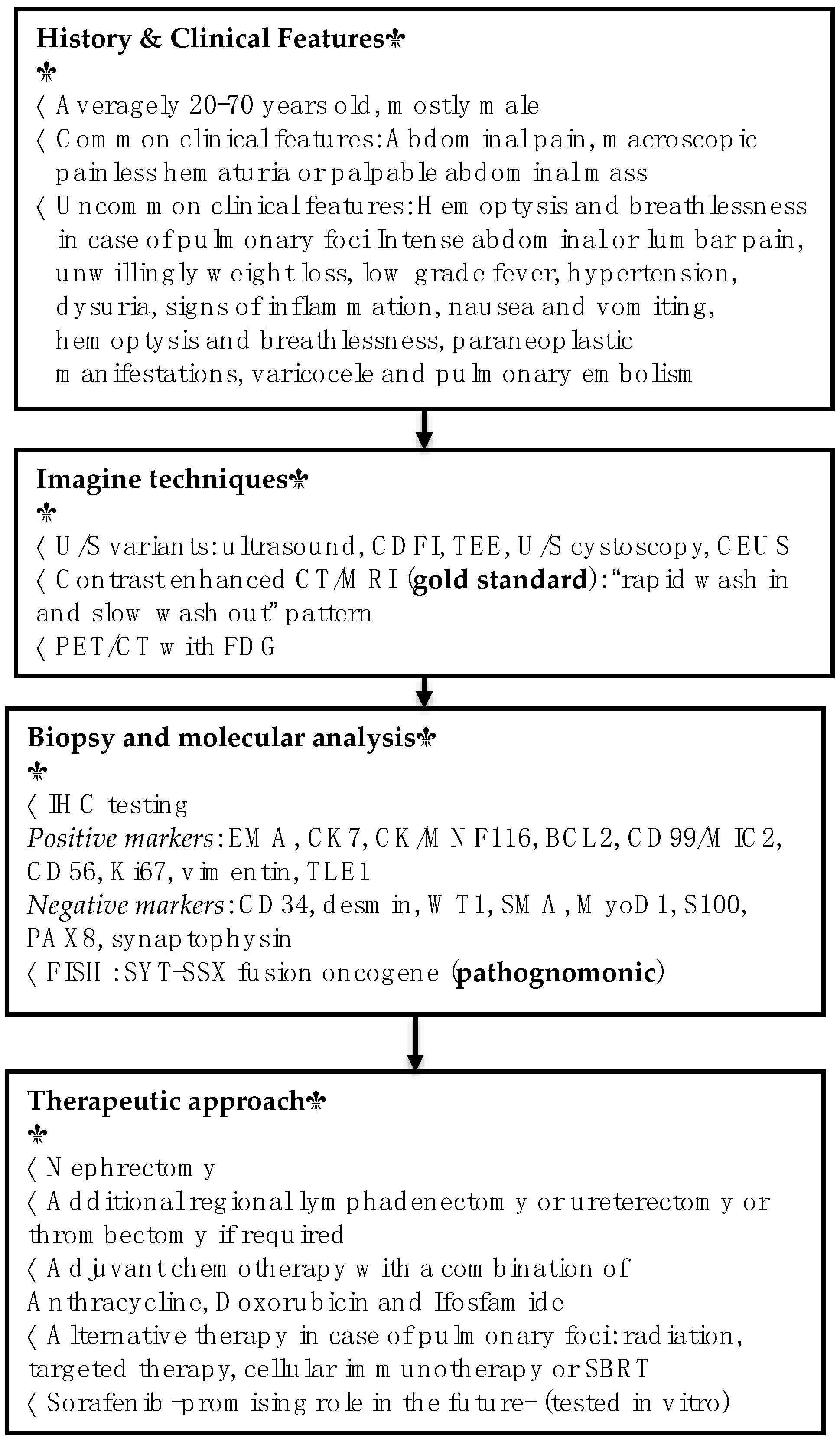
3. Clinical Features

The clinical features of PRSS are not specific, making its differential diagnosis from other renal tumors challenging at an early stage. The most commonly reported symptom is gross, macroscopic hematuria, which sometimes occurs painlessly [25,33,34,35,36]. Another clinical sign is intermittent mild to intense abdominal or lumbar pain [37]. In asymptomatic patients, a bimanually palpable non-tender abdominal mass, with smooth surface and firm to hard consistency, can be detected incidentally during routine evaluation. The lump may be located at the right or left hypochondrium as well as the lumbar, extending into the iliac region [1,30,31]. Other, less frequent symptoms include discomfort, fullness, unwilling weight loss, low-grade fever, hypertension, discolored urine, dysuria, pallor in the sclera, signs of inflammation, nausea and vomiting [19,21,26,35,38]. A rare case of jaundice in a patient with Stauffer’s syndrome has been reported in the literature as a paraneoplastic manifestation of PRSS [37].
The most frequent sites of metastasis are the lungs (36.4% of the patients regardless of the fusion type), with hemoptysis and breathlessness as the main symptoms. Other metastatic foci occur at the liver (50% of patients with SYT-SSX2 fusion type), bones, perirenal adipose tissue and brain [20,22,25,29,39,40]. The tumor metastasizes via the blood and rarely via the lymphatic system. It mainly appears at renal hilar, aortocaval and aorto-iliac lymph nodes [20,22,25]. Moreover, approximately 15% of patients are diagnosed with formed thrombus in adjacent vessels including the renal vein, inferior vena cava (IVC) and portal vein or have a thrombus in the right atrium. Symptoms including non-pitting pedal edema over the shin, non-reducing varicocele and pulmonary embolism are the result of vascular metastasis [14,22,25,35,41]. Nevertheless, despite achievement of adequate resection margins, PRSS is shown to recur in adjacent and distant parts of the body at a percentage of 30–50% [30]. Disease-free survivals range from 5 to 32 months [22].

4. Histopathology and Immunochemistry

Macroscopically, PRSS presents as a yellow-brown, jelly-like, well-encapsulated mass [1,15,42] consisting of cystic, necrotic and hemorrhagic areas [30]. Tumor size ranges variably from 1–35 cm, and most commonly varies from 5–20 cm [12,25,31,43]. It can replace the whole kidney, as well as penetrate the nearby focal renal capsule [10]. Although enlargement of lymph nodes is rare, vascular invasion can be encountered [10,23]. The most common microscopic components are the spindle cells arranged in solid, short, intersecting fascicles with oval to spindle hyperchromatic mitotically active, pleomorphic nuclei and scant to moderate eosinophilic cytoplasm. The nucleus to cytoplasm rate is high. Frequently, the cells develop a perivascular pattern. Thick wall cystic areas lined by hobnailed epithelium are also typical. The tumor is characterized by dilated renal tubules, necrosis, hemorrhage, minimal fat and extensive neovascularization [22,23,24,27,30,35,44]. In the BSS type, secondarily, a degree of epithelial differentiation with microcyst formation coexists, while in PDSS, immature cells with irregular nuclei replace the renal parenchyma [3,36].
In addition, there is an optimal immunohistochemical (IHC) panel of markers that can be used to establish SS diagnosis (Figure 1). The markers, which are usually positive in an IHC test, are epithelial membrane antigen (EMA), cytokeratin (CK) 7, CK/MNF116, B cell lymphoma 2 (BCL2- associated with the SYT-SS2 fusion and may indicate ineffectiveness of chemoradiation after surgery), cluster of differentiation molecule 99 (CD99/MIC2), CD56, Ki67, vimentin and transducin-like enhancer of split 1 (TLE1). Mostly negative markers are CD34, Desmin, Wilms’ tumor protein (WT-1), smooth muscle antibody (SMA), MyoD1, S100, paired-box factor 8 (PAX8—regularly positive in renal cell carcinoma) and synaptophysin [17,31,43,44,45,46,47,48].
Especially, TLE1 has the highest negative predictive value, specificity and sensitivity among all currently available IHC test markers (Figure 1). Nevertheless, it can be detected in many other mesenchymal tumors. This means that a TLE1 negative result excludes PRSS from the differential diagnosis [20,49]. However, FISH still remains the gold standard technique for spotting the pathognomonic SYT-SSX fusion oncogene [1,50,51].

5. Diagnostic Modalities

Several imaging techniques have been proposed to more reliably diagnose PRSS [10,15]. In ultrasound (U/S) images, SS is usually depicted as a single or multiple solid hypo-echoic masses, with unclear or irregular boundary, with additional calcifications, lymph node infiltration or pseudocapsule. Alternative U/S variants are highly recommended; color Doppler flow imaging (CDFI) is indicative of hypovascularity, dotted blood flow signal inside the neoplasm, and on occasion, renal vein thrombosis [14]. Intraoperative transesophageal echocardiography (TEE) can detect extensions such as cardiac metastasis. A common early finding is also bleeding of the ureter in U/S cystoscopy. Lastly, although contrast enhanced ultrasound (CEUS) reveals “slow in and fast out” enhancement, which is unusual in other renal tumors, parametric imaging with software such as “SonoLiver CAP” is proposed to conduct further quantitative analysis of the SS’s enhancement pattern [16,23,52].
Computed tomography (CT) and magnetic resonance imaging (MRI) scans usually reveal an enhancing renal mass with solid and cystic components and unclear margins that may extend to the perinephric area [53]. Additional intravenous contrast administration helps distinguish solid components with heterogeneous enhancement that lasts during nephrography and excretory phase and follows the “rapid wash in and slow wash out” pattern. However, MRI is the gold standard imaging modality for soft-tissue tumors and, therefore, is preferred for sarcomas [54]. On T1-weighted MRI sequences, the tumor is hypointense, similarly to paraspinal muscles. T2-weighted MRI depicts hyperintense areas with the “triple sign” (hemorrhage/necrosis, calcification/fibrosis and air-fluid levels), which can raise suspicions for a preoperative diagnosis. Both CT and MRI can provide critical information on the disease extent, including presence of lymphadenopathy, formation of pseudocapsule or subcapsular hematomas, thrombus in renal veins and local infiltration. The role of nuclear imaging for detecting PRSSs remains ill-determined. A few studies assessed the superiority of positron emission tomography (PET)/CT for the detection of local recurrence and metastasis and, in particular, they indicate multiple hyper-density entities with increased fluorodeoxyglucose (FDG) metabolism in the primary tumor and metastatic lymph nodes, especially in the delayed phase [55]. Diagnostic as well as therapeutic algorithms are summarized in Figure 2.
Tissue biopsy and molecular analysis suggest useful diagnostic approaches [56,57]. Fine needle aspiration cytology (FNAC) or biopsy (FNB) is useful for preoperative diagnosis. Tissue biopsies, though, with IHC markers, are safer to confirm suspicion of PRSS, especially in cases of retroperitoneal masses without typical imaging characteristics. It is indisputable that marking the SYT-SSX oncogene with FISH is a pathognomonic tool for establishing SS diagnosis [58,59]. However, it is reported that some cases of SS do not express the characteristic translocation, and the combination of histopathological features with the clinical picture and imaging is warranted. In one case without the SYT-SSX gene and in some cases with certain SYT-SS2 genes, the prognosis for inducing remission was better after chemotherapy [60].
Differential diagnosis includes primary renal tumors including fibrosarcoma, sarcomatoid renal cell carcinoma, solitary fibrous tumor, adult Wilms’ tumor, primary renal primitive NETs, undifferentiated carcinoma, congenital mesoblastic nephroma, sarcomatoid transitional cell carcinoma of the renal pelvis, and angiomyolipoma [15,16]. Secondary tumors that should also be considered are primary retroperitoneal sarcoma involving the kidney, renal metastatic or Ewing sarcoma, and NETs. By definition, fibrosarcomas are only immunohistochemically reactive for vimentin and rarely for SMA. Biphasic and monophasic SS can be closely similar to sarcomatoid renal cell carcinoma, which has a honeycomb pattern of enhanced signal in CEUS. On the other hand, SS mitotic activity is elevated compared to solitary fibrous tumor, which is positive for CD34 and pSTAT6 and negative for CK [61]. The ETV6-NTRK3 fusion gene can be detected by polymerase chain reaction (PCR) to confirm the diagnosis of congenital mesoblastic nephroma [62,63].

6. Therapeutic Approach

Due to the rarity of PRSS and the limited cases reported in the literature, there are no specific guidelines for its therapeutic management. All cases of suspected sarcoma need to be reviewed by a multidisciplinary sarcoma team/center before initiating treatment (Figure 2).
The main approach consists of surgical treatment followed by adjuvant chemotherapy. The vast majority of centers would consider pre-operative chemotherapy and radiotherapy for SS of the retroperitoneum such as PRSS, if it is likely to reduce the morbidity of a radical surgical resection (organ preserving or aiding marginality against a vital structure). Patients undergo nephrectomy to alleviate the symptoms of PRSS and prevent local relapse with radical resection [29,37,64]. A thoracoabdominal incision with partial resection of the diaphragm and ascending colon is sometimes indicated for radical surgical intervention. Emergency laparotomy may also be performed in case of severe abdominal pain or hemorrhage. Nevertheless, in cases of major vessel invasion or gross hematuria with active bleeding, an embolization may be performed preoperatively. Finally, laparoscopic surgery has been suggested as an alternative treatment option for selected cases.
More than 90% of patients with completely resected PRSSs will relapse locally. Unfortunately, from published series, it is unclear which tumors will recur after total resection even with negative microscopic margins. The optimal extent of surgical margins required to achieve tumor clearance remains largely unknown. Additionally, the role of partial resection in these highly vascular and frequently necrotic tumors remains to be determined [1,13,65]. Intra-operatively, the adrenal gland is preserved when feasible [37,38]. Additional regional lymphadenectomy or ureterectomy is performed as required [6,45,55]. As previously mentioned, vascular thrombi are common and may extend into the infra-hepatic IVC and left lumbar vein [14,23,29,30,52]. In the latter case, they must be excised through thrombectomy.
The need to establish certain diagnostic and therapeutic guidelines (including examination of imaging, radiomics, tumor biology, and surgical and chemotherapeutic tools for retroperitoneal sarcomas, such as PRSS) led to the establishment of the retroperitoneal sarcoma registry “RESAR” between centers in Europe, North America, Asia, Australia and South America. New prospectively collected registry data are expected to be used in auxiliary studies [66].
The clinical benefit of adjuvant chemotherapy remains controversial and is studied with new registry data. Initial studies included anthracycline- and ifosfamide-based chemotherapy and revealed a small benefit in terms of survival, which, however, was not reproduced in a subsequent, large clinical trial [59,66]. Thus, no consensus has been yet achieved, and the debate is still ongoing, whereas the chemotherapeutic management of PRSS varies between institutions and countries. So far, there is no convincing evidence on survival benefits of chemotherapy (for the different histological subtypes of SS), the quality of treatment, and the criteria for selection of patients for adjuvant systemic therapy [26]. Therefore, recent surveys recommend the use of adjuvant chemotherapy only for younger patients and/or larger tumors where clinical advantages could rather be expected. In general, RSS is sensitive to chemotherapy in up to 53% of cases, leading to an overall survival benefit of 8–10% [56]. The use of chemotherapy preoperatively reduces the size of the mass up to 50%, facilitating the following surgical manipulations. Postoperative chemotherapy increased the time of relapse, as well as general survival [14,16]. The most common chemotherapeutic regimen consists of a combination of anthracycline, doxorubicin and ifosfamide in a total of 3–6 cycles [20,21]. Recent investigations indicated that doxorubicin-based chemotherapy significantly increased time of local and distant recurrence, as well as overall recurrence-free survival in comparison to patients who were just observed without therapy. However, an increase in overall survival was not statistically significant [58].
Adjuvant radiotherapy may be used to help local control of high-grade, superficial lesions of the tumor. Radiation can be beneficial in decreasing local recurrence rates and metastases but may not be a feasible option if there is a risk of radiation injury to an adjacent organ. The benefit of radiotherapy in synovial sarcoma is less clear than for chemotherapy. Nevertheless, in case of pulmonary metastatic foci, stereotactic body radiotherapy (SBRT) constitutes an additional therapeutic approach with comparable results and prognosis to surgical resection. In the SBRT regimen, three-dimensional image-guided high-dose radiation is administered within a course of treatment of up to five fractions [9,10,15,40].
Alternative therapeutic options include Anlotinib as a targeted therapy with adequate response in decreasing the lung metastatic nodules. Anlotinib is a novel tyrosine kinase inhibitor targeting multiple factors involved in tumor proliferation, vasculature and tumor microenvironment (Figure 3). Anlotinib inhibits VEGF/VEGFR signaling by selectively targeting VEGFR-2, -3 and FGFR-1, -2, -3,-4 with high affinity, leading to significant inhibition of tumor proliferation (Figure 2). Therefore, in patients with several metastatic SS entities who were refractory to previous anthracycline-based chemotherapy, Anlotinib was proved to have broad-spectrum antitumor activity, while its toxicity was manageable and acceptable [67]. Similarly to treatment for other types of tumors, immunotherapy is also used for patients with STS [68,69]. In this case, autologous T-cells, transduced with a T-cell receptor on tumor-infiltrating lymphocytes, target the NY-ESO-1 antigen. The latter has been detected in 80% of SS affected patients and revealed as an excellent target for NY-ESO-1 positive sarcomas [51].
Sorafenib is another potential future therapeutic choice that could be per-os administered (Figure 3). Mitogen-activated protein kinase (MAPK) cascades have been shown to play a pivotal role in SS survival. Sorafenib, a potent recombinant activated factor (RAF) inhibitor, effectively inhibits the MAPK signaling pathway and cellular proliferation and induces apoptosis, downregulating cyclin D1 and Rb levels (Figure 3). In addition, the above-mentioned compound inhibits the tyrosine kinase receptors VEGF and PDGFR and blocks the serine/threonine kinase RAF, a mediator of IGF-induced signal transduction. The transcription of IGF-2 is activated by SS18-SSX fusion proteins and may be encountered in aggressive types of SS. Therefore, the abeyance of the RAF/MEK/ERK signaling pathway inhibits the proliferation of SS cell lines [15,70,71]. Finally, sorafenib downregulates cyclin D1, whose accumulation is also promoted by SS18-SSX fusion proteins, resulting in G1 arrest and S phase decrease (Figure 2). In conclusion, sorafenib seems to be effective for growth inhibition of SS cell lines in vitro and may become a new therapeutic option for patients with synovial sarcoma [72]. However, more clinicals trials are warranted in order to estimate the efficacy of sorafenib.

7. Conclusions

PRSS is a rare, rapidly growing tumor, with potential metastatic dissemination. Tumor size and histologic grade are the main prognostic factors of PRSS, while aggressive surgical resection of the tumor remains the treatment of choice. The role of preoperative or postoperative chemotherapy remains uncertain. Neoadjuvant chemotherapy may reduce the size of the tumor and simplify subsequent surgical operations, especially in large and high-grade tumors. In addition, patients who receive adjuvant chemotherapy exhibit improved disease-free survival rates. Radiation therapy alone as a complementary treatment modality to surgery either before or after resection results in non-significant differences in survival rates, whereas neoadjuvant administration of chemotherapy combined with radiation may be beneficial. So far, there is no convincing evidence on survival benefits of chemotherapy (for the different histological subtypes of SS), the quality of treatment and the criteria for selection of patients for adjuvant systemic therapy. Finally, emerging data support resection of pulmonary and hepatic metastases, which may improve survival in selected cases. The prognosis of patients with PRSS remains dismal, as the 5-year survival rate is only 20–30% and deteriorates when high mitotic activity is presented. Local recurrence even after complete R0 surgical excision remains the most common cause of death. Evaluation of patients from experienced centers in the context of MDT decision-making is of cardinal importance in order to provide the optimal treatment planning and subsequently the best possible long-term outcomes.

Author Contributions

Conceptualization, A.M. (Aikaterini Mastoraki) and D.S.; methodology, A.M. (Aikaterini Mastoraki), D.S., D.T. and G.A.; writing—original draft preparation, D.M.K., A.S., N.M., M.V. and A.M. (Adamantios Michalinos).; writing—review and editing, A.M. (Aikaterini Mastoraki), I.A., D.S. and G.A.; supervision, D.S. All authors have read and agreed to the published version of the manuscript.

Funding

This research received no external funding.

Institutional Review Board Statement

Not applicable.

Informed Consent Statement

Not applicable.

Data Availability Statement

Not applicable.

Conflicts of Interest

The authors declare no conflict of interest.

References

  1. Cai, H.J.; Cao, N.; Wang, W.; Kong, F.L.; Sun, X.X.; Huang, B. Primary renal synovial sarcoma: A case report. World J. Clin. Cases 2019, 7, 3098–3103. [Google Scholar] [CrossRef] [PubMed]
  2. Pitino, A.; Squillaci, S.; Spairani, C.; Cosimi, M.F.; Feyles, E.; Ricci, D.; Bardari, F.; Graziano, M.; Morabito, F.; Cesarani, F.; et al. Primary synovial sarcoma of the kidney. A case report with pathologic appraisal investigation and literature review. Pathologica 2011, 103, 271–278. [Google Scholar] [PubMed]
  3. Romero-Rojas, A.E.; Díaz-Pérez, J.A.; Messa-Botero, O.A.; Neira-Mejia, F.E. Early age renal synovial sarcoma. Arch. Esp. Urol. 2010, 63, 464–471. [Google Scholar] [CrossRef] [PubMed]
  4. Kageyama, S.; Tsuru, T.; Okamoto, K.; Narita, M.; Okada, Y. Primary synovial sarcoma arising from a crossed ectopic kidney with fusion. Int. J. Urol. 2010, 17, 96–98. [Google Scholar] [CrossRef]
  5. Wang, Z.H.; Wang, X.C.; Xue, M. Clinicopathologic analysis of 4 cases of primary renal synovial sarcoma. Chin. J. Cancer 2010, 29, 212–216. [Google Scholar] [CrossRef]
  6. Gulum, M.; Yeni, E.; Savas, M.; Ozardali, I.; Ozdemir, I.; Mil, D.; Altunkol, A.; Ciftci, H. Primary renal synovial sarcoma. Case Rep. Urol. 2011, 2011. [Google Scholar] [CrossRef]
  7. Kim, D.-H.; Sohn, J.H.; Lee, M.C.; Lee, G.; Yoon, G.-S.; Hashimoto, H.; Sonobe, H.; Ro, J.Y. Primary synovial sarcoma of the kidney. Am. J. Surg. Pathol. 2000, 24, 1097–1104. [Google Scholar] [CrossRef]
  8. Ozkan, E.E.; Mertsoylu, H.; Ozardali, H.I. A case of renal synovial sarcoma treated with adjuvant ifosfamide and doxorubicin. Intern. Med. 2011, 50, 1575–1580. [Google Scholar] [CrossRef]
  9. Chen, S.; Bhuiya, T.; Liatsikos, E.N.; Alexianu, M.D.; Weiss, G.H.; Kahn, L.B. Primary synovial sarcoma of the kidney: A case report with literature review. Int. J. Surg. Pathol. 2011, 9, 335–339. [Google Scholar] [CrossRef]
  10. Xu, R.F.; He, E.H.; Yi, Z.X.; Lin, J.; Zhang, Y.N.; Qian, L.X. Multimodality-imaging manifestations of primary renal-allograft synovial sarcoma: First case report and literature review. World J. Clin. Cases 2019, 27, 1677–1685. [Google Scholar] [CrossRef]
  11. Divetia, M.; Karpate, A.; Basak, R.; Desai, S.B. Synovial sarcoma of the kidney. Ann. Diagn. Pathol. 2008, 12, 333–339. [Google Scholar] [CrossRef] [PubMed]
  12. Wang, Z.; Zhong, Z.; Zhu, L.; Xiong, W.; Pan, C.; Wang, X.; Huang, Z.; Zhao, X. Primary synovial sarcoma of the kidney: A case report. Oncol. Lett. 2015, 10, 3542–3544. [Google Scholar] [CrossRef] [PubMed]
  13. Scarpato, K.R.; Makari, J.H.; Agaronov, M.; Balarezo, F.; Parikh, N.; Finck, C.M.; Ferrer, F.A. Primary renal synovial sarcoma in a 13-year-old boy. J. Pediatric Surg. 2011, 46, 1849–1851. [Google Scholar] [CrossRef] [PubMed]
  14. Dutt, U.K.; Manikandan, R.; Dorairajan, L.N.; Srinivas, B.H. Biphasic renal synovial sarcoma with extensive venous tumor thrombosis: A rare presentation. Urol. Ann. 2018, 10, 339–341. [Google Scholar]
  15. Huang, Y.; Liu, D.; Luo, J.; Chen, W. Primary renal synovial sarcoma: A case report and literature review. J. Cancer Res. Ther. 2018, 14, 267–269. [Google Scholar]
  16. Vedana, M.; Fuenfschilling, M.; Tzankov, A.; Zellweger, T. Primary synovial cell sarcoma of the kidney: Case report and review of the literature. Case Rep. Oncol. 2015, 8, 128–132. [Google Scholar] [CrossRef]
  17. Yang, L.; Wang, K.; Hong, L.; Wang, Y.; Li, X. The value of immunohistochemistry in diagnosing primary renal synovial sarcoma: A case report and literature review. Int. Sur. 2012, 97, 177–181. [Google Scholar] [CrossRef]
  18. Nishida, T.; Inamoto, T.; Uehara, H.; Ibuki, N.; Koyama, K.; Komura, K.; Fujisue, Y.; Kurisu, Y.; Tsuji, M.; Azuma, H.; et al. Monophasic primary renal synovial sarcoma accompanied with a hemorrhagic cyst. Urol. J. 2011, 8, 244–247. [Google Scholar]
  19. Wang, Y.; Wang, K.; Zhang, Z.; Chen, L.; Xu, F. Primary renal synovial sarcoma-a case report. West Indian Med. J. 2011, 60, 354–356. [Google Scholar]
  20. El Chediak, A.; Mukherji, D.; Temraz, S.; Temraz, S.; Nassif, S.; Sinno, S.; Mahfouz, R.; Shamseddine, A. Primary synovial sarcoma of the kidney: A case report of complete pathological response at a Lebanese tertiary care center. BMC Urol. 2018, 18. [Google Scholar] [CrossRef]
  21. Modi, G.; Madabhavi, I.; Panchal, H.; Anand, A.; Patel, A.; Parikh, S.; Revannasiddaiah, S. Primary synovial sarcoma of kidney: A rare differential diagnosis of renomegaly. Case Rep. Pathol. 2014, 2014. [Google Scholar] [CrossRef] [PubMed]
  22. Rose, L.; Grignon, D.; Cheng, L.; Fan, R.; Zhang, S.; Alruwaii, F.; Chen, S. Primary Renal Synovial Sarcomas: PAX 8 Immunostaining and Unusual Molecular Findings. Appl. Immunohistochem. Mol. Morphol. 2020, 28, 221–228. [Google Scholar] [CrossRef] [PubMed]
  23. Chandrasekaran, D.; Narayanaswamy, K.; Sundersingh, S.; Senniappan, K.; Raja, A. Primary Synovial Sarcoma of the Kidney with Inferior Vena Caval Thrombus. Indian J. Surg. Oncol. 2016, 7, 345–348. [Google Scholar] [CrossRef] [PubMed]
  24. Gong, J.; Kang, W.; Li, S.; Yang, Z.; Xu, J. CT findings of synovial sarcomas of the kidney with pathological correlation. Clin. Imaging 2013, 37, 1033–1036. [Google Scholar] [CrossRef]
  25. Blas, L.; Roberti, J. Primary Renal Synovial Sarcoma and Clinical and Pathological Findings: A Systematic Review. Curr. Urol. Rep. 2021, 22, 25. [Google Scholar] [CrossRef]
  26. Abbas, M.; Dämmrich, M.E.; Braubach, P.; Meinardus, A.; Kramer, M.W.; Merseburger, A.S.; Herrmann, T.R.W.; Grünwald, V.; Kreipe, H.-H. Synovial sarcoma of the kidney in a young patient with a review of the literature. Rare Tumors 2014, 6, 5393. [Google Scholar] [CrossRef]
  27. Marković-Lipkovski, J.; Sopta, J.; Vjestica, J.; Vujanić, G.; Tulić, C. Rapidly progressive course of primary renal synovial sarcoma-case report. Srp. Arh. Za Celok. Lek. 2013, 141, 814–818. [Google Scholar] [CrossRef]
  28. Dassi, V.; Das, K.; Singh, B.P.; Swain, S.K. Primary synovial sarcoma of kidney: A rare tumor with an atypical presentation. Indian J. Urol. 2009, 25, 269–271. [Google Scholar] [CrossRef]
  29. Iacovelli, R.; Altavilla, A.; Ciardi, A.; Urbano, F.; Manai, C.; Gentile, V.; Cortesi, E. Clinical and pathological features of primary renal synovial sarcoma: Analysis of 64 cases from 11 years of medical literature. BJU Int. 2012, 110, 1449–1454. [Google Scholar] [CrossRef]
  30. Pathrose, G.; John, N.T.; Hariharan, P. Renal Synovial Sarcoma in a Young Pregnant Lady: A Case Report and Clinico-Pathological Profile. J Clin. Diagn. Res. 2017, 11. [Google Scholar] [CrossRef]
  31. Bakhshi, G.D.; Khan, A.S.; Shaikh, A.S.; Khan, M.A.; Khan, M.A.; Jamadar, N.M. Primary renal synovial sarcoma. Clins Pract. 2012, 2, e44. [Google Scholar] [CrossRef] [PubMed]
  32. Öztürk, H. Prognostic features of renal sarcomas (Review). Oncol. Lett. 2015, 9, 1034–1038. [Google Scholar] [CrossRef] [PubMed]
  33. Vesoulis, Z.; Rahmeh, T.; Nelson, R.; Clarke, R.; Lu, Y.; Dankoff, J. Fine needle aspiration biopsy of primary renal synovial sarcoma. A case report. Acta. Cytol. 2003, 47, 668–672. [Google Scholar] [CrossRef] [PubMed]
  34. Perlmutter, A.E.; Saunders, S.E.; Zaslau, S.; Chang, W.W.; Farivar-Mohseni, H. Primary synovial sarcoma of the kidney. Int. J. Urol. 2005, 12, 760–762. [Google Scholar] [CrossRef] [PubMed]
  35. Lv, X.F.; Qiu, Y.W.; Han, L.J.; Cao, J.; Zhang, C.; Liu, Z.-Y.; Zhang, X.-L.; Cai, P.-Q.; Li, L. Primary renal synovial sarcoma: Computed tomography imaging findings. Acta Radiol. 2015, 56, 493–499. [Google Scholar] [CrossRef] [PubMed]
  36. Wezel, F.; Ströbel, P.; Michaely, H.; Michel, M.S.; Häcker, A. Biphasisches primäres Synovialsarkom der Niere. Der Urol. 2010, 49, 411–414. [Google Scholar] [CrossRef]
  37. Dewana, S.K.; Parmar, K.M.; Sharma, G.; Bansal, A.; Panwar, P.; Mavuduru, R.S. Paraneoplastic hepatic dysfunction with jaundice in a case of primary renal synovial sarcoma: A very rare scenario. Urol. Case Rep. 2019, 24. [Google Scholar] [CrossRef]
  38. Lakshmaiah, K.C.; Saini, K.S.; Singh, T.; Jain, A.; Kumar, R.V.; Sajeevan, K.V.; Lokanatha, D.; Jacob, L.A. Primary synovial sarcoma of kidney-a report of 2 cases and review of literature. J. Egypt. Natl. Cancer Inst. 2010, 22, 149–153. [Google Scholar]
  39. Radhakrishnan, V.; Dhanushkodi, M.; Narayanswamy, K.; Raja, A.; Sundersingh, S.; Sagar, T. Synovial sarcoma of kidney in a child: A rare presentation. J. Indian Assoc. Pediatr. Surg. 2016, 21, 75–77. [Google Scholar] [CrossRef]
  40. Kataria, T.; Janardhan, N.; Abhishek, A.; Sharan, G.K.; Mitra, S. Pulmonary metastasis from renal synovial sarcoma treated by stereotactic body radiotherapy: A case report and review of the literature. J. Cancer Res. Ther. 2010, 6, 75–79. [Google Scholar] [CrossRef]
  41. Trolliet, S.; Lindner, V.; Krzisch, S.; Schneider, M.; Jung, J.L. Synovialosarcome rénal primitive. Prog. Urol. 2014, 24, 156–160. [Google Scholar] [CrossRef] [PubMed]
  42. Puj, K.S.; Pandya, S.J.; Warikoo, V.; Chauhan, T.; Samanta, S.T. Primary Synovial Sarcoma of the Kidney. A Rare Presentat. Urol. 2018, 116. [Google Scholar]
  43. Lopes, H.; Pereira, C.A.; Zucca, L.E.; Serrano, S.V.; Silva, S.R.M.; Camparoto, M.L.; Cárcano, F.M. Primary monophasic synovial sarcoma of the kidney: A case report and review of literature. Clin. Med. Insights Oncol. 2013, 7, 257–262. [Google Scholar] [CrossRef] [PubMed]
  44. Majumder, A.; Dey, S.; Khandakar, B.; Medda, S.; Chandra Paul, P. Primary renal synovial sarcoma: A rare tumor with an atypical presentation. Arch. Iran. Med. 2014, 17, 726–728. [Google Scholar] [PubMed]
  45. Mishra, S.; Awasthi, N.; Hazra, S.P.; Bera, M.K. Primary synovial sarcoma of the kidney. Saudi J. Kidney Dis. Transpl. 2015, 26, 996–999. [Google Scholar] [CrossRef]
  46. Schoolmeester, J.K.; Cheville, J.C.; Folpe, A.L. Synovial sarcoma of the kidney: A clinicopathologic, immunohistochemical, and molecular genetic study of 16 cases. Am. J. Surg. Pathol. 2014, 38, 60–65. [Google Scholar] [CrossRef]
  47. Ozkanli, S.S.; Yildirim, A.; Zemheri, E.; Gucer, F.I.; Aydin, A.; Caskurlu, T. Primary synovial sarcoma of the kidney. Urol. Int. 2012, 92, 369–372. [Google Scholar] [CrossRef]
  48. Karafin, M.; Parwani, A.V.; Netto, G.J.; Illei, P.B.; Epstein, J.I.; Ladanyi, M.; Argani, P. Diffuse expression of PAX2 and PAX8 in the cystic epithelium of mixed epithelial stromal tumor, angiomyolipoma with epithelial cysts, and primary renal synovial sarcoma: Evidence supporting renal tubular differentiation. Am. J. Surg. Pathol. 2011, 35, 1264–1273. [Google Scholar] [CrossRef]
  49. Rekhi, B.; Basak, R.; Desai, S.B.; Jambhekar, N.A. Immunohistochemical validation of TLE1, a novel marker, for synovial sarcomas. Indian J. Med. Res. 2012, 136, 766–775. [Google Scholar]
  50. Bella, A.J.; Winquist, E.W.; Perlman, E.J. Primary synovial sarcoma of the kidney diagnosed by molecular detection of SYT-SSX fusion transcripts. J. Urol. 2002, 168, 1092–1093. [Google Scholar] [CrossRef]
  51. Koyama, S.; Morimitsu, Y.; Morokuma, F.; Hashimoto, H. Primary synovial sarcoma of the kidney: Report of a case confirmed by molecular detection of the SYT-SSX2 fusion transcripts. Pathol. Int. 2001, 51, 385–391. [Google Scholar] [CrossRef]
  52. Chen, P.C.; Chang, Y.H.; Yen, C.C.; Pan, C.C.; Chiang, H. Primary renal synovial sarcoma with inferior vena cava and right atrium invasion. Int. J. Urol. 2003, 10, 657–660. [Google Scholar] [CrossRef] [PubMed]
  53. Kawahara, T.; Sekiguchi, Z.; Makiyama, K.; Nakayama, T.; Nagashima, Y.; Kita, K.; Namura, K.; Itou, H.; Sano, F.; Hayashi, N.; et al. Primary Synovial Sarcoma of the Kidney. Case Rep. Oncol. 2009, 2, 189–193. [Google Scholar] [CrossRef] [PubMed]
  54. Zakhary, M.M.; Elsayes, K.M.; Platt, J.F.; Francis, I.R. Magnetic resonance imaging features of renal synovial sarcoma: A case report. Cancer Imaging 2008, 8, 45–47. [Google Scholar] [CrossRef] [PubMed]
  55. Erturhan, S.; Seçkiner, I.; Zincirkeser, S.; Erbaǧci, A.; Çelik, M.; Yaǧci, F.; Karakok, M. Primary synovial sarcoma of the kidney: Use of PET/CT in diagnosis and follow-up. Ann. Nucl. Med. 2008, 22, 225–229. [Google Scholar] [CrossRef]
  56. Pereira, E.; Silva, R.; Leitão, T.; Correia, L.; Martins, F.; Palma Dos Reis, J.; Lopes, T. Primary synovial sarcoma of the kidney with unusual follow up findings. Can. J. Urol. 2013, 20, 6734–6736. [Google Scholar]
  57. Tan, Y.S.; Ng, L.G.; Yip, S.K.; Tay, M.H.; Lim, A.S.; Tien, S.L.; Cheng, L.; Tan, P.H. Synovial sarcoma of the kidney: A report of 4 cases with pathologic appraisal and differential diagnostic review. Anal. Quant. Cytol. Histol. 2010, 32, 239–245. [Google Scholar]
  58. Park, S.J.; Kim, H.K.; Kim, C.K.; Park, S.-K.; Go, E.-S.; Kim, M.-E.; Hong, D.S. A case of renal synovial sarcoma: Complete remission was induced by chemotherapy with doxorubicin and ifosfamide. Korean J. Intern. Med. 2004, 19, 62–65. [Google Scholar] [CrossRef]
  59. Mirza, M.; Zamilpa, I.; Bunning, J. Primary renal synovial sarcoma. Urology 2008, 72, 716.e11–716.e12. [Google Scholar] [CrossRef] [PubMed]
  60. Törnkvist, M.; Wejde, J.; Ahlén, J.; Brodin, B.; Larsson, O. A novel case of synovial sarcoma of the kidney: Impact of SS18/SSX analysis of renal hemangiopericytoma-like tumors. Diagn. Mol. Pathol. 2004, 13, 47–51. [Google Scholar] [CrossRef]
  61. Paláu, L.M.A.; Thu Pham, T.; Barnard, N.; Merino, M.J. Primary synovial sarcoma of the kidney with rhabdoid features. Int. J. Surg. Pathol. 2007, 15, 421–428. [Google Scholar] [CrossRef] [PubMed]
  62. Shannon, B.A.; Murch, A.; Cohen, R.J. Primary renal synovial sarcoma confirmed by cytogenetic analysis: A lesion distinct from sarcomatoid renal cell carcinoma. Arch. Pathol. Lab. Med. 2005, 12, 238–240. [Google Scholar] [CrossRef] [PubMed]
  63. Argani, P.; Faria, P.A.; Epstein, J.I.; Reuter, V.E.; Perlman, E.J.; Beckwith, J.B.; Ladanyi, M. Primary renal synovial sarcoma: Molecular and morphologic delineation of an entity previously included among embryonal sarcomas of the kidney. Am. J. Surg. Pathol. 2000, 24, 1087–1096. [Google Scholar] [CrossRef]
  64. Grampurohit, V.U.; Myageri, A.; Rao, R.V. Primary renal synovial sarcoma. Urol. Ann. 2011, 3, 110–113. [Google Scholar] [CrossRef] [PubMed]
  65. Chung, S.D.; Huang, K.H.; Chueh, S.C.; Lai, M.K.; Lin, W.C. Primary synovial sarcoma of the kidney. J. Med. Assoc. 2008, 107, 344–347. [Google Scholar] [CrossRef]
  66. Houdta, W.J.; Rautb, C.P.; Bonvalotc, S.; Swallowd, C.J.; Haase, R.; Gronchi, A. New research strategies in retroperitoneal sarcoma. The case of TARPSWG, STRASS and RESAR: Making progress through collaboration. Curr. Opin. Oncol. 2019, 31, 310–316. [Google Scholar] [CrossRef]
  67. Chi, Y.; Fang, Z.; Hong, X.; Yao, Y.; Sun, P.; Wang, G.; Du, F.; Sun, Y.; Wu, Q.; Qu, G.; et al. Safety and Efficacy of Anlotinib, a Multikinase Angiogenesis Inhibitor, in Patients with Refractory Metastatic Soft-Tissue Sarcoma. Clin. Cancer Res. 2018, 24, 5233–5238. [Google Scholar] [CrossRef]
  68. Machairas, N.; Tsilimigras, D.I.; Pawlik, T.M. Current Landscape of Immune Checkpoint Inhibitor Therapy for Hepatocellular Carcinoma. Cancers 2022, 14, 2018. [Google Scholar] [CrossRef]
  69. Mosca, L.; de Angelis, A.; Ronchi, A.; de Chiara, A.; Fazioli, F.; Ruosi, C.; Altucci, L.; Conte, M.; de Nigris, F. Sarcoma Common MHC-I Haplotype Restricts Tumor-Specific CD8+ T Cell Response. Cancers 2022, 14, 3414. [Google Scholar] [CrossRef]
  70. Basso, U.; Brunello, A.; Bertuzzi, A.; Santoro, A. Sorafenib is active on lung metastases from synovial sarcoma. Ann. Oncol. 2009, 20, 386–387. [Google Scholar] [CrossRef]
  71. Peng, C.L.; Guo, W.; Ji, T.; Ren, T.; Yang, Y.; Li, D.-S.; Qu, H.y.; Li, X.; Tang, S.; Yan, T.-Q.; et al. Sorafenib induces growth inhibition and apoptosis in human synovial sarcoma cells via inhibiting the RAF/MEK/ERK signaling pathway. Cancer Biol. Ther. 2009, 8, 1729–1736. [Google Scholar] [CrossRef] [PubMed]
  72. Mastoraki, A.; Schizas, D.; Papanikolaou, I.S.; Bagias, G.; Machairas, N.; Agrogiannis, G.; Liakakos, T.; Liakakos, T.; Arkadopoulos, N. Management of primary retroperitoneal synovial sarcoma: A case report and review of literature. World. J. Gastrointest. Surg. 2019, 11, 27–33. [Google Scholar] [CrossRef] [PubMed]
Figure 1. A monophasic PRSS. (a) Hematoxylin & eosin stain (x200). (b) TLE-1 stain (x200). (c) CD56 stain (x200).
Jpm 12 01450 g001
Figure 2. Diagnostic and therapeutic algorithm for PRSS.
Jpm 12 01450 g002
Figure 3. Genetic pathways and regulatory mechanisms modified by the administration of immunotherapeutic agents.
Jpm 12 01450 g003
Publisher’s Note: MDPI stays neutral with regard to jurisdictional claims in published maps and institutional affiliations.

