Abstract
Ankylosing spondylitis (AS) is an inflammatory disease that involves the axial skeleton and can present with extra-articular manifestations. However, there are scarce reports describing the link between AS and pulmonary arterial hypertension (PAH). Herein, we report on a 58-year-old man with a history of AS for 32 years who developed PAH as confirmed by echocardiography and right cardiac catheterization. To our knowledge, this is the first case of AS associated with PAH 32 years after the AS diagnosis with a detailed clinical description. We are recommended that physicians should be aware of this rare comorbidity in AS patients. Early echocardiographic screening is necessary for symptomatic patients.
1. Background
An extra-articular presentation is common in patients with AS, including uveitis, inflammatory bowel disease, and psoriasis [1]. Cardiovascular involvement has also been described in AS, manifested as aortitis, cardiac conduction disturbances, and valvular heart disease [2]. Pulmonary manifestations in AS, such as interstitial lung disease, apical fibrobullous disease, obstructive sleep apnea, and spontaneous pneumothorax, are less frequent [3]. However, very few reports describe the link between AS and pulmonary arterial hypertension (PAH). Herein, we report on a middle-aged man with a history of young-aged AS for 32 years associated with PAH.
2. Case Presentation
A 58-year-old man had a history of AS with stable disease activity following administration of the disease-modifying antirheumatic drug sulfasalazine at a dose of 5 mg twice per day for 32 years. He complained of progressive exertional dyspnea and leg edema and felt shortness of breath for the last 3 months after climbing two floors of stairs. He was admitted to our hospital under the impression of PAH and the World Health Organization’s (WHO) functional class IV [4]. This patient was never a smoker and denied any history of exposure to toxic chemicals.
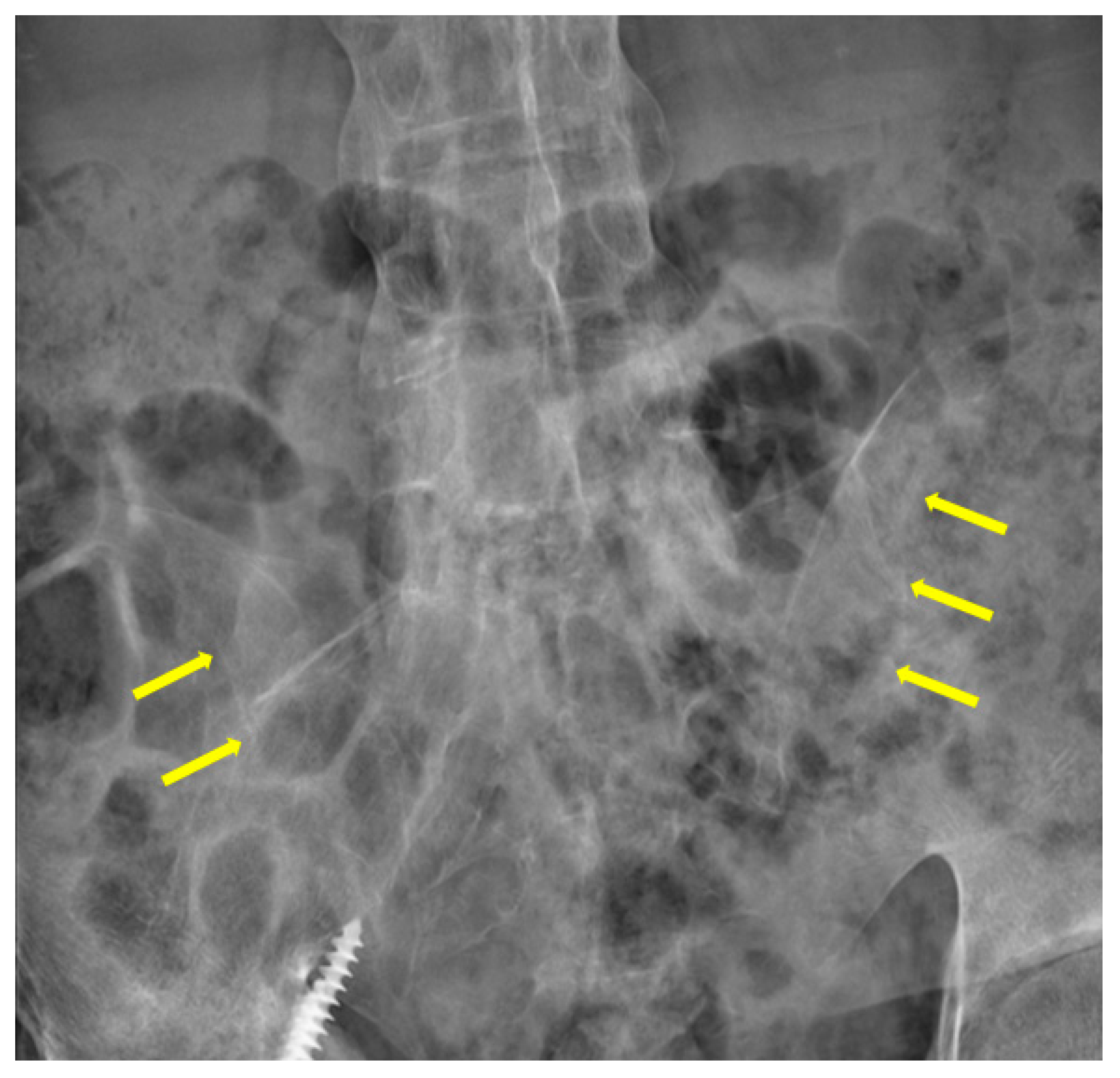
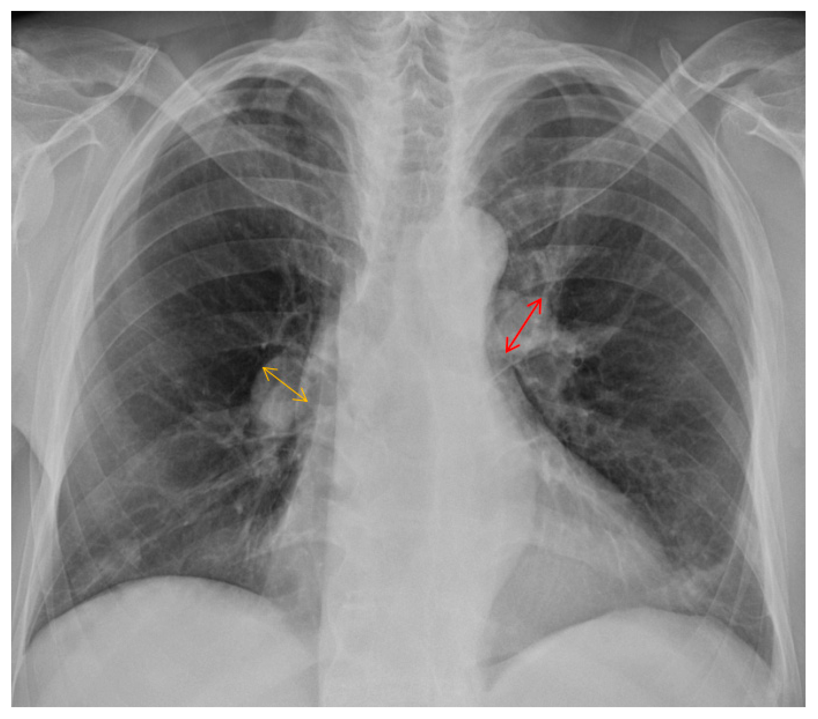
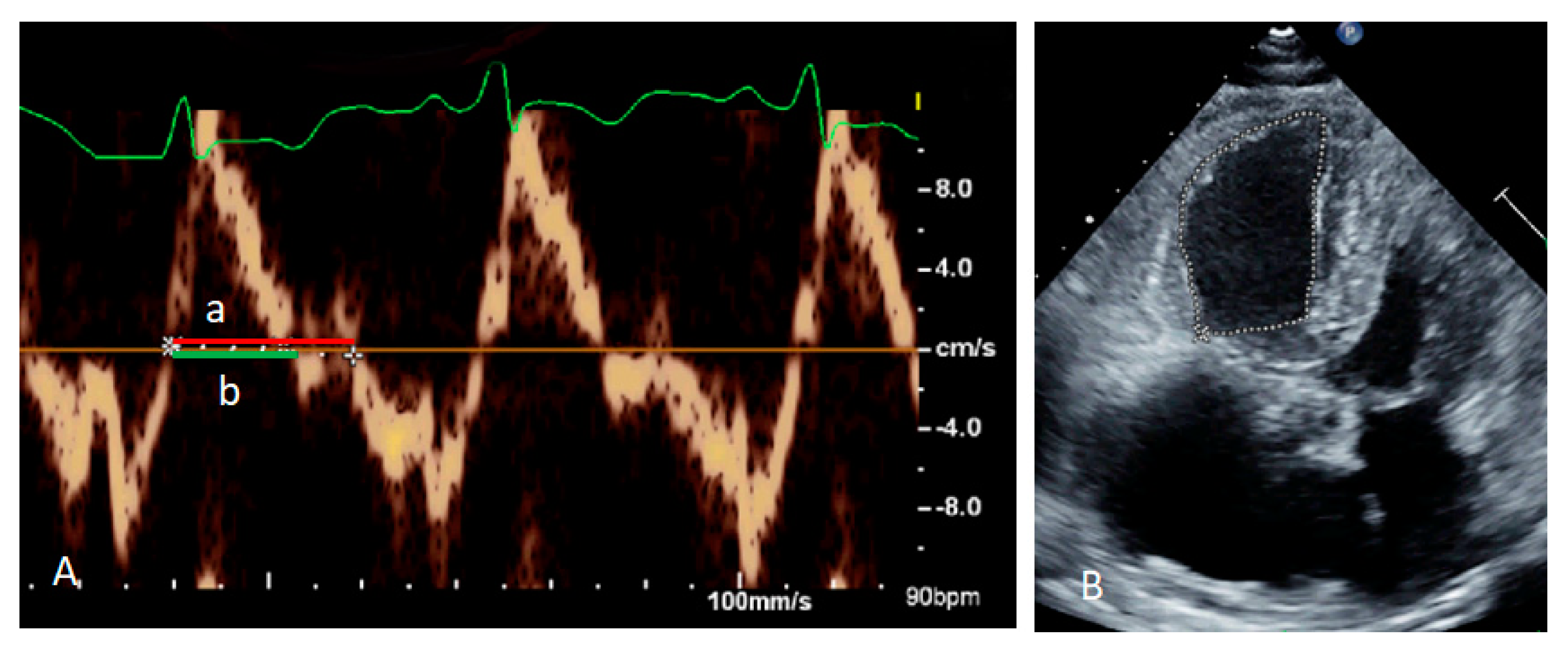
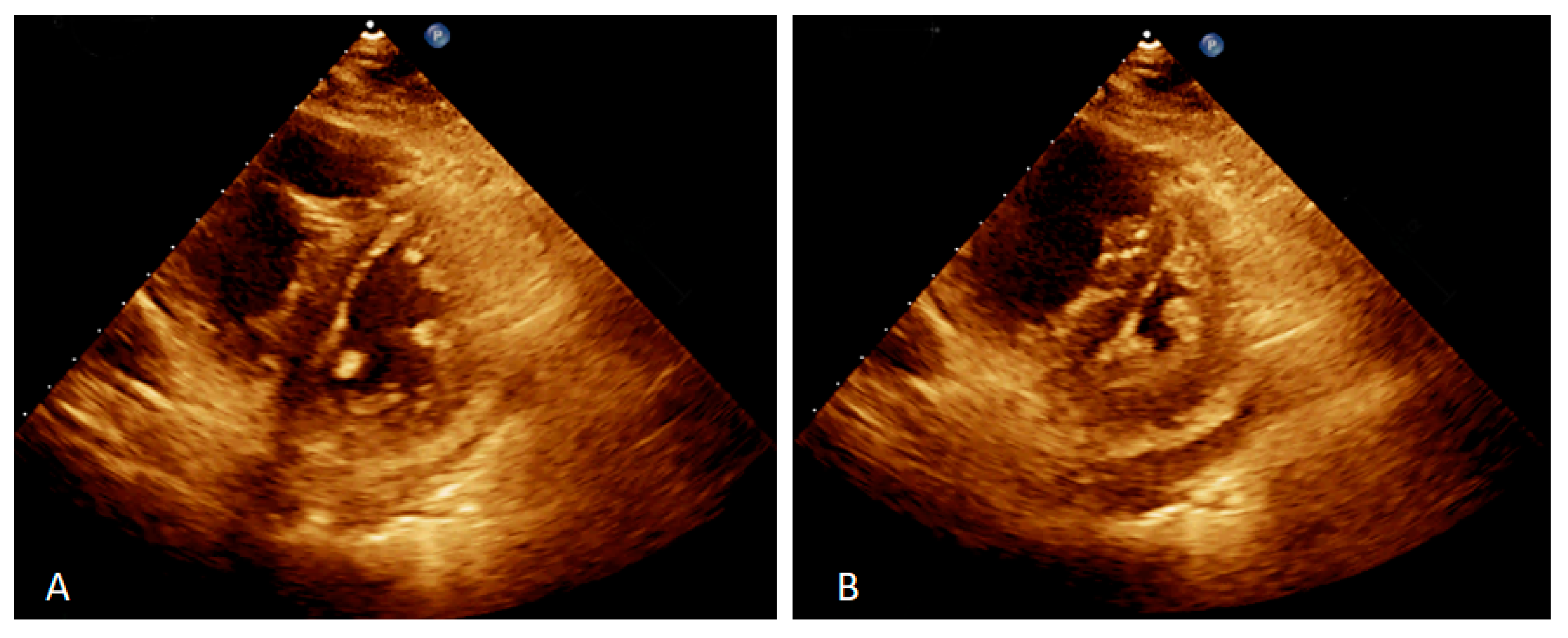
Split-second pulmonary heart sounds with bilateral lower extremity edema were discovered during the examination. Laboratory testing (white blood cell, hemoglobin, platelet, and liver function tests) results were within normal limits, except for blood urea nitrogen of 27 mg/dL (reference range <20 mg/dL), creatinine of 1.42 mg/dL (reference range <1.30 mg/dL), high-sensitivity troponin I of 48.8 pg/mL(reference range <17.5 pg/mL), and N-terminal pro-brain natriuretic peptide (NT-pro-BNP) of 4528 pg/mL (reference range <300 pg/mL) (Table 1). The 6 min walk distance (6MWD) was 340 m, desaturation from 98% to 82%. Grade 3 bilateral sacroiliitis was diagnosed according to the modified New York classification on a plain radiograph examination of the pelvis (Figure 1) [5]. A chest film demonstrated enlarged bilateral main pulmonary arteries with abrupt tapering of the peripheral pulmonary vasculature (Figure 2). The electrocardiograph disclosed an inverted T-wave in the precordial leads, and the inferior leads II, III, aVF, and aVF suggested right ventricular strain. Echocardiography revealed preserved left ventricular systolic function (left ventricular ejection fraction = 59.7%). No significant valvular heart disease was detected except for severe tricuspid regurgitation. The estimated systolic pulmonary artery pressure was approximately 96 mmHg. Dilated right ventricle (RV), a high RV index of myocardial performance (RIMP, 0.64; reference range <0.54) (Figure 3A) and low fractional area change (FAC) of the RV (21%; reference range >35%) (Figure 3B) suggested impaired global right ventricular function (Table 2). A D-shaped left ventricle evidenced during systole and diastole can be the result of high right ventricular pressure and volume overload (Figure 4A,B). No intracardiac shunt finding was detected.

Table 1.
Laboratory data.
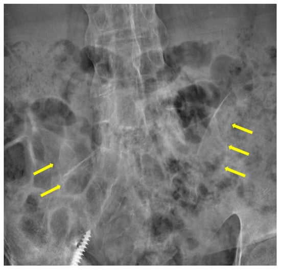
Figure 1.
A plain radiograph of the pelvis demonstrated sclerosis, partial ankylosis and narrowing (yellowish arrows) over the sacroiliac joints, suggesting grade-3 bilateral sacroiliitis.
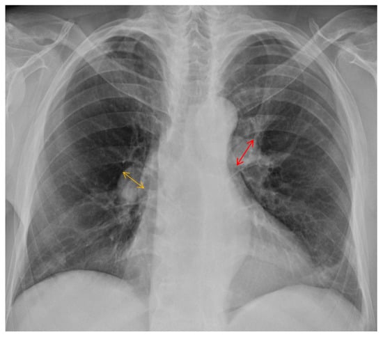
Figure 2.
Chest radiograph showed engorged bilateral main pulmonary arteries and pruning of peripheral pulmonary vessels. The orange and reddish lines indicate the diameter of the right (21 mm) and left (24 mm) main pulmonary artery, respectively.
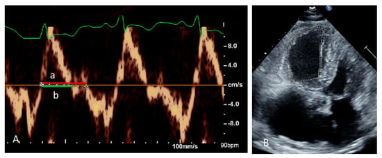
Figure 3.
Right Ventricular Index of Myocardial Performance (RIMP) equals (a − b)/b, and the value was 0.64; “a” means tricuspid closure-open time, and “b” means ejection time, respectively. (A) The fractional area change of the right ventricle (RV FAC) on the four−chamber view was 21% (B).

Table 2.
Echocardiographic results.
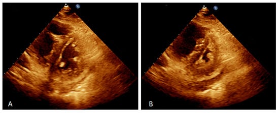
Figure 4.
Two-dimensional echocardiography on the parasternal short-axis view showed D-shaped left ventricle during diastole, indicated for fluid overload (A) and systole, indicated for pressure overload (B).
Thyroid function was normal, and a test for the human immunodeficiency virus was negative. Pulmonary function testing revealed normal but mild impairment of the diffusing capacity of the lung for carbon monoxide (DLCO), which was 15.6% of the predicted value. Abdominal sonography conducted negative findings for portal hypertension or cirrhosis. No evidence of pulmonary embolism, pulmonary emphysema, vasculitis, associated interstitial lung disease, or granulomatous infection was observed according to a high-resolution computed tomography scan. A right cardiac catheterization study demonstrated main pulmonary artery pressure of 85/23 mmHg (mean = 43 mmHg), RV pressure of 84/11 mmHg, mean right atrial pressure of 18 mmHg, pulmonary artery wedge pressure of 18 mmHg, pulmonary vascular resistance of 9.39 WU, and a cardiac index of 2.11 L/min/m2. No definite causes for the development of PAH were disclosed in this case. Hence, the patient was diagnosed with AS associated with PAH.
During hospitalization, the patient was treated with diuretics and was discharged with symptomatic improvement on the 6MWD (430 m) and the NT-pro-BNP level (1171 pg/mL). During the outpatient clinic follow-up, 62.5 mg of bosentan twice per day was initiated. Then, the prescription was switched to 20 mg of sildenafil three times per day following approval from the National Health Insurance Bureau. One month later, 10 mg of macitentan every day was added to the sildenafil because the patient still suffered from dyspnea (Table 3). The NT-pro-BNP level decreased from 1171 to 98 pg/mL, and the 6MWD increased from 430 to 550 m 3 months after starting the macitentan treatment. The patient was under outpatient departmental follow-up in stable condition.

Table 3.
Timeline of the patient’s treatments.
3. Discussion
To our knowledge, this is the first case of AS associated with PAH 32 years after the AS diagnosis. The diagnosis of PAH was confirmed by transthoracic echocardiography and right heart catheterization.
Autoimmune diseases such as systemic sclerosis, systemic lupus erythematosus, and mixed connective tissue disease are common etiologies of PAH [6]. However, only a few case reports have described the association between AS and PAH. Karoli et al. illustrated the high incidence of pulmonary hypertension in AS patients [7]. However, their study made the diagnosis of PAH by echocardiography without right heart catheterization data. The coexistence of another connective tissue disease was not excluded, which may have contributed to pulmonary hypertension.
In contrast, Hung et al. described a rare case of a 27-year-old man with a 12-year history of AS who developed PAH. The diagnosis was confirmed by echocardiography and right heart catheterization [8]. Another observational study reported that older age, a long smoking history, AS duration, and poor spinal mobility increase the risk of PAH in patients with AS [9]. Endothelial dysfunction may play an essential role in pathogenesis [9].
In the present case, we reported a middle-aged man with AS associated with PAH 32 years after the AS diagnosis. The abnormal RIMP and RV FAC indicated RV dysfunction, which may have been caused by pulmonary hypertension. The diagnoses of thyroid disease, HIV-associated PAH, cirrhosis, portal hypertension, lung disease, left heart failure, and pulmonary emboli were excluded because of negative laboratory findings. The high pulmonary capillary wedge pressure suggested that the elevated left ventricular end-diastolic pressure may result from fluid overload related to kidney injury. Left-sided valvular heart disease is another etiology of PAH [10,11], and we excluded left-sided valvular heart disease in our case by echocardiography. Thus, PAH was diagnosed as associated with AS.
In summary, we report an AS patient with PAH. An echocardiogram is suggested for all patients with symptomatic who have heart failure. Physicians should be aware of this rare comorbidity in AS patients. Early screening is necessary for symptomatic patients using modalities such as echocardiography. Further epidemiologic studies are needed to disclose the association between AS and PAH.
Author Contributions
Conceptualization and writing—original draft preparation, T.-Y.Y., Y.-H.C., W.-Z.S. and G.-P.J.; Writing—review and editing, T.-Y.Y., W.-Z.S. and G.-P.J.; visualization, T.-Y.Y. and W.-Z.S.; supervision, G.-P.J.; project administration, T.-Y.Y. and W.-Z.S. All authors have read and agreed to the published version of the manuscript.
Funding
This research received no external funding.
Institutional Review Board Statement
Not applicable.
Informed Consent Statement
We have de-identified all the details of this case. Written informed consent was obtained from the patient for the publication of this report and the publication of any potentially identifiable images or data included in this article.
Data Availability Statement
Not applicable.
Conflicts of Interest
The authors declare that the research was conducted in the absence of any commercial or financial relationships that could be construed as a potential conflict of interest.
References
- Stolwijk, C.; van Tubergen, A.; Castillo-Ortiz, J.D.; Boonen, A. Prevalence of extra-articular manifestations in patients with ankylosing spondylitis: A systematic review and meta-analysis. Ann. Rheum. Dis. 2015, 74, 65–73. [Google Scholar] [CrossRef] [PubMed]
- Ward, M.M. Lifetime risks of valvular heart disease and pacemaker use in patients with ankylosing spondylitis. J. Am. Heart Assoc. 2018, 7, e010016. [Google Scholar] [CrossRef] [PubMed]
- Kanathur, N.; Lee-Chiong, T. Pulmonary manifestations of ankylosing spondylitis. Clin. Chest Med. 2010, 31, 547–554. [Google Scholar] [CrossRef] [PubMed]
- Maron, B.A.; Abman, S.H.; Elliott, C.G.; Frantz, R.P.; Hopper, R.K.; Horn, E.M.; Nicolls, M.R.; Shlobin, O.A.; Shah, S.J.; Kovacs, G.; et al. Pulmonary arterial hypertension: Diagnosis, treatment, and novel advances. Am. J. Respir. Crit. Care Med. 2021, 203, 1472–1487. [Google Scholar] [CrossRef] [PubMed]
- Palazzi, C.; Salvarani, C.; D’Angelo, S.; Olivieri, I. Aortitis and periaortitis in ankylosing spondylitis. Jt. Bone Spine 2011, 78, 451–455. [Google Scholar] [CrossRef] [PubMed]
- Galiè, N.; Humbert, M.; Vachiery, J.L.; Gibbs, S.; Lang, I.; Torbicki, A.; Simonneau, G.; Peacock, A.; Vonk Noordegraaf, A.; Beghetti, M.; et al. 2015 ESC/ERS Guidelines for the diagnosis and treatment of pulmonary hypertension: The Joint Task Force for the Diagnosis and Treatment of Pulmonary Hypertension of the European Society of Cardiology (ESC) and the European Respiratory Society (ERS): Endorsed by: Association for European Paediatric and Congenital Cardiology (AEPC), International Society for Heart and Lung Transplantation (ISHLT). Eur. Heart J. 2016, 37, 67–119. [Google Scholar] [CrossRef] [PubMed]
- Karoli, N.; Rebrov, A. Pulmonary hypertension, involvement of the right and left cardiac parts in patients with ankylosing spondylarthritis. Klin. Med. 2004, 82, 31–34. [Google Scholar]
- Hung, Y.M.; Cheng, C.C.; Wann, S.R.; Lin, S.L. Ankylosing spondylitis associated with pulmonary arterial hypertension. Intern. Med. 2015, 54, 431–434. [Google Scholar] [CrossRef] [PubMed]
- Poddubnyĭ, D.; Rebrov, A. Pulmonary hypertension in patients with ankylosing spondilitis: Main factors of development. Ter. Arkh. 2008, 80, 72–75. [Google Scholar] [PubMed]
- Magne, J.; Pibarot, P.; Sengupta, P.P.; Donal, E.; Rosenhek, R.; Lancellotti, P. Pulmonary hypertension in valvular disease: A comprehensive review on pathophysiology to therapy from the HAVEC Group. JACC Cardiovasc. Imaging. 2015, 8, 83–99. [Google Scholar] [CrossRef] [PubMed]
- Siao, W.Z.; Liu, C.H.; Wang, Y.H.; Wei, J.C.; Jong, G.P. Increased risk of valvular heart disease in patients with ankylosing spondylitis: A nationwide population-based longitudinal cohort study. Ther. Adv. Musculoskelet. Dis. 2021, 13, 1759720X211021676. [Google Scholar] [CrossRef] [PubMed]
Disclaimer/Publisher’s Note: The statements, opinions and data contained in all publications are solely those of the individual author(s) and contributor(s) and not of MDPI and/or the editor(s). MDPI and/or the editor(s) disclaim responsibility for any injury to people or property resulting from any ideas, methods, instructions or products referred to in the content. |
© 2022 by the authors. Licensee MDPI, Basel, Switzerland. This article is an open access article distributed under the terms and conditions of the Creative Commons Attribution (CC BY) license (https://creativecommons.org/licenses/by/4.0/).

