

Imaging-Based Patterns of Failure following Re-Irradiation for Recurrent/Progressive High-Grade Glioma †
 , , , ,
Abstract
1. Introduction
2. Methods and Materials
2.1. Patient Selection
2.2. Re-Irradiation Volume Delineation and Techniques
2.3. Adjuvant Treatment and Follow-Up Schedule
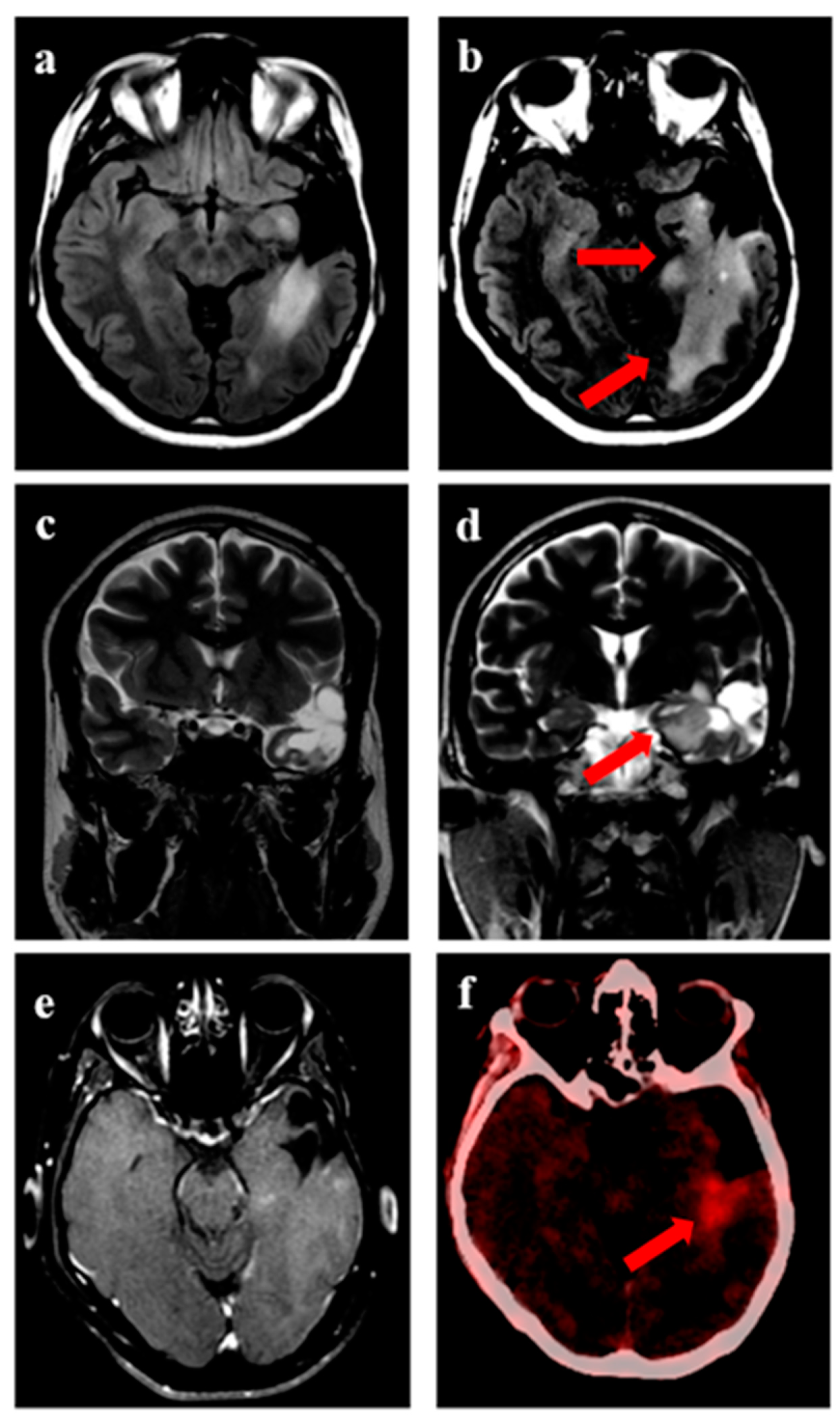
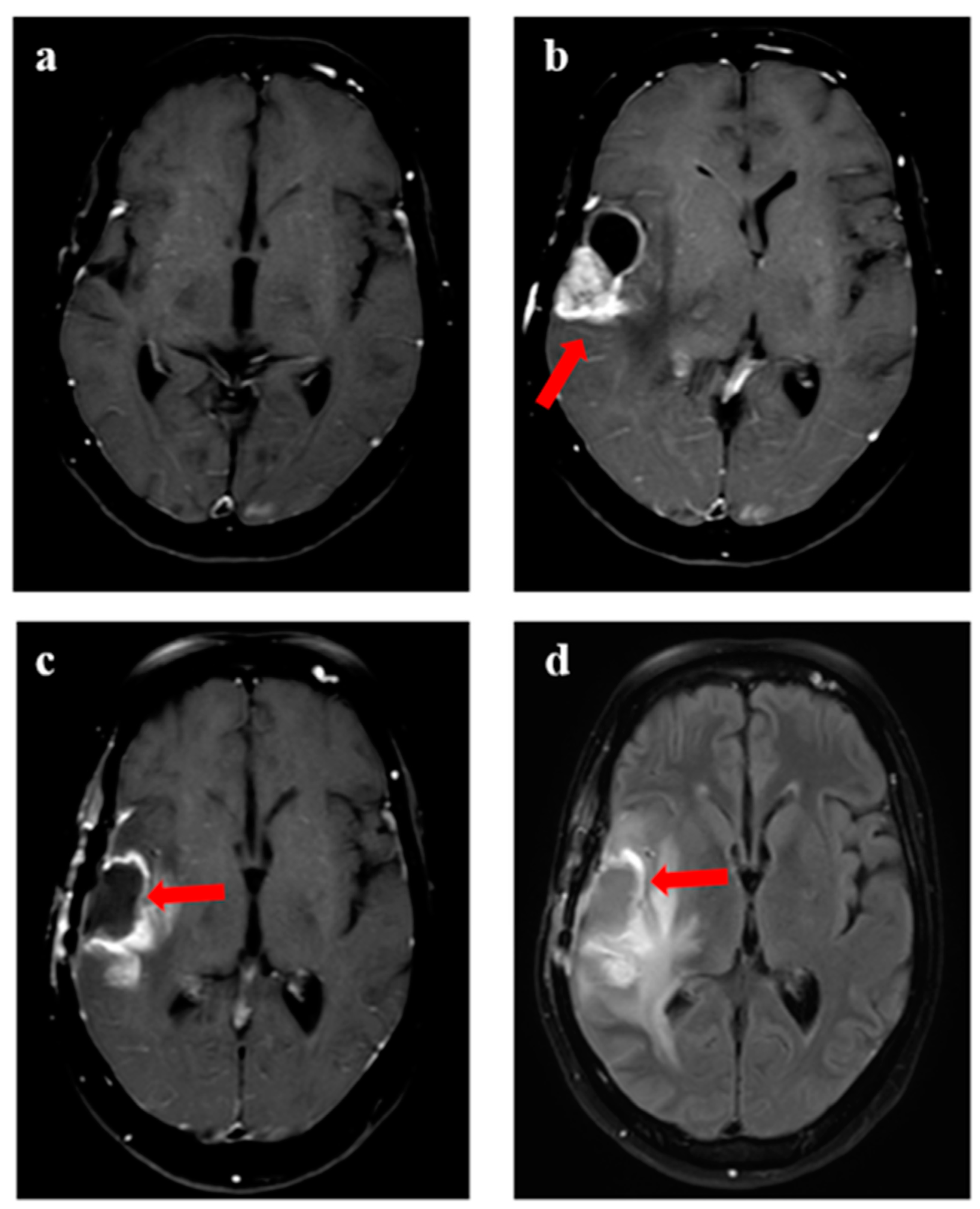
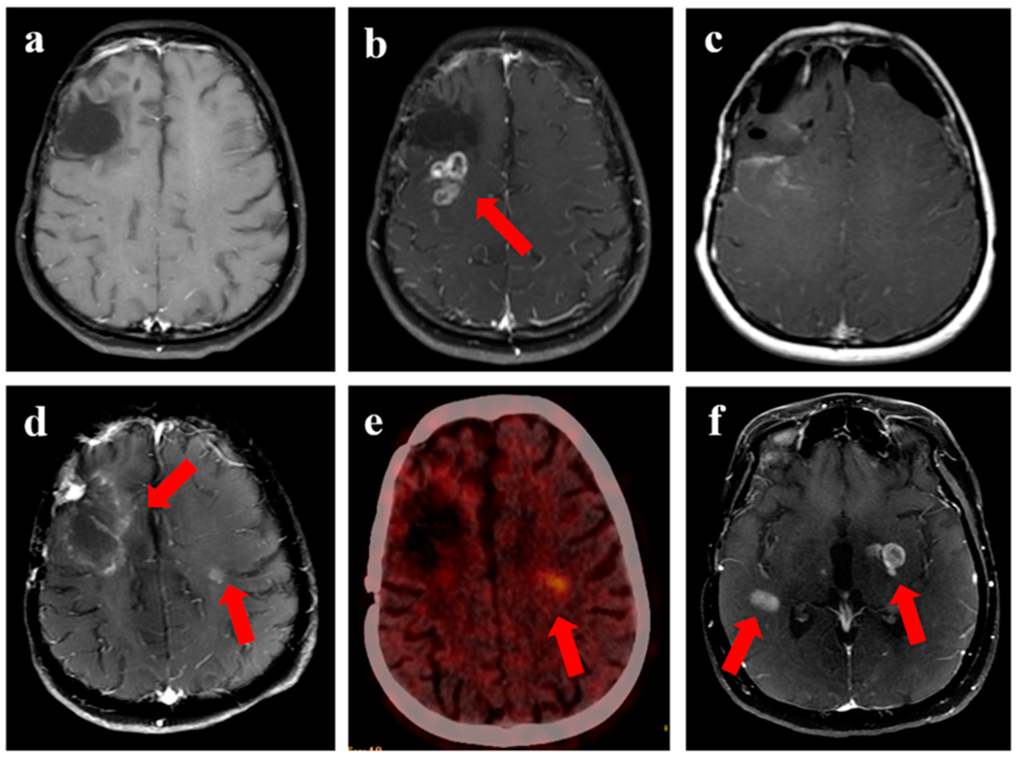
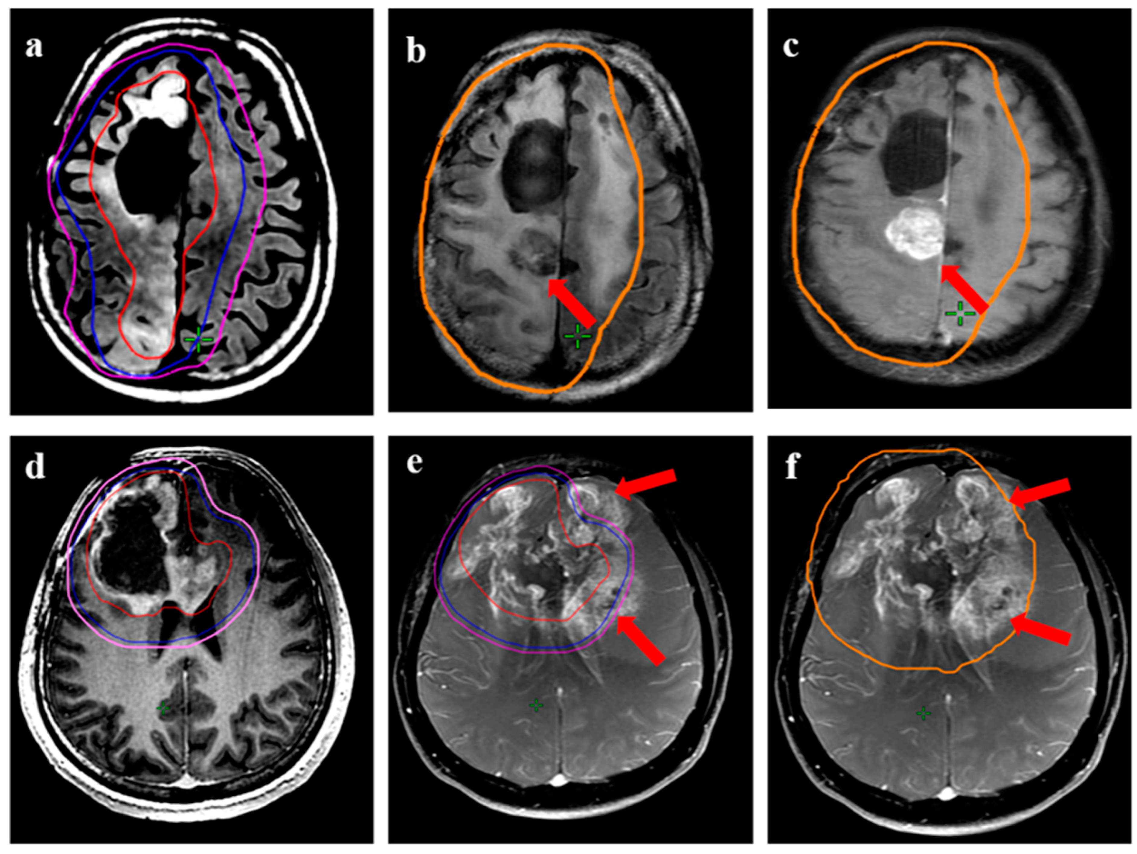
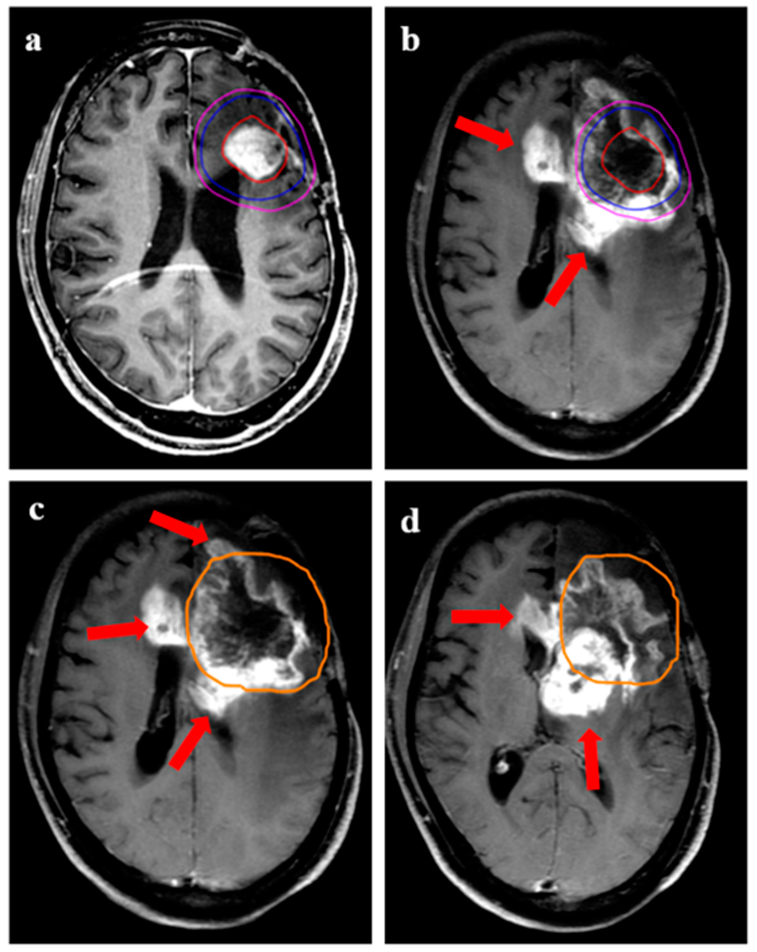
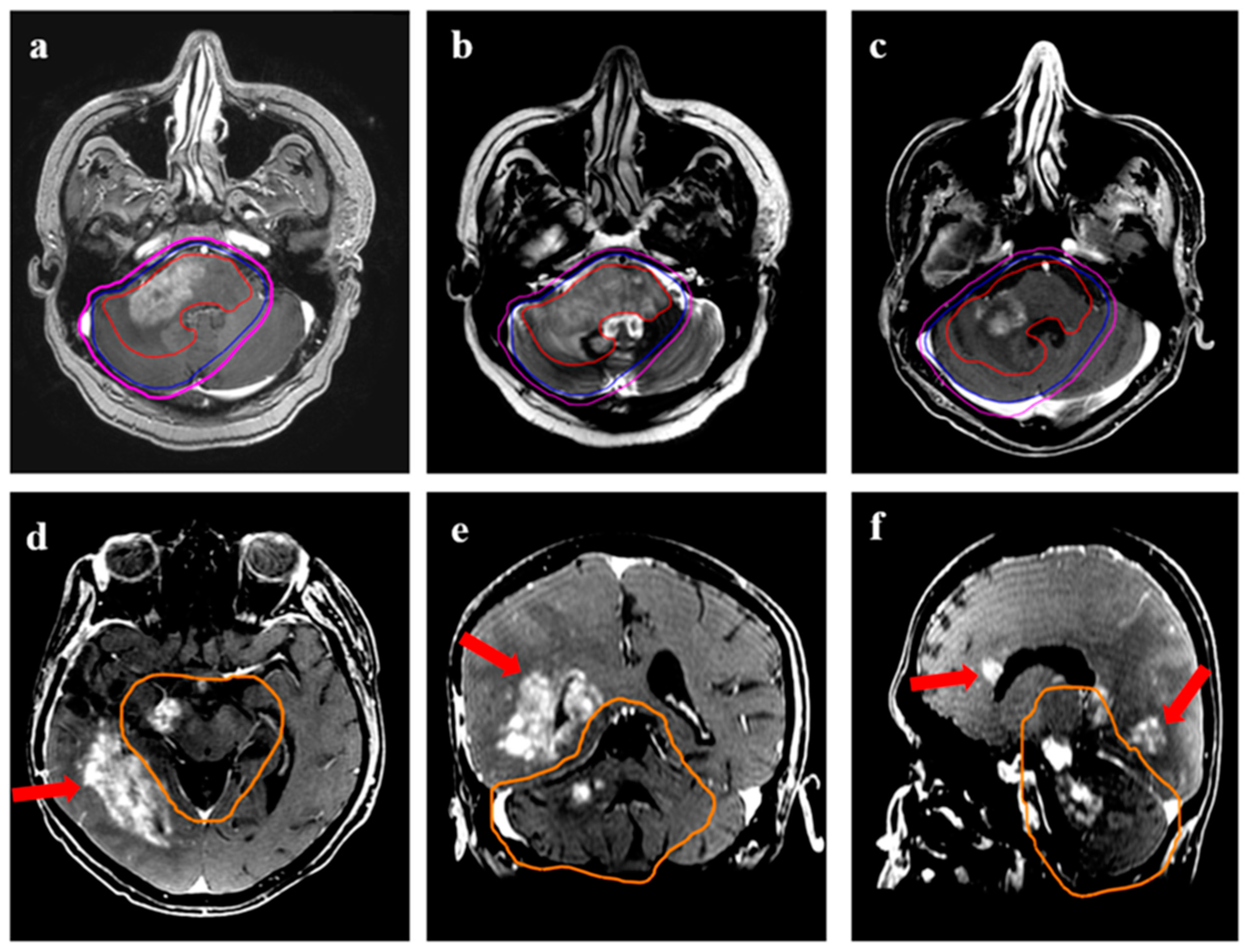
2.4. Analysis of Failure Patterns
2.5. Statistical Analysis
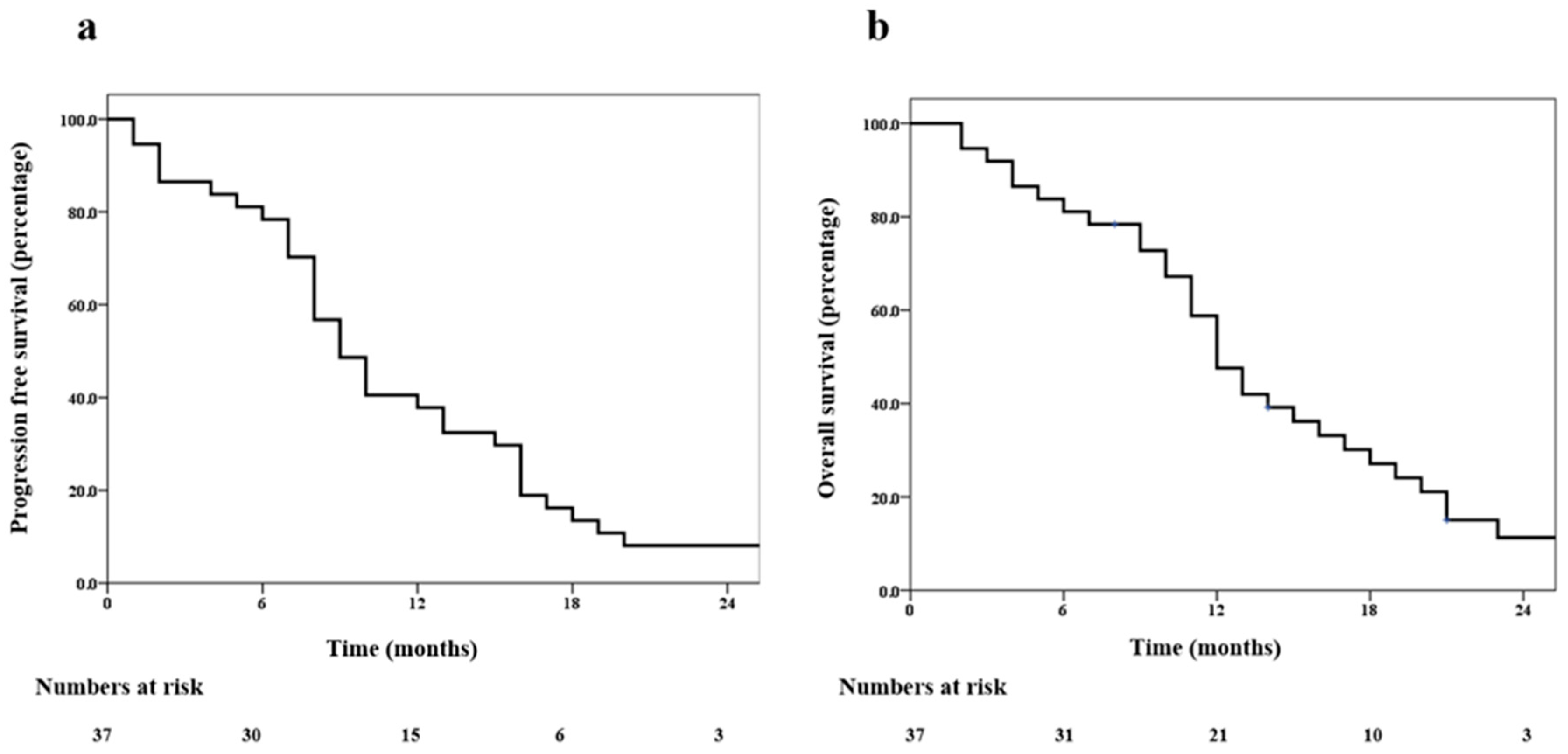
3. Results
4. Discussion
5. Conclusions
Author Contributions
Funding
Institutional Review Board Statement
Informed Consent Statement
Data Availability Statement
Acknowledgments
Conflicts of Interest
References
- Stupp, R.; Brada, M.; van den Bent, M.J.; Tonn, J.C.; Pentheroudakis, G. High-Grade Glioma: ESMO Clinical Practice Guidelines for Diagnosis, Treatment and Follow-Up. Ann. Oncol. 2014, 25, 93–101. [Google Scholar] [CrossRef] [PubMed]
- Weller, M.; van den Bent, M.; Preusser, M.; Le Rhun, E.; Tonn, J.C.; Minniti, G.; Bendszus, M.; Balana, C.; Chinot, O.; Dirven, L.; et al. EANO Guidelines on the Diagnosis and Treatment of Diffuse Gliomas of Adulthood. Nat. Rev. Clin. Oncol. 2021, 18, 170–186. [Google Scholar] [CrossRef]
- Mohile, N.A.; Messersmith, H.; Gatson, N.T.; Hottinger, A.F.; Lassman, A.; Morton, J.; Ney, D.; Nghiemphu, P.L.; Olar, A.; Olson, J.; et al. Therapy for Diffuse Astrocytic and Oligodendroglial Tumors in Adults: ASCO-SNO Guideline. J. Clin. Oncol. 2022, 40, 403–426. [Google Scholar] [CrossRef]
- Stupp, R.; Mason, W.P.; van den Bent, M.J.; Weller, M.; Fisher, B.; Taphoorn, M.J.B.; Belanger, K.; Brandes, A.A.; Marosi, C.; Bogdahn, U.; et al. Radiotherapy plus Concomitant and Adjuvant Temozolomide for Glioblastoma. N. Engl. J. Med. 2005, 352, 987–996. [Google Scholar] [CrossRef]
- Louis, D.N.; Perry, A.; Wesseling, P.; Brat, D.J.; Cree, I.A.; Figarella-Branger, D.; Hawkins, C.; Ng, H.K.; Pfister, S.M.; Reifenberger, G.; et al. The 2021 WHO Classification of Tumors of the Central Nervous System: A Summary. Neuro-Oncology 2021, 23, 1231–1251. [Google Scholar] [CrossRef] [PubMed]
- Milano, M.T.; Okunieff, P.; Donatello, R.S.; Mohile, N.A.; Sul, J.; Walter, K.A.; Korones, D.N. Patterns and Timing of Recurrence after Temozolomide-Based Chemoradiation for Glioblastoma. Int. J. Radiat. Oncol. Biol. Phys. 2010, 78, 1147–1155. [Google Scholar] [CrossRef] [PubMed]
- Rapp, M.; Baernreuther, J.; Turowski, B.; Steiger, H.-J.; Sabel, M.; Kamp, M.A. Recurrence Pattern Analysis of Primary Glioblastoma. World Neurosurg. 2017, 103, 733–740. [Google Scholar] [CrossRef]
- Zhou, X.; Liao, X.; Zhang, B.; He, H.; Shui, Y.; Xu, W.; Jiang, C.; Shen, L.; Wei, Q. Recurrence Patterns in Patients with High-Grade Glioma Following Temozolomide-Based Chemoradiotherapy. Mol. Clin. Oncol. 2016, 5, 289–294. [Google Scholar] [CrossRef]
- Seystahl, K.; Wick, W.; Weller, M. Therapeutic Options in Recurrent Glioblastoma—An Update. Crit. Rev. Oncol./Hematol. 2016, 99, 389–408. [Google Scholar] [CrossRef]
- Krauze, A.; Attia, A.; Braunstein, S.; Chan, M.; Combs, S.; Fietkau, R.; Fiveash, J.; Flickinger, J.; Grosu, A.; Howard, S.; et al. Expert Consensus on Re-Irradiation for Recurrent Glioma. Radiat. Oncol. 2017, 12, 194. [Google Scholar] [CrossRef]
- Maitre, M.; Gupta, T.; Maitre, P.; Chatterjee, A.; Dasgupta, A.; Moiyadi, A.; Shetty, P.; Epari, S.; Sahay, A.; Patil, V.; et al. High-Dose Salvage Re-Irradiation in Recurrent/Progressive Adult Diffuse Gliomas: Development of a Novel Prognostic Scoring System. Cancer Radiother. 2022, 26, 994–1001. [Google Scholar] [CrossRef] [PubMed]
- Shen, C.J.; Kummerlowe, M.N.; Redmond, K.J.; Martinez-Gutierrez, J.C.; Usama, S.M.; Holdhoff, M.; Grossman, S.A.; Laterra, J.J.; Strowd, R.E.; Kleinberg, L.R. Re-Irradiation for Malignant Glioma: Toward Patient Selection and Defining Treatment Parameters for Salvage. Adv. Radiat. Oncol. 2018, 3, 582–590. [Google Scholar] [CrossRef]
- Maitre, P.; Gupta, T.; Maitre, M.; Goda, J.; Krishnatry, R.; Chatterjee, A.; Sridhar, E.; Sahay, A.; Mokal, S.; Moiyadi, A.; et al. Prospective Longitudinal Assessment of Quality of Life and Activities of Daily Living as Patient-Reported Outcome Measures in Recurrent/Progressive Glioma Treated with High-Dose Salvage Re-Irradiation. Clin. Oncol. 2021, 33, e155–e165. [Google Scholar] [CrossRef] [PubMed]
- Gupta, T.; Maitre, M.; Maitre, P.; Goda, J.S.; Krishnatry, R.; Chatterjee, A.; Moiyadi, A.; Shetty, P.; Epari, S.; Sahay, A.; et al. High-Dose Salvage Re-Irradiation for Recurrent/Progressive Adult Diffuse Glioma: Healing or Hurting? Clin. Transl. Oncol. 2021, 23, 1358–1367. [Google Scholar] [CrossRef] [PubMed]
- Tsien, C.I.; Pugh, S.L.; Dicker, A.P.; Raizer, J.J.; Matuszak, M.M.; Lallana, E.C.; Huang, J.; Algan, O.; Deb, N.; Portelance, L.; et al. NRG Oncology/RTOG1205: A Randomized Phase II Trial of Concurrent Bevacizumab and Reirradiation Versus Bevacizumab Alone as Treatment for Recurrent Glioblastoma. J. Clin. Oncol. 2022, 41, 1285–1295. [Google Scholar] [CrossRef]
- Shanker, M.; Chua, B.; Bettington, C.; Foote, M.C.; Pinkham, M.B. Re-Irradiation for Recurrent High-Grade Gliomas: A Systematic Review and Analysis of Treatment Technique with Respect to Survival and Risk of Radionecrosis. Neuro-Oncol. Pract. 2019, 6, 144–155. [Google Scholar] [CrossRef]
- Srinivasan, S.; Dasgupta, A.; Chatterjee, A.; Baheti, A.; Engineer, R.; Gupta, T.; Murthy, V. The Promise of Magnetic Resonance Imaging in Radiation Oncology Practice in the Management of Brain, Prostate, and GI Malignancies. JCO Glob. Oncol. 2022, 8, e2100366. [Google Scholar] [CrossRef]
- Quartuccio, N.; Laudicella, R.; Vento, A.; Pignata, S.; Mattoli, M.V.; Filice, R.; Comis, A.D.; Arnone, A.; Baldari, S.; Cabria, M.; et al. The Additional Value of 18F-FDG PET and MRI in Patients with Glioma: A Review of the Literature from 2015 to 2020. Diagnostics 2020, 10, 357. [Google Scholar] [CrossRef]
- Galldiks, N.; Niyazi, M.; Grosu, A.L.; Kocher, M.; Langen, K.-J.; Law, I.; Minniti, G.; Kim, M.M.; Tsien, C.; Dhermain, F.; et al. Contribution of PET Imaging to Radiotherapy Planning and Monitoring in Glioma Patients—A Report of the PET/RANO Group. Neuro-Oncology 2021, 23, 881–893. [Google Scholar] [CrossRef]
- Johnson, B.E.; Mazor, T.; Hong, C.; Barnes, M.; Aihara, K.; McLean, C.Y.; Fouse, S.D.; Yamamoto, S.; Ueda, H.; Tatsuno, K.; et al. Mutational Analysis Reveals the Origin and Therapy-Driven Evolution of Recurrent Glioma. Science 2014, 343, 189–193. [Google Scholar] [CrossRef]
- Birzu, C.; French, P.; Caccese, M.; Cerretti, G.; Idbaih, A.; Zagonel, V.; Lombardi, G. Recurrent Glioblastoma: From Molecular Landscape to New Treatment Perspectives. Cancers 2020, 13, 47. [Google Scholar] [CrossRef] [PubMed]
- Shapiro, L.Q.; Beal, K.; Goenka, A.; Karimi, S.; Iwamoto, F.M.; Yamada, Y.; Zhang, Z.; Lassman, A.B.; Abrey, L.E.; Gutin, P.H. Patterns of Failure after Concurrent Bevacizumab and Hypofractionated Stereotactic Radiation Therapy for Recurrent High-Grade Glioma. Int. J. Radiat. Oncol. Biol. Phys. 2013, 85, 636–642. [Google Scholar] [CrossRef] [PubMed]
- Niyazi, M.; Jansen, L.L.; Rottler, M.; Ganswindt, U.; Belka, C. Recurrence Pattern Analysis after Re-Irradiation with Bevacizumab in Recurrent Malignant Glioma Patients. Radiat. Oncol. 2014, 9, 299. [Google Scholar] [CrossRef] [PubMed]
- Dasgupta, A.; Geraghty, B.; Maralani, P.J.; Malik, N.; Sandhu, M.; Detsky, J.; Tseng, C.-L.; Soliman, H.; Myrehaug, S.; Husain, Z.; et al. Quantitative Mapping of Individual Voxels in the Peritumoral Region of IDH-Wildtype Glioblastoma to Distinguish between Tumor Infiltration and Edema. J. Neurooncol. 2021, 153, 251–261. [Google Scholar] [CrossRef] [PubMed]
- Malik, N.; Geraghty, B.; Dasgupta, A.; Maralani, P.J.; Sandhu, M.; Detsky, J.; Tseng, C.-L.; Soliman, H.; Myrehaug, S.; Husain, Z.; et al. MRI Radiomics to Differentiate between Low Grade Glioma and Glioblastoma Peritumoral Region. J. Neurooncol. 2021, 155, 181–191. [Google Scholar] [CrossRef]
- Laack, N.N.; Pafundi, D.; Anderson, S.K.; Kaufmann, T.; Lowe, V.; Hunt, C.; Vogen, D.; Yan, E.; Sarkaria, J.; Brown, P.; et al. Initial Results of a Phase 2 Trial of 18F-DOPA PET-Guided Dose-Escalated Radiation Therapy for Glioblastoma. Int. J. Radiat. Oncol. Biol. Phys. 2021, 110, 1383–1395. [Google Scholar] [CrossRef]
- Celli, M.; Caroli, P.; Amadori, E.; Arpa, D.; Gurrieri, L.; Ghigi, G.; Cenni, P.; Paganelli, G.; Matteucci, F. Diagnostic and Prognostic Potential of 18F-FET PET in the Differential Diagnosis of Glioma Recurrence and Treatment-Induced Changes After Chemoradiation Therapy. Front. Oncol. 2021, 11, 721821. [Google Scholar] [CrossRef]
- Kazmi, F.; Soon, Y.Y.; Leong, Y.H.; Koh, W.Y.; Vellayappan, B. Re-Irradiation for Recurrent Glioblastoma (GBM): A Systematic Review and Meta-Analysis. J. Neuro-Oncol. 2019, 142, 79–90. [Google Scholar] [CrossRef]
- Kulinich, D.P.; Sheppard, J.P.; Nguyen, T.; Kondajji, A.M.; Unterberger, A.; Duong, C.; Enomoto, A.; Patel, K.; Yang, I. Radiotherapy versus Combination Radiotherapy-Bevacizumab for the Treatment of Recurrent High-Grade Glioma: A Systematic Review. Acta Neurochir. 2021, 163, 1921–1934. [Google Scholar] [CrossRef]
- Fazzari, F.G.T.; Rose, F.; Pauls, M.; Guay, E.; Ibrahim, M.F.K.; Basulaiman, B.; Tu, M.; Hutton, B.; Nicholas, G.; Ng, T.L. The Current Landscape of Systemic Therapy for Recurrent Glioblastoma: A Systematic Review of Randomized-Controlled Trials. Crit. Rev. Oncol. Hematol. 2022, 169, 103540. [Google Scholar] [CrossRef]
- Baviskar, Y.; Dasgupta, A.; Chatterjee, A.; Kanan, S.; Epari, S.; Sahu, A.; Puranik, A.; Patil, V.; Moiyadi, A.; Gupta, T. MMAP-08 CHEMO-REIRRADIATION (NORMOFRACTIONATED VS. HYPOFRACTIONATED) WITH OR WITHOUT BEVACIZUMAB IN RECURRENT ADULT DIFFUSE HIGH-GRADE GLIOMA (COBRA): PHASE III RANDOMIZED CONTROLLED TRIAL WITH A 2 X 2 FACTORIAL DESIGN. Neuro-Oncol. Adv. 2022, 4, i16. [Google Scholar] [CrossRef]


| Features | Frequency |
|---|---|
| Gender | |
| Male | 28 (76%) |
| Female | 9 (24%) |
| Grade | |
| Grade 3 | 23 (62%) |
| Grade 4 | 14 (38%) |
| Histology | |
| Astrocytoma | 35 (95%) |
| Oligodendroglioma | 2 (5%) |
| IDH a | |
| Mutant | 15 (41%) |
| Negative | 9 (24%) |
| Non-contributory/ Unknown | 13 (35%) |
| ATRX a | |
| Lost | 16 (43%) |
| Retained | 7 (19%) |
| Non-contributory | 14 (38%) |
| Dose: first course of radiation | |
| Median (range) | 56 (54–60) Gy |
| Technique | |
| 2D-conventional | 15 (41%) |
| 3D-conformal | 20 (54%) |
| IMRT | 2 (5%) |
| Concurrent chemotherapy | |
| Yes | 23 (62%) |
| No | 14 (38%) |
| Adjuvant chemotherapy | |
| Yes | 21 (57%) |
| No | 16 (43%) |
| Features | Frequency |
|---|---|
| Age (during re-irradiation) | |
| Median (range) | 40 (24–60) years |
| Time from first radiation | |
| Median (range) | 44 (18–169) months |
| Surgery | |
| No surgery | 3 (8%) |
| Biopsy | 3 (8%) |
| Subtotal resection | 22 (59%) |
| Gross total resection | 9 (24%) |
| Grade during recurrence a | |
| Grade 3 | 4 (12%) |
| Grade 4 | 33 (88%) |
| Performance status during re-irradiation | |
| KPS ≥ 80 | 21 (57%) |
| KPS 50–70 | 16 (43%) |
| Dose during reirradiation | |
| Median (range) | 54 (45–59.4) Gy |
| Cumulative EQD2 | |
| Median (range) | 106.2 (96.2–113.3) |
| PTV volume during re-irradiation | |
| Median (range) | 290 (75–704) cc |
| Concurrent chemotherapy during re-irradiation | |
| Yes | 25 (68%) |
| No | 12 (32%) |
| Chemotherapy | |
| Concurrent chemotherapy alone during re-irradiation | 2 (5%) |
| After reirradiation (with or without concurrent) | 29 (78%) |
| No chemotherapy | 6 (16%) |
| Features | Frequency |
|---|---|
| Imaging modality during recurrence | |
| MRI alone | 27 (73%) |
| PET alone | 3 (8%) |
| MRI and PET | 7 (19%) |
| Time to recurrence after re-irradiation | |
| Median (range) | 9 (1–47) months |
| Volume of recurrence following re-irradiation | |
| Median (range) | 69.5 (3.3–221) cc |
| Patterns of recurrence (with respect to 95% isodose) | |
| Central | 27 (73%) |
| Marginal | 4 (11%) |
| Distant | 6 (16%) |
| Time to recurrence (for different patterns) | |
| Central | |
| Median (range) | 10 (1–47) months |
| Marginal | |
| Median (range) | 11 (6–16) months |
| Distant | |
| Median (range) | 8 (2–19) months |
Disclaimer/Publisher’s Note: The statements, opinions and data contained in all publications are solely those of the individual author(s) and contributor(s) and not of MDPI and/or the editor(s). MDPI and/or the editor(s) disclaim responsibility for any injury to people or property resulting from any ideas, methods, instructions or products referred to in the content. |
© 2023 by the authors. Licensee MDPI, Basel, Switzerland. This article is an open access article distributed under the terms and conditions of the Creative Commons Attribution (CC BY) license (https://creativecommons.org/licenses/by/4.0/).
Share and Cite
Datta, D.; Dasgupta, A.; Chatterjee, A.; Sahu, A.; Bhattacharya, K.; Meena, L.; Joshi, K.; Puranik, A.; Dev, I.; Moiyadi, A.; et al. Imaging-Based Patterns of Failure following Re-Irradiation for Recurrent/Progressive High-Grade Glioma. J. Pers. Med. 2023, 13, 685. https://doi.org/10.3390/jpm13040685
Datta D, Dasgupta A, Chatterjee A, Sahu A, Bhattacharya K, Meena L, Joshi K, Puranik A, Dev I, Moiyadi A, et al. Imaging-Based Patterns of Failure following Re-Irradiation for Recurrent/Progressive High-Grade Glioma. Journal of Personalized Medicine. 2023; 13(4):685. https://doi.org/10.3390/jpm13040685
Chicago/Turabian StyleDatta, Debanjali, Archya Dasgupta, Abhishek Chatterjee, Arpita Sahu, Kajari Bhattacharya, Lilawati Meena, Kishore Joshi, Ameya Puranik, Indraja Dev, Aliasgar Moiyadi, and et al. 2023. "Imaging-Based Patterns of Failure following Re-Irradiation for Recurrent/Progressive High-Grade Glioma" Journal of Personalized Medicine 13, no. 4: 685. https://doi.org/10.3390/jpm13040685
APA StyleDatta, D., Dasgupta, A., Chatterjee, A., Sahu, A., Bhattacharya, K., Meena, L., Joshi, K., Puranik, A., Dev, I., Moiyadi, A., Shetty, P., Singh, V., Patil, V., Menon, N., Sridhar, E., Sahay, A., & Gupta, T. (2023). Imaging-Based Patterns of Failure following Re-Irradiation for Recurrent/Progressive High-Grade Glioma. Journal of Personalized Medicine, 13(4), 685. https://doi.org/10.3390/jpm13040685