

Chest Tube Placement of Secondary Tracheoesophageal Voice Prosthesis: Overcoming Challenging Anatomy in the Laryngectomy Patient

 ,
, 
Abstract
1. Background
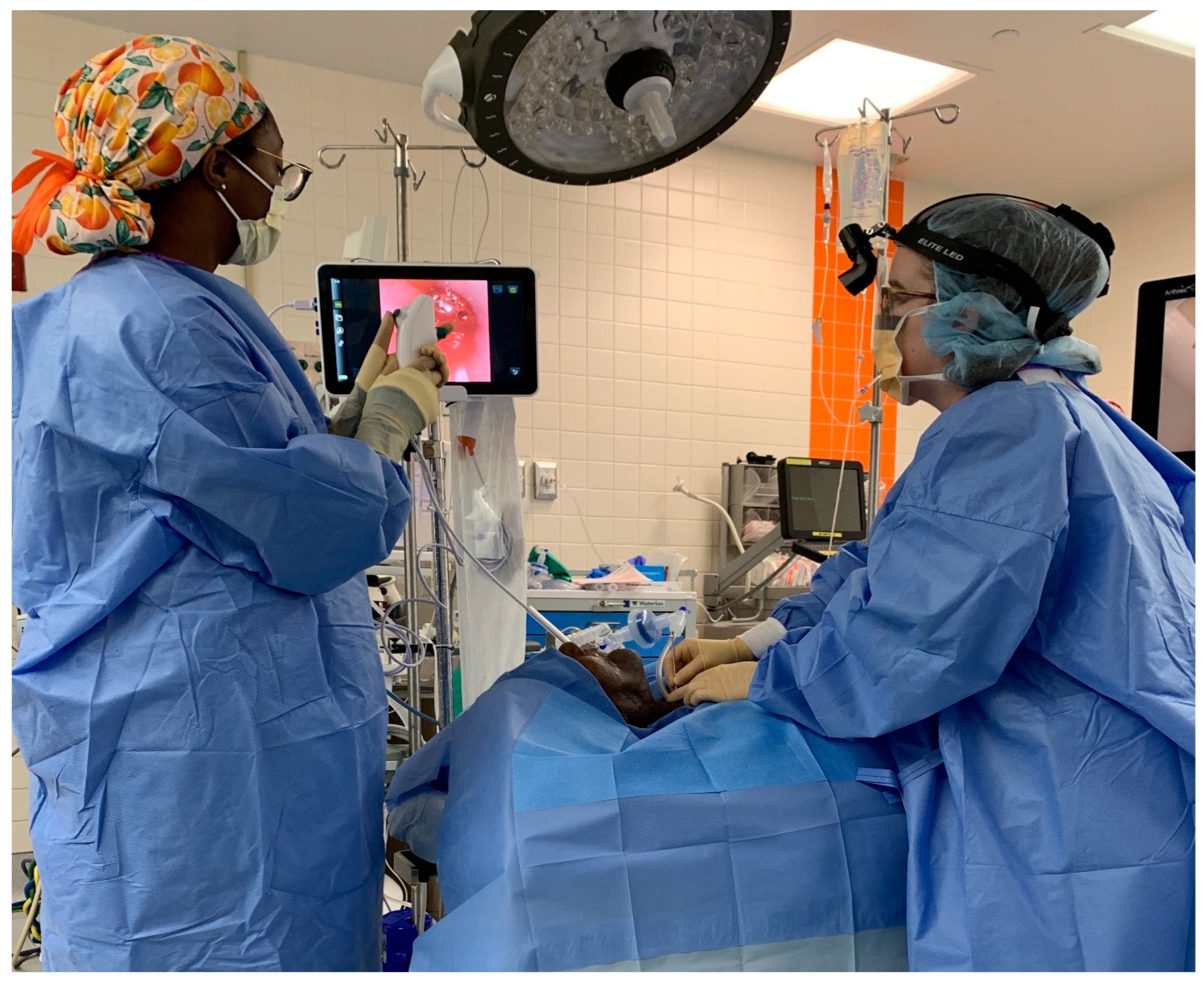
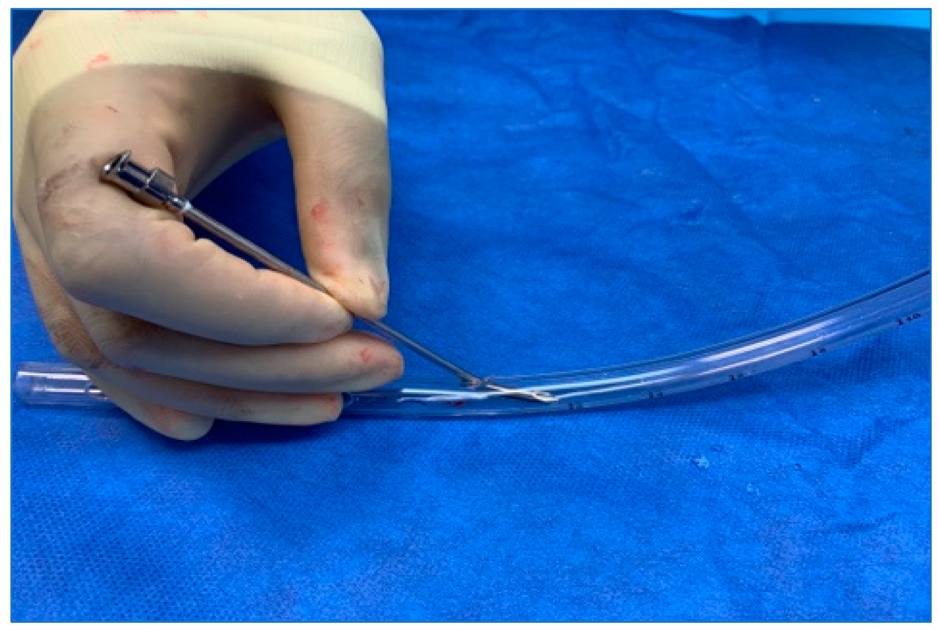
2. Methods
3. Results
4. Discussion
5. Conclusions
Author Contributions
Funding
Institutional Review Board Statement
Informed Consent Statement
Data Availability Statement
Conflicts of Interest
Abbreviations
References
- Attieh, A.Y.; Searl, J.; Shahaltough, N.H.; Wreikat, M.M.; Lundy, D.S. Voice restoration following total laryngectomy by tracheoesophageal prosthesis: Effect on patients’ QOL and voice handicap in Jordan. Health Qual Life Outcomes 2008, 6, 26. [Google Scholar] [CrossRef] [PubMed]
- Clements, K.S.; Rassekh, C.H.; Seikaly, H.; Hokanson, J.A.; Calhoun, K.H. Communication After Laryngectomy: An Assessment of Patient Satisfaction. Arch. Otolaryngol. Neck Surg. 1997, 123, 493–496. [Google Scholar] [CrossRef] [PubMed]
- Singer, M.I.; Blom, E.D. An endoscopic technique for restoration of voice after laryngectomy. Ann. Otol. Rhinol. Laryngol. 1980, 89, 529–533. [Google Scholar] [CrossRef] [PubMed]
- Chakravarty, P.D.; McMurran, A.E.L.; Banigo, A.; Shakeel, M.; Ah-See, K.W. Primary versus secondary tracheoesophageal puncture: Systematic review and meta-analysis. J. Laryngol. Otol. 2018, 132, 14–21. [Google Scholar] [CrossRef]
- Neto, J.C.B.; Dedivitis, R.A.; Aires, F.T.; Pfann, R.Z.; Matos, L.L.; Cernea, C.R. Comparison between Primary and Secondary Tracheoesophageal Puncture Prosthesis: A Systematic Review. ORL J. Otorhinolaryngol. Relat. Spec. 2017, 79, 222–229. [Google Scholar] [CrossRef]
- Chone, C.T.; Gripp, F.M.; Spina, A.L.; Crespo, A.N. Primary versus secondary tracheoesophageal puncture for speech rehabilitation in total laryngectomy: Long-term results with indwelling voice prosthesis. Otolaryngol. Head Neck Surg. 2005, 133, 89–93. [Google Scholar] [CrossRef]
- Gitomer, S.A.; Hutcheson, K.A.; Christianson, B.L.; Samuelson, M.B.; Barringer, D.A.; Roberts, D.B.; Hessel, A.C.; Weber, R.S.; Lewin, J.S.; Zafereo, M.E. Influence of timing, radiation, and reconstruction on complications and speech outcomes with tracheoesophageal puncture. Head Neck 2016, 38, 1765–1771. [Google Scholar] [CrossRef]
- Sinclair, C.F.; Rosenthal, E.L.; McColloch, N.L.; Magnuson, J.S.; Desmond, R.A.; Peters, G.E.; Carroll, W.R. Primary versus delayed tracheoe sophageal puncture for laryngopharyngectomy with free flap reconstruction. Laryngoscope 2011, 121, 1436–1440. [Google Scholar] [CrossRef]
- Emerick, K.S.; Tomycz, L.; Bradford, C.R.; Lyden, T.H.; Chepeha, D.B.; Wolf, G.T.; Teknos, T.N. Primary versus secondary tracheoesophageal puncture in salvage total laryngectomy following chemoradiation. Otolaryngol. Head Neck Surg. 2009, 140, 386–390. [Google Scholar] [CrossRef]
- Dowthwaite, S.A.; Penhearow, J.; Szeto, C.; Nichols, A.; Franklin, J.; Fung, K.; Yoo, J. Postlaryngectomy pharyngocutaneous fistula: Determining the risk of preoperative tracheostomy and primary tracheoesophageal puncture. J. Otolaryngol. Head Neck Surg. 2012, 41, 169–175. [Google Scholar]
- Parikh, S.R.; Irish, J.C.; Curran, A.J.; Gullane, P.J.; Brown, D.H.; Rotstein, L.E. Pharyngocutaneous fistulae in laryngectomy patients: The Toronto Hospital experience. J. Otolaryngol. 1998, 27, 136–140. [Google Scholar] [PubMed]
- Sayles, M.; Grant, D.G. Preventing pharyngo-cutaneous fistula in total laryngectomy: A systematic review and meta-analysis. Laryngoscope 2014, 124, 1150–1163. [Google Scholar] [CrossRef] [PubMed]
- Clancy, K.; Lujan, J.S.; Xia, T.; Zender, C.; Maronian, N. Outcomes of tracheoesophageal puncture in twice-radiated patients. Am. J. Otolaryngol. 2019, 40, 102272. [Google Scholar] [CrossRef] [PubMed]
- Shires, C.B.; Latour, M.; Sebelik, M.; Dewan, K. The use of Montgomery salivary bypass tubes and pharyngocutaneous fistula following salvage laryngectomy. World J. Otorhinolaryngol.—Head Neck Surg. 2024, 10, 43–48. [Google Scholar] [CrossRef]
- Azar, S.S.; Shires, C.B.; Dewan, K.; Chhetri, D.K. Total tracheoesophageal puncture failure: A scoping review of patient characteristics and etiologies. Head Neck 2024. [Google Scholar] [CrossRef]
- Bianco, M.R.; Saita, V.; Occhiuzzi, F.; Modica, D.M.; Latella, D.; Azzolina, A.; Galfano, M.; Allegra, E. Long-Term Complications of Tracheoesophageal Voice Prosthesis. J. Clin. Med. 2024, 13, 1912. [Google Scholar] [CrossRef]
- Noordiana, S.H.; Fang, F.T.H.; Kamarudin, N.A.; Azman, M. Sealing the Breach: A Surgical Solution for Tracheoesophageal Fistula with a Simple Two-Layer Closure. Cureus 2024, 16, e61934. [Google Scholar] [CrossRef]
- Suehiro, A.; Honda, K.; Kishimoto, Y.; Iwanaga, K.; Fujimura, S.; Kawai, Y.; Kojima, T.; Hamaguchi, K.; Omori, K. Modified method for tracheoesophageal fistula closure in intractable cases. Auris Nasus Larynx 2024, 51, 774–778. [Google Scholar] [CrossRef]
- Escandón, J.M.; Mohammad, A.; Mathews, S.; Bustos, V.P.; Santamaría, E.; Chen, H.C.; Langstein, H.N.; Manrique, O.J. Definitive Closure of the Tracheoesophageal Puncture Site after Oncologic Laryngectomy: A Systematic Review and Meta-Analysis. Arch. Plast. Surg. 2022, 49, 617–632. [Google Scholar] [CrossRef]
- Yavuz, H.; Vural, O. Tracheoesophageal puncture closure with annular mucosal flap. Head Neck 2021, 43, 1705–1710. [Google Scholar] [CrossRef]
- Patel, R.S.; Mohr, T.; Hartman, C.; Stach, C.; Sikora, A.G.; Zevallos, J.P.; Sandulache, V.C. Tracheoesophageal Prosthesis Use Is Associated with Improved Overall Quality of Life in Veterans with Laryngeal Cancer. Ann. Otol. Rhinol. Laryngol. 2018, 127, 421–428. [Google Scholar] [CrossRef] [PubMed]
- Ringash, J.; Bernstein, L.J.; Devins, G.; Dunphy, C.; Giuliani, M.; Martino, R.; McEwen, S. Head and Neck Cancer Survivorship: Learning the Needs, Meeting the Needs. In Seminars in Radiation Oncology; W.B. Saunders: Philadelphia, PA, USA, 2018; Volume 28, pp. 64–74. [Google Scholar]
- Galli, A.; Giordano, L.; Biafora, M.; Tulli, M.; Di Santo, D.; Bussi, M. Voice prosthesis rehabilitation after total laryngectomy: Are satisfaction and quality of life maintained over time? Acta Otorhinolaryngol. Ital. 2019, 39, 162–168. [Google Scholar] [CrossRef] [PubMed]
- Jiang, N.; Kearney, A.; Damrose, E.J. Tracheoesophageal fistula length decreases over time. Eur. Arch. Otorhinolaryngol. 2016, 273, 1819–1824. [Google Scholar] [CrossRef] [PubMed]
- Brook, I.; Goodman, J.F. Tracheoesophageal Voice Prosthesis Use and Maintenance in Laryngectomees. Int. Arch. Otorhinolaryngol. 2020, 24, e535–e538. [Google Scholar] [CrossRef]
- Lewin, J.S.; Baumgart, L.M.; Barrow, M.P.; Hutcheson, K.A. Device life of the trachesophageal voice prosthesis revisited. JAMA Otolaryngol. Head Neck Surg. 2017, 143, 65–71. [Google Scholar] [CrossRef]
- Silver, F.M.; Gluckman, J.L.; Donegan, J.O. Operative complications of tracheoesophageal puncture. Laryngoscope 1985, 95, 1360–1362. [Google Scholar] [CrossRef]
- Andrews, J.C.; Mickel, R.A.; Monahan, G.P.; Hanson, D.G.; Ward, P.H. Major complications following tracheoesophageal puncture for voice rehabilitation. Laryngoscope 1987, 97, 562–567. [Google Scholar] [CrossRef]
- Padhya, T.A.; Athavale, S.M.; Morgan, J.M.; McCaffrey, T.V. An Alternative Approach for Secondary Tracheoesophageal Puncture in the Difficult Laryngectomy Neck. Laryngoscope 2008, 118, 266–269. [Google Scholar] [CrossRef]
- Mohr, R.M.; Paddock, B.H.; Boehler, J. An adaptation of tracheoesophageal puncture. Laryngoscope 1983, 93, 1086–1088. [Google Scholar] [CrossRef]
- Ananthapadmanabhan, S.; Jabbour, J.; Lai, S.; Suruliraj, A.; Smith, M.; Riffat, F.; Devadas, M.; Liem, H.; Sritharan, N. A Multidisciplinary Approach to Secondary Tracheoesophageal Puncture for Voice Prosthesis Insertion Using Flexible Esophagoscopy. J. Voice 2022, 38, 1255.e1–1255.e8. [Google Scholar] [CrossRef]
- LeBert, B.; McWhorter, A.J.; Kunduk, M.; Walvekar, R.R.; Lewin, J.S.; Hutcheson, K.A.; Barringer, D.A.; Hessel, A.C.; Holsinger, F.C. Secondary Tracheoesophageal Puncture with In-Office Transnasal Esophagoscopy. Arch. Otolaryngol. Neck Surg. 2009, 135, 1190–1194. [Google Scholar] [CrossRef] [PubMed]
- Spofford, B.; Jafek, B.; Barcz, D. An improved method for creating tracheoesophageal fistulas for Blom-Singer or Panje voice prostheses. Laryngoscope 1984, 94 Pt 1, 257–258. [Google Scholar] [CrossRef] [PubMed]
- Maniglia, A.J. Newer technique of tracheoesophageal fistula for vocal rehabilitation after total laryngectomy. Laryngoscope 1985, 95, 1064–1066. [Google Scholar] [CrossRef] [PubMed]
- Smith, A.; Thimmappa, V.; Jones, J.; Shires, C.; Sebelik, M. The Use of Ultrasound for Sizing Tracheoesophageal Speech Prostheses. Otolaryngol.–Head Neck Surg. 2016, 157, 1075–1078. [Google Scholar] [CrossRef]
- Shires, C.B.; Boughter, J.D., Jr.; Smith, A.; Sebelik, M.E. Head and Neck Ultrasound Utilization Rates: 2012 to 2019. OTO Open 2023, 7, e97. [Google Scholar] [CrossRef]
- Glazer, T.A.; Meraj, T.; Lyden, T.H.; Spector, M.E. In-Office Secondary Tracheoesophageal Puncture with Immediate Prosthesis Placement. Otolaryngol. Head Neck Surg. 2016, 155, 360–363. [Google Scholar] [CrossRef]
- Tkaczuk, A.T.; Taylor, R.J.; Wolf, J.S. A Novel Device for Placement of a Secondary Tracheoesophageal Voice Prosthesis: A Preliminary Feasibility Study. ORL J. Otorhinolaryngol. Relat. Spec. 2018, 80, 36–40. [Google Scholar] [CrossRef]
- Ricci, E.; Riva, G.; Dagna, F.; Seglie, E.; Cavalot, A. In-clinic secondary tracheoesophageal puncture and voice prosthesis placement in laryngectomees. Eur. Ann. Otorhinolaryngol. Head Neck Dis. 2018, 135, 349–352. [Google Scholar] [CrossRef]
- Wu, M.P.; Sullivan, C.; Deschler, D.G. Modified Technique for Retrograde Placement of the Tracheoesophageal Voice Prosthesis in the Office. Laryngoscope 2024, ahead of print. [Google Scholar] [CrossRef]
- Inhealth Technologies. Blom-Singer Indwelling Voice Prosthesis for Primary, Secondary, Replacement TEP Procedures: Medical Professional Instructoins for Use; Freudenberg Medical: Beverly, MA, USA, 2018. [Google Scholar]
- Atos Medical. Provox ActiValve: Clinician Instructions for Use; Atos Medical AB: Malmö, Sweden, 2020. [Google Scholar]
- Maniglia, A.J. Vocal rehabilitation after total laryngectomy: A flexible fiberoptic endoscopic technique for tracheoesophageal fistula. Laryngoscope 1982, 92, 1437–1439. [Google Scholar] [CrossRef]
- Abdul-Aziz, D.; Zenga, J.; Deschler, D.G. The Difficult Secondary Tracheoesophageal Puncture: A Technique for Safe Insertion. ORL J. Otorhinolaryngol. Relat. Spec. 2019, 81, 10–15. [Google Scholar] [CrossRef] [PubMed]
- Scherl, C.; Kauffels, J.; Schützenberger, A.; Döllinger, M.; Bohr, C.; Dürr, S.; Fietkau, R.; Haderlein, M.; Koch, M.; Traxdorf, M.; et al. Secondary Tracheoesophageal Puncture After Laryngectomy Increases Complications with Shunt and Voice Prosthesis. Laryngoscope 2020, 130, E865–E873. [Google Scholar] [CrossRef] [PubMed]
- Neumann, A.; Schultz-Coulon, H.J. Management of complications after prosthetic voice rehabilitation. HNO 2000, 48, 508–516. [Google Scholar] [CrossRef] [PubMed]
- Izdebski, K.; Reed, C.G.; Ross, J.C.; Hilsinger, R.L. Problems with tracheoesophageal fistula voice restoration in totally laryngectomized patients. A review of 95 cases. Arch. Otolaryngol. Head Neck Surg. 1994, 120, 840–845. [Google Scholar] [CrossRef]


| Patient Characteristics (N = 76) | |
|---|---|
| Average age | 62 years |
| Average time from TL to TEP | 197 days |
| Sex (M:F) | 3.1:1.2 |
| Prior attempt with another technique | 74% (n = 56) |
| Prior XRT | 92% (n = 70) |
Disclaimer/Publisher’s Note: The statements, opinions and data contained in all publications are solely those of the individual author(s) and contributor(s) and not of MDPI and/or the editor(s). MDPI and/or the editor(s) disclaim responsibility for any injury to people or property resulting from any ideas, methods, instructions or products referred to in the content. |
© 2024 by the authors. Licensee MDPI, Basel, Switzerland. This article is an open access article distributed under the terms and conditions of the Creative Commons Attribution (CC BY) license (https://creativecommons.org/licenses/by/4.0/).
Share and Cite
Shires, C.B.; Schertzer, J.S.; Ottenstein, L.; Harris, T.; Sebelik, M.E. Chest Tube Placement of Secondary Tracheoesophageal Voice Prosthesis: Overcoming Challenging Anatomy in the Laryngectomy Patient. J. Pers. Med. 2024, 14, 1021. https://doi.org/10.3390/jpm14101021
Shires CB, Schertzer JS, Ottenstein L, Harris T, Sebelik ME. Chest Tube Placement of Secondary Tracheoesophageal Voice Prosthesis: Overcoming Challenging Anatomy in the Laryngectomy Patient. Journal of Personalized Medicine. 2024; 14(10):1021. https://doi.org/10.3390/jpm14101021
Chicago/Turabian StyleShires, Courtney B., Joseph S. Schertzer, Lauren Ottenstein, Tricia Harris, and Merry E. Sebelik. 2024. "Chest Tube Placement of Secondary Tracheoesophageal Voice Prosthesis: Overcoming Challenging Anatomy in the Laryngectomy Patient" Journal of Personalized Medicine 14, no. 10: 1021. https://doi.org/10.3390/jpm14101021
APA StyleShires, C. B., Schertzer, J. S., Ottenstein, L., Harris, T., & Sebelik, M. E. (2024). Chest Tube Placement of Secondary Tracheoesophageal Voice Prosthesis: Overcoming Challenging Anatomy in the Laryngectomy Patient. Journal of Personalized Medicine, 14(10), 1021. https://doi.org/10.3390/jpm14101021