Review

The cGAS/STING Pathway—A New Potential Biotherapeutic Target for Gastric Cancer?

1
Department of General Surgery, Xiangya Hospital, Central South University, 87 Xiangya Road, Changsha 410017, China
2
National Clinical Research Center for Geriatric Disorders, Xiangya Hospital, Central South University, Changsha 410017, China
*
Author to whom correspondence should be addressed.
J. Pers. Med. 2024, 14(7), 736; https://doi.org/10.3390/jpm14070736
Submission received: 7 June 2024 / Revised: 5 July 2024 / Accepted: 8 July 2024 / Published: 9 July 2024

Abstract

:
Gastric cancer ranks among the top five deadliest tumors worldwide, both in terms of prevalence and mortality rates. Despite mainstream treatments, the efficacy in treating gastric cancer remains suboptimal, underscoring the urgency for novel therapeutic approaches. The elucidation of tumor immunosuppressive microenvironments has shifted focus towards cancer biotherapeutics, which leverage the patient’s immune system or biologics to target tumor cells. Biotherapy has emerged as a promising alternative for tumors resistant to traditional chemotherapy, radiation, and immunotherapy. Central to this paradigm is the cGAS-STING pathway, a pivotal component of the innate immune system. This pathway recognizes aberrant DNA, such as that from viral infections or tumor cells, and triggers an immune response, thereby reshaping the immunosuppressive tumor microenvironment into an immune-stimulating milieu. In the context of gastric cancer, harnessing the cGAS-STING pathway holds significant potential for biotherapeutic interventions. This review provides a comprehensive overview of the latest research on cGAS-STING in gastric cancer, including insights from clinical trials involving STING agonists. Furthermore, it assesses the prospects of targeting the cGAS-STING pathway as a novel biotherapeutic strategy for gastric cancer.

1. Backgrounds

Gastric cancer stands as one of the most pervasive malignancies globally, characterized by its significant morbidity and mortality rates, posing a formidable challenge to public health worldwide. Ranked as the fifth most common cancer globally, gastric cancer ranks third in cancer-related deaths due to its aggressive nature [1,2]. Its primary risk factors encompass Helicobacter pylori infection, dietary patterns, smoking, Epstein-Barr virus (EBV) infection, and genetic predispositions [3]. The burden of gastric cancer is particularly pronounced in East Asia and South America. In China, digestive system cancers contribute to 41.6% of new cancer cases and 49.3% of cancer-related deaths, with gastric cancer representing a substantial portion of these statistics [4,5,6]. Recent guidelines from the National Comprehensive Cancer Network (NCCN) emphasize the importance of precision therapy alongside conventional treatments such as surgical resection, radiotherapy, chemotherapy, targeted therapy, and immunotherapy. Precision therapy is particularly pertinent for cases of gastric cancer with poor prognoses [7,8]. Therefore, the quest for novel targets in gastric cancer treatment assumes paramount importance, aiming to address the prevalent issue of resistance to immune checkpoint inhibitor monotherapy among gastric cancer patients.
Cancer biotherapy harnesses biological agents to trigger the host’s innate anti-tumor response, effectively impeding tumor cell proliferation. The human immune system assumes a pivotal role in the initiation, progression, and metastasis of malignant tumors, exerting its tumor-suppressing effects by activating robust anti-tumor responses. This immune-mediated mechanism is closely linked to the prognosis of various cancer therapies [9]. Currently, biotherapeutic interventions targeting T cells have demonstrated promising therapeutic outcomes against both solid tumors and hematological malignancies [10]. Consequently, the prospect of achieving durable tumor treatment and control via modulation of the innate immune system holds significant promise [11,12].
The cyclic GMP–AMP synthase (cGAS)–stimulator of interferon genes (STING) pathway serves as a pivotal mediator of innate immune responses, uniquely activated by the detection of DNA from both foreign microbial sources and the host itself. This pathway plays a critical role in diverse physiological processes, including inflammation, stress responses, autoimmune disorders, and tumorigenesis [13,14,15,16].
This review outlines the expression of cGAS-STING in gastric cancer and its association with key genetic markers related to gastric cancer, elucidating its potential therapeutic implications in this context. Furthermore, it delves into future research prospects pertaining to the transduction of the cGAS-STING pathway and its pivotal role in facilitating the clinical translation of novel therapies for gastric cancer.

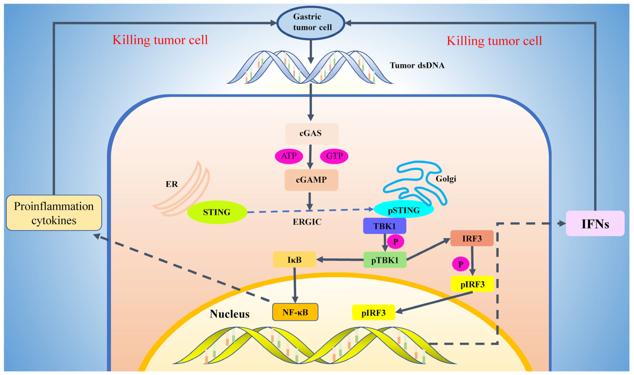
2. Activation Mechanism of cGAS-STING Pathway

Exogenous viruses, bacteria, sterile inflammation, tumors, chromosome instability, and chemotherapy can all inflict cellular damage, leading to the release of double-stranded DNA (dsDNA) [17]. Ordinarily confined within the nucleus and mitochondria, the presence of dsDNA in the cytoplasm serves as a damage-associated molecular pattern (DAMP). Consequently, dsDNA that escapes lysosomal degradation becomes implicated in the initiation and progression of various inflammatory conditions and tumors through the activation of the cGAS-STING signaling pathway.
cGAS, short for “cyclic GMP-AMP synthetase”, is situated within the cytoplasm, where it detects the presence of dsDNA—an anomaly in this cellular compartment—and catalyzes the conversion of GTP and ATP into “cyclic GMP-AMP” (cGAMP) [18,19]. This enzymatic process involves two functional domains within the cGAS protein: the N-terminal nucleotide transferase domain (NTase domain) and the C-terminal nucleotide transferase domain (CTD). The NTase domain primarily recognizes and binds dsDNA molecules, whereas the CTD facilitates the synthesis of cGAMP from ATP and GTP substrates. Notably, the resulting cGAMP, containing two phosphodiester bonds, exhibits heightened affinity for STING [20,21]. Upon cGAMP binding to STING, an intracellular immune response is triggered, activating pathways that culminate in the production of interferon and other immune-related molecules essential for combating infections [22]. Hence, cGAS functions as an “accelerator” for the immune system, recognizing both endogenous and exogenous DNA to initiate immune responses. This mechanism fortifies the body’s “attack force” against tumors and pathogenic microorganisms.
STING (Stimulator of Interferon Genes) is a transmembrane protein consisting of 379 amino acid residues, predominantly localized within the endoplasmic reticulum (ER). Its structure comprises distinct functional domains crucial for its role in cellular signaling. The N-terminal region encompasses the first 137 amino acid residues, forming a transmembrane domain that anchors STING to the ER membrane. Residues 138 to 340 constitute the cyclic dinucleotide binding domain, essential for STING’s interaction with cGAMP and initiating downstream signaling cascades. The remaining residues (341 to 379) constitute the C-terminal tails (CTT), potentially modulating STING function or interactions [23,24,25]. The ligand-binding domain of the STING protein is located in its C-terminal domain and is responsible for recognizing and binding intracellular cyclic dinucleotides (such as cGAMP). Upon binding with cGAMP, the ligand-binding domain of STING undergoes a conformational change, leading to the dissociation of the CTT. Subsequently, activated STING undergoes aggregation and translocates to the Golgi apparatus via the endoplasmic reticulum Golgi intermediate compartment (ERGIC) [26]. Following activation, STING recruits and activates tank-binding kinase 1 (TBK1) via a specific binding motif (PLPLRT/SD) within its CTT. TBK1, an inhibitor of kappa B kinase (IKK)-related kinase, upon binding to STING, phosphorylates STING itself, augmenting its activity and facilitating prolonged signal transduction. Concurrently, TBK1 phosphorylates interferon regulatory factor 3 (IRF3), a pivotal transcription factor in the interferon signaling pathway [27,28]. Phosphorylated IRF3 undergoes conformational changes, enabling its dimerization and nuclear translocation. This process enhances the transcription of interferon (IFN) and other immune-related genes, thereby bolstering the cellular immune response [29].
In addition to the classical IFN pathway, the nuclear factor kappa B (NF-kB) serves as another crucial downstream effector of STING, contributing to the regulation of inflammatory and immune responses [30,31]. Recent investigations have unveiled that STING activation triggers the activation of TGF-beta-activated kinase 1 (TAK1) and I kappa B kinase (IKK) complexes, culminating in the phosphorylation and subsequent degradation of I kappa B [32,33]. Consequently, the degradation of IkB permits NF-kB to translocate into the nucleus, initiating NF-kB-mediated gene transcription. This transcriptional activation induces the production of inflammatory factors vital for sustaining immune and inflammatory responses.
Upon completion of the immune response, the STING protein undergoes ubiquitination. The ubiquitinated STING is then recognized and transported to the lysosome, where it undergoes degradation, effectively curtailing excessive immune responses [34] (Figure 1).

3. The Role of cGAS-STING Signaling Pathway in Tumor Biotherapy

The tumor microenvironment (TME) is a complex and heterogeneous milieu comprising various cell types aside from tumor cells, encompassing fibroblasts, stromal cells, immune cells, and the vascular system. Within this intricate landscape, the TME fosters tumor growth and progression through mechanisms such as angiogenesis induction, the establishment of an immunosuppressive milieu, and the facilitation of tumor cell evasion. These processes significantly curtail the efficacy of conventional tumor treatments like surgery, radiotherapy, and chemotherapy [35,36]. Notably, the activation of the cGAS-STING signaling pathway assumes a pivotal role in tumor biotherapy.
Upon activation of the cGAS-STING signaling pathway, a significant production of interferons (IFNs) ensues. IFNs represent a pivotal class of immunomodulatory molecules crucial for anti-tumor immunity. They facilitate several key processes: Firstly, IFNs promote the recognition and processing of tumor-associated antigens (TAAs) by antigen-presenting cells, such as dendritic cells, initiating the activation of tumor-specific T cells for the targeted elimination of tumor cells. Additionally, IFNs bolster the recruitment and activation of immune effectors like T cells and natural killer cells within tumor tissues, augmenting their tumor-killing capabilities and fortifying immune responses. Furthermore, IFNs orchestrate a shift in the tumor microenvironment by diminishing the production of immunosuppressive factors, such as anti-inflammatory cytokines and immunosuppressive cytokines, while amplifying the generation of immunostimulatory factors like pro-inflammatory cytokines and chemokines. This recalibration enhances immune cell functionality against tumors [37,38]. Clinical investigations have substantiated the correlation between IFN-γ-induced heightened levels of infiltrating T cells and improved overall survival in patients afflicted with colorectal cancer [39,40]. Nonetheless, tumors deploy various strategies to thwart IFN-mediated immune responses. They may inhibit IFN formation via the JAK-STAT signaling pathway or release inhibitory factors. Moreover, tumors secrete chemokines like lectins to curtail the recruitment of T cells to the tumor microenvironment, thereby evading immune surveillance and minimizing the likelihood of immune system attacks [41,42].
The cGAS-STING signaling pathway has the capability to reverse this detrimental phenomenon. For instance, research has unveiled that activating STING signaling via constitutively active STING in both human and murine colorectal cancer (CRC) models can induce endogenous interferon signaling within CRC cells, even in those with intact mismatch repair (MMR) signaling. This activation leads to the initiation of local interferon signaling in cancer cells, the recruitment of antigen-presenting cells, the mobilization of effector lymphocytes, and enhances the responsiveness of previously “cold” tumor microenvironments (TMEs) to in vivo immune checkpoint inhibitor (ICI) therapy [43]. Thus, the cGAS-STING signaling pathway has the potential to counteract the inhibitory effects exerted by tumor cells on interferons, by continuously releasing substantial amounts of interferons. This transformation effectively converts a “cold tumor” into a “hot tumor”, thereby shifting the tumor microenvironment from an immunosuppressive state to an immune-promoting one. Consequently, this process not only impedes tumor development and metastasis but also enhances the efficacy of other therapeutic modalities [44,45,46] (Figure 2).

4. The cGAS-STING Pathway in Gastric Cancer

As mentioned earlier, the primary risk factors for gastric cancer include infections by Helicobacter pylori and Epstein-Barr virus (EBV). As exogenous pathogens, their DNA can bind to cGAS and activate the cGAS-STING signaling pathway [47]. Researchers observed that after 20 weeks of Helicobacter pylori infection, immunohistochemical analysis revealed a significant increase in STING protein expression in the gastric epithelial cells of mice [48]. Additionally, quantitative PCR results showed elevated mRNA expression levels of interferons (IFNs) and the downstream inflammatory factor IL-6 in gastric tissue, confirming the activation of the cGAS-STING pathway by Helicobacter pylori infection [48]. Building upon this observation, further investigations demonstrated significantly higher STING protein expression in normal tissues adjacent to cancer compared to tumor tissues in surgical resection samples from clinical gastric cancer patients. Patients exhibiting lower STING protein expression levels in gastric tumor tissues exhibited more advanced TNM stages and lower overall survival rates [48]. Regarding EBV infection, STING expression was notably higher in the surgical specimens of patients with EBV-positive gastric cancer compared to EBV-negative cases. Interestingly, patients with STING-positive expression demonstrated better overall survival rates regardless of EBV infection status [49]. Moreover, a predictive model based on cGAS-STING pathway-associated genes (CSRs) was developed to forecast overall survival in gastric cancer patients using bioinformatics techniques applied to The Cancer Genome Atlas (TCGA) and Gene Expression Omnibus (GEO) databases [50]. Additionally, a study by Gao et al. identified prognosis and immunotherapy biomarkers (M2GO), including STING downstream molecules TBK1 and IRF3, utilizing transcriptome sequencing data from clinical gastric cancer patients with ovarian metastasis, which were externally validated using the TCGA-STAD database. Patients with lower M2GO expression levels exhibited relatively poorer prognoses [51]. Furthermore, Japanese researchers found that STING protein expression levels and CD8+ T cell infiltration in gastric cancer samples from HER2-positive patients were significantly lower compared to HER2-negative patients. Inhibition of HER2 resulted in upregulated expression levels of STING protein and CD8+ T cell infiltration, suggesting that HER2 positivity may promote a tumor immunosuppressive microenvironment in gastric cancer by inhibiting STING expression and CD8+ T cell infiltration, thereby worsening patient prognosis [52] (Table 1).
While there are currently no ongoing clinical trials investigating STING agonists specifically for gastric cancer, promising results from experimental studies conducted in cell and animal models instill confidence in their potential efficacy. In gastric cancer cell lines, for instance, metformin has demonstrated the ability to not only inhibit the mTOR1 signaling pathway via classical AMPK-dependent or independent mechanisms but also to exhibit anti-tumor effects by activating the cGAS-STING pathway [55]. Similarly, in both gastric cancer cells and mouse-transplanted tumor models, Anlotinib has shown efficacy in reducing tumor proliferation and metastasis by activating the cGAS-STING signaling pathway [53]. Additionally, it has been observed to enhance the anti-tumor efficacy of anti-PD-L1 therapy. It is noteworthy that while gastric cancer cells undergoing chemotherapy, radiotherapy, or other stress responses often experience cell damage or apoptosis, leading to the release of endogenous dsDNA into the cytoplasm and activating anti-tumor immune responses via the cGAS-STING pathway, these cells may also evade this immune surveillance through various mechanisms, such as reducing their DNA sensing function. However, combination therapy may counteract this effect. In studies utilizing Mouse Forestomach Carcinoma (MFC) cells and MFC tumor-bearing mice, it was found that radiotherapy significantly upregulated the gene expression of the STING signaling pathway, as well as the expression of PD-1/PD-L1 [54]. Additionally, Hosseinzadeh et al.’s recent study found that STING agonists did not directly affect the viability of gastric cancer cells. However, the combined treatment of anti-PD-1, IFN-γ, and STING agonists increased the apoptosis rate in the treatment group. This further suggests that the combination of IFN-γ, anti-PD-1, and STING agonists could provide a promising approach for gastric cancer immunotherapy [56].

5. Types of STING Agonists and Their Related Clinical Trials

STING agonists, heralded as prominent agents in contemporary tumor biotherapy, function by stimulating the release of interferons (IFNs) through the activation of STING. This activation fosters an immune-reactive tumor microenvironment, reverses immune tolerance, amplifies the efficacy of immunotherapy, and constrains tumor progression. Broadly categorized, STING agonists comprise cyclic dinucleotides (CDN) and non-cyclic dinucleotides (NCDN) [57,58] (Table 2).
CDN agonists represent the natural ligands of STING and were among the earliest STING agonists discovered. These cyclic dinucleotides, such as 3′3′-cGAMP and 2′3′-cGAMP, are synthesized by bacteria or fungi and are released into host cells during infection to activate the STING pathway [62]. However, initial CDN candidates exhibited poor in vivo experimental results, prompting the development of novel CDN drugs [59,60]. Currently, a diverse array of CDN STING agonists has advanced into clinical trials. For instance, MIW815, also known as ADU-S100 and developed by Aduro Biotech, belongs to the cyclic GMP-AMP (cGAMP) class of STING agonists [63]. Notably, MIW815 has demonstrated efficacy in inhibiting colon cancer peritoneal metastasis in animal models [64]. Clinical trials have further shown that MIW815, either alone or in combination with the PD-1 inhibitor Spartalizumab, improves outcomes in patients with advanced metastatic lymphoma [64,65]. Similarly, Merck has developed MK-1454, a cGAMP STING agonist, which, when combined with Pembrolizumab, enhances outcomes in patients with advanced solid tumors or lymphomas [66]. Furthermore, BMS has developed BMS-986301, based on IFM Therapeutics’ research [67]. Although clinical trial results are pending, the significant efficacy observed in CRC animal models when combined with PD-1 inhibitors has instilled confidence in researchers. Additionally, Spring Bank Pharmaceuticals has developed SB-11285, currently undergoing clinical trials in combination with atezolizumab, an anti-PD-L1 monoclonal antibody, in patients with advanced solid tumors [68].
When compared to CDN agonists, NCDN agonists offer potential advantages, including oral delivery feasibility and lower-cost production [69]. Despite the promise of the most classic NCDN agonist, DMXAA (Vadimezan), in numerous clinical trials, its failure stemmed from an inability to effectively engage with human STING [70]. Consequently, there has been a shift towards developing NCDN agonists specifically targeting human STING, exemplified by compounds like GSK-3745417, TAK-676, SNX281, and MK-2118. These novel agents are currently undergoing evaluation in combination with other drugs, such as Pembrolizumab, in Phase I trials for advanced solid tumors or non-small cell lung cancer [58,71] (Table 3).

6. Application of Nano Drug Delivery Technology

While STING agonists exhibit significant promise in eliciting anti-tumor immunity, their clinical application is hindered by several limitations, including a short serum half-life, inadequate cytoplasmic delivery, and tissue specificity [72]. Nano drug carrier technology, leveraging nanomaterials to encapsulate drugs into nanoparticles, offers a solution to enhance drug delivery, release, and targeting, thereby addressing these challenges in the clinical application of STING agonists [73,74]. Various nanocarrier drug delivery technologies have been developed to facilitate the delivery of STING agonists. These include hydrogels, self-assembly systems, exosomes, liposome preparations, and polymer nanoparticles, all of which have found extensive application in STING agonist delivery systems [75]. For instance, liposome preparation technology has been utilized to encapsulate hydrophilic and negatively charged STING agonists within the hydrophobic core of micro-nanoparticles. This approach not only prolongs the half-life of STING agonists but also ensures more effective cytosolic delivery [76,77]. Additionally, exosome and polymer nanoparticle delivery techniques have been employed to enhance the low cell membrane penetration efficiency of STING agonists, thereby improving therapeutic efficacy [78,79,80,81].

7. Conclusions and Discussions

As an immune surveillance mechanism, the cGAS-STING pathway primarily elicits the production of interferons and other immune regulatory factors by detecting abnormalities in endogenous and exogenous DNA, including viral infections and DNA damage in tumor cells. Consequently, it promotes an anti-tumor immune response and assumes a crucial role in tumor biotherapy.
Several clinical studies have found that STING expression is downregulated in gastric cancer patients, and the loss of STING is positively correlated with tumor progression (possibly due to a reduced DNA sensing function), thereby affecting anti-tumor immune responses and cancer progression [48]. These studies also highlight the prognostic and therapeutic significance of the cGAS-STING signaling pathway in gastric cancer. Specifically, the expression level of STING correlates positively with the prognosis of gastric cancer patients, particularly those with Helicobacter pylori or EBV infection, as well as HER2-positive cases. Although clinical trials investigating STING agonists for gastric cancer are currently lacking, encouraging therapeutic effects have been observed with agents like metformin and Anlotinib in animal models through STING activation. Given the rapid advancements in the nanomaterial delivery of STING agonists and promising results from clinical trials evaluating STING agonists in combination with other anti-tumor agents for advanced solid tumors or lymphomas, we advocate for the utilization of STING agonists in conjunction with other therapies—such as chemotherapy, radiotherapy, anti-angiogenic therapy, and immunotherapy—to enhance the clinical efficacy of anti-tumor therapy for gastric cancer.
However, it must be acknowledged that although studies involving clinical samples, experimental animal models, and in vitro cell assays have demonstrated that low STING expression is associated with poor prognosis in gastric cancer, STING activation could be a promising therapeutic strategy to inhibit tumor growth by enhancing immune responses. Nonetheless, STING agonists might trigger systemic inflammatory responses, leading to increased side effects. Additionally, there could be significant variability in patient responses to STING agonists, necessitating further research to identify which patients are most likely to benefit from this therapy. Overall, targeting STING in the treatment of gastric cancer is a promising field of research. Current studies are also exploring the potential of combining STING agonists with other therapies to improve outcomes for gastric cancer patients. However, further clinical trials and mechanistic studies are required to validate the mechanisms and potential value of STING-based treatments for gastric cancer. These efforts will provide a more solid scientific foundation for developing more effective treatment strategies for gastric cancer.

Author Contributions

M.T., F.T. and S.Z. offered main direction and significant guidance of this manuscript. M.T. drafted the manuscript and illustrated the figures and tables for the manuscript. All authors have read and agreed to the published version of the manuscript.

Funding

This research received no external funding.

Institutional Review Board Statement

Not applicable.

Informed Consent Statement

Not applicable.

Data Availability Statement

Not applicable.

Conflicts of Interest

The authors declare no conflict of interest.

Abbreviations

TMETumor microenvironment
cGAScyclic GMP–AMP synthase
STINGstimulator of interferon genes
DAMPdamage-associated molecular pattern
dsDNAdouble-stranded DNA
cGAMPcyclic GMP-AMP
ATPAdenosine Triphosphate
CTDC-terminal nucleotide transferase domain
EREndoplasmic reticulum
CTTC-terminal tail
ERGICendoplasmic reticulum Golgi intermediate chamber
TBK1tank-binding kinase 1
IKKinhibitor of kappa B kinas
IRF3interferon regulatory factor 3
IFNinterferon
NF-kBnuclear factor kappa B
TAK1TGF-beta-activated kinase 1
IKKI kappa B kinase
NKcells natural killer cells
CRCcolorectal cancer
CDNcyclic dinucleotides
NCDNnon- cyclic dinucleotides
OSoverall survival
HER2human epidermalal growth factor receptor receptor 2
PD-1programmed death-1
PD-L1Programmed cell death ligand 1
MFCmouse forestomach carcinoma

References

  1. Siegel, R.L.; Giaquinto, A.N.; Jemal, A. Cancer statistics, 2024. CA A Cancer J. Clin. 2024, 74, 12–49. [Google Scholar] [CrossRef]
  2. Smyth, E.C.; Nilsson, M.; Grabsch, H.I.; van Grieken, N.C.; Lordick, F. Gastric cancer. Lancet 2020, 396, 635–648. [Google Scholar] [CrossRef] [PubMed]
  3. Yoo, H.W.; Hong, S.J.; Kim, S.H. Helicobacter pylori Treatment and Gastric Cancer Risk After Endoscopic Resection of Dysplasia: A Nationwide Cohort Study. Gastroenterology 2024, 166, 313–322.e3. [Google Scholar] [CrossRef]
  4. He, S.; Xia, C.; Li, H.; Cao, M.; Yang, F.; Yan, X.; Zhang, S.; Teng, Y.; Li, Q.; Chen, W. Cancer profiles in China and comparisons with the USA: A comprehensive analysis in the incidence, mortality, survival, staging, and attribution to risk factors. Sci. China Life Sci. 2024, 67, 122–131. [Google Scholar] [CrossRef] [PubMed]
  5. Wang, F.H.; Zhang, X.T.; Li, Y.F.; Tang, L.; Qu, X.J.; Ying, J.E.; Zhang, J.; Sun, L.Y.; Lin, R.B.; Qiu, H.; et al. The Chinese Society of Clinical Oncology (CSCO): Clinical guidelines for the diagnosis and treatment of gastric cancer, 2021. Cancer Commun. 2021, 41, 747–795. [Google Scholar] [CrossRef] [PubMed]
  6. Russo, A.E.; Strong, V.E. Gastric Cancer Etiology and Management in Asia and the West. Annu. Rev. Med. 2019, 70, 353–367. [Google Scholar] [CrossRef]
  7. Ajani, J.A.; D’Amico, T.A.; Bentrem, D.J.; Chao, J.; Cooke, D.; Corvera, C.; Das, P.; Enzinger, P.C.; Enzler, T.; Fanta, P.; et al. Gastric Cancer, Version 2.2022, NCCN Clinical Practice Guidelines in Oncology. J. Natl. Compr. Cancer Netw. 2022, 20, 167–192. [Google Scholar] [CrossRef] [PubMed]
  8. Huang, C.Y.; Wu, X.B. Pembrolizumab plus chemotherapy for advanced gastric cancer. Lancet Oncol. 2024, 25, e50. [Google Scholar] [CrossRef]
  9. Chiossone, L.; Dumas, P.Y.; Vienne, M.; Vivier, E. Natural killer cells and other innate lymphoid cells in cancer. Nat. Rev. Immunol. 2018, 18, 671–688. [Google Scholar] [CrossRef]
  10. Waldman, A.D.; Fritz, J.M.; Lenardo, M.J. A guide to cancer immunotherapy: From T cell basic science to clinical practice. Nat. Rev. Immunol. 2020, 20, 651–668. [Google Scholar] [CrossRef]
  11. Demaria, O.; Cornen, S.; Daëron, M.; Morel, Y.; Medzhitov, R.; Vivier, E. Harnessing innate immunity in cancer therapy. Nature 2019, 574, 45–56. [Google Scholar] [CrossRef]
  12. Wu, S.Y.; Fu, T.; Jiang, Y.Z.; Shao, Z.M. Natural killer cells in cancer biology and therapy. Mol. Cancer 2020, 19, 120. [Google Scholar] [CrossRef] [PubMed]
  13. Decout, A.; Katz, J.D.; Venkatraman, S.; Ablasser, A. The cGAS-STING pathway as a therapeutic target in inflammatory diseases. Nat. Rev. Immunol. 2021, 21, 548–569. [Google Scholar] [CrossRef] [PubMed]
  14. Hopfner, K.P.; Hornung, V. Molecular mechanisms and cellular functions of cGAS-STING signalling. Nat. Rev. Mol. Cell Biol. 2020, 21, 501–521. [Google Scholar] [CrossRef] [PubMed]
  15. Kwon, J.; Bakhoum, S.F. The Cytosolic DNA-Sensing cGAS-STING Pathway in Cancer. Cancer Discov. 2020, 10, 26–39. [Google Scholar] [CrossRef]
  16. Samson, N.; Ablasser, A. The cGAS-STING pathway and cancer. Nat. Cancer 2022, 3, 1452–1463. [Google Scholar] [CrossRef] [PubMed]
  17. Zhang, X.; Bai, X.C.; Chen, Z.J. Structures and Mechanisms in the cGAS-STING Innate Immunity Pathway. Immunity 2020, 53, 43–53. [Google Scholar] [CrossRef]
  18. Ergun, S.L.; Li, L. Structural Insights into STING Signaling. Trends Cell Biol. 2020, 30, 399–407. [Google Scholar] [CrossRef]
  19. Vashi, N.; Bakhoum, S.F. The Evolution of STING Signaling and Its Involvement in Cancer. Trends Biochem. Sci. 2021, 46, 446–460. [Google Scholar] [CrossRef]
  20. Sun, L.; Wu, J.; Du, F.; Chen, X.; Chen, Z.J. Cyclic GMP-AMP synthase is a cytosolic DNA sensor that activates the type I interferon pathway. Science 2013, 339, 786–791. [Google Scholar] [CrossRef]
  21. Wu, J.; Sun, L.; Chen, X.; Du, F.; Shi, H.; Chen, C.; Chen, Z.J. Cyclic GMP-AMP is an endogenous second messenger in innate immune signaling by cytosolic DNA. Science 2013, 339, 826–830. [Google Scholar] [CrossRef] [PubMed]
  22. Ablasser, A.; Goldeck, M.; Cavlar, T.; Deimling, T.; Witte, G.; Röhl, I.; Hopfner, K.P.; Ludwig, J.; Hornung, V. cGAS produces a 2′-5′-linked cyclic dinucleotide second messenger that activates STING. Nature 2013, 498, 380–384. [Google Scholar] [CrossRef] [PubMed]
  23. Liu, S.; Cai, X.; Wu, J.; Cong, Q.; Chen, X.; Li, T.; Du, F.; Ren, J.; Wu, Y.T.; Grishin, N.V.; et al. Phosphorylation of innate immune adaptor proteins MAVS, STING, and TRIF induces IRF3 activation. Science 2015, 347, aaa2630. [Google Scholar] [CrossRef]
  24. Tanaka, Y.; Chen, Z.J. STING specifies IRF3 phosphorylation by TBK1 in the cytosolic DNA signaling pathway. Sci. Signal. 2012, 5, ra20. [Google Scholar] [CrossRef]
  25. Bowie, A. The STING in the tail for cytosolic DNA-dependent activation of IRF3. Sci. Signal. 2012, 5, pe9. [Google Scholar] [CrossRef] [PubMed]
  26. Zhang, B.C.; Nandakumar, R.; Reinert, L.S.; Huang, J.; Laustsen, A.; Gao, Z.L.; Sun, C.L.; Jensen, S.B.; Troldborg, A.; Assil, S.; et al. STEEP mediates STING ER exit and activation of signaling. Nat. Immunol. 2020, 21, 868–879. [Google Scholar] [CrossRef] [PubMed]
  27. Ma, M.; Dang, Y.; Chang, B.; Wang, F.; Xu, J.; Chen, L.; Su, H.; Li, J.; Ge, B.; Chen, C.; et al. TAK1 is an essential kinase for STING trafficking. Mol. Cell 2023, 83, 3885–3903.e3885. [Google Scholar] [CrossRef]
  28. Zhang, C.; Shang, G.; Gui, X.; Zhang, X.; Bai, X.C.; Chen, Z.J. Structural basis of STING binding with and phosphorylation by TBK1. Nature 2019, 567, 394–398. [Google Scholar] [CrossRef]
  29. King, K.R.; Aguirre, A.D.; Ye, Y.X.; Sun, Y.; Roh, J.D.; Ng, R.P., Jr.; Kohler, R.H.; Arlauckas, S.P.; Iwamoto, Y.; Savol, A.; et al. IRF3 and type I interferons fuel a fatal response to myocardial infarction. Nat. Med. 2017, 23, 1481–1487. [Google Scholar] [CrossRef]
  30. Balka, K.R.; Louis, C.; Saunders, T.L.; Smith, A.M.; Calleja, D.J.; D’Silva, D.B.; Moghaddas, F.; Tailler, M.; Lawlor, K.E.; Zhan, Y.; et al. TBK1 and IKKε Act Redundantly to Mediate STING-Induced NF-κB Responses in Myeloid Cells. Cell Rep. 2020, 31, 107492. [Google Scholar] [CrossRef]
  31. Liu, S.; Yang, B.; Hou, Y.; Cui, K.; Yang, X.; Li, X.; Chen, L.; Liu, S.; Zhang, Z.; Jia, Y.; et al. The mechanism of STING autoinhibition and activation. Mol. Cell 2023, 83, 1502–1518.e10. [Google Scholar] [CrossRef] [PubMed]
  32. Bulgakova, O.; Kausbekova, A.; Kussainova, A.; Kalibekov, N.; Serikbaiuly, D.; Bersimbaev, R. Involvement of Circulating Cell-Free Mitochondrial DNA and Proinflammatory Cytokines in Pathogenesis of Chronic Obstructive Pulmonary Disease and Lung Cancer. Asian Pac. J. Cancer Prev. 2021, 22, 1927–1933. [Google Scholar] [CrossRef] [PubMed]
  33. Oduro, P.K.; Zheng, X.; Wei, J.; Yang, Y.; Wang, Y.; Zhang, H.; Liu, E.; Gao, X.; Du, M.; Wang, Q. The cGAS-STING signaling in cardiovascular and metabolic diseases: Future novel target option for pharmacotherapy. Acta Pharm. Sin. B 2022, 12, 50–75. [Google Scholar] [CrossRef] [PubMed]
  34. Gonugunta, V.K.; Sakai, T.; Pokatayev, V.; Yang, K.; Wu, J.; Dobbs, N.; Yan, N. Trafficking-Mediated STING Degradation Requires Sorting to Acidified Endolysosomes and Can Be Targeted to Enhance Anti-tumor Response. Cell Rep. 2017, 21, 3234–3242. [Google Scholar] [CrossRef] [PubMed]
  35. Bader, J.E.; Voss, K.; Rathmell, J.C. Targeting Metabolism to Improve the Tumor Microenvironment for Cancer Immunotherapy. Mol. Cell 2020, 78, 1019–1033. [Google Scholar] [CrossRef]
  36. Xiao, Y.; Yu, D. Tumor microenvironment as a therapeutic target in cancer. Pharmacol. Ther. 2021, 221, 107753. [Google Scholar] [CrossRef]
  37. Lorenzi, S.; Mattei, F.; Sistigu, A.; Bracci, L.; Spadaro, F.; Sanchez, M.; Spada, M.; Belardelli, F.; Gabriele, L.; Schiavoni, G. Type I IFNs control antigen retention and survival of CD8α(+) dendritic cells after uptake of tumor apoptotic cells leading to cross-priming. J. Immunol. 2011, 186, 5142–5150. [Google Scholar] [CrossRef]
  38. Broz, M.L.; Binnewies, M.; Boldajipour, B.; Nelson, A.E.; Pollack, J.L.; Erle, D.J.; Barczak, A.; Rosenblum, M.D.; Daud, A.; Barber, D.L.; et al. Dissecting the tumor myeloid compartment reveals rare activating antigen-presenting cells critical for T cell immunity. Cancer Cell 2014, 26, 638–652. [Google Scholar] [CrossRef]
  39. Simpson, J.A.; Al-Attar, A.; Watson, N.F.; Scholefield, J.H.; Ilyas, M.; Durrant, L.G. Intratumoral T cell infiltration, MHC class I and STAT1 as biomarkers of good prognosis in colorectal cancer. Gut 2010, 59, 926–933. [Google Scholar] [CrossRef] [PubMed]
  40. Laengle, J.; Stift, J.; Bilecz, A.; Wolf, B.; Beer, A.; Hegedus, B.; Stremitzer, S.; Starlinger, P.; Tamandl, D.; Pils, D.; et al. DNA damage predicts prognosis and treatment response in colorectal liver metastases superior to immunogenic cell death and T cells. Theranostics 2018, 8, 3198–3213. [Google Scholar] [CrossRef]
  41. Gordon-Alonso, M.; Hirsch, T.; Wildmann, C.; van der Bruggen, P. Galectin-3 captures interferon-gamma in the tumor matrix reducing chemokine gradient production and T-cell tumor infiltration. Nat. Commun. 2017, 8, 793. [Google Scholar] [CrossRef]
  42. Clancy-Thompson, E.; Perekslis, T.J.; Croteau, W.; Alexander, M.P.; Chabanet, T.B.; Turk, M.J.; Huang, Y.H.; Mullins, D.W. Melanoma Induces, and Adenosine Suppresses, CXCR3-Cognate Chemokine Production and T-cell Infiltration of Lungs Bearing Metastatic-like Disease. Cancer Immunol. Res. 2015, 3, 956–967. [Google Scholar] [CrossRef]
  43. Vornholz, L.; Isay, S.E.; Kurgyis, Z.; Strobl, D.C.; Loll, P.; Mosa, M.H.; Luecken, M.D.; Sterr, M.; Lickert, H.; Winter, C.; et al. Synthetic enforcement of STING signaling in cancer cells appropriates the immune microenvironment for checkpoint inhibitor therapy. Sci. Adv. 2023, 9, eadd8564. [Google Scholar] [CrossRef]
  44. McLaughlin, M.; Patin, E.C.; Pedersen, M.; Wilkins, A.; Dillon, M.T.; Melcher, A.A.; Harrington, K.J. Inflammatory microenvironment remodelling by tumour cells after radiotherapy. Nat. Rev. Cancer 2020, 20, 203–217. [Google Scholar] [CrossRef]
  45. Xu, W.; Atkinson, V.G.; Menzies, A.M. Intratumoural immunotherapies in oncology. Eur. J. Cancer 2020, 127, 1–11. [Google Scholar] [CrossRef] [PubMed]
  46. Wang, W.; Yang, F.; Zhang, L.; Wang, M.; Yin, L.; Dong, X.; Xiao, H.; Xing, N. Targeting DNA Damage and Repair Machinery via Delivering WEE1 Inhibitor and Platinum (IV) Prodrugs to Stimulate STING Pathway for Maximizing Chemo-Immunotherapy in Bladder Cancer. Adv. Mater. 2024, 36, e2308762. [Google Scholar] [CrossRef]
  47. Dooyema, S.D.R.; Noto, J.M.; Wroblewski, L.E.; Piazuelo, M.B.; Krishna, U.; Suarez, G.; Romero-Gallo, J.; Delgado, A.G.; Peek, R.M. Helicobacter pylori actively suppresses innate immune nucleic acid receptors. Gut Microbes 2022, 14, 2105102. [Google Scholar] [CrossRef] [PubMed]
  48. Song, S.; Peng, P.; Tang, Z.; Zhao, J.; Wu, W.; Li, H.; Shao, M.; Li, L.; Yang, C.; Duan, F.; et al. Decreased expression of STING predicts poor prognosis in patients with gastric cancer. Sci. Rep. 2017, 7, 39858. [Google Scholar] [CrossRef] [PubMed]
  49. Sun, Q.; Fu, Y.; Chen, X.; Li, L.; Wu, H.; Liu, Y.; Xu, H.; Zhou, G.; Fan, X.; Xia, H. Prognostic Perspectives of STING and PD-L1 Expression and Correlation with the Prognosis of Epstein-Barr Virus-Associated Gastric Cancers. Gut Liver 2022, 16, 875–891. [Google Scholar] [CrossRef] [PubMed]
  50. Yang, K.S.; Xu, C.Q.; Lv, J. Identification and validation of the prognostic value of cyclic GMP-AMP synthase-stimulator of interferon (cGAS-STING) related genes in gastric cancer. Bioengineered 2021, 12, 1238–1250. [Google Scholar] [CrossRef] [PubMed]
  51. Gao, J.; Zhao, Z.; Zhang, H.; Huang, S.; Xu, M.; Pan, H. Transcriptomic characterization and construction of M2 macrophage-related prognostic and immunotherapeutic signature in ovarian metastasis of gastric cancer. Cancer Immunol. Immunother. CII 2023, 72, 1121–1138. [Google Scholar] [CrossRef]
  52. Fukai, S.; Nakajima, S.; Saito, M.; Saito, K.; Kase, K.; Nakano, H.; Sato, T.; Sakuma, M.; Kaneta, A.; Okayama, H.; et al. Down-regulation of stimulator of interferon genes (STING) expression and CD8(+) T-cell infiltration depending on HER2 heterogeneity in HER2-positive gastric cancer. Gastric Cancer 2023, 26, 878–890. [Google Scholar] [CrossRef] [PubMed]
  53. Yuan, M.; Guo, X.L.; Chen, J.H.; He, Y.; Liu, Z.Q.; Zhang, H.P.; Ren, J.; Xu, Q. Anlotinib suppresses proliferation, migration, and immune escape of gastric cancer cells by activating the cGAS-STING/IFN-β pathway. Neoplasma 2022, 69, 807–819. [Google Scholar] [CrossRef]
  54. Hong, S.; Bi, M.; Yu, H.; Yan, Z.; Wang, H. Radiation therapy enhanced therapeutic efficacy of anti-PD1 against gastric cancer. J. Radiat. Res. 2020, 61, 851–859. [Google Scholar] [CrossRef]
  55. Shen, Q.; Yang, L.; Li, C.; Wang, T.; Lv, J.; Liu, W.; Lin, Y.; Yin, Y.; Tao, K. Metformin promotes cGAS/STING signaling pathway activation by blocking AKT phosphorylation in gastric cancer. Heliyon 2023, 9, e18954. [Google Scholar] [CrossRef] [PubMed]
  56. Hosseinzadeh, S.; Imani, M.; Pourfarzi, F.; Jafari, N.; AbedianKenari, S.; Safarzadeh, E. Combination of IFN-gamma with STING agonist and PD-1 immune checkpoint blockade: A potential immunotherapy for gastric cancer. Med. Oncol. 2024, 41, 110. [Google Scholar] [CrossRef]
  57. Jiang, M.; Chen, P.; Wang, L.; Li, W.; Chen, B.; Liu, Y.; Wang, H.; Zhao, S.; Ye, L.; He, Y.; et al. cGAS-STING, an important pathway in cancer immunotherapy. J. Hematol. Oncol. 2020, 13, 81. [Google Scholar] [CrossRef] [PubMed]
  58. Wu, J.J.; Zhao, L.; Hu, H.G.; Li, W.H.; Li, Y.M. Agonists and inhibitors of the STING pathway: Potential agents for immunotherapy. Med. Res. Rev. 2020, 40, 1117–1141. [Google Scholar] [CrossRef]
  59. Yang, J.; Luo, Z.; Ma, J.; Wang, Y.; Cheng, N. A next-generation STING agonist MSA-2: From mechanism to application. J. Control. Release 2024, 371, 273–287. [Google Scholar] [CrossRef]
  60. Zaver, S.A.; Woodward, J.J. Cyclic dinucleotides at the forefront of innate immunity. Curr. Opin. Cell Biol. 2020, 63, 49–56. [Google Scholar] [CrossRef]
  61. Xin, G.F.; Chen, N.N.; Li, L.L.; Liu, X.C.; Che, C.C.; Wu, B.D.; You, Q.D.; Xu, X.L. An updated patent review of stimulator of interferon genes agonists (2021-present). Expert Opin. Ther. Pat. 2024, 34, 297–313. [Google Scholar] [CrossRef] [PubMed]
  62. Che, X.; Zhang, J.; Quan, H.; Yang, L.; Gao, Y.Q. CDNs-STING Interaction Mechanism Investigations and Instructions on Design of CDN-Derivatives. J. Phys. Chem. B 2018, 122, 1862–1868. [Google Scholar] [CrossRef] [PubMed]
  63. Meric-Bernstam, F.; Sweis, R.F.; Hodi, F.S.; Messersmith, W.A.; Andtbacka, R.H.I.; Ingham, M.; Lewis, N.; Chen, X.; Pelletier, M.; Chen, X.; et al. Phase I Dose-Escalation Trial of MIW815 (ADU-S100), an Intratumoral STING Agonist, in Patients with Advanced/Metastatic Solid Tumors or Lymphomas. Clin. Cancer Res. 2022, 28, 677–688. [Google Scholar] [CrossRef] [PubMed]
  64. Lee, S.J.; Yang, H.; Kim, W.R.; Lee, Y.S.; Lee, W.S.; Kong, S.J.; Lee, H.J.; Kim, J.H.; Cheon, J.; Kang, B.; et al. STING activation normalizes the intraperitoneal vascular-immune microenvironment and suppresses peritoneal carcinomatosis of colon cancer. J. Immunother. Cancer 2021, 9, e002195. [Google Scholar] [CrossRef] [PubMed]
  65. Meric-Bernstam, F.; Sweis, R.F.; Kasper, S.; Hamid, O.; Bhatia, S.; Dummer, R.; Stradella, A.; Long, G.V.; Spreafico, A.; Shimizu, T.; et al. Combination of the STING Agonist MIW815 (ADU-S100) and PD-1 Inhibitor Spartalizumab in Advanced/Metastatic Solid Tumors or Lymphomas: An Open-Label, Multicenter, Phase Ib Study. Clin. Cancer Res. 2023, 29, 110–121. [Google Scholar] [CrossRef] [PubMed]
  66. Chang, W.; Altman, M.D.; Lesburg, C.A.; Perera, S.A.; Piesvaux, J.A.; Schroeder, G.K.; Wyss, D.F.; Cemerski, S.; Chen, Y.; DiNunzio, E.; et al. Discovery of MK-1454: A Potent Cyclic Dinucleotide Stimulator of Interferon Genes Agonist for the Treatment of Cancer. J. Med. Chem. 2022, 65, 5675–5689. [Google Scholar] [CrossRef]
  67. Amouzegar, A.; Chelvanambi, M.; Filderman, J.N.; Storkus, W.J.; Luke, J.J. STING Agonists as Cancer Therapeutics. Cancers 2021, 13, 2695. [Google Scholar] [CrossRef]
  68. Challa, S.V.; Zhou, S.; Sheri, A.; Padmanabhan, S.; Meher, G.; Gimi, R.; Schmidt, D.; Cleary, D.; Afdhal, N.; Iyer, R. Preclinical studies of SB 11285, a novel STING agonist for immuno-oncology. J. Clin. Oncol. 2017, 35, e14616. [Google Scholar] [CrossRef]
  69. Cherney, E.C.; Zhang, L.; Lo, J.; Huynh, T.; Wei, D.; Ahuja, V.; Quesnelle, C.; Schieven, G.L.; Futran, A.; Locke, G.A.; et al. Discovery of Non-Nucleotide Small-Molecule STING Agonists via Chemotype Hybridization. J. Med. Chem. 2022, 65, 3518–3538. [Google Scholar] [CrossRef]
  70. Shih, A.Y.; Damm-Ganamet, K.L.; Mirzadegan, T. Dynamic Structural Differences between Human and Mouse STING Lead to Differing Sensitivity to DMXAA. Biophys. J. 2018, 114, 32–39. [Google Scholar] [CrossRef]
  71. Tian, X.; Ai, J.; Tian, X.; Wei, X. cGAS-STING pathway agonists are promising vaccine adjuvants. Med. Res. Rev. 2024, 44, 1768–1799. [Google Scholar] [CrossRef] [PubMed]
  72. Garland, K.M.; Sheehy, T.L.; Wilson, J.T. Chemical and Biomolecular Strategies for STING Pathway Activation in Cancer Immunotherapy. Chem. Rev. 2022, 122, 5977–6039. [Google Scholar] [CrossRef] [PubMed]
  73. Guo, J.; Huang, L. Nanodelivery of cGAS-STING activators for tumor immunotherapy. Trends Pharmacol. Sci. 2022, 43, 957–972. [Google Scholar] [CrossRef] [PubMed]
  74. Li, Y.; Li, X.; Yi, J.; Cao, Y.; Qin, Z.; Zhong, Z.; Yang, W. Nanoparticle-Mediated STING Activation for Cancer Immunotherapy. Adv. Healthc. Mater. 2023, 12, e2300260. [Google Scholar] [CrossRef] [PubMed]
  75. Huang, C.; Shao, N.; Huang, Y.; Chen, J.; Wang, D.; Hu, G.; Zhang, H.; Luo, L.; Xiao, Z. Overcoming challenges in the delivery of STING agonists for cancer immunotherapy: A comprehensive review of strategies and future perspectives. Mater. Today. Bio 2023, 23, 100839. [Google Scholar] [CrossRef] [PubMed]
  76. Dane, E.L.; Belessiotis-Richards, A.; Backlund, C.; Wang, J.; Hidaka, K.; Milling, L.E.; Bhagchandani, S.; Melo, M.B.; Wu, S.; Li, N.; et al. STING agonist delivery by tumour-penetrating PEG-lipid nanodiscs primes robust anticancer immunity. Nat. Mater. 2022, 21, 710–720. [Google Scholar] [CrossRef] [PubMed]
  77. Nakamura, T.; Sato, T.; Endo, R.; Sasaki, S.; Takahashi, N.; Sato, Y.; Hyodo, M.; Hayakawa, Y.; Harashima, H. STING agonist loaded lipid nanoparticles overcome anti-PD-1 resistance in melanoma lung metastasis via NK cell activation. J. Immunother. Cancer 2021, 9, e002852. [Google Scholar] [CrossRef] [PubMed]
  78. Cheng, L.; Zhang, P.; Liu, Y.; Liu, Z.; Tang, J.; Xu, L.; Liu, J. Multifunctional hybrid exosomes enhanced cancer chemo-immunotherapy by activating the STING pathway. Biomaterials 2023, 301, 122259. [Google Scholar] [CrossRef] [PubMed]
  79. McAndrews, K.M.; Che, S.P.Y.; LeBleu, V.S.; Kalluri, R. Effective delivery of STING agonist using exosomes suppresses tumor growth and enhances antitumor immunity. J. Biol. Chem. 2021, 296, 100523. [Google Scholar] [CrossRef]
  80. Wang, Y.; Li, S.; Hu, M.; Yang, Y.; McCabe, E.; Zhang, L.; Withrow, A.M.; Ting, J.P.; Liu, R. Universal STING mimic boosts antitumour immunity via preferential activation of tumour control signalling pathways. Nat. Nanotechnol. 2024, 19, 856–866. [Google Scholar] [CrossRef]
  81. Zhang, C.; Cao, Q.; Li, Y.; Lu, J.; Xiong, S.; Yue, Y. Exosome co-delivery of a STING agonist augments immunogenicity elicited by CVB3 VP1 vaccine via promoting antigen cross-presentation of CD8 (+) DCs. Int. J. Biol. Macromol. 2024, 261 Pt 1, 129518. [Google Scholar] [CrossRef] [PubMed]
Figure 1. cGAS-STING pathway in tumors. In tumors, where cells tend to accumulate significant DNA damage, cGAS plays a pivotal role by catalyzing the production of cGAMP upon the detection of tumor-derived double-stranded DNA (dsDNA). Subsequently, cGAMP binds to the STING protein located on the endoplasmic reticulum (ER), initiating a cascade of events. Activated STING is translocated from the ER to the Golgi apparatus via the endoplasmic reticulum-Golgi intermediate compartment. Once activated, STING triggers the activation of key signaling molecules such as TBK1 (TANK-binding kinase 1) and IRF3 (Interferon Regulatory Factor 3). This activation culminates in the production of transcription factors NF-κB and interferon upon their translocation into the nucleus. Ultimately, these molecular responses orchestrate the immune-mediated destruction of tumor cells, thereby contributing to tumor suppression. cGAS cyclic GMP–AMP synthase; STING stimulator of interferon genes; dsDNA double-stranded DNA; cGAMP cyclic GMP-AMP; ATP Adenosine Triphosphate; ER Endoplasmic reticulum; ERGIC endoplasmic reticulum Golgi intermediate chamber; TBK1 tank-binding kinase 1; IKK inhibitor of kappa B kinas; IRF3 interferon regulatory factor 3; IFN interferon; NF-kB nuclear factor kappa B. The role of the cGAS-STING signaling pathway in tumor biotherapy.
Jpm 14 00736 g001
Figure 2. cGAS-STING can transform a cold tumor into a hot tumor and promote tumor cell apoptosis. The immunosuppressive microenvironment comprises primarily immature dendritic cells, immunosuppressive Treg cells, and M2 tumor-associated macrophages, alongside chemical factors such as IL-10 and TGF-β released by tumor cells. These immunosuppressive elements collaborate to curtail the efficacy of targeted therapies like traditional chemotherapy and radiotherapy, thereby fostering tumor progression, dissemination, and evasion of immune surveillance. Through the release of IFNs and NF-κB, the cGAS-STING pathway facilitates the maturation of dendritic cells, the recruitment of cytotoxic CD8+ T cells, the polarization of M2 tumor-associated macrophages toward the M1 phenotype, and the augmentation of inflammatory cytokine secretion. Consequently, the immunosuppressive microenvironment undergoes a transition into an immune-supportive milieu, thereby enhancing the therapeutic outcomes against tumors and prompting tumor cell apoptosis. cGAS cyclic GMP–AMP synthase; STING stimulator of interferon genes; IFN interferon; NF-kB nuclear factor kappa B; NK cells natural killer cells; TAM tumor-associated macrophages. The cGAS-STING pathway in gastric cancer.
Jpm 14 00736 g002
Table 1. Role of cGAS-STING pathway in gastric cancer.
Sample Source (Cells, Animal Model or Clinical Patients)Specific InformationExpression Level of cGAS-STINGFunction of cGAS-STINGReference
miceHelicobacter pylori infectionThe expression of STING protein increasedSuppression of infection[48]
Xenograft tumorSTING expression was lowAnlotinib activates STING and improves the efficacy of anti-PD-L1[53]
MFCSTING expression was lowRadiation therapy activates STING, thereby promoting the expression of PD-1/PD-L1[54]
cellsBGC823, AGS, and SGC7901STING expression was lowMetformin activates STING to inhibit cell proliferation and invasion[55]
AGS
and HS746T
STING expression was lowAnlotinib activates STING and inhibits tumor cell proliferation and invasion[53]
clinical patientsnoSTING expression was increased in tumor tissue of patientsSTING-positive patients had better TNM stage and OS[48]
EBV infectionSTING expression was increased in tumor tissue of patients with EBV positive gastric cancerSTING positive expression showed better OS rate regardless of EBV infection[49]
Metastatic ovarian carcinomaThe expression of TBK1 and IRF3 was lowPatients with lower M2GO, which includes TBK1 and IRF3, had worse outcomes[51]
HER-2The expression of STING was lower in HER2-positive patientsPatients with lower STING expression had worse prognosis[52]
TBK1 tank-binding kinase 1; IRF3 interferon regulatory factor 3; HER2 human epidermal growth factor receptor 2; PD-1 programmed death-1; PD-L1 Programmed cell death ligand 1; EBV Epstein-Barr virus; OS overall survival; MFC mouse forestomach carcinoma.
Table 2. Types and characteristics of STING agonists.
TypeSTING AgonistAdvantageInsufficiencyReference
CDNADU-S100/MIW815, MK-1454, SB11285, BMS-986301, BI-1387446, IMSA-101, JNJ-67544412, et al.It belongs to natural ligand and has obvious effect; it is suitable for intratumoral drug delivery therapyIt has poor stability, poor cell targeting, and low cell uptake efficiency[59,60]
NCDNDMXAA, MK-2118, GSK-3745417, SNX281, TAK-676, E7766, SR-717, RVU3128603, et al.It has better oral absorption prospects and lower production costsThe clinical trial of DMXAA failed, and the clinical trial results of other drugs were not clear[61]
CDN cyclic dinucleotides; NCDN non-cyclic dinucleotides.
Table 3. Clinical trials of STING agonists combined with other antitumor drugs.
Type of AgonistsNameTumorCombination TherapyPhaseNCT Code
CDNMIW815Advanced/Metastatic solid tumors or lymphomasSpartalizumab(PD-1 Inhibitor)INCT03172936
Head and neck squamous cell carcinomaPembrolizumab(PD-1 Inhibitor)IINCT03937141
MK-1454Advanced/Metastatic solid tumors or lymphomasPembrolizumabINCT03010176
Head and neck squamous cell carcinomaPembrolizumabIINCT04220866
BMS-986301Advanced solid tumorsNivolumabINCT03956680
SB-11285Advanced solid tumorsAtezolizumab (PD-L1 Inhibitor)INCT04096638
NCDNDMXAA Non-small cell lung cancer Docetaxel or PCIIINCT00738387, NCT00662597, NCT00832494
Advanced solid tumors Docetaxel or PCINCT01290380, NCT01278849, NCT01240642,
NCT01031212
MK-2118Advanced/Metastatic solid tumors or lymphomasPembrolizumabINCT03249792
GSK-3745417 Advanced solid tumors DostarlimabIINCT03843359
SNX281Advanced/Metastatic solid tumors or lymphomasPembrolizumabINCT04609579
TAK-676 Advanced solid tumors PembrolizumabINCT04420884
Non-small cell lung cancer PembrolizumabINCT04879849
CDN cyclic dinucleotides; NCDN non-cyclic dinucleotides; PD-1 programmed cell death protein 1; PD-L1 Programmed cell death ligand 1.
Disclaimer/Publisher’s Note: The statements, opinions and data contained in all publications are solely those of the individual author(s) and contributor(s) and not of MDPI and/or the editor(s). MDPI and/or the editor(s) disclaim responsibility for any injury to people or property resulting from any ideas, methods, instructions or products referred to in the content.

