
Recruitment-Potential-Oriented Mechanical Ventilation Protocol and Narrative Review for Patients with Acute Respiratory Distress Syndrome

 , , , and
Abstract
:1. Introduction
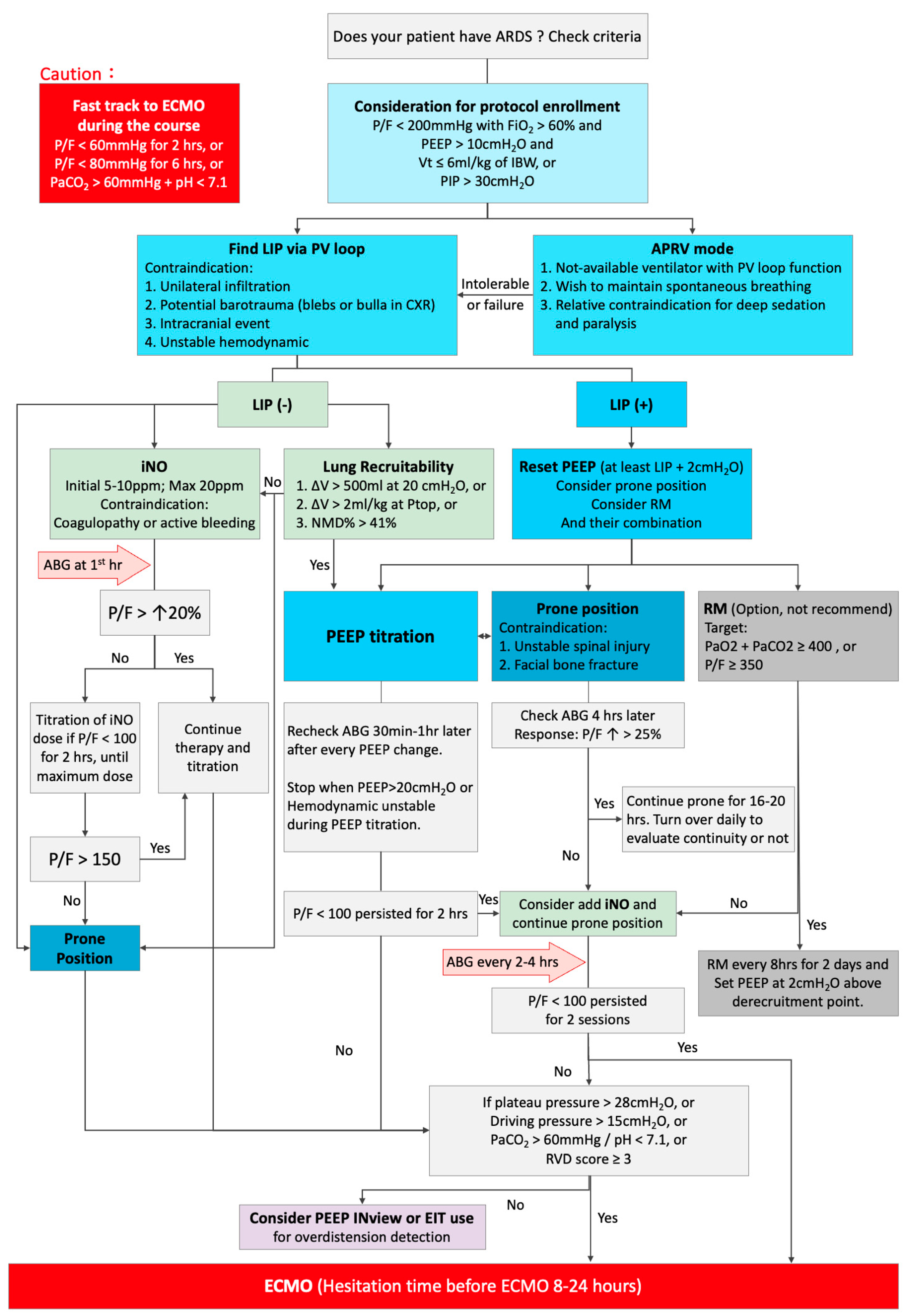
2. Recognize That the Patient Has ARDS
3. Choose the Appropriate PEEP
3.1. General Consideration
3.2. How to Perform P-V Curve and PEEP Titration
- Sedate patients;
- Use succinylcholine 1 mg/kg IV and repeat until the breathing has been controlled;
- Always start from 0 cm H2O, targeting 35–40 cm H2O;
- Hold at the top for 5–15 s and then passively exhale;
- Pressure ramp: 3 cm H2O.
3.3. Alternative Approaches
3.4. Paralysis Strategy
4. Integrated Multiple Modalities for Severe Hypoxia: Prone, iNO, RM, and ECMO
4.1. Recruitment Maneuver
4.2. Meticulous PEEP Titration
4.3. iNO
4.4. Prone
4.5. ECMO
5. Lung Mechanics Safety Check
6. Conclusions
Author Contributions
Funding
Institutional Review Board Statement
Informed Consent Statement
Data Availability Statement
Conflicts of Interest
References
- Ashbaugh, D.G.; Bigelow, D.B.; Petty, T.L.; Levine, B.E. Acute respiratory distress in adults. Lancet 1967, 2, 319–323. [Google Scholar] [CrossRef]
- Matthay, M.A.; Zemans, R.L.; Zimmerman, G.A.; Arabi, Y.M.; Beitler, J.R.; Mercat, A.; Herridge, M.; Randolph, A.G.; Calfee, C.S. Acute respiratory distress syndrome. Nat. Rev. Dis. Primers 2019, 5, 18. [Google Scholar] [CrossRef] [PubMed]
- Fan, E.; Del Sorbo, L.; Goligher, E.C.; Hodgson, C.L.; Munshi, L.; Walkey, A.J.; Adhikari, N.K.J.; Amato, M.B.P.; Branson, R.; Brower, R.G.; et al. An Official American Thoracic Society/European Society of Intensive Care Medicine/Society of Critical Care Medicine Clinical Practice Guideline: Mechanical Ventilation in Adult Patients with Acute Respiratory Distress Syndrome. Am. J. Respir. Crit. Care Med. 2017, 195, 1253–1263. [Google Scholar] [CrossRef] [PubMed]
- Brower, R.G.; Matthay, M.A.; Morris, A.; Schoenfeld, D.; Thompson, B.T.; Wheeler, A. Ventilation with lower tidal volumes as compared with traditional tidal volumes for acute lung injury and the acute respiratory distress syndrome. N. Engl. J. Med. 2000, 342, 1301–1308. [Google Scholar] [PubMed]
- Guérin, C.; Reignier, J.; Richard, J.C.; Beuret, P.; Gacouin, A.; Boulain, T.; Mercier, E.; Badet, M.; Mercat, A.; Baudin, O.; et al. Prone positioning in severe acute respiratory distress syndrome. N. Engl. J. Med. 2013, 368, 2159–2168. [Google Scholar] [CrossRef] [PubMed]
- Brower, R.G.; Lanken, P.N.; MacIntyre, N.; Matthay, M.A.; Morris, A.; Ancukiewicz, M.; Schoenfeld, D.; Thompson, B.T. Higher versus lower positive end-expiratory pressures in patients with the acute respiratory distress syndrome. N. Engl. J. Med. 2004, 351, 327–336. [Google Scholar]
- Talmor, D.; Sarge, T.; Malhotra, A.; O’Donnell, C.R.; Ritz, R.; Lisbon, A.; Novack, V.; Loring, S.H. Mechanical ventilation guided by esophageal pressure in acute lung injury. N. Engl. J. Med. 2008, 359, 2095–2104. [Google Scholar] [CrossRef]
- Kacmarek, R.M.; Villar, J.; Sulemanji, D.; Montiel, R.; Ferrando, C.; Blanco, J.; Koh, Y.; Soler, J.A.; Martínez, D.; Hernández, M.; et al. Open Lung Approach for the Acute Respiratory Distress Syndrome: A Pilot, Randomized Controlled Trial. Crit. Care Med. 2016, 44, 32–42. [Google Scholar] [CrossRef]
- Briel, M.; Meade, M.; Mercat, A.; Brower, R.G.; Talmor, D.; Walter, S.D.; Slutsky, A.S.; Pullenayegum, E.; Zhou, Q.; Cook, D.; et al. Higher vs lower positive end-expiratory pressure in patients with acute lung injury and acute respiratory distress syndrome: Systematic review and meta-analysis. JAMA 2010, 303, 865–873. [Google Scholar] [CrossRef]
- Amato, M.B.; Barbas, C.S.; Medeiros, D.M.; Magaldi, R.B.; Schettino, G.P.; Lorenzi-Filho, G.; Kairalla, R.A.; Deheinzelin, D.; Munoz, C.; Oliveira, R.; et al. Effect of a protective-ventilation strategy on mortality in the acute respiratory distress syndrome. N. Engl. J. Med. 1998, 338, 347–354. [Google Scholar] [CrossRef]
- Meade, M.O.; Cook, D.J.; Guyatt, G.H.; Slutsky, A.S.; Arabi, Y.M.; Cooper, D.J.; Davies, A.R.; Hand, L.E.; Zhou, Q.; Thabane, L.; et al. Ventilation strategy using low tidal volumes, recruitment maneuvers, and high positive end-expiratory pressure for acute lung injury and acute respiratory distress syndrome: A randomized controlled trial. JAMA 2008, 299, 637–645. [Google Scholar] [CrossRef]
- Suzumura, E.A.; Figueiró, M.; Normilio-Silva, K.; Laranjeira, L.; Oliveira, C.; Buehler, A.M.; Bugano, D.; Passos Amato, M.B.; Ribeiro Carvalho, C.R.; Berwanger, O.; et al. Effects of alveolar recruitment maneuvers on clinical outcomes in patients with acute respiratory distress syndrome: A systematic review and meta-analysis. Intensive Care Med. 2014, 40, 1227–1240. [Google Scholar] [CrossRef]
- Cavalcanti, A.B.; Suzumura, É.A.; Laranjeira, L.N.; Paisani, D.M.; Damiani, L.P.; Guimarães, H.P.; Romano, E.R.; Regenga, M.M.; Taniguchi, L.N.T.; Teixeira, C.; et al. Effect of Lung Recruitment and Titrated Positive End-Expiratory Pressure (PEEP) vs. Low PEEP on Mortality in Patients with Acute Respiratory Distress Syndrome: A Randomized Clinical Trial. JAMA 2017, 318, 1335–1345. [Google Scholar] [PubMed]
- Papazian, L.; Forel, J.M.; Gacouin, A.; Penot-Ragon, C.; Perrin, G.; Loundou, A.; Jaber, S.; Arnal, J.M.; Perez, D.; Seghboyan, J.M.; et al. Neuromuscular blockers in early acute respiratory distress syndrome. N. Engl. J. Med. 2010, 363, 1107–1116. [Google Scholar] [CrossRef]
- Moss, M.; Huang, D.T.; Brower, R.G.; Ferguson, N.D.; Ginde, A.A.; Gong, M.N.; Grissom, C.K.; Gundel, S.; Hayden, D.; Hite, R.D.; et al. Early Neuromuscular Blockade in the Acute Respiratory Distress Syndrome. N. Engl. J. Med. 2019, 380, 1997–2008. [Google Scholar]
- Peek, G.J.; Mugford, M.; Tiruvoipati, R.; Wilson, A.; Allen, E.; Thalanany, M.M.; Hibbert, C.L.; Truesdale, A.; Clemens, F.; Cooper, N.; et al. Efficacy and economic assessment of conventional ventilatory support versus extracorporeal membrane oxygenation for severe adult respiratory failure (CESAR): A multicentre randomised controlled trial. Lancet 2009, 374, 1351–1363. [Google Scholar] [CrossRef]
- Combes, A.; Hajage, D.; Capellier, G.; Demoule, A.; Lavoué, S.; Guervilly, C.; Da Silva, D.; Zafrani, L.; Tirot, P.; Veber, B.; et al. Extracorporeal Membrane Oxygenation for Severe Acute Respiratory Distress Syndrome. N. Engl. J. Med. 2018, 378, 1965–1975. [Google Scholar] [CrossRef] [PubMed]
- Zhou, Y.; Jin, X.; Lv, Y.; Wang, P.; Yang, Y.; Liang, G.; Wang, B.; Kang, Y. Early application of airway pressure release ventilation may reduce the duration of mechanical ventilation in acute respiratory distress syndrome. Intensive Care Med. 2017, 43, 1648–1659. [Google Scholar] [CrossRef]
- Fredericks, A.S.; Bunker, M.P.; Gliga, L.A.; Ebeling, C.G.; Ringqvist, J.R.; Heravi, H.; Manley, J.; Valladares, J.; Romito, B.T. Airway Pressure Release Ventilation: A Review of the Evidence, Theoretical Benefits, and Alternative Titration Strategies. Clin. Med. Insights. Circ. Respir. Pulm. Med. 2020, 14, 1179548420903297. [Google Scholar] [CrossRef]
- Michael, J.R.; Barton, R.G.; Saffle, J.R.; Mone, M.; Markewitz, B.A.; Hillier, K.; Elstad, M.R.; Campbell, E.J.; Troyer, B.E.; Whatley, R.E.; et al. Inhaled nitric oxide versus conventional therapy: Effect on oxygenation in ARDS. Am. J. Respir. Crit. Care Med. 1998, 157, 1372–1380. [Google Scholar] [CrossRef] [PubMed]
- Adhikari, N.K.; Dellinger, R.P.; Lundin, S.; Payen, D.; Vallet, B.; Gerlach, H.; Park, K.J.; Mehta, S.; Slutsky, A.S.; Friedrich, J.O. Inhaled nitric oxide does not reduce mortality in patients with acute respiratory distress syndrome regardless of severity: Systematic review and meta-analysis. Crit. Care Med. 2014, 42, 404–412. [Google Scholar] [CrossRef] [PubMed]
- Gebistorf, F.; Karam, O.; Wetterslev, J.; Afshari, A. Inhaled nitric oxide for acute respiratory distress syndrome (ARDS) in children and adults. Cochrane Database Syst. Rev. 2016, 2016, CD002787. [Google Scholar] [CrossRef]
- Young, D.; Lamb, S.E.; Shah, S.; MacKenzie, I.; Tunnicliffe, W.; Lall, R.; Rowan, K.; Cuthbertson, B.H. High-frequency oscillation for acute respiratory distress syndrome. N. Engl. J. Med. 2013, 368, 806–813. [Google Scholar] [CrossRef]
- Ferguson, N.D.; Cook, D.J.; Guyatt, G.H.; Mehta, S.; Hand, L.; Austin, P.; Zhou, Q.; Matte, A.; Walter, S.D.; Lamontagne, F.; et al. High-frequency oscillation in early acute respiratory distress syndrome. N. Engl. J. Med. 2013, 368, 795–805. [Google Scholar] [CrossRef] [PubMed]
- Sud, S.; Sud, M.; Friedrich, J.O.; Wunsch, H.; Meade, M.O.; Ferguson, N.D.; Adhikari, N.K. High-frequency oscillatory ventilation versus conventional ventilation for acute respiratory distress syndrome. Cochrane Database Syst. Rev. 2016, 4, CD004085. [Google Scholar] [CrossRef] [PubMed]
- Bellani, G.; Laffey, J.G.; Pham, T.; Fan, E.; Brochard, L.; Esteban, A.; Gattinoni, L.; van Haren, F.; Larsson, A.; McAuley, D.F.; et al. Epidemiology, Patterns of Care, and Mortality for Patients with Acute Respiratory Distress Syndrome in Intensive Care Units in 50 Countries. JAMA 2016, 315, 788–800. [Google Scholar] [CrossRef]
- Fuller, B.M.; Mohr, N.M.; Drewry, A.M.; Carpenter, C.R. Lower tidal volume at initiation of mechanical ventilation may reduce progression to acute respiratory distress syndrome: A systematic review. Crit. Care 2013, 17, R11. [Google Scholar] [CrossRef]
- Sahetya, S.K.; Mallow, C.; Sevransky, J.E.; Martin, G.S.; Girard, T.D.; Brower, R.G.; Checkley, W. Association between hospital mortality and inspiratory airway pressures in mechanically ventilated patients without acute respiratory distress syndrome: A prospective cohort study. Crit. Care 2019, 23, 367. [Google Scholar] [CrossRef] [PubMed]
- Webb, H.H.; Tierney, D.F. Experimental pulmonary edema due to intermittent positive pressure ventilation with high inflation pressures. Protection by positive end-expiratory pressure. Am. Rev. Respir. Dis. 1974, 110, 556–565. [Google Scholar]
- Hodgson, C.L.; Tuxen, D.V.; Davies, A.R.; Bailey, M.J.; Higgins, A.M.; Holland, A.E.; Keating, J.L.; Pilcher, D.V.; Westbrook, A.J.; Cooper, D.J.; et al. A randomised controlled trial of an open lung strategy with staircase recruitment, titrated PEEP and targeted low airway pressures in patients with acute respiratory distress syndrome. Crit. Care 2011, 15, R133. [Google Scholar] [CrossRef]
- Gattinoni, L.; Caironi, P.; Cressoni, M.; Chiumello, D.; Ranieri, V.M.; Quintel, M.; Russo, S.; Patroniti, N.; Cornejo, R.; Bugedo, G. Lung recruitment in patients with the acute respiratory distress syndrome. N. Engl. J. Med. 2006, 354, 1775–1786. [Google Scholar] [CrossRef] [PubMed]
- Bikker, I.; Miranda, R.D.; Van Bommel, J.; Bakker, J.; Gommers, D. Functional residual capacity measurement during mechanical ventilation in order to find the optimal positive end-expiratory pressure. Crit. Care 2007, 11, 205. [Google Scholar] [CrossRef]
- Richard, J.C.; Pouzot, C.; Pinzón, A.M.; González, J.S.; Orkisz, M.; Neyran, B.; Hoyos, M.H.; Lavenne, F.; Guerin, C. Reliability of the nitrogen washin-washout technique to assess end-expiratory lung volume at variable PEEP and tidal volumes. Intensive Care Med. Exp. 2014, 2, 10. [Google Scholar] [CrossRef] [PubMed]
- Gómez-Laberge, C.; Arnold, J.H.; Wolf, G.K. A unified approach for EIT imaging of regional overdistension and atelectasis in acute lung injury. IEEE Trans. Med. Imaging 2012, 31, 834–842. [Google Scholar] [CrossRef] [PubMed]
- Jonkman, A.H.; Alcala, G.C.; Pavlovsky, B.; Roca, O.; Spadaro, S.; Scaramuzzo, G.; Chen, L.; Dianti, J.; Sousa, M.L.A.; Sklar, M.C.; et al. Lung Recruitment Assessed by Electrical Impedance Tomography (RECRUIT): A Multicenter Study of COVID-19 Acute Respiratory Distress Syndrome. Am. J. Respir. Crit. Care Med. 2023, 208, 25–38. [Google Scholar] [CrossRef]
- Beitler, J.R.; Sarge, T.; Banner-Goodspeed, V.M.; Gong, M.N.; Cook, D.; Novack, V.; Loring, S.H.; Talmor, D. Effect of Titrating Positive End-Expiratory Pressure (PEEP) with an Esophageal Pressure-Guided Strategy vs an Empirical High PEEP-Fio2 Strategy on Death and Days Free From Mechanical Ventilation Among Patients with Acute Respiratory Distress Syndrome: A Randomized Clinical Trial. JAMA 2019, 321, 846–857. [Google Scholar]
- Pelosi, P.; Goldner, M.; McKibben, A.; Adams, A.; Eccher, G.; Caironi, P.; Losappio, S.; Gattinoni, L.; Marini, J.J. Recruitment and derecruitment during acute respiratory failure: An experimental study. Am. J. Respir. Crit. Care Med. 2001, 164, 122–130. [Google Scholar] [CrossRef]
- Kallet, R.H.; Branson, R.D. Respiratory controversies in the critical care setting. Do the NIH ARDS Clinical Trials Network PEEP/FIO2 tables provide the best evidence-based guide to balancing PEEP and FIO2 settings in adults? Respir. Care 2007, 52, 461–475; discussion 475–477. [Google Scholar] [PubMed]
- Walkey, A.J.; Del Sorbo, L.; Hodgson, C.L.; Adhikari, N.K.J.; Wunsch, H.; Meade, M.O.; Uleryk, E.; Hess, D.; Talmor, D.S.; Thompson, B.T.; et al. Higher PEEP versus Lower PEEP Strategies for Patients with Acute Respiratory Distress Syndrome. A Systematic Review and Meta-Analysis. Ann. Am. Thorac. Soc. 2017, 14, S297–S303. [Google Scholar] [CrossRef]
- Chen, L.; Del Sorbo, L.; Grieco, D.L.; Junhasavasdikul, D.; Rittayamai, N.; Soliman, I.; Sklar, M.C.; Rauseo, M.; Ferguson, N.D.; Fan, E.; et al. Potential for Lung Recruitment Estimated by the Recruitment-to-Inflation Ratio in Acute Respiratory Distress Syndrome. A Clinical Trial. Am. J. Respir. Crit. Care Med. 2020, 201, 178–187. [Google Scholar] [CrossRef]
- Grieco, D.L.; Pintaudi, G.; Bongiovanni, F.; Anzellotti, G.M.; Menga, L.S.; Cesarano, M.; Dell’Anna, A.M.; Rosá, T.; Delle Cese, L.; Bello, G.; et al. Recruitment-to-inflation Ratio Assessed through Sequential End-expiratory Lung Volume Measurement in Acute Respiratory Distress Syndrome. Anesthesiology 2023, 139, 801–814. [Google Scholar] [CrossRef]
- Luecke, T.; Corradi, F.; Pelosi, P. Lung imaging for titration of mechanical ventilation. Curr. Opin. Anaesthesiol. 2012, 25, 131–140. [Google Scholar] [CrossRef]
- Stefanidis, K.; Dimopoulos, S.; Tripodaki, E.S.; Vitzilaios, K.; Politis, P.; Piperopoulos, P.; Nanas, S. Lung sonography and recruitment in patients with early acute respiratory distress syndrome: A pilot study. Crit. Care 2011, 15, R185. [Google Scholar] [CrossRef] [PubMed]
- Bouhemad, B.; Brisson, H.; Le-Guen, M.; Arbelot, C.; Lu, Q.; Rouby, J.J. Bedside ultrasound assessment of positive end-expiratory pressure-induced lung recruitment. Am. J. Respir. Crit. Care Med. 2011, 183, 341–347. [Google Scholar] [CrossRef] [PubMed]
- Demory, D.; Arnal, J.M.; Wysocki, M.; Donati, S.; Granier, I.; Corno, G.; Durand-Gasselin, J. Recruitability of the lung estimated by the pressure volume curve hysteresis in ARDS patients. Intensive Care Med. 2008, 34, 2019–2025. [Google Scholar] [CrossRef] [PubMed]
- Gattinoni, L.; Pesenti, A.; Avalli, L.; Rossi, F.; Bombino, M. Pressure-volume curve of total respiratory system in acute respiratory failure. Computed tomographic scan study. Am. Rev. Respir. Dis. 1987, 136, 730–736. [Google Scholar] [CrossRef]
- Millington, S.J.; Cardinal, P.; Brochard, L. Setting and Titrating Positive End-Expiratory Pressure. Chest 2022, 161, 1566–1575. [Google Scholar] [CrossRef]
- Grasso, S.; Terragni, P.; Mascia, L.; Fanelli, V.; Quintel, M.; Herrmann, P.; Hedenstierna, G.; Slutsky, A.S.; Ranieri, V.M. Airway pressure-time curve profile (stress index) detects tidal recruitment/hyperinflation in experimental acute lung injury. Crit. Care Med. 2004, 32, 1018–1027. [Google Scholar] [CrossRef]
- Grasso, S.; Stripoli, T.; De Michele, M.; Bruno, F.; Moschetta, M.; Angelelli, G.; Munno, I.; Ruggiero, V.; Anaclerio, R.; Cafarelli, A.; et al. ARDSnet ventilatory protocol and alveolar hyperinflation: Role of positive end-expiratory pressure. Am. J. Respir. Crit. Care Med. 2007, 176, 761–767. [Google Scholar] [CrossRef]
- Putensen, C.; Mutz, N.J.; Putensen-Himmer, G.; Zinserling, J. Spontaneous breathing during ventilatory support improves ventilation-perfusion distributions in patients with acute respiratory distress syndrome. Am. J. Respir. Crit. Care Med. 1999, 159, 1241–1248. [Google Scholar] [CrossRef]
- van Haren, F.; Pham, T.; Brochard, L.; Bellani, G.; Laffey, J.; Dres, M.; Fan, E.; Goligher, E.C.; Heunks, L.; Lynch, J.; et al. Spontaneous Breathing in Early Acute Respiratory Distress Syndrome: Insights from the Large Observational Study to UNderstand the Global Impact of Severe Acute Respiratory FailurE Study. Crit. Care Med. 2019, 47, 229–238. [Google Scholar] [CrossRef] [PubMed]
- Spinelli, E.; Mauri, T.; Beitler, J.R.; Pesenti, A.; Brodie, D. Respiratory drive in the acute respiratory distress syndrome: Pathophysiology, monitoring, and therapeutic interventions. Intensive Care Med. 2020, 46, 606–618. [Google Scholar] [CrossRef] [PubMed]
- Thille, A.W.; Rodriguez, P.; Cabello, B.; Lellouche, F.; Brochard, L. Patient-ventilator asynchrony during assisted mechanical ventilation. Intensive Care Med. 2006, 32, 1515–1522. [Google Scholar] [CrossRef] [PubMed]
- Brochard, L.; Slutsky, A.; Pesenti, A. Mechanical Ventilation to Minimize Progression of Lung Injury in Acute Respiratory Failure. Am. J. Respir. Crit. Care Med. 2017, 195, 438–442. [Google Scholar] [CrossRef]
- Bertoni, M.; Telias, I.; Urner, M.; Long, M.; Del Sorbo, L.; Fan, E.; Sinderby, C.; Beck, J.; Liu, L.; Qiu, H.; et al. A novel non-invasive method to detect excessively high respiratory effort and dynamic transpulmonary driving pressure during mechanical ventilation. Crit. Care 2019, 23, 346. [Google Scholar] [CrossRef] [PubMed]
- Fan, E.; Checkley, W.; Stewart, T.E.; Muscedere, J.; Lesur, O.; Granton, J.T.; Freitag, A.P.; Jacka, M.; Ferguson, N.D.; Meade, M.O. Complications from recruitment maneuvers in patients with acute lung injury: Secondary analysis from the lung open ventilation study. Respir. Care 2012, 57, 1842–1849. [Google Scholar] [CrossRef] [PubMed]
- Morán, I.; Blanch, L.; Fernández, R.; Fernández-Mondéjar, E.; Zavala, E.; Mancebo, J. Acute physiologic effects of a stepwise recruitment maneuver in acute respiratory distress syndrome. Minerva Anestesiol. 2011, 77, 1167–1175. [Google Scholar] [PubMed]
- Borges, J.B.; Okamoto, V.N.; Matos, G.F.; Caramez, M.P.; Arantes, P.R.; Barros, F.; Souza, C.E.; Victorino, J.A.; Kacmarek, R.M.; Barbas, C.S.; et al. Reversibility of lung collapse and hypoxemia in early acute respiratory distress syndrome. Am. J. Respir. Crit. Care Med. 2006, 174, 268–278. [Google Scholar] [CrossRef]
- Mergoni, M.; Volpi, A.; Bricchi, C.; Rossi, A. Lower inflection point and recruitment with PEEP in ventilated patients with acute respiratory failure. J. Appl. Physiol. (Bethesda, Md. 1985) 2001, 91, 441–450. [Google Scholar] [CrossRef]
- Chiumello, D.; Arnal, J.M.; Umbrello, M.; Cammaroto, A.; Formenti, P.; Mistraletti, G.; Bolgiaghi, L.; Gotti, M.; Novotni, D.; Reidt, S.; et al. Hysteresis and Lung Recruitment in Acute Respiratory Distress Syndrome Patients: A CT Scan Study. Crit. Care Med. 2020, 48, 1494–1502. [Google Scholar] [CrossRef]
- Alqahtani, J.S.; Aldhahir, A.M.; Al Ghamdi, S.S.; AlBahrani, S.; AlDraiwiesh, I.A.; Alqarni, A.A.; Latief, K.; Raya, R.P.; Oyelade, T. Inhaled Nitric Oxide for Clinical Management of COVID-19: A Systematic Review and Meta-Analysis. Int. J. Environ. Res. Public Health 2022, 19, 12803. [Google Scholar] [CrossRef] [PubMed]
- Lotz, C.; Muellenbach, R.M.; Meybohm, P.; Mutlak, H.; Lepper, P.M.; Rolfes, C.B.; Peivandi, A.; Stumpner, J.; Kredel, M.; Kranke, P.; et al. Effects of inhaled nitric oxide in COVID-19-induced ARDS—Is it worthwhile? Acta Anaesthesiol. Scand. 2021, 65, 629–632. [Google Scholar] [CrossRef] [PubMed]
- Hurford, W.E. Inhaled nitric oxide. Respir. Care Clin. N. Am. 2002, 8, 261–279. [Google Scholar] [CrossRef] [PubMed]
- Gattinoni, L.; Pelosi, P.; Vitale, G.; Pesenti, A.; D’Andrea, L.; Mascheroni, D. Body position changes redistribute lung computed-tomographic density in patients with acute respiratory failure. Anesthesiology 1991, 74, 15–23. [Google Scholar] [CrossRef] [PubMed]
- Pelosi, P.; D’Andrea, L.; Vitale, G.; Pesenti, A.; Gattinoni, L. Vertical gradient of regional lung inflation in adult respiratory distress syndrome. Am. J. Respir. Crit. Care Med. 1994, 149, 8–13. [Google Scholar] [CrossRef] [PubMed]
- Blanch, L.; Mancebo, J.; Perez, M.; Martinez, M.; Mas, A.; Betbese, A.J.; Joseph, D.; Ballús, J.; Lucangelo, U.; Bak, E. Short-term effects of prone position in critically ill patients with acute respiratory distress syndrome. Intensive Care Med. 1997, 23, 1033–1039. [Google Scholar] [CrossRef] [PubMed]
- Cornejo, R.A.; Díaz, J.C.; Tobar, E.A.; Bruhn, A.R.; Ramos, C.A.; González, R.A.; Repetto, C.A.; Romero, C.M.; Gálvez, L.R.; Llanos, O.; et al. Effects of prone positioning on lung protection in patients with acute respiratory distress syndrome. Am. J. Respir. Crit. Care Med. 2013, 188, 440–448. [Google Scholar] [CrossRef]
- Gattinoni, L.; Busana, M.; Giosa, L.; Macrì, M.M.; Quintel, M. Prone Positioning in Acute Respiratory Distress Syndrome. Semin. Respir. Crit. Care Med. 2019, 40, 94–100. [Google Scholar] [CrossRef]
- Pelosi, P.; Tubiolo, D.; Mascheroni, D.; Vicardi, P.; Crotti, S.; Valenza, F.; Gattinoni, L. Effects of the prone position on respiratory mechanics and gas exchange during acute lung injury. Am. J. Respir. Crit. Care Med. 1998, 157, 387–393. [Google Scholar] [CrossRef]
- Le, M.Q.; Rosales, R.; Shapiro, L.T.; Huang, L.Y. The Down Side of Prone Positioning: The Case of a Coronavirus 2019 Survivor. Am. J. Phys. Med. Rehabil. 2020, 99, 870–872. [Google Scholar] [CrossRef]
- Taccone, P.; Pesenti, A.; Latini, R.; Polli, F.; Vagginelli, F.; Mietto, C.; Caspani, L.; Raimondi, F.; Bordone, G.; Iapichino, G.; et al. Prone positioning in patients with moderate and severe acute respiratory distress syndrome: A randomized controlled trial. JAMA 2009, 302, 1977–1984. [Google Scholar] [CrossRef] [PubMed]
- Walter, T.; Ricard, J.D. Extended prone positioning for intubated ARDS: A review. Crit. Care 2023, 27, 264. [Google Scholar] [CrossRef] [PubMed]
- Tang, Y.H.; Hsu, S.F.; Chung, H.P.; Chen, C.H.; Chang, W.K.; Kuo, K.C.; Chen, Y.T.; Wu, J.C.; Lin, C.Y.; Wang, C.J. Use of the Prone Position in Critically Ill COVID-19 Patients: Can the Response Be Predicted? Int. J. Gerontol. 2022, 16, 191–195. [Google Scholar]
- Douglas, I.S.; Rosenthal, C.A.; Swanson, D.D.; Hiller, T.; Oakes, J.; Bach, J.; Whelchel, C.; Pickering, J.; George, T.; Kearns, M.; et al. Safety and Outcomes of Prolonged Usual Care Prone Position Mechanical Ventilation to Treat Acute Coronavirus Disease 2019 Hypoxemic Respiratory Failure. Crit. Care Med. 2021, 49, 490–502. [Google Scholar] [CrossRef] [PubMed]
- Sud, S.; Friedrich, J.O.; Taccone, P.; Polli, F.; Adhikari, N.K.; Latini, R.; Pesenti, A.; Guérin, C.; Mancebo, J.; Curley, M.A.; et al. Prone ventilation reduces mortality in patients with acute respiratory failure and severe hypoxemia: Systematic review and meta-analysis. Intensive Care Med. 2010, 36, 585–599. [Google Scholar] [CrossRef] [PubMed]
- Schmidt, M.; Bailey, M.; Sheldrake, J.; Hodgson, C.; Aubron, C.; Rycus, P.T.; Scheinkestel, C.; Cooper, D.J.; Brodie, D.; Pellegrino, V.; et al. Predicting survival after extracorporeal membrane oxygenation for severe acute respiratory failure. The Respiratory Extracorporeal Membrane Oxygenation Survival Prediction (RESP) score. Am. J. Respir. Crit. Care Med. 2014, 189, 1374–1382. [Google Scholar] [CrossRef] [PubMed]
- Schmidt, M.; Zogheib, E.; Rozé, H.; Repesse, X.; Lebreton, G.; Luyt, C.E.; Trouillet, J.L.; Bréchot, N.; Nieszkowska, A.; Dupont, H.; et al. The PRESERVE mortality risk score and analysis of long-term outcomes after extracorporeal membrane oxygenation for severe acute respiratory distress syndrome. Intensive Care Med. 2013, 39, 1704–1713. [Google Scholar] [CrossRef] [PubMed]
- Li, X.; Scales, D.C.; Kavanagh, B.P. Unproven and Expensive before Proven and Cheap: Extracorporeal Membrane Oxygenation versus Prone Position in Acute Respiratory Distress Syndrome. Am. J. Respir. Crit. Care Med. 2018, 197, 991–993. [Google Scholar] [CrossRef] [PubMed]
- Quintel, M.; Busana, M.; Gattinoni, L. Breathing and Ventilation during Extracorporeal Membrane Oxygenation: How to Find the Balance between Rest and Load. Am. J. Respir. Crit. Care Med. 2019, 200, 954–956. [Google Scholar] [CrossRef]
- Tonna, J.E.; Abrams, D.; Brodie, D.; Greenwood, J.C.; Rubio Mateo-Sidron, J.A.; Usman, A.; Fan, E. Management of Adult Patients Supported with Venovenous Extracorporeal Membrane Oxygenation (VV ECMO): Guideline from the Extracorporeal Life Support Organization (ELSO). ASAIO J. (Am. Soc. Artif. Internal Organs 1992) 2021, 67, 601–610. [Google Scholar] [CrossRef]
- Araos, J.; Alegria, L.; Garcia, P.; Cruces, P.; Soto, D.; Erranz, B.; Amthauer, M.; Salomon, T.; Medina, T.; Rodriguez, F.; et al. Near-Apneic Ventilation Decreases Lung Injury and Fibroproliferation in an Acute Respiratory Distress Syndrome Model with Extracorporeal Membrane Oxygenation. Am. J. Respir. Crit. Care Med. 2019, 199, 603–612. [Google Scholar] [CrossRef]
- Jardin, F.; Vieillard-Baron, A. Is there a safe plateau pressure in ARDS? The right heart only knows. Intensive Care Med. 2007, 33, 444–447. [Google Scholar] [CrossRef]
- Mekontso Dessap, A.; Boissier, F.; Charron, C.; Bégot, E.; Repessé, X.; Legras, A.; Brun-Buisson, C.; Vignon, P.; Vieillard-Baron, A. Acute cor pulmonale during protective ventilation for acute respiratory distress syndrome: Prevalence, predictors, and clinical impact. Intensive Care Med. 2016, 42, 862–870. [Google Scholar] [CrossRef] [PubMed]
- Amato, M.B.; Meade, M.O.; Slutsky, A.S.; Brochard, L.; Costa, E.L.; Schoenfeld, D.A.; Stewart, T.E.; Briel, M.; Talmor, D.; Mercat, A.; et al. Driving pressure and survival in the acute respiratory distress syndrome. N. Engl. J. Med. 2015, 372, 747–755. [Google Scholar] [CrossRef]
- Gattinoni, L.; Pesenti, A. ARDS: The non-homogeneous lung; facts and hypothesis. Intensive Crit. Care Dig. 1987, 6, 1–4. [Google Scholar]
- Lu, Q.; Rouby, J.J. Measurement of pressure-volume curves in patients on mechanical ventilation: Methods and significance. Crit. Care 2000, 4, 91–100. [Google Scholar] [CrossRef]
- Servillo, G.; Svantesson, C.; Beydon, L.; Roupie, E.; Brochard, L.; Lemaire, F.; Jonson, B. Pressure-volume curves in acute respiratory failure: Automated low flow inflation versus occlusion. Am. J. Respir. Crit. Care Med. 1997, 155, 1629–1636. [Google Scholar] [CrossRef] [PubMed]
- Harris, R.S.; Hess, D.R.; Venegas, J.G. An objective analysis of the pressure-volume curve in the acute respiratory distress syndrome. Am. J. Respir. Crit. Care Med. 2000, 161, 432–439. [Google Scholar] [CrossRef]
- Ward, N.S.; Lin, D.Y.; Nelson, D.L.; Houtchens, J.; Schwartz, W.A.; Klinger, J.R.; Hill, N.S.; Levy, M.M. Successful determination of lower inflection point and maximal compliance in a population of patients with acute respiratory distress syndrome. Crit. Care Med. 2002, 30, 963–968. [Google Scholar] [CrossRef]
- Chen, L.; Del Sorbo, L.; Grieco, D.L.; Shklar, O.; Junhasavasdikul, D.; Telias, I.; Fan, E.; Brochard, L. Airway Closure in Acute Respiratory Distress Syndrome: An Underestimated and Misinterpreted Phenomenon. Am. J. Respir. Crit. Care Med. 2018, 197, 132–136. [Google Scholar] [CrossRef]
- Jonson, B.; Svantesson, C. Elastic pressure-volume curves: What information do they convey? Thorax 1999, 54, 82–87. [Google Scholar] [CrossRef] [PubMed]
- Rackley, C.R. Monitoring During Mechanical Ventilation. Respir. Care 2020, 65, 832–846. [Google Scholar] [CrossRef] [PubMed]

| Method | Advantage | Disadvantage |
|---|---|---|
| PEEP/FiO2 table [4,38] |
|
|
| PEEPIN view [32,33] |
|
|
| EIT [34,35] |
|
|
| Esophageal pressure-guided PEEP setting [7,36] |
|
|
| Recruitment-to-inflation ratio method [40,41] |
|
|
| Lung ultrasound [42,43,44] |
|
|
| P-V curve [45] |
|
|
Disclaimer/Publisher’s Note: The statements, opinions and data contained in all publications are solely those of the individual author(s) and contributor(s) and not of MDPI and/or the editor(s). MDPI and/or the editor(s) disclaim responsibility for any injury to people or property resulting from any ideas, methods, instructions or products referred to in the content. |
© 2024 by the authors. Licensee MDPI, Basel, Switzerland. This article is an open access article distributed under the terms and conditions of the Creative Commons Attribution (CC BY) license (https://creativecommons.org/licenses/by/4.0/).
Share and Cite
Wang, C.-J.; Wang, I.-T.; Chen, C.-H.; Tang, Y.-H.; Lin, H.-W.; Lin, C.-Y.; Wu, C.-L. Recruitment-Potential-Oriented Mechanical Ventilation Protocol and Narrative Review for Patients with Acute Respiratory Distress Syndrome. J. Pers. Med. 2024, 14, 779. https://doi.org/10.3390/jpm14080779
Wang C-J, Wang I-T, Chen C-H, Tang Y-H, Lin H-W, Lin C-Y, Wu C-L. Recruitment-Potential-Oriented Mechanical Ventilation Protocol and Narrative Review for Patients with Acute Respiratory Distress Syndrome. Journal of Personalized Medicine. 2024; 14(8):779. https://doi.org/10.3390/jpm14080779
Chicago/Turabian StyleWang, Chieh-Jen, I-Ting Wang, Chao-Hsien Chen, Yen-Hsiang Tang, Hsin-Wei Lin, Chang-Yi Lin, and Chien-Liang Wu. 2024. "Recruitment-Potential-Oriented Mechanical Ventilation Protocol and Narrative Review for Patients with Acute Respiratory Distress Syndrome" Journal of Personalized Medicine 14, no. 8: 779. https://doi.org/10.3390/jpm14080779
APA StyleWang, C.-J., Wang, I.-T., Chen, C.-H., Tang, Y.-H., Lin, H.-W., Lin, C.-Y., & Wu, C.-L. (2024). Recruitment-Potential-Oriented Mechanical Ventilation Protocol and Narrative Review for Patients with Acute Respiratory Distress Syndrome. Journal of Personalized Medicine, 14(8), 779. https://doi.org/10.3390/jpm14080779