Subcutaneous versus Transvenous Implantable Cardioverter Defibrillator in Patients with End-Stage Renal Disease Requiring Dialysis: Extended Long-Term Retrospective Multicenter Follow-Up

 , , , , , and
Abstract
1. Introduction
2. Methods
2.1. Implantation Procedure
2.2. Patient Follow-Up
2.3. Data Collection
2.4. Statistical Analysis
3. Results
3.1. Patient Population and Implant Procedure
3.2. Follow-Up
4. Discussion
Limitations
5. Conclusions
Author Contributions
Funding
Institutional Review Board Statement
Informed Consent Statement
Data Availability Statement
Conflicts of Interest
Abbreviations
| BMI | Body mass index |
| CIED | Cardiac implantable electronic device |
| CKD | Chronic kidney disease |
| CRP | C-reactive proteins |
| CRT | Cardiac resynchronization therapy |
| ESRD | End-stage renal disease |
| GFR | Glomerular filtration rate |
| HD | Hemodialysis |
| HF | Heart failure |
| HFrEF | Heart failure with reduced ejection fraction |
| ICD | Implantable cardioverter defibrillator |
| LV-EF | Left ventricular ejection fraction |
| Min | Minutes |
| NYHA | New York Heart Association |
| SCD | Sudden cardiac death |
| S-ICD | Subcutaneous implantable cardioverter defibrillator |
| TLE | Transvenous lead extraction |
| TV-ICD | Transvenous implantable cardioverter defibrillator |
References
- Zeppenfeld, K.; Tfelt-Hansen, J.; De Riva, M.; Winkel, B.G.; Behr, E.R.; Blom, N.A.; Charron, P.; Corrado, D.; Dagres, N.; De Chillou, C.; et al. 2022 ESC Guidelines for the management of patients with ventricular arrhythmias and the prevention of sudden cardiac death. Eur. Heart J. 2022, 43, 3997–4126. [Google Scholar] [CrossRef]
- McDonagh, T.A.; Metra, M.; Adamo, M.; Gardner, R.S.; Baumbach, A.; Böhm, M.; Burri, H.; Butler, J.; Čelutkienė, J.; Chioncel, O.; et al. 2021 ESC Guidelines for the diagnosis and treatment of acute and chronic heart failure. Eur. Heart J. 2021, 42, 3599–3726. [Google Scholar] [CrossRef]
- Fong, K.Y.; Ng, C.J.R.; Wang, Y.; Yeo, C.; Tan, V.H. Subcutaneous Versus Transvenous Implantable Defibrillator Therapy: A Systematic Review and Meta-Analysis of Randomized Trials and Propensity Score–Matched Studies. J. Am. Heart Assoc. 2022, 11, e024756. [Google Scholar] [CrossRef]
- Guha, A.; Maddox, W.R.; Colombo, R.; Nahman, N.S.; Kintziger, K.W.; Waller, J.L.; Diamond, M.; Murphy, M.; Kheda, M.; Litwin, S.E.; et al. Cardiac implantable electronic device infection in patients with end-stage renal disease. Heart Rhythm 2015, 12, 2395–2401. [Google Scholar] [CrossRef]
- Mehta, V.S.; Elliott, M.K.; Sidhu, B.S.; Gould, J.; Kemp, T.; Vergani, V.; Kadiwar, S.; Shetty, A.K.; Blauth, C.; Gill, J.; et al. Long-term survival following transvenous lead extraction: Importance of indication and comorbidities. Heart Rhythm 2021, 18, 1566–1576. [Google Scholar] [CrossRef]
- Knops, R.E.; Pepplinkhuizen, S.; Delnoy, P.P.H.M.; Boersma, L.V.A.; Kuschyk, J.; El-Chami, M.F.; Bonnemeier, H.; Behr, E.R.; Brouwer, T.F.; Kaab, S.; et al. Device-related complications in subcutaneous versus transvenous ICD: A secondary analysis of the PRAETORIAN trial. Eur. Heart J. 2022, 43, 4872–4883. [Google Scholar] [CrossRef]
- Schiedat, F.; Meuterodt, B.; Prull, M.; Aweimer, A.; Gotzmann, M.; O’Connor, S.; Perings, C.; Korth, J.; Lawo, T.; El-Battrawy, I.; et al. Comparison of infection and complication rates associated with transvenous vs. subcutaneous defibrillators in patients with stage 4 chronic kidney disease: A multicenter long-term retrospective follow-up. Front. Cardiovasc. Med. 2024, 11, 1397138. [Google Scholar] [CrossRef]
- Winter, J.; Siekiera, M.; Shin, D.-I.; Meyer, C.; Kröpil, P.; Clahsen, H.; O’Connor, S. Intermuscular technique for implantation of the subcutaneous implantable cardioverter defibrillator: Long-term performance and complications. EP Eur. 2017, 19, 2036–2041. [Google Scholar] [CrossRef]
- Kloppe, A.; Winter, J.; Prull, M.; Aweimer, A.; El-Battrawy, I.; Hanefeld, C.; O’Connor, S.; Mügge, A.; Schiedat, F. Subcutaneous cardioverter defibrillator implanted intermuscularly in patients with end-stage renal disease requiring hemodialysis: 5-year follow-up. J. Interv. Card. Electrophysiol. 2024, 1–8. [Google Scholar] [CrossRef]
- Kirkfeldt, R.E.; Johansen, J.B.; Nohr, E.A.; Jorgensen, O.D.; Nielsen, J.C. Complications after cardiac implantable electronic device implantations: An analysis of a complete, nationwide cohort in Denmark. Eur. Heart J. 2014, 35, 1186–1194. [Google Scholar] [CrossRef]
- Bongiorni, M.G.; Burri, H.; Deharo, J.C.; Starck, C.; Kennergren, C.; Saghy, L.; Rao, A.; Tascini, C.; Lever, N.; Kutarski, A.; et al. 2018 EHRA expert consensus statement on lead extraction: Recommendations on definitions, endpoints, research trial design, and data collection requirements for clinical scientific studies and registries: Endorsed by APHRS/HRS/LAHRS. EP Eur. 2018, 20, 1217. [Google Scholar] [CrossRef]
- Jankowski, J.; Floege, J.; Fliser, D.; Böhm, M.; Marx, N. Cardiovascular Disease in Chronic Kidney Disease: Pathophysiological Insights and Therapeutic Options. Circulation 2021, 143, 1157–1172. [Google Scholar] [CrossRef]
- Cozzolino, M.; Mangano, M.; Stucchi, A.; Ciceri, P.; Conte, F.; Galassi, A. Cardiovascular disease in dialysis patients. Nephrol. Dial. Transplant. 2018, 33 (Suppl. S3), iii28–iii34. [Google Scholar] [CrossRef]
- Bardy, G.H.; Lee, K.L.; Mark, D.B.; Poole, J.E.; Packer, D.L.; Boineau, R.; Domanski, M.; Troutman, C.; Anderson, J.; Johnson, G.; et al. Amiodarone or an Implantable Cardioverter–Defibrillator for Congestive Heart Failure. N. Engl. J. Med. 2005, 352, 225–237. [Google Scholar] [CrossRef]
- Weiss, R.; Knight, B.P.; Gold, M.R.; Leon, A.R.; Herre, J.M.; Hood, M.; Rashtian, M.; Kremers, M.; Crozier, I.; Lee, K.L.; et al. Safety and Efficacy of a Totally Subcutaneous Implantable-Cardioverter Defibrillator. Circulation 2013, 128, 944–953. [Google Scholar] [CrossRef] [PubMed]
- El-Chami, M.F.; Burke, M.C.; Herre, J.M.; Shah, M.H.; Sadhu, A.; Niebauer, M.J.; Kutalek, S.P.; Carter, N.; Gold, M.R. Outcomes of subcutaneous implantable cardioverter-defibrillator in dialysis patients: Results from the S-ICD post-approval study. Heart Rhythm 2020, 17, 1566–1574. [Google Scholar] [CrossRef]
- Ayoub, K.; Fry, E.; Marji, M.; Masri, A.; Hesselson, A.; Ellison, K. Implantable cardioverter-defibrillators with end stage renal disease: Nationwide inpatient sample database results. Pacing Clin. Electrophysiol. 2022, 45, 124–131. [Google Scholar] [CrossRef] [PubMed]
- Dreger, H.; Grohmann, A.; Bondke, H.; Gast, B.; Baumann, G.; Melzer, C. Is Antiarrhythmia Device Implantation Safe under Dual Antiplatelet Therapy? Pacing Clin. Electrophysiol. 2010, 33, 394–399. [Google Scholar] [CrossRef]
- Tonelli, M.; Wiebe, N.; Culleton, B.; House, A.; Rabbat, C.; Fok, M.; McAlister, F.; Garg, A.X. Chronic Kidney Disease and Mortality Risk: A Systematic Review. J. Am. Soc. Nephrol. 2006, 17, 2034–2047. [Google Scholar] [CrossRef]
- Genovesi, S.; Boriani, G.; Covic, A.; Vernooij, R.W.M.; Combe, C.; Burlacu, A.; Davenport, A.; Kanbay, M.; Kirmizis, D.; Schneditz, D.; et al. Sudden cardiac death in dialysis patients: Different causes and management strategies. Nephrol. Dial. Transplant. 2021, 36, 396–405. [Google Scholar] [CrossRef]
- Wan, C.; Herzog, C.A.; Zareba, W.; Szymkiewicz, S.J. Sudden Cardiac Arrest in Hemodialysis Patients with Wearable Cardioverter Defibrillator. Ann. Noninvasive Electrocardiol. 2014, 19, 247–257. [Google Scholar] [CrossRef] [PubMed]
- Gold, M.R.; El-Chami, M.F.; Burke, M.C.; Upadhyay, G.A.; Niebauer, M.J.; Prutkin, J.M.; Herre, J.M.; Kutalek, S.; Dinerman, J.L.; Knight, B.P.; et al. Postapproval Study of a Subcutaneous Implantable Cardioverter-Defibrillator System. J. Am. Coll. Cardiol. 2023, 82, 383–397. [Google Scholar] [CrossRef] [PubMed]
- Roy-Chaudhury, P.; Tumlin, J.A.; Koplan, B.A.; Costea, A.I.; Kher, V.; Williamson, D.; Pokhariyal, S.; Charytan, D.M.; Williamson, D.; Roy-Chaudhury, P.; et al. Primary outcomes of the Monitoring in Dialysis Study indicate that clinically significant arrhythmias are common in hemodialysis patients and related to dialytic cycle. Kidney Int. 2018, 93, 941–951. [Google Scholar] [CrossRef]
- Olsen, T.; Jørgensen, O.D.; Nielsen, J.C.; Thøgersen, A.M.; Philbert, B.T.; Johansen, J.B. Incidence of device-related infection in 97 750 patients: Clinical data from the complete Danish device-cohort (1982–2018). Eur. Heart J. 2019, 40, 1862–1869. [Google Scholar] [CrossRef]
- Asif, A.; Salman, L.; Lopera, G.; Haqqie, S.S.; Carrillo, R. Transvenous Cardiac Implantable Electronic Devices and Hemodialysis Catheters: Recommendations to Curtail a Potentially Lethal Combination. Semin. Dial. 2012, 25, 582–586. [Google Scholar] [CrossRef]
- Lekkerkerker, J.C.; Van Nieuwkoop, C.; Trines, S.A.; Van Der Bom, J.G.; Bernards, A.; Van De Velde, E.T.; Bootsma, M.; Zeppenfeld, K.; Jukema, J.W.; Borleffs, J.-W.; et al. Risk factors and time delay associated with cardiac device infections: Leiden device registry. Heart 2009, 95, 715–720. [Google Scholar] [CrossRef] [PubMed]
- Jukema, J.W.; Timal, R.J.; Rotmans, J.I.; Hensen, L.C.R.; Buiten, M.S.; De Bie, M.K.; Putter, H.; Zwinderman, A.H.; Van Erven, L.; Krol-van Straaten, M.J.; et al. Prophylactic Use of Implantable Cardioverter-Defibrillators in the Prevention of Sudden Cardiac Death in Dialysis Patients: The Prospective, Randomized, Controlled ICD2 Trial. Circulation 2019, 139, 2628–2638. [Google Scholar] [CrossRef]
- Boersma, L.; Burke, M.C.; Neuzil, P.; Lambiase, P.; Friehling, T.; Theuns, D.A.; Garcia, F.; Carter, N.; Stivland, T.; Weiss, R. Infection and mortality after implantation of a subcutaneous ICD after transvenous ICD extraction. Heart Rhythm 2016, 13, 157–164. [Google Scholar] [CrossRef]
- Nowak, K.; Kusztal, M. Cardiac Implantable Electronic Devices in Hemodialysis and Chronic Kidney Disease Patients—An Experience-Based Narrative Review. J. Clin. Med. 2021, 10, 1745. [Google Scholar] [CrossRef]
- Markewitz, A.; Bundesfachgruppe Herzschrittmacher und Defibrillatoren. Jahresbericht 2019 des Deutschen Herzschrittmacher- und Defibrillator-Registers–Teil 2: Implantierbare Kardioverter-Defibrillatoren (ICD): Fachgruppe Herzschrittmacher und Defibrillatoren beim IQTIG–Institut für Qualitätssicherung und Transparenz im Gesundheitswesen. Herzschrittmacher. Elektrophysiol. 2021, 32, 524–540. [Google Scholar] [CrossRef]


| TV-ICD (n = 17) | S-ICD (n = 26) | p-Value | |
|---|---|---|---|
| Age (years) | 65.1 (54.5–73.0) | 64.9 (56.5–79.0) | 0.967, ns |
| BMI (kg/m2) | 27.0 (24.1–30.2) | 24.8 (22.0–28.3) | 0.177, ns |
| Female gender, n (%) | 3 (17.6) | 5 (19.2) | 0.892, ns |
| Primary prophylactic, n (%) | 10 (58.8) | 18 (69.2) | 0.386, ns |
| Anemia, n (%) | 10 (58.8) | 5 (19.2) | 0.128, ns |
| Hyperparathyroidism, n (%) | 5 (29.4) | 3 (11.5) | 0.148, ns |
| Previous renal transplant, n (%) | 0 | 4 (15.4) | 0.087, ns |
| Ischemic cardiomyopathy, n (%) | 10 (58.8) | 18 (69.2) | 0.108, ns |
| NYHA class | 2.3 ± 1.3 | 2.5 ± 0.5 | 0.381, ns |
| Coronary artery disease, n (%) | 12 (70.6) | 21 (80.8) | 0.301, ns |
| Myocardial infarction, n (%) | 11 (64.7) | 11 (42.3) | 0.195, ns |
| History of heart surgery, n (%) | 6 (35.3) | 8 (30.8) | 0.831, ns |
| Atrial arrhythmia, n (%) | 8 (47.1) | 10 (38.5) | 0.387, ns |
| Arterial hypertension, n (%) | 13 (76.5) | 20 (76.9) | 0.791, ns |
| Diabetes, n (%) | 10 (58.8) | 10 (38.4) | 0.269, ns |
| Hyperlipoproteinemia, n (%) | 11 (64.7) | 16 (61.5) | 0.964, ns |
| Chronic obstructive lung disease, n (%) | 1 (5.9) | 1 (3.8) | 0.785, ns |
| Stroke of ischemic and non-ischemic etiology, n (%) | 1 (5.9) | 4 (15.4) | 0.365, ns |
| Liver disease, n (%) | 3 (17.6) | 0 | 0.083, ns |
| Peripheral artery disease, n (%) | 3 (17.6) | 1 (3.8) | 0.146, ns |
| History of vascular surgery, n (%) | 1 (5.9) | 1 (3.8) | 0.785, ns |
| Potassium (mmol/L) | 4.8 ± 0.5 | 4.9 ± 0.6 | 0.515, ns |
| Hemoglobin (g/dL) | 11.4 ± 1.4 | 11.6 ± 2.3 | 0.811, ns |
| CRP (mg/dL) | 2.9 ± 2.9 | 2.1 ± 2.0 | 0.286, ns |
| Betablocker, n (%) | 9 (52.9) | 5 (19.2) | <0.05 |
| ACE inhibitor/ARB, n (%) | 4 (23.5) | 2 (11.5) | 0.216, ns |
| MRA, n (%) | 1 (5.9) | 0 | 0.332, ns |
| Diuretics, n (%) | 8 (47.1) | 4 (15.4) | <0.05 |
| Anti-arrhythmic medication, n (%) | 6 (35.3) | 3 (11.5) | <0.05 |
| Cardiac glycosides, n (%) | 1 (5.9) | 1 (0.4) | 0.785, ns |
| ASS, n (%) | 7 (41.2) | 22 (84.6) | <0.05 |
| DAPT, n (%) | 3 (17.6) | 12 (46.2) | <0.05 |
| OAK, n (%) | 7 (41.2) | 6 (23.1) | 0.642, ns |
| Corticosteroid, n (%) | 2 (11.8) | 4 (15.4) | 0.496, ns |
| Immunosuppression medication, n (%) | 0 | 4 (15.4) | <0.05 |
| Insulin, n (%) | 7 (41.2) | 5 (19.2) | 0.143, ns |
| LVEF (%) | 31.3 (26.3–43.8) | 27.9 (25.0–30.5) | 0.243, ns |
| Implant duration (min) | 54.8 (37.5–72.5) | 71.4 (55.4–87.4) | <0.05 |
| Minor perioperative complications, n (%) | 5 (29.4) | 1 (3.8) | <0.05 |
| Perioperative vascular complications | 0 | 0 | ns |
| TV-ICD (n = 17) | S-ICD (n = 26) | Hazard Ratio (95% CI) | p | |
|---|---|---|---|---|
| Duration follow-up, months | 84.4 (25.0–165.5) | 103.3 (100.0–123.0) | 0.305, ns | |
| Infection requiring whole system removal, n (%) | 5 (29.4) | 0 | <0.005 | |
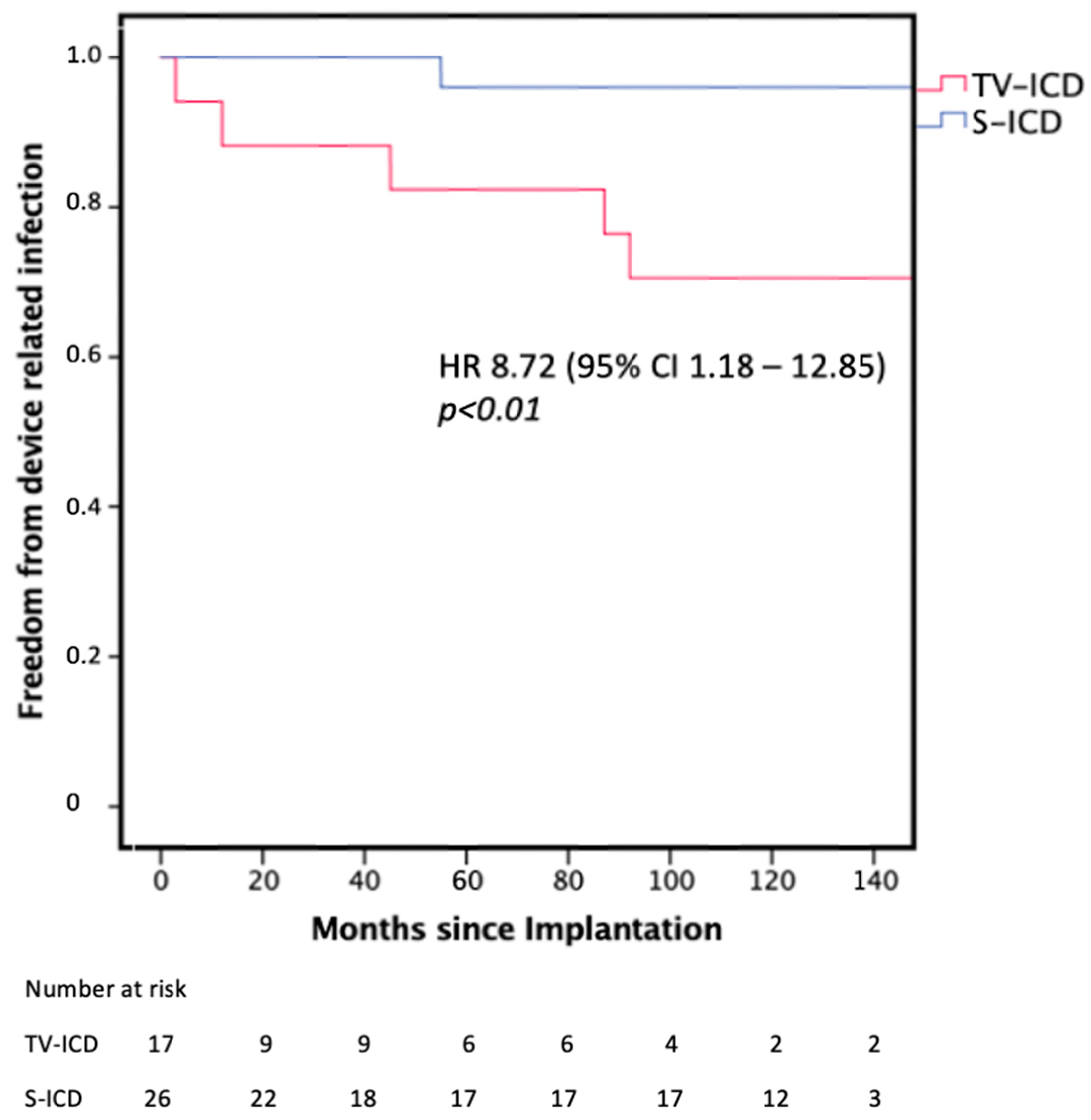
| Device-related infection, n (%) | 5 (29.4) | 1 (3.8) | 8.72 (1.18–12.85) | <0.01 |
| Time to first device infection, (months) | 47.8 (7.5–89.5) | - | ||
| Lead associated complication | 2 (11.7) | 0 | 0.188, ns | |
| Overall mortality, n (%) | 6 (35.3) | 6 (23.1) | 1.92 (0.96–6.15) | 0.274, ns |
| Cardiovascular mortality, n (%) | 4 (23.5) | 1 (3.8) | 9.17 (1.12–8.33) | <0.05 |
| Patients experiencing ventricular arrhythmia | 3 (17.6) | 2 (7.7) | 0.355, ns | |
| Mean number ventricular arrhythmia episodes | 0.7 ± 1.8 | 0.1 ± 0.3 | 0.150, ns | |
| Average ATP | 0.2 ± 0.5 | 0 | ns | |
| Patients receiving a shock, n (%) | 2 (11.7) | 4 (15.4) | 0.388, ns | |
| Patients receiving appropriate shocks | 2 (11.7) | 2 (7.7) | 0.605, ns | |
| Patients receiving inappropriate shocks, n (%) | 0 | 2 (7.7) | 0.242, ns | |
| Patients receiving more than one shock | 0 | 0 | ||
| Hospitalization rate, n (%) | 11 (64.7) | 8 (30.8) | 2.59 (1.12–6.41) | <0.05 |
| Mean number hospitalizations per patients | 3.4 ± 2.9 | 0.8 ± 1.8 | <0.05 | |
| Patients with cardiac hospitalization, n (%) | 7 (41.2) | 3 (11.5) | 5.99 (1.24–28.9) | <0.05 |
| Mean cardiac hospitalizations | 1.3 ± 2.4 | 0.2 ± 0.9 | <0.05 | |
| Patients with non-cardiac hospitalization, n (%) | 7 (41.2) | 4 (15.4) | 1.43 (0.86-1.73) | 0.158, ns |
| Number of non-cardiac hospitalizations | 0.8 ± 1.5 | 0.4 ± 1.3 | 0.377, ns | |
| Patients with device hospitalization, n (%) | 7 (41.2) | 1 (3.8) | 10.2 (1.22-84.61) | <0.001 |
| Overall duration hospitalization (days) | 62.0 ± 22.6 | 24.0 ± 18.6 | <0.05 |
Disclaimer/Publisher’s Note: The statements, opinions and data contained in all publications are solely those of the individual author(s) and contributor(s) and not of MDPI and/or the editor(s). MDPI and/or the editor(s) disclaim responsibility for any injury to people or property resulting from any ideas, methods, instructions or products referred to in the content. |
© 2024 by the authors. Licensee MDPI, Basel, Switzerland. This article is an open access article distributed under the terms and conditions of the Creative Commons Attribution (CC BY) license (https://creativecommons.org/licenses/by/4.0/).
Share and Cite
Schiedat, F.; Meuterodt, B.; Winter, J.; Prull, M.; Aweimer, A.; Gotzmann, M.; O’Connor, S.; Perings, C.; Lawo, T.; El-Battrawy, I.; et al. Subcutaneous versus Transvenous Implantable Cardioverter Defibrillator in Patients with End-Stage Renal Disease Requiring Dialysis: Extended Long-Term Retrospective Multicenter Follow-Up. J. Pers. Med. 2024, 14, 870. https://doi.org/10.3390/jpm14080870
Schiedat F, Meuterodt B, Winter J, Prull M, Aweimer A, Gotzmann M, O’Connor S, Perings C, Lawo T, El-Battrawy I, et al. Subcutaneous versus Transvenous Implantable Cardioverter Defibrillator in Patients with End-Stage Renal Disease Requiring Dialysis: Extended Long-Term Retrospective Multicenter Follow-Up. Journal of Personalized Medicine. 2024; 14(8):870. https://doi.org/10.3390/jpm14080870
Chicago/Turabian StyleSchiedat, Fabian, Benjamin Meuterodt, Joachim Winter, Magnus Prull, Assem Aweimer, Michael Gotzmann, Stephen O’Connor, Christian Perings, Thomas Lawo, Ibrahim El-Battrawy, and et al. 2024. "Subcutaneous versus Transvenous Implantable Cardioverter Defibrillator in Patients with End-Stage Renal Disease Requiring Dialysis: Extended Long-Term Retrospective Multicenter Follow-Up" Journal of Personalized Medicine 14, no. 8: 870. https://doi.org/10.3390/jpm14080870
APA StyleSchiedat, F., Meuterodt, B., Winter, J., Prull, M., Aweimer, A., Gotzmann, M., O’Connor, S., Perings, C., Lawo, T., El-Battrawy, I., Hanefeld, C., Korth, J., Mügge, A., & Kloppe, A. (2024). Subcutaneous versus Transvenous Implantable Cardioverter Defibrillator in Patients with End-Stage Renal Disease Requiring Dialysis: Extended Long-Term Retrospective Multicenter Follow-Up. Journal of Personalized Medicine, 14(8), 870. https://doi.org/10.3390/jpm14080870