Article

Effect of Phase Distribution on the Antibacterial Property and Cytotoxicity of Ti-5Al-2.5Cu Alloy after Heat Treatment at Various Temperatures

1
Ph. D. Program, School of Dentistry, College of Dental Medicine, Kaohsiung Medical University, Kaohsiung 80708, Taiwan
2
Department of Mechanical Engineering, National Pingtung University of Science and Technology, Pingtung 91201, Taiwan
3
Department of Dentistry, Kaohsiung Medical University Hospital, Kaohsiung 80708, Taiwan
4
School of Dentistry, College of Dental Medicine, Kaohsiung Medical University, Kaohsiung 80708, Taiwan
*
Author to whom correspondence should be addressed.
Metals 2020, 10(7), 858; https://doi.org/10.3390/met10070858
Submission received: 9 June 2020 / Revised: 25 June 2020 / Accepted: 26 June 2020 / Published: 29 June 2020

Abstract

:
Titanium alloys are widely employed for the fabrication of biomedical devices. In this study, we designed and developed a Ti-5Al-2.5Cu alloy, which exhibited antibacterial properties. Microstructure and elemental analyses were performed using X-ray diffraction (XRD), scanning electron microscopy (SEM), energy dispersive X-ray spectroscopy (EDS), and transmission electron microscopy (TEM). We evaluated the alloy’s antibacterial properties using Escherichia coli in the plate-count method. The cytotoxicity was examined using the MG-63 cell response by 3-(4,5-dimethylthiazol-2-yl)-2,5-diphenyl tetrazolium bromide (MTT) and lactate dehydrogenase (LDH) assays. Microstructural analysis revealed that Ti-5Al-2.5Cu exhibited an equiaxed α’ martensite structure after short-term annealing. The heterogeneous and homogeneous α → α + Ti2Cu phase transitions occurred at ~840 and 920 °C, respectively. The antibacterial property for Ti-5Al-2.5Cu was varied by volume fraction in the Ti2Cu and Cu-rich phase, which was obtained using different heat treatments. The high volume fraction of the Ti2Cu and Cu-rich phase was observed after long-term annealing at 720–840 °C and thus exhibited a higher antibacterial rate. The relationship between phase distribution and the antibacterial property could be satisfied by a positive linear regression equation. Cytotoxicity results showed that heat treatments at different temperatures for Ti-5Al-2.5Cu alloys had no effect on cell viability. The optimal heat treatment for Ti-5Al-2.5Cu alloy was annealing at 760 °C for 24 h. After, the alloy exhibited both promising antibacterial performance and good cytocompatibility.

1. Introduction

Titanium and titanium alloys (Ti and Ti alloys) possess superior biocompatibility and high specific strength [1,2,3]. Therefore, these alloys are widely applied for the fabrication of biomedical devices. Commercial pure Ti (CP-Ti) is commonly applied as a dental material and is suitable for dental prosthetic applications [4,5]. Because of its poor mechanical properties, the content of oxygen in titanium is used to improve the mechanical properties, and the alloy is graded as G1 to G4 pure Ti.
Additionally, the Ti-Al-V alloy series such as Ti-6Al-4V (Ti-64) alloys have been widely applied for the fabrication of specific biomedical materials because of their excellent mechanical properties, especially when compared with those of CP-Ti. An additional 3–6% Al is considered to enhance the mechanical properties of pure Ti. Some alloys such as Ti-15Mo-5Zr-3Al (mass%) [6] and Ti-5Al-2.5Fe [7] have been developed for application in the biomedical field to withstand stress. Bolzoni [8] investigated the mechanical properties of Ti-xAl alloys (x = 1–6 wt%), showing that both the ultimate tensile strength and yield strength continuously increased with an increasing Al content. The strengthening mechanism caused after adding Al to Ti could be explained by the simultaneous contribution of the substitutional strengthening effect of Al atoms in the hexagonal closest packed (HCP) α-Ti lattice. The use of Al and V as biomaterials has been questioned from a biocompatibility viewpoint. To eliminate doubts regarding the usage of implantable materials, many β stabilizing elements such as Ta, Nb, Mo, Fe, Cu, Co, and Ag have been used to replace V [9]. Moreover, Al is the main strengthening element of Ti, so determining whether its benefit is either excessive or a drawback is a major issue in the biomaterial field. Among these β stabilizing elements, Cu is well known as an alloying element with potential bactericidal effects. It has been previously applied as an antibacterial agent. The material exhibits antibacterial functions through the release of metal ions or nanoparticles from the surface [10]. Consequently, Cu has been widely added to Ti because of its antibacterial behavior [11,12,13]. Meanwhile, Ti alloyed with a small amount of Cu (1–5 wt%) is reported to exhibit adequate mechanical properties, biocompatibility, and corrosion resistance for medicinal purposes [14,15].
In addition to ion release, Liu [16] claimed that the Ti-5Cu alloy, which has a rough surface (i.e., sandblasted, large grit, and etched), could exhibit better antibacterial properties than mechanically ground alloys. Furthermore, our previous study [13] showed that spherical and acicular Ti2Cu alloys exhibited different Cu contents, which was a factor impacting antibacterial properties. The antibacterial rate had a linear relationship with the type and proportion of the precipitate. In addition, Ma [10] stated that the antibacterial rate increased with a higher distribution of Cu on the surface after heat treatment. These results showed that the interface between bacteria and substrates may be an important factor in the antibacterial behavior.
In this study, the binary Ti-Cu alloy presented a eutectoid transformation with 7.1% Cu (wt%) content at 790 °C. The α phase and Ti2Cu were formed under these conditions. It has been previously shown that the eutectoid structure of the Ti-Cu alloy is α + Ti2Cu [3,13,15,17,18,19]. The shape of Ti2Cu has been found to vary depending on its heat treatment and forging temperature [13,19]. Ti2Cu intermetallic compounds were observed to have formed two shapes in the Ti-2.5Cu alloy: spherical particles due to decomposition of the supersaturated α phase during annealing at 790 °C and acicular ones produced by aging after solution treatment [20]. Additionally, the mechanical behavior of Ti-Cu alloys was improved by controlling Ti2Cu precipitation [21].
To achieve superior mechanical properties and antibacterial properties, 5 wt% Al and 2.5 wt% Cu were designed, respectively. The purpose of this study was to investigate the microstructure and antibacterial property of the Ti-5Al-2.5Cu alloy after a series of heat treatments in order to clarify the relationship of its microstructure with its antibacterial properties. To confirm whether the addition of Al was safe for biomedical materials, we also evaluated its biocompatibility. Based on these results, a titanium alloy with antibacterial properties was developed for application in dental implant materials.

2. Materials and Methods

2.1. Sample Preparation

A Ti-4.98mass%Al-2.48mass%Cu-0.16mass%O-0.03mass%C-0.02mass%N (Ti-5Al-2.5Cu) alloy was prepared using a high-purity sponge titanium, 99.7% pure electrolytic Al, and pure Cu in a vacuum arc melting furnace. The ingot (5 kg) was re-melted twice to ensure compositional homogeneity. The ingot was cylindrical with a diameter of 100 mm and height of 150 mm. After homogenization at 1000 °C for 4 h in a protective Ar2 atmosphere, the specimen was hot forged, cold rolled to a rod, and cut by a cutting machine to a final thickness of 1.0 mm. The specimens were heat treated at 720, 760, 840, 880, 920, and 1040 °C for various times (1, 12, and 24 h) in a vacuum furnace and then water quenched. In this study, a total of 270 samples were produced and divided into each heat treatment group (n = 15) and randomly assigned to subsequent experiments. The specimens subjected to antibacterial testing were prepared by grinding with a series of #100- to #1500-grit SiC (Silicon Carbide) sandpapers to ensure consistent roughness values.

2.2. Microstructure Characterization

Specimen microstructures were observed by scanning electron microscopy (SEM, JEOL JSM-6380, Tokyo, Japan) at 20 kV after etching in 1% HF, 2.5% HNO3, 1.5% HCl, and 95% H2O for 3 min at room temperature (approximately 25 ± 0.5 °C). In addition, energy dispersive X-ray spectrometry (EDS) was performed using a high resolution-SEM (HITACHI SU8000, Tokyo, Japan). The crystal structure of the present alloy was confirmed using a D8 X-ray diffraction (XRD) with Cu-Kα radiation (λ = 1.5406 Å), which operated at 40 kV and 30 mA at room temperature, and scanned from 30° to 80° with a step rate of 0.02°/s. Specimens used for electron microscopy were prepared using a double-jet electropolisher with an electrolyte containing 60% methanol, 35% n-butyl alcohol, and 5% perchloric acid at a temperature of −15 to −5 °C for 90 s. The voltage was maintained at 20 V. Electron microscopy of the specimens was performed using a transmission electron microscope (TEM, PHILPLIES CM200, Tokyo, Japan) at 200 kV. The Ti53 specimens were annealed at various temperatures for 24 h and were selected for the cytotoxicity test. The crystallographic analyses of the precipitates and matrix were accompanied by a selected area diffraction pattern (SADP). An energy dispersive X-ray spectrometer was used for compositional analyses.
SEM images were quantified using the ImageJ software (version 1.52q, National Institutes of Health, Bethesda, MD, USA) which revealed the distribution of the matrix and precipitates of the Ti-5Al-2.5Cu alloy after different heat treatments.

2.3. Antibacterial Test

The antibacterial properties of Ti-5Al-2.5Cu (n = 3) were analyzed according to the JIS Z2801-2000 specifications [13]. The experimental specimens were sterilized at approximately 135 °C for 30 min by steam autoclaving. Then, 0.4 mL of Escherichia coli (E. coli [ATCC-25922]) suspension with a density of 106 CFU/mL was dripped onto the specimens, as well as on a control group (broth only). The area of the bacteria in contact with the specimens was 10 × 10 mm2. To avoid evaporation, the specimens with the suspension were inoculated in a humid environment at 37 °C for 24 h. After incubation, the specimens were carefully washed with 1× diluted phosphate-buffered saline to remove bacteria from the specimens. Then, 0.02 mL of the washing solution was spread onto agar plates and the agar plates were incubated at 37 °C for an additional 24 h. After incubation, the number of colonies was counted. The procedure was repeated three times and the average was obtained for comparison. The antibacterial rate (AR) was calculated using the following formula [10]:
AR = (NcontrolNspecimen)/Ncontrol × 100%
where Ncontrol and Nspecimen are the average numbers of bacterial colonies of the broth only and Ti-5Al-2.5Cu specimens, respectively.

2.4. Culture of Cell

MG-63 human osteoblast cells were used to evaluate the biocompatibility of the specimens. The cells were collected and cultured in Dulbecco’s modified Eagle’s medium (DMEM) containing 10% fetal bovine serum and 1% penicillin/streptomycin in an environment with 5% CO2 at 37 °C. Each sample (n = 3) was extracted in DMEM for 3 days at 72 °C. The cells were seeded at a density of 1.0 × 10⁴ cells/well in a 96-well plate and cultured for 24, 72, and 120 h, respectively.

2.5. Cell Viability Assay

The 3-(4,5-dimethylthiazol-2-yl)-2,5-diphenyl tetrazolium bromide (MTT) assay was performed to evaluate cell viability. After 24, 72, and 120 h of culturing, 100 μL of the MTT solution was added into the well of the 96-well plate and incubated for 2 h at 37 °C. Then, the medium was removed and replaced with 1 mL dimethyl sulfoxide (DMSO) to dissolve the MTT formazan. The optical density (OD) of the solution (100 μL) was read after 10 min using an ELISA reader (Infinite F50, Tecan, Switzerland) at 570 nm. The assay was performed in triplicate. The relative growth rate (RGR) was calculated using the following formula [10]:
RGR = (ODcontrolODspecimen)/ODcontrol × 100%
Lactate dehydrogensae (LDH) assays were performed by assessing the LDH released into the media as a marker of dead cells. After 24 h of incubation, 100 μL of eluate was transferred to another 96-well cell culture plate. Extracellular LDH activity was measured using an LDH detection kit. The absorbance of this colored solution was quantified at a certain wavelength (490 nm) using an ELISA reader.

3. Results

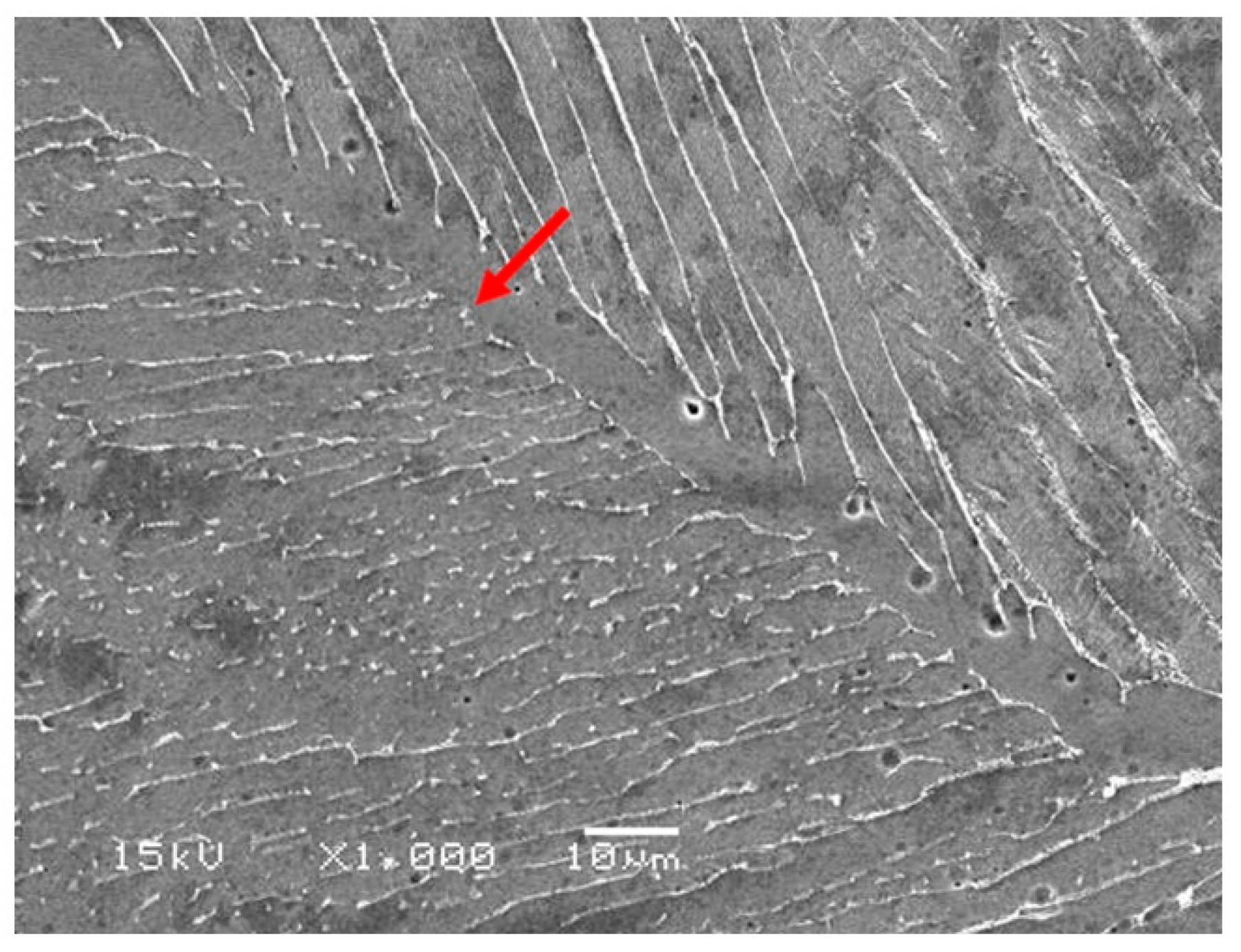
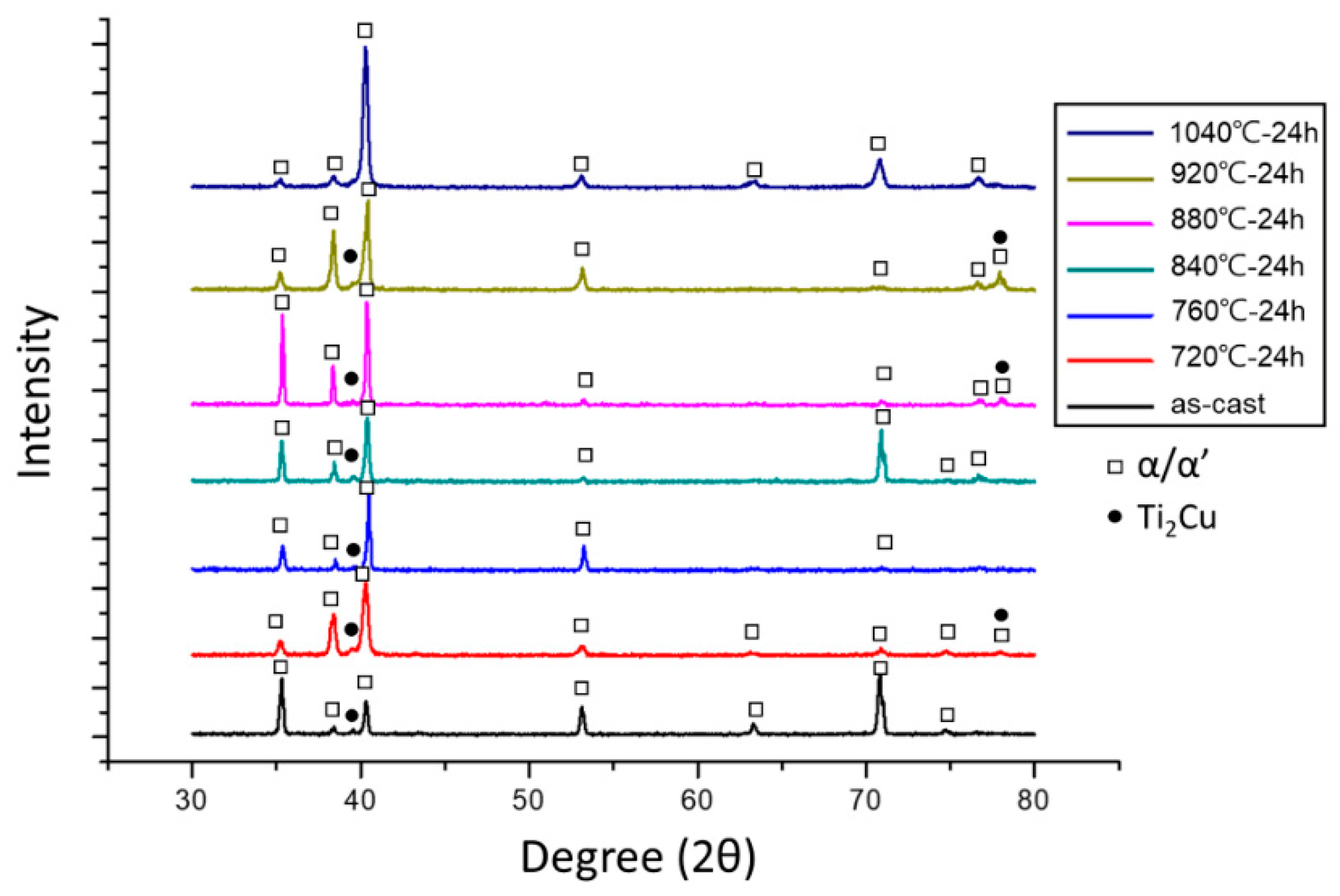
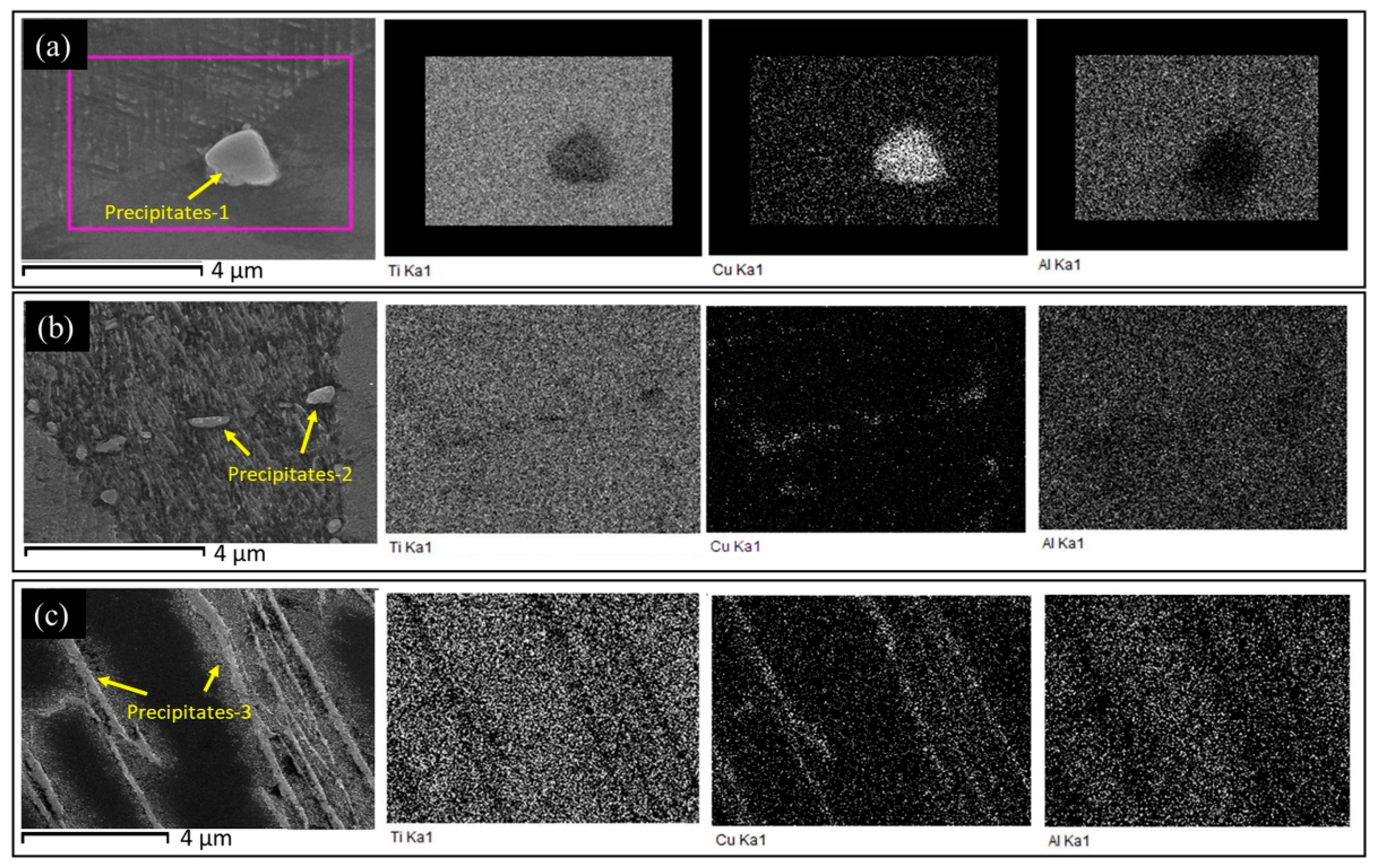
3.1. Microstructural Observations

Figure 1 shows the SEM micrographs of the as-cast Ti-5Al-2.5Cu alloy and reveals that the microstructure of the as-cast specimen exhibited an essentially equiaxed structure with acicular α’ martensite and some precipitates formed at the boundaries. To observe the precipitation of the Ti2Cu phase of the Ti-5Al-2.5Cu alloy after annealing, various heat treatments were performed. Figure 2 shows the SEM electron micrographs of the Ti-5Al-2.5Cu annealed at 720–920 °C for 1, 12, and 24 h, respectively. No significant difference was observed in the SEM micrographs of the Ti-5Al-2.5Cu alloy when treated for 1 h at different temperatures. Additionally, some precipitates were formed on the interface of the martensite and within the matrix, especially when the heat treatment time increased to 24 h. Some fine Ti2Cu precipitates were found within the matrix when the alloy was annealed at 760 °C and 840 °C for 12 and 24 h.
Figure 3 shows the XRD patterns obtained from Ti-5Al-2.5Cu alloy after different heat treatments, showing only the α/α’ and Ti2Cu phases. Based on the XRD data, the lattice parameters for each phase in the three alloys were carefully gathered and these data were additionally confirmed by TEM diffractions.
TEM electron micrographs of the Ti-5Al-2.5Cu alloy obtained after annealing at 920 °C for 24 h clearly showed the Ti2Cu precipitates, as shown in Figure 4a. The SADPs of the precipitate and matrix are shown in Figure 4b. Figure 4c shows that g = [ 2 1 ¯ 1 ¯ 0 ] for the matrix and g = [ 01 1 ¯ 0 ] for another matrix. This suggests that the orientation relationship (OR) between the matrices was [ 2 1 ¯ 1 ¯ 0 ] α // [ 01 1 ¯ 0 ] α . Figure 4d shows the value of the foil normal of the SADP was g = [ 1 ¯ 1 ¯ 23 ] and no extra reflection spots of Ti2Cu precipitates were found. It is clear that the SADP taken from region C belonged to the Ti2Cu phase and that the foil normal was g = [100]Ti2Cu (Figure 4e). The lattice parameters of the Ti2Cu and α matrix were a = 0.296 ± 0.006 and c = 1.062 ± 0.009 nm, and a = 0.295 ± 0.005 nm and c = 0.465 ± 0.008 nm, respectively. Microstructures in the alloy were a hexagonal α phase and a body-centered-tetragonal (BCT) Ti2Cu intermetallic compound. From such patterns, the orientation relation between α and Ti2Cu was established as [ [ 1 ¯ 1 ¯ 23 ] α//[100]Ti2Cu. The indexed SADP for Ti2Cu phase is illustrated in Figure 4f.
When the Ti-5Al-2.5Cu alloy was annealed at 1040 °C, the microstructure of the alloy was an equiaxed α lamellar phase with a width of 2–5 μm, as shown in Figure 5a. The TEM electron micrographs of the Ti-5Al-2.5Cu (annealed at 1040 °C for 1 h) obviously showed that some sub-grain structures were found within the α matrix and some dislocations were observed either on the interface or within the sub-grain (Figure 5b). Furthermore, no β phase was observed during these heat treatments based on the XRD and TEM examinations.
To analyze the precipitation of the Ti2Cu phases annealed at various temperatures, SEM-EDS was performed. Figure 6 shows the EDS maps for the specimens annealed at 720, 920, and 1040 °C for 24 h. The chemical compositions of the Ti2Cu precipitates are shown in Table 1. The Cu content of the Ti2Cu phase formed on the interface after annealing at 740 °C was approximately 34.12 wt%. Figure 6 shows the EDS maps, which clearly show that the precipitate was a high copper phase and aluminum was evenly distributed in the α matrix. When the annealed temperature increased to 920 °C, the copper content of the Ti2Cu phase formed either on the interphase or within the matrix, decreasing to 23.33 wt%. Similarly, EDS-mapping showed that Cu distribution was consistent with the position of the precipitates. Furthermore, Ti2Cu was not observed in the specimen annealed at 1040 °C. Instead, a Cu-rich phase with a lamellar structure with 7.23 wt% Cu content was observed.

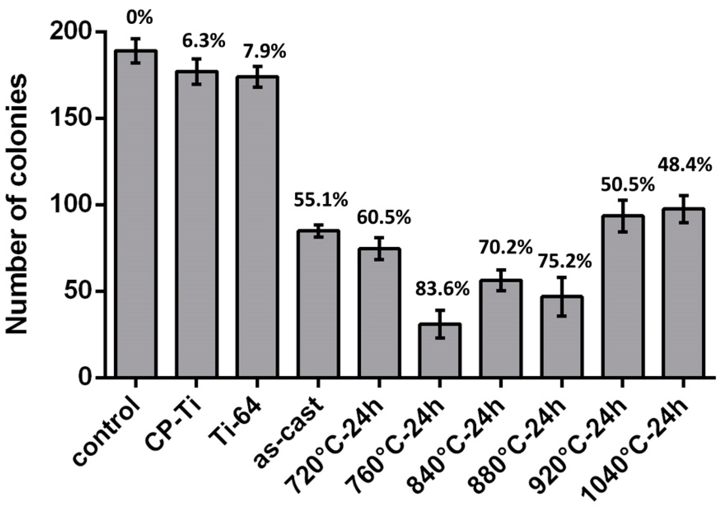
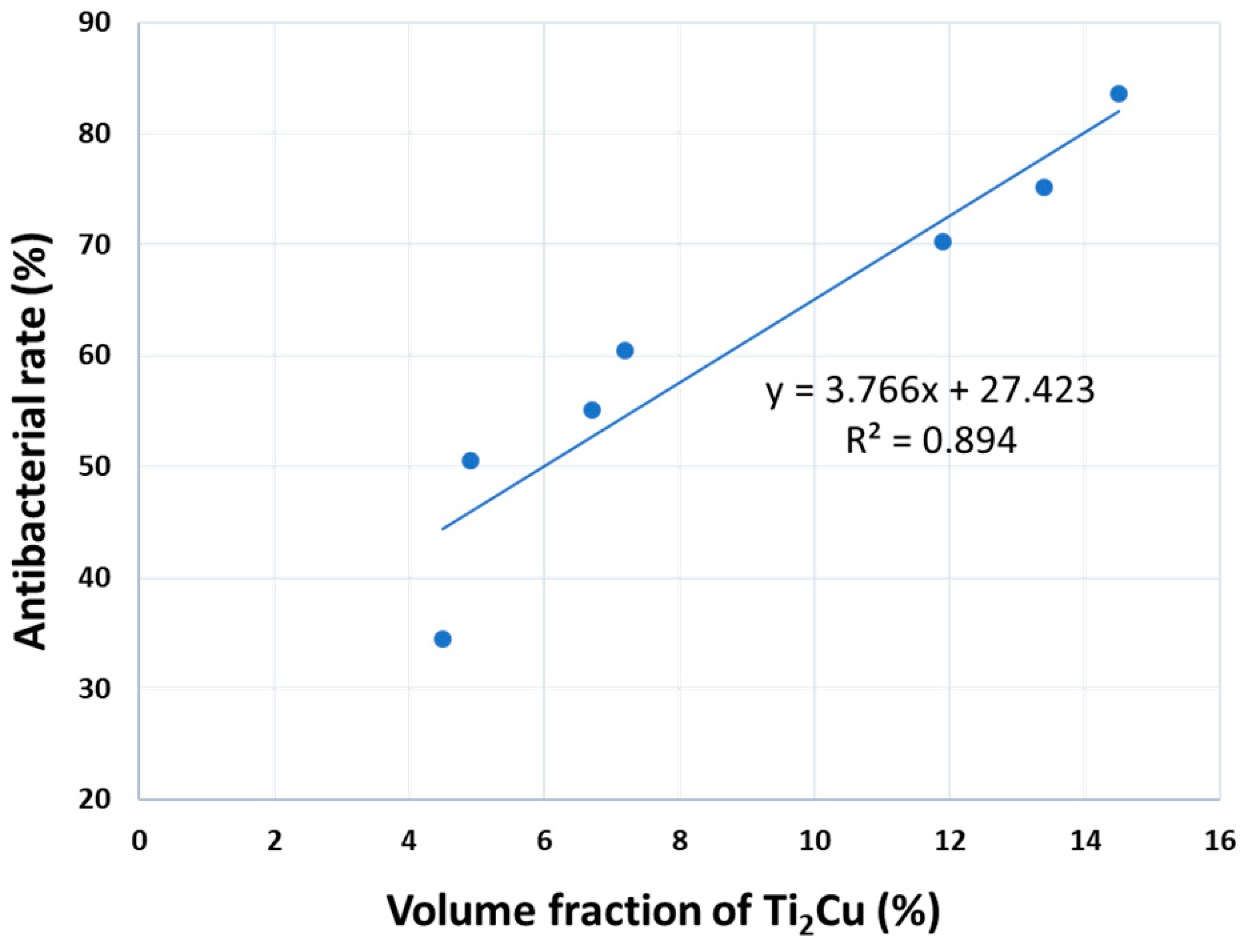
3.2. Antibacterial Property

The Ti-5Al-2.5Cu specimens annealed at various temperatures for 24 h underwent antibacterial analysis. Figure 7 shows the typical E. coli bacterial colonies observed on the surface of the CP-Ti, Ti-64, and Ti-5Al-2.5Cu alloy after 24 h. A large amount of E. coli cells was observed on the control (broth only), CP-Ti, and Ti-64, indicating that CP-Ti and Ti-64 did not exhibit any antibacterial properties. It is obvious that the antibacterial property of the Ti-5Al-2.5Cu alloy varied because of heat treatment. The as-cast Ti-5Al-2.5Cu alloy did not exhibit a strong antibacterial property. However, the Ti-5Al-2.5Cu samples annealed at 720, 760, 840, and 880 °C exhibited antibacterial properties, and fewer bacterial colonies were observed. The Ti-5Al-2.5Cu alloy annealed at 760 °C exhibited the highest AR (83.6%). In addition, the antibacterial property decreased when the annealing temperature was increased to 920 and 1040 °C.

3.3. Cytotoxicity Evaluation

The Ti-5Al-2.5Cu specimens annealed at various temperatures for 24 h underwent the cytotoxicity test. Figure 8a shows the results of the MTT assay and the cell viability on the CP-Ti, Ti-64, and Ti-5Al-2.5Cu alloys after culturing for 24, 72, and 120 h. It is obvious that the RGR values of the MG-63 cells of each Ti-5Al-2.5Cu sample were between 91.54% and 99.71%, showing no significant difference from either CP-Ti or Ti-64. When the incubation time was extended to 72 and 120 h, the RGR values of each alloy were ranged from 90.39% to 96.06% and 90.67% to 97.31%, respectively. No significant difference was observed in cell survival among the different samples, which confirms the good cytocompatibility of the Ti-5Al-2.5Cu alloys after heat treatment at different temperatures.
After incubation for 24 h, the LDH assay showed that the LDH values of the Ti-5Al-2.5Cu alloy after annealing at 720, 840, and 880 °C, which, when compared to CP-Ti and Ti-64, were slightly increased (Figure 8b). When the incubation time was extended to 72 h, the Ti-5Al-2.5Cu specimens annealed at 720, 760, and 840 °C, exhibiting higher LDH values. In addition, the Ti-5Al-2.5Cu specimens annealed at only 720 and 760 °C showed slightly higher LDH values after 120 h of incubation.

4. Discussion

Pure titanium exhibits excellent biocompatibility and corrosion resistance, which is why it is widely applied for the fabrication of biomedical materials. When an implant is required for strength, such as for hip joints, knee joints, elbow joints, and bone plates, it must have superior mechanical properties. The mechanical properties of pure Ti are significantly affected by the presence of interstitial elements, such as oxygen and nitrogen. Particularly, the amount of oxygen in metallic pure Ti is used to classify Ti into four different grades [22]. In contrast, the mechanical behavior of Ti can be enhanced via alloying. In particular, the addition of Al has received significant attention because Al is cheaper and lighter than Ti. Furthermore, the addition of Al increases the deformability of the alloy as compared to other α stabilizers [8]. Dental implants are developed based on the ‘osseointegration’ of the peri-implant bone. They are embedded in the maxilla and/or mandible for the management of tooth loss and to aid in the replacement of lost orofacial structures. Unfortunately, failures are largely due to implant-associated infection [23]. Residual materials, protruding implants, poorly seated cover screws, and trauma from inadequately relieved dentures or occlusal trauma from opposing teeth are some of the most frequent causes of soft tissue infection during the healing period [24]. Infections that originate from soft tissues may potentially progress deeper into the bone and undermine the osseointegration process around the dental implant. Consequently, the bacteria colonized on the implant surface can lead to a loss of the protection of the osseointegrated part of the implant against bacterial invasion [25]. According to a American Academy of Orthodontic Medicine 2008 report, approximately 80% of clinicians with one to three years of experience in orthodontics have used temporary anchorage devices (TAD) as a clinical orthodontic treatment tool, and some cases of TAD failure have been attributed to infection, force, material strength, and corrosion [25]. Additionally, soft tissue inflammation and swelling around the bone screws, or soft tissue friction and hyperplasia, reduce the protection mechanism of the mucosa itself, resulting in an increased chance of infection [26].
However, currently available bone screws/implants do not exhibit antibacterial properties. The human oral cavity is a complex ecological environment because the saliva in the oral cavity contains water, amino acids, proteins, lipids, sugars, and some inorganic substances. Such an environment is suitable for bacteria growth such as anaerobic spirochetes, Vibrio, Staphylococcus, Lactobacillus, and Streptococcus. These bacteria are easily adsorbed on the surface of the material. Under normal conditions, these microorganisms are nonpathogenic. However, when changes occur in the oral environment, these microorganisms will overgrow and cause infection and inflammation. In addition, when the surface of the material interacts with bacteria for a long time, the bacteria will release oxides of H2O2 (hydrogen peroxide) on the surface of the material [27,28]. This substance can easily corrode the surface of the material. The structural damage causes loosening, breaking, or failure of the bone screw/mini-implant which, in turn, causes an increase in treatment time and cost or ion release due to material corrosion, which affects human health.
Chen et al. investigated the use of 492 Ti TADs in orthodontic treatment and found three statistically significant reasons for TAD failure, including (1) severe infection of the surrounding soft tissues of TAD; (2) premature loading within three weeks after implantation; and (3) mandibular retraction of the treated patients [29]. Therefore, implant infection is a problem that needs to be urgently solved [26]. Thus, in this study, Ti-5Al-2.5Cu alloys were fabricated to obtain a new implant material with antibacterial properties.
After heat treatment at different temperatures, the microstructure, surface element distribution, and distribution of phase and grain sizes were strongly affected in the Ti alloy. The Ti-5Al-2.5Cu alloy exhibited different distribution of the α phase precipitates after annealing at different temperatures. In eutectoid alloys, such precipitation occurred due to martensite decomposition in response to aging heat treatment. Cardoso et al. [27] reported that the microstructure of the Ti-Cu alloy consists of Ti2Cu and the α phase; no evidence of beta phase stabilization has been observed in any rapidly quenched samples. Based on the present study, we found that an α → α + Ti2Cu phase transition occurred when the as-cast alloy was annealed at 920 °C. Note that this result has never been reported before. Furthermore, when the annealing temperature decreased to 840 °C, some Ti2Cu precipitates were found within the matrix. Based on these results, the heterogeneous and homogeneous temperatures of the α → α + Ti2Cu phase transition were found to be approximately 920 and 840 °C, respectively. According to the Ti-Cu binary phase diagram, the eutectoid temperature of the Ti-Cu binary alloy was 790 °C [28]. Furthermore, the Ti2Cu precipitate could precipitate at ≦790 °C when the Cu content was below 7.0 wt% in the Ti-Cu alloy. Therefore, the addition of 5.0 wt% Al increased the occurrence temperature of the Ti2Cu phase in the Ti-2.5Cu alloy, indicating that the stable temperature of the Ti2Cu phase increased.
The amounts of the α-Ti and Ti2Cu precipitates were calculated using the ImageJ software, as detailed in Table 2. By substituting the ratios of the different phases and AR into a general linear equation, a new equation can be obtained to illustrate the relationship between the microstructure and antibacterial properties, as shown in Figure 9. The inferential AR (IAR) can be separated into a based AR (BAR) plus the contribution of precipitates as follows:
IAR = (Dp × F × 100%) + BAR
where Dp and F are the distribution of precipitates and the impact factors (F = 3.766) of antibacterial properties and BAR = 27.423%. Thus, if the volume fraction of Ti2Cu and Cu-rich phases is 0, Ti-5Al-2.5Cu could exhibit basic antibacterial properties (27.423%). However, in this study, Cu-rich and/or Ti2Cu phases were found along the grain matrix, boundaries, and lamellar structure after both casting and heat treatments at different temperatures. The main reason for this formation was that the Ti-5Al-2.5Cu alloy was decomposed into a block with high Cu content because of the addition of Al or segregation in the casting state. To verify the contribution of precipitates to the Ti-Al-Cu alloy, a binary Ti-2Cu alloy (Ti-1.89 wt% Cu) solution treated at 880 °C was used to verify the BAR. This design was based on the Cu content in the matrix ranging from 2.02 to 2.29 wt% (Table 1). The AR of the Ti-2Cu alloy was 30.4%, which was close to the calculated BAR of 27.423%. Thus, the inferential AR could be calculated by a linear regression equation based on the different amounts of phases. There was a linear relationship between the proportion of phases in the Ti-5Al-2.5Cu alloy and antibacterial properties. We hypothesized that the concentration of Cu in the granular Ti2Cu precipitate was higher and therefore it caused the release of more Cu2+ ions. This could explain the reason why the best antibacterial property observed in the Ti-5Al-2.5Cu alloy was annealed at 760 °C for 24 h. The diffusion and distribution of Cu in the alloy play an important role in the Ti2Cu precipitation and affect the antibacterial property. The Ti-5Al-2.5Cu alloy annealed at 1040 °C for 24 h exhibited a gap from the expected theoretical antibacterial properties (Table 2). Because the alloy did not have Ti2Cu precipitates, the antibacterial ability of the alloy was approximately equal to the basic antibacterial value of the Ti-2Cu alloy. This also indirectly proves that the AR can depend on the amount of Ti2Cu precipitates [10]. It is well known that Ti-Cu based alloys with higher Cu content can exhibit better antibacterial properties [29]. However, based on the same copper content, the antibacterial properties of the Ti-5Al-2.5Cu alloy could also be enhanced through different microstructures and phase distributions on the surface.
According to the above microstructure and antibacterial analyses, it can be seen that antibacterial properties for different Ti-5Al-2.5Cu alloys mainly depend on the Cu element content on the surfaces given by different phases. Thus, we can conclude that the Ti-5Al-2.5Cu alloy with a high proportion of Ti2Cu can release more copper ions when exposed to bacterial suspensions [10]. Researchers believe that released Cu2+ ions react with bacteria and kill them when Cu2+ ions combine with the plasma membrane, via electrostatic attraction and then penetrate the cell membrane through the opening or closing of the membrane channels [30,31]. Conversely, if there is less Ti2Cu and Cu-rich in the same Cu content range, it will result in poor antibacterial properties (such as 920 °C, 24 h, and 1040 °C, 24 h).
For new implant materials, biocompatibility is an important requirement for application in the biomedical field. In addition to evaluating the antibacterial ability of the Ti-5Al-2.5Cu alloy, this study also evaluated biocompatibility using MG-63 cells. Ma [10] investigated the cytotoxicity of Ti-6Al-4V-5Cu alloys after solution and aging treatments. They believed that more interstitial Cu atoms occur when the alloy is treated at a high temperature. Thus, this caused a higher release of Cu ions, which slightly affected the cytotoxicity of the Ti-6Al-4V-5Cu alloy. However, in the present study, the content of Cu was 2.5 wt% and no significant difference was observed in the cell survival among groups (Figure 8), which confirms the good cytocompatibility of the Ti-5Al-2.5Cu alloy.

5. Conclusions

In this study, 2.5% Cu was added to a Ti-5Al alloy in an attempt to obtain a dental implant with antibacterial properties. Different microstructures of the Ti-5Al-2.5Cu alloy were obtained using various heat treatments. Thus, their corresponding antibacterial properties and biocompatibility were discussed. In the future, the response of this alloy to in vivo study still needs to be evaluated. Based on the results, the following conclusions can be drawn:
  • The microstructure of the present Ti-5Al-2.5Cu alloy was essentially an acicular α’ martensite. Some Ti2Cu-phase precipitates were also observed, which improved the antibacterial property of the alloy.
  • During various heat treatments, a high volume fraction of the Ti2Cu phase was obtained after annealing at 760–880 °C. The addition of 5.0 wt% Al increased the temperature at which the Ti2Cu-phase precipitates occurred.
  • The antibacterial property was linearly related to the ratio of precipitates and was accumulated using a basic antibacterial value. The optimal heat treatment for the Ti-5Al-2.5Cu alloy was annealing at 760 °C for 24 h, at which it exhibited promising antibacterial properties.
  • The Ti-5Al-2.5Cu alloy heat treated under different conditions exhibited promising non-cytotoxicity when cultured with MG-63 cells for 1, 4, and 7 days.

Author Contributions

Conceptualization, J.-K.D. and C.-Y.C.; methodology, Y.-H.C. and C.-Y.C.; software, Y.-H.C. and Y.-T.C.; validation, Y.-H.C., C.-Y.C., and J.-K.D.; formal analysis, C.-Y.C. and Y.-T.C.; investigation, Y.-H.C. and Y.-T.C.; resources, J.-K.D.; data curation, C.-Y.C.; writing—original draft preparation, Y.-H.C. and Y.-T.C.; writing—review and editing, J.-K.D.; visualization, J.-K.D.; supervision, C.-Y.C.; project administration, J.-K.D.; funding acquisition, J.-K.D. All authors have read and agreed to the published version of the manuscript.

Funding

This study was financially supported by a fund from the Ministry of Science and Technology MOST106-2314-B-037-013- and 107-2314-B-037-46- and Kaohsiung Medical University Hospital, KMUH107-7R76 and KMUH108-8M62.

Conflicts of Interest

The authors declare no conflict of interest.

References

  1. Ou, K.-L.; Weng, C.-C.; Lin, Y.-H.; Huang, M.-S. A promising of alloying modified beta-type titanium-niobium implant for biomedical applications: Microstructural characteristics, in vitro biocompatibility and antibacterial performance. J. Alloys Compd. 2017, 697, 231–238. [Google Scholar] [CrossRef]
  2. Zhang, E.; Zheng, L.; Liu, J.; Bai, B.; Liu, C. Influence of cu content on the cell biocompatibility of Ti-Cu sintered alloys. Mater. Sci. Eng. C 2015, 46, 148–157. [Google Scholar] [CrossRef] [PubMed]
  3. Song, Z.Y.; Sun, Q.Y.; Xiao, L.; Sun, J.; Zhang, L.C.; Guo, X.D.; Li, X.D. Age hardening and its modeling of Ti–2.5cualloy. Mater. Sci. Eng. A 2013, 568, 118–122. [Google Scholar] [CrossRef]
  4. Elias, C.N.; Fernandes, D.J.; Souza, F.M.; Monteiro, E.S.; Biasi, R.S. Mechanical and clinical properties of titanium and titanium-based alloys (Ti g2, Ti g4 cold worked nanostructured and Ti g5) for biomedical applications. J. Mater. Res. Technol. 2019, 8, 1060–1069. [Google Scholar] [CrossRef]
  5. Elias, C.; Lima, J.; Valiev, R. Biomedical applications of titanium and its alloys. JOM 2008, 60, 46–49. [Google Scholar] [CrossRef]
  6. Lee, S.H.; Todai, M.; Tane, M.; Hagihara, K.; Nakajima, H.; Nakano, T. Biocompatible low young’s modulus achieved by strong crystallographic elastic anisotropy in Ti–15Mo–5Zr–3Al alloy single crystal. J. Mech. Behav. Biomed. Mater. 2012, 14, 48–54. [Google Scholar] [CrossRef]
  7. Bordji, K.; Jouzeau, J.Y.; Mainard, D.; Payan, E.; Netter, P.; Rie, K.T.; Stucky, T.; Hage-Ali, M. Cytocompatibility of Ti-6Al-4V and Ti-5Al-2.5Fe alloys according to three surface treatments, using human fibroblasts and osteoblasts. Biomaterials 1996, 17, 929–940. [Google Scholar] [CrossRef]
  8. Bolzoni, L.; Raynova, S.; Yang, F. Strengthening mechanisms of Ti via Al addition. J. Alloys Compd. 2020, 820, 153447. [Google Scholar] [CrossRef]
  9. Niinomi, M. Recent metallic materials for biomedical applications. Metall. Mater. Trans. A 2002, 33, 477. [Google Scholar] [CrossRef]
  10. Ma, Z.; Ren, L.; Liu, R.; Yang, K.; Zhang, Y.; Liao, Z.; Liu, W.; Qi, M.; Misra, R.D.K. Effect of heat treatment on cu distribution, antibacterial performance and cytotoxicity of Ti–6Al–4V–5Cu alloy. J. Mater. Sci. Technol. 2015, 31, 723–732. [Google Scholar] [CrossRef]
  11. Jin, X.; Gao, L.; Liu, E.; Yu, F.; Shu, X.; Wang, H. Microstructure, corrosion and tribological and antibacterial properties of Ti-Cu coated stainless steel. J. Mech. Behav. Biomed. Mater. 2015, 50, 23–32. [Google Scholar] [CrossRef] [PubMed] [Green Version]
  12. Wojcieszak, D.; Kaczmarek, D.; Antosiak, A.; Mazur, M.; Rybak, Z.; Rusak, A.; Osekowska, M.; Poniedzialek, A.; Gamian, A.; Szponar, B. Influence of Cu-Ti thin film surface properties on antimicrobial activity and viability of living cells. Mater. Sci. Eng. C 2015, 56, 48–56. [Google Scholar] [CrossRef] [PubMed]
  13. Wu, J.-H.; Chen, K.-K.; Chao, C.-Y.; Chang, Y.-H.; Du, J.-K. Effect of Ti2Cu precipitation on antibacterial property of Ti-5Cu alloy. Mater. Sci. Eng. C 2020, 108, 110433. [Google Scholar] [CrossRef] [PubMed]
  14. Zhang, E.; Wang, X.; Chen, M.; Hou, B. Effect of the existing form of cu element on the mechanical properties, bio-corrosion and antibacterial properties of Ti-Cu alloys for biomedical application. Mater. Sci. Eng. C 2016, 69, 1210–1221. [Google Scholar] [CrossRef] [PubMed]
  15. Liu, J.; Li, F.; Liu, C.; Wang, H.; Ren, B.; Yang, K.; Zhang, E. Effect of cu content on the antibacterial activity of titanium-copper sintered alloys. Mater. Sci. Eng. C 2014, 35, 392–400. [Google Scholar] [CrossRef] [PubMed]
  16. Liu, H.; Liu, R.; Ullah, I.; Zhang, S.; Sun, Z.; Ren, L.; Yang, K. Rough surface of copper-bearing titanium alloy with multifunctions of osteogenic ability and antibacterial activity. J. Mater. Sci. Technol. 2020, 48, 130–139. [Google Scholar] [CrossRef]
  17. Zhang, E.; Li, F.; Wang, H.; Liu, J.; Wang, C.; Li, M.; Yang, K. A new antibacterial titanium-copper sintered alloy: Preparation and antibacterial property. Mater. Sci. Eng. C 2013, 33, 4280–4287. [Google Scholar] [CrossRef]
  18. Souza, S.A.; Afonso, C.R.M.; Ferrandini, P.L.; Coelho, A.A.; Caram, R. Effect of cooling rate on Ti–Cu eutectoid alloy microstructure. Mater. Sci. Eng. C 2009, 29, 1023–1028. [Google Scholar] [CrossRef]
  19. Chen, Y.; Wei, J.F.; Zhao, Y.; Sun, J. Effect of semi-solid forging temperature on microstructure and mechanical properties of Ti14 alloy. J. Alloys Compd. 2009, 487, 314–320. [Google Scholar] [CrossRef]
  20. Yao, X.; Sun, Q.Y.; Xiao, L.; Sun, J. Effect of Ti2Cu precipitates on mechanical behavior of Ti–2.5cu alloy subjected to different heat treatments. J. Alloys Compd. 2009, 484, 196–202. [Google Scholar] [CrossRef]
  21. Sun, Q.Y.; Yu, Z.T.; Zhu, R.H.; Gu, H.C. Mechanical behavior and deformation mechanisms of Ti–2.5cu alloy reinforced by nano-scale precipitates at 293 and 77 k. Mater. Sci. Eng. A 2004, 364, 159–165. [Google Scholar] [CrossRef]
  22. Gerdemann, S.J. Titanium process technologies. Adv. Mater. Processes 2001, 159, 41–43. [Google Scholar]
  23. Campoccia, D.; Montanaro, L.; Arciola, C.R. A review of the clinical implications of anti-infective biomaterials and infection-resistant surfaces. Biomaterials 2013, 34, 8018–8029. [Google Scholar] [CrossRef] [PubMed]
  24. Esposito, M.; Hirsch, J.; Lekholm, U.; Thomsen, P. Differential diagnosis and treatment strategies for biologic complications and failing oral implants: A review of the literature. Int. J. Oral Maxillofac. Implants 1999, 14, 473–490. [Google Scholar]
  25. Ren, X.; van der Mei, H.C.; Ren, Y.; Busscher, H.J. Keratinocytes protect soft-tissue integration of dental implant materials against bacterial challenges in a 3D-tissue infection model. Acta Biomater. 2019, 96, 237–246. [Google Scholar] [CrossRef]
  26. Heublein, B.; Rohde, R.; Kaese, V.; Niemeyer, M.; Hartung, W.; Haverich, A. Biocorrosion of magnesium alloys: A new principle in cardiovascular implant technology? Heart 2003, 89, 651. [Google Scholar] [CrossRef] [Green Version]
  27. Cardoso, F.F.; Cremasco, A.; Contieri, R.J.; Lopes, E.S.N.; Afonso, C.R.M.; Caram, R. Hexagonal martensite decomposition and phase precipitation in Ti–Cu alloys. Mater. Des. 2011, 32, 4608–4613. [Google Scholar] [CrossRef]
  28. Kikuchi, M.; Takada, Y.; Kiyosue, S.; Yoda, M.; Woldu, M.; Cai, Z.; Okuno, O.; Okabe, T. Mechanical properties and microstructures of cast Ti-Cu alloys. Dent. Mater. 2003, 19, 174–181. [Google Scholar] [CrossRef]
  29. Ren, L.; Ma, Z.; Li, M.; Zhang, Y.; Liu, W.; Liao, Z.; Yang, K. Antibacterial properties of Ti–6Al–4V–xCu alloys. J. Mater. Sci. Technol. 2014, 30, 699–705. [Google Scholar] [CrossRef]
  30. Raffi, M.; Mehrwan, S.; Bhatti, T.M.; Akhter, J.I.; Hameed, A.; Yawar, W.; ul Hasan, M.M. Investigations into the antibacterial behavior of copper nanoparticles against escherichia coli. Ann. Microbiol. 2010, 60, 75–80. [Google Scholar] [CrossRef]
  31. Thurman, R.B.; Gerba, C.P.; Bitton, G. The molecular mechanisms of copper and silver ion disinfection of bacteria and viruses. Crit. Rev. Environ. Sci. Technol. 1989, 18, 295–315. [Google Scholar] [CrossRef]
Figure 1. Scanning electron micrographs (SEM) of the as-cast Ti-5Al-2.5Cu alloy (the red arrow indicates the fine Ti2Cu precipitate).
Metals 10 00858 g001
Figure 2. SEM electron micrographs of the specimens annealed at 720–920 °C for 1, 12, and 24 h (the red arrows indicate fine Ti2Cu precipitates).
Metals 10 00858 g002
Figure 3. The X-ray diffraction (XRD) patterns of the Ti-5Al-2Cu alloys after different heat treatments.
Metals 10 00858 g003
Figure 4. Transmission electron micrographs (TEM) were taken from the specimen annealed at 920 °C for 24 h. (a) Bright field (BF) image; (b) selected area diffraction pattern (SADP) of the matrix marked as A; the foil normal is [ 2 1 ¯ 1 ¯ 0 ] (c) schematic diagram of SADP from (b); (d) SADP of the matrix marked as B, the foil normal is [ 1 ¯ 1 ¯ 23 ]; (e) SADP of the matrix and precipitates marked as B, the foil normal is [100]Ti2Cu and (f) schematic diagram of SADP from (c).
Metals 10 00858 g004
Figure 5. SEM electron micrographs of the specimen annealed at 1040 °C for 24 h. (a) SEM image and (b) TEM BF image.
Metals 10 00858 g005
Figure 6. Energy dispersive X-ray spectroscopy (EDS) maps of the specimens annealed at (a) 720 °C, (b) 920 °C, and (c) 1040 °C for 24 h.
Metals 10 00858 g006
Figure 7. Numbers of bacterial colonies and the corresponding antibacterial properties (%) of the CP-Ti, Ti-64, and Ti-5Al-2.5Cu specimens annealed at various temperatures for 24 h.
Metals 10 00858 g007
Figure 8. Cytotoxicity evaluation of the CP-Ti, Ti-64, and Ti-5Al-2.5Cu specimens annealed at various temperatures after 24, 72, and 120 h of incubation. (a) The tetrazolium bromide (MTT) assay and (b) lactate dehydrogensae (LDH) assay.
Metals 10 00858 g008
Figure 9. Antibacterial rate versus volume fraction of the Ti2Cu and Cu-rich phases.
Metals 10 00858 g009
Table 1. Chemical compositions of the precipitates investigated by EDS, as marked in Figure 6 (wt%).
Heat TreatmentPhaseTiAl * (Mean ± SD)Cu * (Mean ± SD)
720 °C, 24 hMatrixBal.5.13 ± 0.232.29 ± 0.13
Precipitate-1 (Ti2Cu phase)Bal.3.99 ± 0.2034.12 ± 0.27
920 °C, 24 hMatrixBal.5.42 ± 0.142.16 ± 0.19
Precipitate-2 (Ti2Cu phase)Bal.4.32 ± 0.1723.33 ± 0.21
1040 °C, 24 hMatrixBal.5.35 ± 0.192.04 ± 0.15
Precipitate-3 (Cu-rich phase)Bal.4.87 ± 0.227.23 ± 0.17
* The average of the chemical composition of each phase was obtained by measuring five positions.
Table 2. Distribution of microstructures and antibacterial rate (%) of the Ti-5Al-2.5Cu alloy in this study.
SpecimensPhase (%)Experimental Antibacterial RateInferential Antibacterial Rate
α-TiTi2Cu + Cu-rich
As-castBal.6.755.152.7
720 °C, 24 hBal.7.260.554.5
760 °C, 24 hBal.14.583.682.0
840 °C, 24 hBal.11.970.272.2
880 °C, 24 hBal.13.475.277.9
920 °C, 24 hBal.4.950.545.9
1040 °C, 24 hBal.4.534.544.4
Inferential antibacterial rate (IAR) was calculated using the linear regression equation: IAR = BAR + (Dp × Fp × 100%)

