Article

Gold Compounds Inhibit the Ca2+-ATPase Activity of Brain PMCA and Human Neuroblastoma SH-SY5Y Cells and Decrease Cell Viability

by
Maria Berrocal
1,2,
Juan J. Cordoba-Granados
1,2,
Sónia A. C. Carabineiro
3,
Carlos Gutierrez-Merino
1,2,
Manuel Aureliano
4,5,* and
Ana M. Mata
1,2,*
1
Departamento de Bioquímica y Biología Molecular y Genética, Facultad de Ciencias, Universidad de Extremadura, 06006 Badajoz, Spain
2
Instituto de Biomarcadores de Patologías Moleculares, Universidad de Extremadura, 06006 Badajoz, Spain
3
LAQV-REQUIMTE, Department of Chemistry, NOVA School of Science and Technology, Universidade NOVA de Lisboa, 2829-516 Caparica, Portugal
4
Faculdade de Ciências e Tecnologia (FCT), Universidade do Algarve, 8005-139 Faro, Portugal
5
Centro de Ciências do Mar (CCMar), FCT, Universidade do Algarve, 8005-139 Faro, Portugal
*
Authors to whom correspondence should be addressed.
Metals 2021, 11(12), 1934; https://doi.org/10.3390/met11121934
Submission received: 1 November 2021 / Revised: 23 November 2021 / Accepted: 27 November 2021 / Published: 30 November 2021
(This article belongs to the Special Issue The Biological Applications of Metals and Metal Complexes)

Abstract

:
Plasma membrane calcium ATPases (PMCA) are key proteins in the maintenance of calcium (Ca2+) homeostasis. Dysregulation of PMCA function is associated with several human pathologies, including neurodegenerative diseases, and, therefore, these proteins are potential drug targets to counteract those diseases. Gold compounds, namely of Au(I), are well-known for their therapeutic use in rheumatoid arthritis and other diseases for centuries. Herein, we report the ability of dichloro(2-pyridinecarboxylate)gold(III) (1), chlorotrimethylphosphinegold(I) (2), 1,3-bis(2,6-diisopropylphenyl)imidazol-2-ylidenegold(I) chloride (3), and chlorotriphenylphosphinegold(I) (4) compounds to interfere with the Ca2+-ATPase activity of pig brain purified PMCA and with membranes from SH-SY5Y neuroblastoma cell cultures. The Au(III) compound (1) inhibits PMCA activity with the IC50 value of 4.9 µM, while Au(I) compounds (2, 3, and 4) inhibit the protein activity with IC50 values of 2.8, 21, and 0.9 µM, respectively. Regarding the native substrate MgATP, gold compounds 1 and 4 showed a non-competitive type of inhibition, whereas compounds 2 and 3 showed a mixed type of inhibition. All gold complexes showed cytotoxic effects on human neuroblastoma SH-SY5Y cells, although compounds 1 and 3 were more cytotoxic than compounds 2 and 4. In summary, this work shows that both Au (I and III) compounds are high-affinity inhibitors of the Ca2+-ATPase activity in purified PMCA fractions and in membranes from SH-SY5Y human neuroblastoma cells. Additionally, they exert strong cytotoxic effects.

1. Introduction

Calcium ion (Ca2+) is an essential element in the functioning of several intracellular processes. This ion regulates several and different metabolic systems, such as photosynthesis, muscle contraction, synaptic plasticity, and apoptosis [1]. Processes of Ca2+ transport are involved in many cellular activities, namely, division, development, motility, homeostasis, stress response, secretion, transport, signaling, and interaction with the host [2,3]. Calcium regulation and fixation is, therefore, critical and necessary, so signaling with energy costs is essential [2]. The intracellular Ca2+ homeostasis is achieved by channels, exchangers, and ionic pumps, which are found in cell membranes and/or in sub-cellular organelles, such as the sarcoplasmic reticulum (SR) or endoplasmic reticulum (ER), Golgi complex, or mitochondria [4].
The P-ATPases are characterized by the formation of an aspartyl phosphate intermediate during the reaction cycle and can be specific carriers of other ions, such as H+, Na+, K+, Mg2+, Ag+ and Ag2+, Zn2+, Co2+, Pb2+, Ni2+, and Cu2+, as well as contaminant ion metals, such as Cd2+ and Hg2+ [5]. Therefore, these ATPases have fundamental roles in the regulation of all those ions and/or detoxification in the case of contaminating metals. Regarding Ca2+, there are three types of Ca2+-ATPases involved in Ca2+ homeostasis: the plasma membrane Ca2+-ATPase (PMCA), which pumps the excess of Ca2+ out of the cell, and two intracellular pumps that accumulate Ca2+ into the sarco/endoplasmic reticulum (SERCA) and to the secretory pathway (SPCA), respectively. The three types of pumps are encoded by separate genes, giving rise to four major PMCA isoforms, three SERCA isoforms, and two SPCA isoforms [6,7,8].
Gold is well-known for its biological and medicinal applications, for instance, in rheumatoid arthritis, antibacterial, antivirus, and anti-parasite activity, as well as in Alzheimer’s disease [9,10,11,12,13,14,15]. In 2013, Au(III) compounds were reported for the first time as Na+/K+-ATPase inhibitors and, more recently, Au(I,III) compounds were also described as SERCA inhibitors, both being proposed targets for gold compounds [16,17,18]. In fact, the biological activity of these inorganic compounds can be attributed, at least in part, to the interaction and inhibition of key protein functions, for example, aquaporin, P-type ATPases, and protein tyrosine phosphatases, among others [16,17,18,19,20]. Some of them, such as Na+/K+-ATPases and Ca2+-ATPases, are putative pharmacological targets of a substantial number of drugs, well-known as ion pumps inhibitors [21,22,23,24,25]. Moreover, it has been shown that the PMCA pump is specifically inhibited by the amyloid-β peptide (Aβ) and tau [26,27,28], which are components of senile plaques and neurofibrillary tangles found in brains affected by Alzheimer’s disease. Those effects were also seen in the PMCA from neuroblastoma cells membranes [28]. Several diseases, such as neurological illnesses, are associated with disfunction of SERCA and PMCA. Therefore, their putative inhibition by compounds used in current therapies should be carefully assessed to finely set their dosage in order to prevent collateral and unwanted toxic effects on healthy cells. That is the reason why the search for specific modulators of these P-type ATPases is of upcoming interest. Some modulators are inhibitors of the Na+/K+-ATPases and Ca2+-ATPases and could prevent calcium homeostasis changes in case of specific diseases.
For example, lithium, well-known in treatment of bipolar disease, has been recently described to partially prevent the increase of Na+/K+-ATPase activity induced by sleep privation, as observed in rats [29]. On the other hand, in humans, lithium showed to be a Na+/K+-ATPase regulator once it was verified that impede the decreased of the Na+/K+-ATPase activity observed in Chorea-acanthocytosis patients [30]. However, metal elements that could be a good choice in the treatment of diseases, but when accumulated in brain, they can differently affect astrocytes and neurons, inducing cytotoxicity in brain cells [31].
In the present study, we further explore the potential of gold compounds in P-type ATPases inhibition and, for the first time, in the PMCA activity. Thus, the aims of this work are (i) to test the brain PMCA activity in the presence of gold complexes 14 (Figure 1); (ii) to characterize the type of Ca2+-ATPase inhibition obtained with these compounds; and (iii) to test their effects in a widely used in vitro model system, such as the SH-SY5H human neuroblastoma cell line, in order to compare the functional effects with those found in the purified brain PMCA. Results indicate that all tested compounds inhibit PMCA activity. Compounds 1 and 4 showed a non-competitive inhibition, while compounds 2 and 3 produced a mixed inhibition on purified PMCA activity. Gold complexes, at the assayed concentrations, are shown to be cytotoxic to SH-SY5Y cells, although the degree of toxicity depended on each gold compound. The fact that gold compounds inhibited the plasma membrane Ca2+-ATPase activity in purified preparations of pig brain led us to test an alternative widely used in vitro model system, such as SH-SY5Y neuroblastoma cells, to corroborate the functional effects of gold compounds found in PMCA from pig brain.

2. Materials and Methods

2.1. Gold Complexes

The gold(I) and gold(III) chemical structures used in this work can be found in Figure 1. Dichloro(2-pyridinecarboxylate)gold(III) (1) was purchased from Aldrich. The Au(I) complexes chlorotrimethylphosphinegold(I) (2), 1,3-bis(2,6-diisopropylphenyl)imidazol-2-ylidenegold(I) chloride (3), and chlorotriphenylphosphinegold(I) (4) (Figure 1, Table 1), were purchased from Strem Chemicals.
Stock solutions of the gold compounds (10 mM) were freshly prepared by dissolving the solid compound in 100% of DMSO and keeping the solution at room temperature. Wherever adequate, the gold compounds solutions freshly prepared were diluted also in DMSO to 1 and/or 0.1 mM final concentrations before being used in the enzymatic assays.

2.2. Preparation of Purified Synaptosomal PMCA

The PMCA was purified from pig brain as described in Salvador and Mata [32]. Briefly, fresh cerebrum (~80 g) obtained from a local slaughterhouse was homogenized in 10 vol of 10 mM HEPES/KOH, pH 7.4; 0.32 M sucrose; 0.5 mM MgSO4; 0.1 mM PMSF; and 2 mM 2-mercaptoethanol. After two centrifugation steps at 1500× g and 20,000× g, the pellet was subjected to 40–20% (w/v) discontinuous sucrose gradient, and synaptosomes were obtained at the interface and resuspended in 10 mM HEPES/KOH, pH 7.4, and 0.32 M sucrose. Synaptosomes were lysed to obtain synaptosomal plasma membranes that were further solubilized with 0.6% (w/v) Triton X-100 and loaded onto a calmodulin affinity column. The fraction containing PMCA was eluted free of lipids with a buffer containing 15% glycerol, 0.06% Triton X-100, and 2 mM EDTA. Protein concentration was determined by the Bradford method [33].

2.3. Ca2+-ATPase Activity

Steady-state assays of the PMCA activity were measured spectrophotometrically at 37 °C, using the coupled enzyme pyruvate kinase/lactate dehydrogenase assay, as described elsewhere [26], under the following conditions: 50 mM HEPES (pH 7.4), 100 mM KCl, 2 mM MgCl2, 5 mM NaN3, 3.16 µM free Ca2+, 0.42 mM phosphoenolpyruvate, 0.22 mM NADH, 28 IU lactate dehydrogenase, and 10 IU pyruvate kinase. Briefly, delipidated purified PMCA (2.5 µg) containing 0.06% Triton X-100 was mixed with 13.32 µg of phosphatidylcholine type IIS (PCIIS, from Sigma) previously dried under a N2 atmosphere. The mixture was incubated for 2 min at 37 °C and then diluted to 1 mL assay medium. Activities were measured after subsequent additions of 1 mM ATP (to start the reaction) and increasing concentrations of gold compounds [26,27]. All experiments were performed at least in triplicate.

2.4. Neuroblastoma SH-SY5Y Cell Cultures, Cell Viability, and Membrane Preparation to Measure Ca2+-ATPase Activity

SH-SY5Y human neuroblastoma cells (Sigma-Aldrich, St. Louis, MO, USA) were seeded in a 96-well-plate (30,000 cells per well) in Dulbecco’s modified Eagle’s medium (DMEM) supplemented with 2 mM L-glutamine, 100 mU/mL penicillin, 0.1 mg/mL streptomycin, and 10% heat-inactivated fetal bovine serum (FBS). After 24 h, the FBS was removed from the wells and gold compounds were added and incubated for 12 h in DMEM without FBS.
Cell viability was determined using a colorimetric assay [34] based on the reduction of the yellow tetrazolium MTT (3-(4,5-dimethylthiazol-2-yl)-2,5-diphenyltetrazolium bromide) by NAD(P)H-dependent oxidoreductase enzymes of viable cells to purple formazan crystals that can be measured spectrophotometrically. Briefly, MTT solution was dissolved in phosphate buffer solution to a final concentration of 0.15 mg/mL and, after DMEM medium removal, 200 µL was added to each well. Plates were maintained in the CO2 incubator for 1 h and then the MTT solution was removed, and 60 µL of DMSO was added to each well to dissolve the formazan crystals. Absorbance was read at 490 nm with background subtraction at 650 nm, in a Varioskan Flash fluorescence spectrophotometer (Thermo Scientific, Waltham, MA, USA). Results were expressed as percentage of the control (untreated cells).
Activity assays were performed in membranes prepared from SH-SY5Y cell cultures, incubated in the presence or in the absence of compounds for 12 h, as described above. After that, cells were scrapped and centrifuged at 1700× g twice. Cell membranes were obtained as previously described in [35]. Briefly, samples were homogenized using 10 mM HEPES/KOH, pH 7.4, 0.32 sucrose; 0.5 mM MgSO4; 0.1 mM phenylmethanesulfonyl fluoride; 2 mM 2-mercaptoethanol and protease inhibitor cocktail solution (Roche Diagnostics, Basel, Switzerland). After that, samples were centrifuged at 1500× g for 10 min, followed by 100,000× g centrifugation, and pellets were resuspended in 10 mM HEPES/KOH, pH 7.4, 0.32 sucrose. Protein concentration was determined as above. Activity assays were performed as described in Section 2.3, using the same assay buffer, supplemented with 0.01% saponin (to distort the plasma and intracellular membranes, allowing the access of substrates to protein molecules). The reaction was started by addition of 1 mM ATP, followed by the addition of gold compounds at the indicated concentrations. Activities were determined after subsequent additions of 100 nM Thapsigargin (to inhibit SERCA), 2 µM vanadate (a concentration that selectively inhibits the PMCA), and 3 mM EGTA (to subtract the contribution of Mg2+-ATPase activity) [27].

2.5. Statistical Analysis

Calculations of IC50 values were performed using Microsoft Office 365 Excel (2019) (Microsoft, Redmond, WA, USA). All values shown are presented as averages and standard deviations of measurements taken from triplicate measurements, using three distinct and independent Ca2+-ATPase preparations. The statistical significance of the data was assessed using the Student’s t-test. Differences from controls were considered significant, at p < 0.05.

3. Results

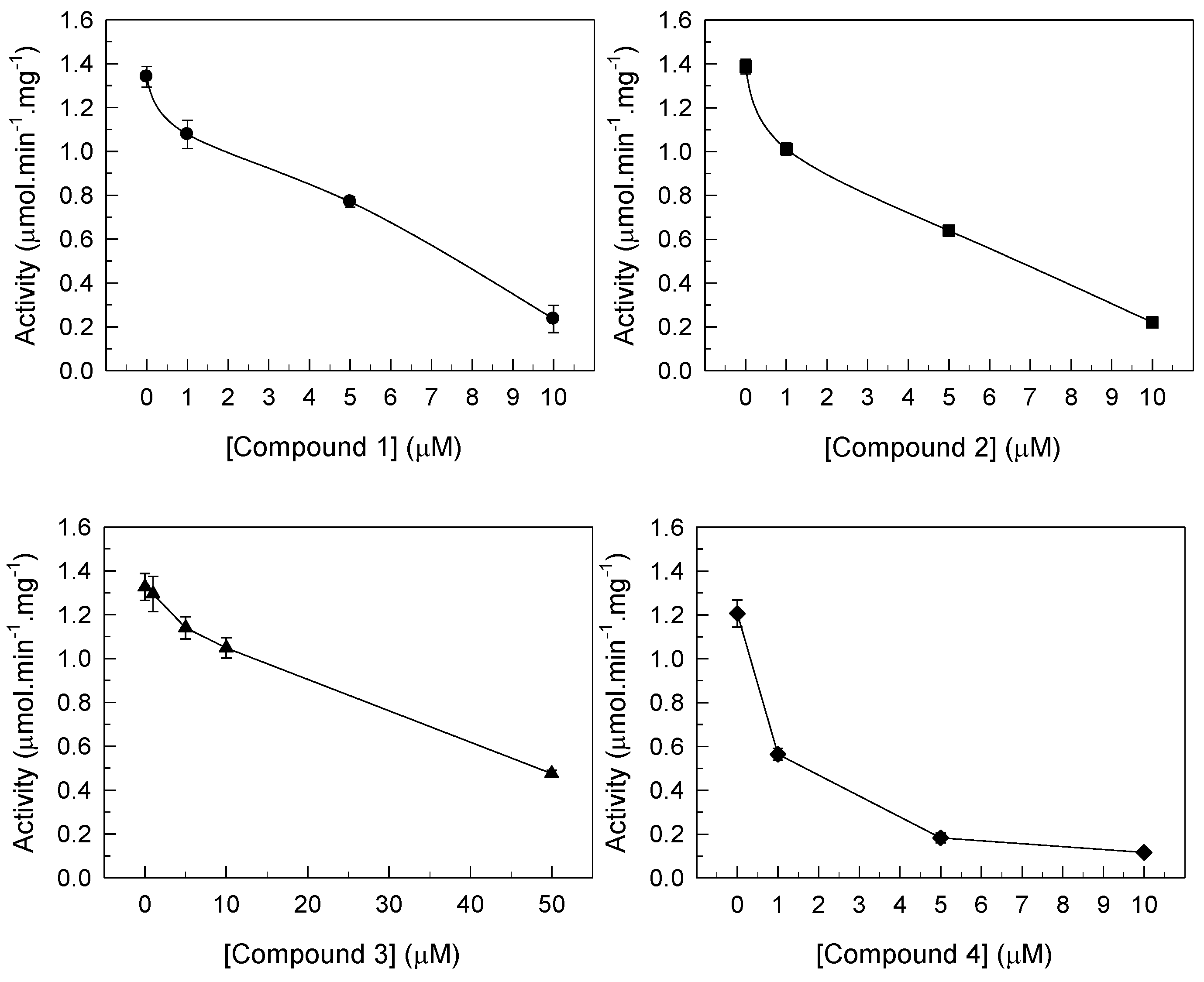
3.1. Inhibition of PMCA Activity by Gold Compounds

The effects of four gold compounds on the activity of the plasma membrane Ca2+-ATPase (PMCA) were investigated for the first time. Compounds were added to the assay after triggering the reaction with ATP, as indicated in the Section 2. As shown in Figure 2, all Au complexes inhibited PMCA activity, depending on the concentration. The inhibitory capacity of the investigated gold compounds was evaluated by the half maximal inhibitory concentration (IC50) values, meaning that the inhibitor concentration induced 50% of Ca2+-ATPase inhibition of the enzyme activity. The IC50 values were calculated using Microsoft Office 365 Excel (Microsoft, Red-mond, WA, USA), and ranged from 0.9 ± 0.1 µM to 21 ± 0.1 µM. Gold(I) compounds 2 and 4 showed the lowest IC50 values: 2.8 ± 0.2 and 0.9 ± 0.1 µM, respectively, indicating a higher inhibition. The gold(III) compound 1 and gold(I) compound 3 exhibited IC50 values in the range of 4.9 ± 0.1 to 21.0 ± 0.1 µM. Thus, the Au(I) complexes 2 and 4 were, respectively, around 6-to 20-fold more powerful inhibitors of the ATPase activity than compounds 1 and 3 (Table 2).
Note that, at these studies, no PMCA incubation with the gold compounds was performed. The compounds were added to the medium seconds after the reaction was initiated. Following the addition of the gold compounds, the rates of the reactions in the presence of different inhibitor concentrations were measured within the following 2–3 min. The gold compounds are stable for at least up to 30 min, as previously observed, in the same medium [18]. In fact, compounds were incubated for 30 min with and without the protein, and the inhibitory effects of the ATPase were the same as without any incubation, suggesting that the Au compounds are stable and the effects observed can be attributed to their addition to the medium [18].
The type of inhibition produced by the gold compounds was analyzed at concentrations around the IC50 by starting the reaction with increasing amounts of the substrate ATP (Figure 3). It was observed that both Au(III) compound 1 and Au(I) compound 4 reduced the Vmax by 60% and 64%, respectively, without affecting the Km, therefore showing the effect of non-competitive inhibition (Figure 3, Table 2).
On the other hand, both Au(I) complexes 2 and 3 decreased the Vmax by 56% and 53%, respectively, and increased the Km by 41% and 52%, respectively, which is indicative of a mixed type inhibition (Figure 3, Table 2). Thus, it can be suggested that both Au(I) compounds 2 and 3 interact with two binding sites of PMCA, one being the ATP binding site.

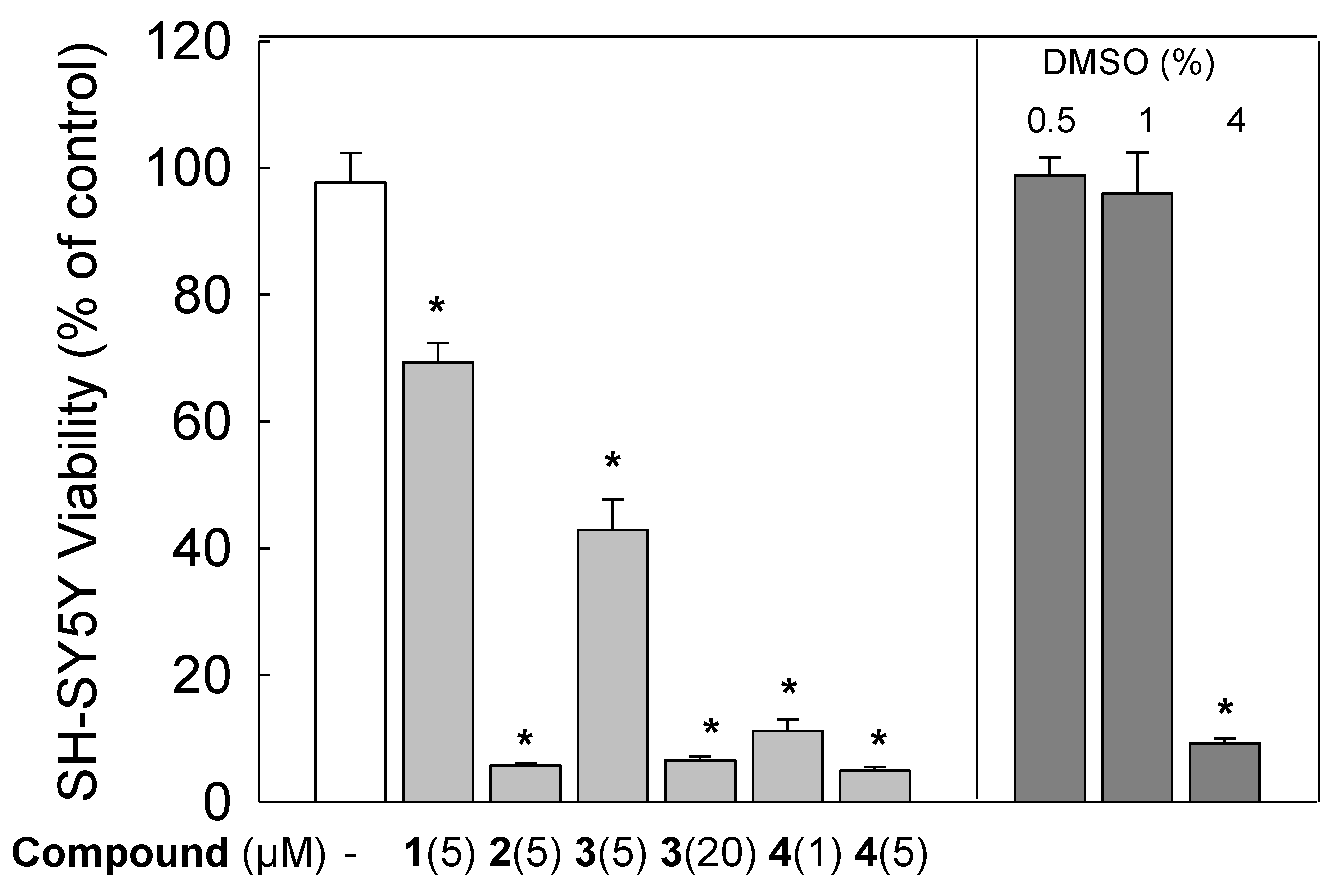
3.2. Cytotoxic Effects of Gold Compounds in SH-SY5Y Cells

The effects of gold compounds were also evaluated in the human neuroblastoma SH-SY5Y cell line in order to see if they could affect cell viability (Figure 4). The relative cell viability was calculated as the percentage of untreated cells (100% viability). Thus, cells treated with 5 µM of compounds 1 and 3 gave the highest cell viability, with values of 69.30 ± 3.06% and 42.85 ± 4.89%, respectively. Nonetheless, cell survival was significantly reduced to 6.50 ± 0.66% when treated with 20 µM of compound 3. When cells were treated with compounds 2 and 4, at a concentration of 5 µM, viability values of 5.73 ± 0.33% and 4.91 ± 0.61%, respectively, were obtained. Regarding this decrease, our results also showed that viability of cells treated with 4% DMSO alone (which is the percentage of this solvent in compound 3 at 20 µM) was significantly decreased down to 9.24 ± 0.72%, suggesting that this reduction could be mostly associated to the toxicity of DMSO. On the contrary, cells incubated with 0.5% and 1% DMSO alone (which were the percentages of solvent in 1 µM and 5 µM of gold compounds, respectively) did not show any significant toxicity, the cell viability being 98.75 ± 2.86% and 95.97 ± 6.51%, respectively.

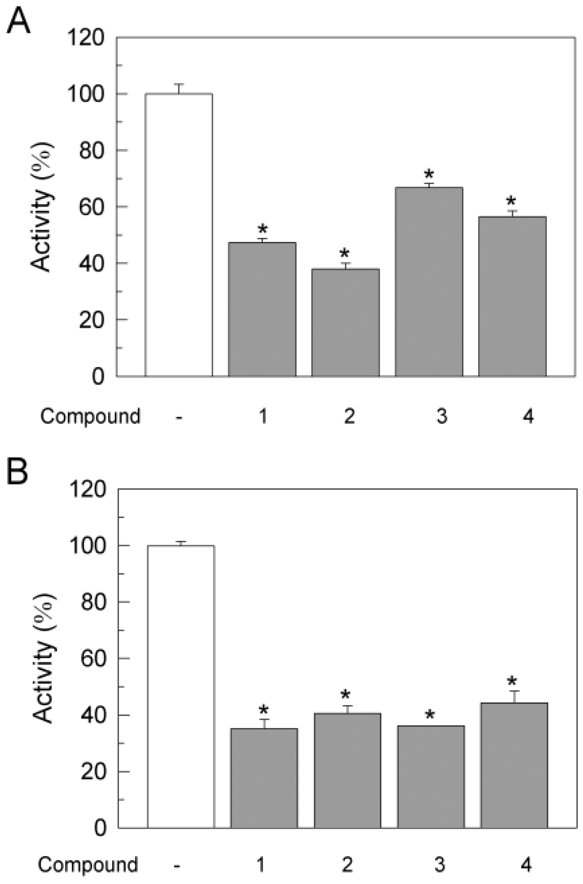
3.3. Effects of Gold Compounds on Ca2+-ATPase Activity in SH-SY5Y Cells

The effects of gold compounds on endogenous Ca2+-ATPase activity were also analyzed in membranes prepared from SH-SY5Y neuroblastoma cells. Figure 5A showed that the Ca2+-ATPase activity of membranes from cells incubated in vitro for 12 h with compounds 1, 2, and 3, at a concentration of 5 μM, and with compound 4 at 1 µM, decreased by 47.31 ± 1.43%, 38.048 ± 1.95%, 66.82 ± 1.46%, and 56.58 ± 1.95%, with respect to untreated cells. Considering that the degree of ATPase activity inhibition was similar to the loss of cell viability, we could not rule out that this inhibition was related to the loss of PMCA protein due to cell death. To experimentally elucidate this question, we also tested the effects of the compounds in membranes prepared from non-treated cells just by adding the compounds after triggering the reaction with ATP. As shown in Figure 5B, the Ca2+-ATPase activity was also inhibited by all compounds in similar percentages to those obtained with treated cells, giving values of 35.02 ± 3.30%, 40.52 ± 2.64%, 36.12 ± 0.12%, and 44.27 ± 4.18% for compounds 1, 2, 3, and 4, respectively. Therefore, we could conclude that the inhibition of the Ca2+-ATPase by gold compounds was due to modulation of PMCA by those compounds and not just a consequence of cell death.

4. Discussion

The Ca2+-ATPase activity was studied in the presence of four gold compounds, either gold(I) or gold(III), with different ligands, namely, dichloro (2-pyridinecarboxylate) gold(III) (abbreviated as 1), chlorotrimethylphosphine gold(I) (2), 1,3-bis(2,6-diisopropylphenyl) imidazole-2-ylidene gold(I) chloride (3), and chlorotriphenylphosphine gold(I) (4) (Figure 1, Table 1). All tested compounds showed a potent inhibitory effect on PMCA activity. This pump showed the highest affinity for the inhibitor compound 4, followed by compounds 2, 1, and 3. This inhibitory sequence was like that previously observed in the case of SERCA protein [18]. The IC50 values obtained in our work with PMCA were similar to those reported for SERCA, except for compound 2, which presented a lower inhibitory potency regarding PMCA (IC50 2.8 µM), in comparison with the value (IC50 0.8 µM) obtained for SERCA [18]. Therefore, these gold(I)-compounds and gold(III)-compounds showed high affinity for PMCA and SERCA. Although PMCA and SERCA are P-type ATPases, they have different features and were previously described to have different sensitivities for specific P-type ATPases inhibitors, such as polyoxometalates. Herein, only compound 2 present a higher inhibition capacity for SERCA, in comparison with PMCA, which was about four times more effective, whereas, for the other compounds, the obtained IC50 values were very similar. This means that not all gold compounds can be used as specific inhibitors for a particular ion pump.
The SERCA vesicle preparations contain mainly the SERCA1 isoform, as analyzed by SDS-PAGE, although minor amounts of sarcolipin could be found in similar preparations from other sources. However, to the best of our knowledge, there are no studies stating that gold compounds bind to this protein known to affect the SERCA function. Therefore, the results described are only due to the effects on the SERCA ATPase activity and not to the interference with sarcolipin that, at these experimental conditions, does not affect the hydrolytic activity. The model used was previously applied to several inorganic compounds, including vanadate. In fact, several studies were described about the interaction of organic and inorganic compounds and their effects on P-type ATPases [23,36,37,38,39,40,41,42,43,44,45,46,47,48,49,50,51]. Therefore, several experiments with several vanadium compounds [45,47,48] at the same experimental conditions for comparison were performed, and the potencies of inhibition are compared in Table 2. In addition, the amyloid-β peptide and tau were used as negative controls, considering that these two molecular markers of the disease have been reported, by some of the authors of this work, as inhibitors of the PMCA pump [26,27,28]. The studies were performed from triplicate measurements, using three distinct and independent Ca2+-ATPase preparations, as described in the Materials and Methods Section 2.3.
Note that P-type ATPases are well known to be inhibited by metal complexes and compounds [17,22,23,24,25], although the structural details of their inhibitory mechanisms remain unresolved. The non-competitive type of inhibition observed for gold(III) compound 1, regarding the native substrate MgATP, is in good agreement with previous studies dealing with SERCA [18] and Na+/K+-ATPase [16]. It was previously described that gold(III) complexes, H[AuCl4], [Au(DMSO)2Cl2]Cl and [Au(bipy)Cl2]Cl (bipy = 2,2′-bipyridine), inhibited the enzymatic activity of purified pig brain Na+/K+-ATPase, with IC50 values of 0.57, 5.5, and 39.8 μM, respectively, in a non-competitive mode of interaction [20]. Besides the non-competitive inhibition observed for gold compounds 1 and 4, a mixed type of inhibition was also observed for compounds 2 and 3. The type of inhibition found for gold compounds 1 (non-competitive) and 3 (mixed) for PMCA was similar to the previously reported results for SERCA [18]. In contrast, gold(I) compounds 2 and 4 inhibited SERCA through non-competitive and mixed inhibition, respectively [18].
These differences led us to suggest that compounds 1 and 3 may share binding sites with similar amino acid residues and charge density on PMCA and SERCA, whereas compounds 2 and 4 could bind to two different sites on SERCA and PMCA. Nevertheless, for the majority of the gold compounds, the mechanisms of inhibition and the protein binding sites for P-ATPases remain to be determined [17,25].
It was suggested that gold(III) compounds can bind to the E1 conformation of the Na+/K+-ATPase [17]. The different binding modes of these gold(III) complexes to the enzyme were explained based on their distinctive structural features. In contrast to gold(III), monomeric vanadate only binds to the E2 conformation of the SERCA, whereas decavanadate (V10) strongly binds to either E1 or E2 conformations, being phosphorylated or not [44]. However, for the majority of the inorganic compounds described to inhibit P-type ATPases, the protein conformations and binding sites are still to be determined [17,18,23,25]. In the near future, protein structural models would help explain such enzymatic results [25,36]. For some drugs, such as thapsigargin (TG) and cyclopiazonic acid (CPA), the mechanisms of action and ATPases binding sites are clearly established [24].
Polyamines, such as spermine, are potential targets for the development of anticancer drugs and have been shown to inhibit the PMCA pump [37]. Moreover, the antipsychotic drug thioridazine and other phenothiazine derivatives also inhibit the PMCA activity [38]. In the case of SERCA, thapsigargin (IC50 = 0.001–0.029 µM), cyclopiazonic acid (IC50 = 0.1–0.2 µM), macrocyclic lactones (IC50 = 66–72 μM), curcuminoids (IC50 = 7–17 μM), and celecoxib (IC50 = 35 μM) are examples of well-known inhibitors [39,40,41,42,43]. These drugs are used in several disease treatments and are also employed as anaesthetics, tumour inhibitors, antibiotics, and insulin mimetic agents [39,40,41,42,43], providing an inhibitory effect not so different from the gold compounds described in the present study (Table 3).
Herein, we aimed to compare the inhibitory capacity of the gold complexes 14 with oxometalates, metal complexes, and polyoxometalates (POTs and POVs), as well as known organic drugs. In fact, several inorganic compounds and complexes inhibit Ca2+-ATPases activity with IC50 inhibition values similar to the well-known Ca2+-ATPases drugs inhibitors, for example decavanadate (IC50 = 15 µM), polyoxotungstates (IC50 = 0.3–200 μM), polyoxovanadates (IC50 = 1 μM), and vanadium complexes, such as BMOV (IC50 = 40 µM), among others [42,43,44,45,46,47,48]. In summary, both gold(I) and gold(III) compounds 14 are potent inhibitors of the PMCA, pointing out this enzyme as a putative target for gold compounds that are promising agents in medicinal chemistry [49,50,51].
This work also showed that both Au (I and III) compounds produced a decline in the viability of SH-SY5Y human neuroblastoma cells, which was dependent on their structures. In addition, the Ca2+-ATPase activity was also inhibited by the presence of these compounds. However, the activity results obtained from membranes of treated cells were comparable to those found with untreated cell membranes, after subsequent addition of ATP and gold compounds to the assay medium. This similarity suggests that the ATPase inhibition is also due to an effect of gold compounds on the protein and not only on the cell viability.
Recently, it was stated that metal contaminants, such as cadmium, induce changes in biochemical parameters in brain, causing neurological dysfunction in rats [52]. However, even essential elements, such as cobalt, proved to be accumulated in the brain, inducing cytotoxicity in SH-SY5Y cells at high concentrations (IC50 = 100.01 ± 5.91 μM, after 72 h exposure) [31]. Cisplatin compounds have also been reported to induce cytotoxicity on SH-SY5Y cells [53]. IC50 values for these platinum compounds of 15–50 μM were observed using the MTT assay, for an incubation time of 24 h. More recently, high citotoxicity was reported for this cell line by two gold(I) complexes containing triphenylphosphine (Au(pben)(PPh3)) (1) or triethylphosphine (Au(pben)(PEt3) (2), showing IC50 values as low as of 2.7 and 1.6 μM, respectively, after 24 h incubation [54], thus having a similar order of magnitude to the values described in the present study for other gold (I and III) compounds.
Altogether, these results suggest that PMCA could be a potential target for the tested gold compounds. In fact, several pathologies, such as those of a neurological nature, are linked with SERCA and PMCA disfunctions. Thus, it is important to find specific modulators of such P-type ATPases that can prevent changes in calcium homeostasis in some diseases. As discussed above, the putative inhibition of these enzymes by the compounds can be used in current neurological therapies [12,13]. However, they should be carefully assessed to finely set their dosage in order to prevent collateral and unwanted toxic effects of healthy neuron cells such as neurons and astrocytes [14].
It is interesting to note that the scientific community in the 21th century has a vast knowledge about genes and proteins but the role of essential elements in life remains somewhat limited, not to mention other elements that are apparently not essential. However, for several metals, and also for gold, the lack of knowledge provides opportunities for discoveries of applications and/or implications in biochemistry and health.

5. Conclusions

The present study shows that PMCA activity is inhibited by both Au(I) or Au(III) compounds. Particularly significant inhibition values were found for the Au(I) compound 4 (IC50 < 1 μM), like those previously described for SERCA. A non-competitive type of inhibition was found for compound 4, which was the most powerful ATPase inhibitor, and for compound 1, whereas a mixed type of inhibition was observed for compounds 2 and 3, revealing distinct modes of interaction with PMCA. Both Au (I and III) compounds also inhibited the endogenous Ca2+-ATPase activity of SH-SY5Y human neuroblastoma cell lines and affected cell viability, this cytotoxicity being higher for gold compounds 2 and 4.
In summary, we have described the interaction of gold(I,III) compounds with key cellular proteins such as SERCA and PMCA, from a kinetic point of view. Considering the importance of gold compounds in inorganic medicinal chemistry, they are very likely to receive much more attention in the near future as putative inhibitors for several other key enzymes with biomedical applications. Thus, the application of gold compounds in biology and biomedical sciences is still a branch in continuous growth.

Author Contributions

Conceptualization: A.M.M. and M.A.; formal analysis: M.B., J.J.C.-G., C.G.-M., M.A. and A.M.M.; funding acquisition: A.M.M., C.G.-M. and M.A.; investigation: M.B., J.J.C.-G., M.A. and A.M.M.; methodology: M.B., J.J.C.-G., M.A. and A.M.M.; resources: S.A.C.C., M.A. and A.M.M., supervision: A.M.M. and M.A.; writing—original draft: A.M.M., J.J.C.-G., M.B. and M.A.; writing—review and editing: A.M.M., M.A., J.J.C.-G., M.B., C.G.-M. and S.A.C.C. All authors have read and agreed to the published version of the manuscript.

Funding

Projects BFU2017-85723-P (to A.M.M. and C.G.-M.), and PID2020-115512GB-I00 (to A.M.M.) funded by MCIN/AEI/10.13039/501100011033 and by “ESF Investing in your future”. We acknowledge Fundação para a Ciência e a Tecnologia (FCT) project UIDB/04326/2020, Associate Laboratory for Green Chemistry–LAQV, financed by national funds from FCT/MCTES (UIDB/50006/2020 and UIDP/50006/2020) and Scientific Employment Stimulus-Institutional Call (CEECINST/00102/2018).

Institutional Review Board Statement

Not applicable.

Informed Consent Statement

Not applicable.

Data Availability Statement

Not applicable.

Conflicts of Interest

The authors declare no conflict of interest. The funders had no role in the design of the study; in the collection, analyses, or interpretation of data; in the writing of the manuscript; or in the decision to publish the results.

Abbreviations

ATPaseAdenosine triphosphatase
Auoxo6[(6,6′-dimethyl-2,2′-bipyridine)2Au2(μ-O)2][PF6]2
BMOVBismaltol oxidovanadium(IV)
CPACyclopyazonic
DMSODimethyl sulfoxide
IC50Half maximal inhibitory concentration
PDC-V(V)Pyridine-2,6-dicarboxylatodioxovanadium(V)
PMCAPlasmatic membrane calcium ATPase
POTs Polyoxotungstates
PV14O40Phosphotetradecavanadate
SERCASarco(endo) plasmatic membrane calcium ATPase
SPCA secretory pathway calcium ATPase
SRSarcoplasmic reticulum
TGThapsigargin
VO4Orthovanadate
V10O28Decavanadate

References

  1. Carafoli, E. The Regulation of Intracellular Calcium; Springer: Boston, MA, USA, 1982; pp. 461–472. [Google Scholar]
  2. Domínguez, D.C.; Guragain, M.; Patrauchan, M. Calcium binding proteins and calcium signaling in prokaryotes. Cell Calcium 2015, 57, 151–165. [Google Scholar] [CrossRef] [PubMed]
  3. Plattner, H.; Verkhratsky, A. The ancient roots of calcium signalling evolutionary tree. Cell Calcium 2015, 57, 123–132. [Google Scholar] [CrossRef] [PubMed] [Green Version]
  4. Wang, S.Q.; Lakatta, E.G.; Cheng, H.; Zhou, Z.Q. Adaptive mechanisms of intracellular calcium homeostasis in mammalian hibernators. J. Exp. Biol. 2002, 10 Pt 19, 2957–2962. [Google Scholar] [CrossRef]
  5. Rappas, M.; Niwa, H.; Zhang, X. Mechanisms of ATPases-A Multi-Disciplinary Approach. Curr. Protein Pept. Sci. 2004, 5, 89–105. [Google Scholar] [CrossRef]
  6. Strehler, E.E.; Zacharias, D.A. Role of alternative splicing in generating isoform diversity among plasma membrane calcium pumps. Physiol. Rev. 2001, 81, 21–50. [Google Scholar] [CrossRef] [Green Version]
  7. Strehler, E.E.; Caride, A.J.; Filoteo, A.G.; Xiong, Y.; Penniston, J.T.; Enyedi, A. Plasma membrane Ca2+-ATPases as dynamic regulators of cellular calcium handling. Ann. N. Y. Acad. Sci. 2007, 1099, 226–236. [Google Scholar] [CrossRef] [Green Version]
  8. Wuytack, F.; Raeymaekers, L.; Missiaen, L. Molecular physiology of the SERCA and SPCA pumps. Cell Calcium 2002, 32, 279–305. [Google Scholar] [CrossRef] [PubMed]
  9. Sadler, P.J.; Sue, R.E. The Chemistry of Gold Drugs. Met. Based Drugs 1994, 1, 107–144. [Google Scholar] [CrossRef] [Green Version]
  10. Mora, M.; Gimeno, M.C.; Visbal, R. Recent advances in gold–NHC complexes with biological properties. Chem. Soc. Rev. 2019, 48, 447–462. [Google Scholar] [CrossRef]
  11. Azharuddin, M.; Zhu, G.H.; Das, D.; Ozgur, E.; Uzun, L.; Turner, A.P.F.; Patra, H.K. A repertoire of biomedical applications of noble metal nanoparticles. Chem. Commun. 2019, 55, 6964–6996. [Google Scholar] [CrossRef]
  12. Pettenuzzo, N.; Brustolin, L.; Coltri, E.; Gambalunga, A.; Chiara, F.; Trevisan, A.; Biondi, B.; Nardon, C.; Fregona, D. Cu II and Au III Complexes with Glycoconjugated Dithiocarbamato Ligands for Potential Applications in Targeted Chemotherapy. ChemMedChem 2019, 14, 1162–1172. [Google Scholar] [CrossRef]
  13. Liu, W.; Gust, R. Metal N-heterocyclic carbene complexes as potential antitumor metallodrugs. Chem. Soc. Rev. 2013, 42, 755–773. [Google Scholar] [CrossRef]
  14. Stratton, M.; Ramachandran, A.; Camacho, E.J.M.; Patil, S.; Waris, G.; Grice, K.A. Anti-fibrotic activity of gold and platinum complexes-Au(I) compounds as a new class of anti-fibrotic agents. J. Inorg. Biochem. 2020, 206, 111023. [Google Scholar] [CrossRef]
  15. Azria, D.; Blanquer, S.; Verdier, J.-M.; Belamie, E. Nanoparticles as contrast agents for brain nuclear magnetic resonance imaging in Alzheimer’s disease diagnosis. J. Mater. Chem. B 2017, 5, 7216–7237. [Google Scholar] [CrossRef] [PubMed]
  16. Petrović, V.; Čolović, M.; Krstić, D.; Vujačić, A.; Petrović, S.; Joksić, G.; Bugarčić, Ž.; Vasić, V. In vitro effects of some gold complexes on Na+/K+ ATPase activity and cell proliferation. J. Inorg. Biochem. 2013, 124, 35–41. [Google Scholar] [CrossRef]
  17. Bondžić, A.M.; Janjić, G.V.; Dramićanin, M.D.; Messori, L.; Massai, L.; Parac Vogt, T.N.; Vasić, V.M. Na/K-ATPase as a target for anticancer metal based drugs: Insights into molecular interactions with selected gold(iii) complexes. Metallomics 2017, 9, 292–300. [Google Scholar] [CrossRef]
  18. Fonseca, C.; Fraqueza, G.; Carabineiro, S.A.C.; Aureliano, M. The Ca2+-ATPase inhibition potential of gold(I,III) compounds. Inorganics 2020, 8, 49. [Google Scholar] [CrossRef]
  19. Aikman, B.; Wenzel, M.; Mósca, A.; de Almeida, A.; Klooster, W.; Coles, S.; Soveral, G.; Casini, A. Gold(III) Pyridine-Benzimidazole Complexes as Aquaglyceroporin Inhibitors and Antiproliferative Agents. Inorganics 2018, 6, 123. [Google Scholar] [CrossRef] [Green Version]
  20. Zou, T.; Lum, C.T.; Lok, C.-N.; Zhang, J.-J.; Che, C.-M. Chemical biology of anticancer gold(iii) and gold(i) complexes. Chem. Soc. Rev. 2015, 44, 8786–8801. [Google Scholar] [CrossRef] [PubMed]
  21. Pedersen, P.L.; Carafoli, E. Ion motive ATPases. II. Energy coupling and work output. Trends Biochem. Sci. 1987, 12, 186–189. [Google Scholar] [CrossRef]
  22. Toyoshima, C.; Nakasako, M.; Nomura, H.; Ogawa, H. Crystal structure of the calcium pump of sarcoplasmic reticulum at 2.6 Å resolution. Nature 2000, 405, 647–655. [Google Scholar] [CrossRef]
  23. Aureliano, M.; Fraqueza, G.; Ohlin, C.A. Ion pumps as biological targets for decavanadate. Dalton Trans. 2013, 42, 11770. [Google Scholar] [CrossRef] [PubMed]
  24. Yatime, L.; Buch-Pedersen, M.J.; Musgaard, M.; Morth, J.P.; Winther, A.-M.L.; Pedersen, B.P.; Olesen, C.; Andersen, J.P.; Vilsen, B.; Schiøtt, B.; et al. P-type ATPases as drug targets: Tools for medicine and science. Biochim. Biophys. Acta-Bioenerg. 2009, 1787, 207–220. [Google Scholar] [CrossRef] [PubMed] [Green Version]
  25. Nikezić, A.V.V.; Janjić, G.V.; Bondžić, A.M.; Zarić, B.L.; Vasić-Anićijević, D.D.; Momić, T.G.; Vasić, V.M. Interaction of Au(III) and Pt(II) complexes with Na/K-ATPase: Experimental and theoretical study of reaction stoichiometry and binding sites. Metallomics 2018, 10, 1003–1015. [Google Scholar] [CrossRef] [PubMed]
  26. Berrocal, M.; Marcos, D.; Sepúlveda, M.R.; Pérez, M.; Ávila, J.; Mata, A.M. Altered Ca2+ dependence of synaptosomal plasma membrane Ca2+-ATPase in human brain affected by Alzheimer’s disease. FASEB J. 2009, 23, 1826–1834. [Google Scholar] [CrossRef] [PubMed]
  27. Berrocal, M.; Corbacho, I.; Gutierrez-Merino, C.; Mata, A.M. Methylene blue activates the PMCA activity and cross-interacts with amyloid beta-peptide, blocking Abeta-mediated PMCA inhibition. Neuropharmacology 2018, 139, 163–172. [Google Scholar] [CrossRef] [PubMed]
  28. Berrocal, M.; Caballero-Bermejo, M.; Gutierrez-Merino, C.; Mata, A.M. Methylene Blue Blocks and Reverses the Inhibitory Effect of Tau on PMCA Function. Int. J. Mol. Sci. 2019, 20, 3521, PMID: 31323781; PMCID: PMC6678728. [Google Scholar] [CrossRef] [Green Version]
  29. Vosahlikova, M.; Roubalova, L.; Cechova, K.; Kaufman, J.; Musil, S.; Miksik, I.; Alda, M.; Svoboda, P. Na+/K+-ATPase and lipid peroxidation in forebrain cortex and hippocampus of sleep-deprived rats treated with therapeutic lithium concentration for different periods of time. Prog. Neuropsychopharmacol. Biol. Psychiatry 2020, 102, 109953. [Google Scholar] [CrossRef]
  30. Hosseinzadeh, Z.; Hauser, S.; Singh, Y.; Pelzl, L.; Schuster, S.; Sharma, Y.; Höflinger, P.; Zacharopoulou, N.; Stournaras, C.; Rathbun, D.L.; et al. Decreased Na+/K+ ATPase Expression and Depolarized Cell Membrane in Neurons Differentiated from Chorea-Acanthocytosis Patients. Sci. Rep. 2020, 10, 8391. [Google Scholar] [CrossRef]
  31. Gómez-Arnaiz, S.; Tate, R.J.; Grant, M.H. Cytotoxicity of cobalt chloride in brain cell lines-A comparison between astrocytoma and neuroblastoma cells. Toxicol. Vitr. 2020, 68, 104958. [Google Scholar] [CrossRef]
  32. Salvador, J.M.; Mata, A.M. Purification of the synaptosomal plasma membrane (Ca2+ + Mg2+)-ATPase from pig brain. Biochem. J. 1996, 315, 183–187. [Google Scholar] [CrossRef] [Green Version]
  33. Bradford, M.M. A rapid and sensitive method for the quantitation of microgram quantities of protein utilizing the principle of protein-dye binding. Anal. Biochem. 1976, 72, 248–254. [Google Scholar] [CrossRef]
  34. Mosmann, T. Rapid colorimetric assay for cellular growth and survival: Application to proliferation and cytotoxicity assays. J. Immunol. Methods 1983, 65, 55–63. [Google Scholar] [CrossRef]
  35. Sepúlveda, M.R.; Hidalgo-Sánchez, M.; Mata, A.M. A developmental profile of the levels of calcium pumps in chick cerebellum. J. Neurochem. 2005, 95, 673–683. [Google Scholar] [CrossRef]
  36. Sciortino, G.; Aureliano, M.; Garribba, E. Rationalizing the decavanadate(V) and oxidovanadium(IV) binding to G-actin and the competition with decaniobate(V) and ATP. Inorg. Chem. 2021, 60, 334–344. [Google Scholar] [CrossRef] [PubMed]
  37. Palacios, J.; Sepúlveda, M.R.; Mata, A.M. Effect of spermine on the activity of synaptosomal plasma membrane Ca2+-ATPase reconstituted in neutral or acidic phospholipids. Biochim. Biophys. Acta (BBA)-Biomembr. 2003, 1611, 197–203. [Google Scholar] [CrossRef] [Green Version]
  38. Palacios, J.; Sepúlveda, M.R.; Lee, A.G.; Mata, A.M. Ca2+ transport by the synaptosomal plasma membrane Ca2+-ATPase and the effect of thioridazine. Biochemistry 2004, 43, 2353–2358. [Google Scholar] [CrossRef]
  39. Moreno, I.; Norambuena, L.; Maturana, D.; Toro, M.; Vergara, C.; Orellana, A.; Zurita-Silva, A.; Ordenes, V.R. AtHMA1 Is a Thapsigargin-sensitive Ca2+/Heavy Metal Pump. J. Biol. Chem. 2008, 283, 9633–9641. [Google Scholar] [CrossRef] [Green Version]
  40. Yard, N.J.; Chiesi, M.; Ball, H.A. Effect of cyclopiazonic acid, an inhibitor of sarcoplasmic reticulum Ca2+-ATPase, on the frequency-dependence of the contraction-relaxation cycle of the guinea-pig isolated atrium. Br. J. Pharmacol. 1994, 113, 1001–1007. [Google Scholar] [CrossRef] [Green Version]
  41. Bilmen, J.G.; Wotton, L.L.; Michelangeli, F. The inhibition of the sarcoplasmic/endoplasmic reticulum Ca2+-ATPase by macrocyclic lactones and cyclosporin A. Biochem. J. 2002, 366, 255–263. [Google Scholar] [CrossRef] [PubMed] [Green Version]
  42. Bilmen, J.G.; Khan, S.Z.; Javed, M.-H.; Michelangeli, F. Inhibition of the SERCA Ca2+ pumps by curcumin. Eur. J. Biochem. 2001, 268, 6318–6327. [Google Scholar] [CrossRef]
  43. Coca, R.; Soler, F.; Cortés-Castell, E.; Gil-Guillén, V.; Fernández-Belda, F. Inhibition mechanism of the intracellular transporter Ca2+-pump from sarco-endoplasmic reticulum by the antitumor agent dimethyl-celecoxib. PLoS ONE 2014, 9, e102083. [Google Scholar] [CrossRef] [Green Version]
  44. Gumerova, N.; Krivosudský, L.; Fraqueza, G.; Breibeck, J.; Al-Sayed, E.; Tanuhadi, E.; Bijelic, A.; Fuentes, J.; Aureliano, M.; Rompel, A. The P-type ATPase inhibiting potential of polyoxotungstates. Metallomics 2018, 10, 287–295. [Google Scholar] [CrossRef] [PubMed] [Green Version]
  45. Fraqueza, G.; Fuentes, J.; Krivosudský, L.; Dutta, S.; Mal, S.S.; Roller, A.; Giester, G.; Rompel, A.; Aureliano, M. Inhibition of Na+/K+- and Ca2+-ATPase activities by phosphotetradecavanadate. J. Inorg. Biochem. 2019, 197, 110700. [Google Scholar] [CrossRef] [PubMed]
  46. Clausen, J.D.; Bublitz, M.; Arnou, B.; Olesen, C.; Andersen, J.P.; Møller, J.V.; Nissen, P. Crystal Structure of the Vanadate-Inhibited Ca2+-ATPase. Structure 2016, 24, 617–623. [Google Scholar] [CrossRef] [PubMed] [Green Version]
  47. Fraqueza, G.; Ohlin, C.A.; Casey, W.H.; Aureliano, M. Sarcoplasmic reticulum calcium ATPase interactions with decaniobate, decavanadate, vanadate, tungstate and molybdate. J. Inorg. Biochem. 2012, 107, 82–89. [Google Scholar] [CrossRef]
  48. Aureliano, M.; Henao, F.; Tiago, T.; Duarte, R.O.; Moura, J.J.G.; Baruah, B.; Crans, D.C. Sarcoplasmic Reticulum Calcium ATPase Is Inhibited by Organic Vanadium Coordination Compounds: Pyridine-2,6-dicarboxylatodioxovanadium(V), BMOV, and an Amavadine Analogue. Inorg. Chem. 2008, 47, 5677–5684. [Google Scholar] [CrossRef] [Green Version]
  49. Machado, J.F.; Correia, J.D.G.; Morais, T.S. Emerging Molecular Receptors for the Specific-Target Delivery of Ruthenium and Gold Complexes into Cancer Cells. Molecules 2021, 26, 3153. [Google Scholar] [CrossRef]
  50. Gamberi, T.; Pratesi, A.; Messori, L.; Massai, L. Proteomics as a tool to disclose the cellular and molecular mechanisms of selected anticancer gold compounds. Coord. Chem. Rev. 2021, 438, 213905. [Google Scholar] [CrossRef]
  51. Rousselle, B.; Bouyer, F.; Bayardon, J.; Laly, M.; Ghiringhelli, F.; Rousselin, Y.; Bodio, E.; Malacea-Kabbara, R. Development of a novel highly anti-proliferative family of gold complexes: Au(i)-phosphonium-phosphines. Dalton Trans. 2021, 50, 4880. [Google Scholar] [CrossRef]
  52. Vijaya, P.; Kaur, H.; Garg, N.; Sharma, S. Protective and therapeutic effects of garlic and tomato on cadmium-induced neuropathology in mice. J. Basic Appl. Zool. 2020, 81, 23. [Google Scholar] [CrossRef]
  53. Donzelli, E.; Carfi, M.; Miloso, M.; Strada, A.; Galbiati, S.; Bayssas, M.; Griffon-Etienne, G.; Cavaletti, G.; Petruccioli, M.G.; Tredici, G. Neurotoxicity of platinum compounds: Comparison of the effects of cisplatin and oxaliplatin on the human neuroblastoma cell line SH-SY5Y. J. Neurooncol. 2004, 67, 65–73. [Google Scholar] [CrossRef] [PubMed]
  54. Rouco, L.; Sánchez-González, Á.; Alvariño, R.; Alfonso, A.; Vázquez-López, E.M.; García-Martínez, E.; Maneiro, M. Combined Effect of Caspase-Dependent and Caspase-Independent Apoptosis in the Anticancer Activity of Gold Complexes with Phosphine and Benzimidazole Derivatives. Pharmaceuticals 2020, 14, 10. [Google Scholar] [CrossRef] [PubMed]
Figure 1. Structures of the gold complexes used in the study: (1) dichloro (2-pyridinecarboxylate) gold(III); (2) chlorotrimethylphosphine gold(I); (3) 1,3-bis(2,6-diisopropylphenyl) imidazole-2-ylidene gold(I) chloride; and (4) chlorotriphenylphosphine gold(I). Formal charge distribution is also shown.
Metals 11 01934 g001
Figure 2. Inhibition of PMCA activity by gold compounds 1, 2, 3, and 4. The Ca2+-ATPase was measured spectrophotometrically at 340 nm and 37 °C, using the coupled enzyme pyruvate kinase/lactate dehydrogenase assay. The experiments were initiated after the addition of 2.5 μg/mL of purifed PMCA reconstituted in PCIIS. Data are plotted as means ± SD. The results shown are the average of triplicate experiments carried out with different preparations.
Metals 11 01934 g002
Figure 3. Lineweaver–Burk plots of Ca2+-ATPase activity in the absence (empty symbols) and in the presence (filled symbols) of gold compounds 1, 2, 3, and 4, at concentrations of 5, 3, 21, and 1 µM, respectively. The plots were used to determine the type of PMCA inhibition. The gold complexes 1 and 4 presented a non-competitive type of inhibition, while compounds 2 and 3 showed a mixed type of inhibition. Data are plotted as means ± standard deviation. The results shown are the average of triplicate experiments carried out in distinct preparations.
Metals 11 01934 g003
Figure 4. Effects of gold compounds on cell viability estimated by the MTT assay. SH-SY5Y cells were treated with gold compounds 1, 2, 3, and 4, at the indicated concentrations (in brackets). The right panel shows viability of cells incubated with 0.5%, 1%, and 4% DMSO alone, which corresponds to final percentages of the solvent in cell treated with 1, 5, and 20 µM gold compounds, respectively. Data are expressed as percentage of untreated cells in the absence of DMSO, as mean ± SEM of at least three independent experiments carried out in triplicate with three different cultures. Symbols (*) represent significant differences when compared to control (ANOVA, Dunnett’s test, p < 0.05).
Metals 11 01934 g004
Figure 5. (A) PMCA activity in membrane extracts (10 μg) from SH-SY5Y cells treated with gold compounds. (B) PMCA activity of non-treated cells before and after addition of compounds to the assay medium, at concentrations of 5, 3, 20, and 1 μM for compounds, 1, 2, 3, and 4, respectively. Data are expressed as percentage of non-treated cells in the absence of DMSO, as mean ± SEM of at least three independent experiments carried out in triplicate with three different cultures. Symbols (*) represent significant differences when compared to control (ANOVA, Dunnett´s test, p < 0.05).
Metals 11 01934 g005
Table 1. Gold compounds used in the present study.
FormulaAbbreviationNet ChargeMW (g/mol)CAS Number
C6H4NAuCl2O21+3389.9788215-41-2
C3H9PAuCl2+1308.5015278-97-4
C27H36AuClN23+1621.01852445-83-1
C18H15PAuCl4+1494.7114243-64-2
Table 2. Km, Vmax, type of inhibition and IC50 values of PMCA inhibition by gold compounds. * p < 0.05 vs. control (without any compound).
Gold CompoundKm (mM)Vmax (µmol·min−1·mg−1)Type of InhibitionIC50 (µM)
0 (µM)0.172 ± 0.0081.92 ± 0.1
15 (µM)0.170 ± 0.0080.763 ± 0.04 *non-competitive4.9 ± 0.1
23 (µM)0.242 ± 0.012 *0.847 ± 0.04 *mixed2.8 ± 0.2
321 (µM)0.262 ± 0.013 *0.909 ± 0.04 *mixed21 ± 0.1
41 (µM)0.174 ± 0.0080.704 ± 0.03 *non-competitive0.9 ± 0.1
Table 3. IC50 values and therapeutic applications of the P-type ATPase inhibitors.
Class of InhibitorsCompoundIC50 (µM)P-Type ATPaseTherapeutical ApplicationsReferences
PolycationSpermine2500PMCAAnticancer[37]
Phenothiazine derivativeThioridazine77PMCAAntipsychotic drug[38]
Gold(I) Compounds


Gold(III) compounds
C3H9PAuCl
C18H15PAuCl
C27H36AuClN2
C6H4NAuCl2O2
H[AuCl4],
[Au(DMSO)2Cl2]Cl
[Au(bipy)Cl2]Cl
0.8
0.9
16.3
4.5
0.7
5.5
39.8
SERCA
SERCA
SERCA
SERCA
Na+/K+-ATPase
Na+/K+-ATPase
Na+/K+-ATPase
Anticancer, antifibrotic[18]
[18]
[18]
[18]
[17]
[17]
[17]
Tungstate compounds and POTsSe2W29
P2W18
P2W18
Sodium tungstate
0.3
0.6
4–200
400
SERCAAnticancer, antibacteria, antivirus[44]
[44]
[44]
[47]
Vanadium (IV, V) compounds and POVsPV14O40
V10O28
VO4
PDC-V(V)
BMOV
0.4
15
50
25
40
SERCAInsulin-mimetic properties[45]
[47]
[47]
[48]
[48]
Sesquiterpene lactoneThapsigargin0.001–0.029SERCAProdrugs for prostate cancer therapy[39]
Indole tetraminic acidCyclopiazoc acid0.2–1.0SERCACardioprotective action in myocardial ischemia[40]
Macrocyclic lactonesCyclosporine A
Rapamycin
62
77
SERCAImmunossupressant agents[41]
CurcuminoidesCurcumin7–17SERCAAntioxidants, antitumoral[42]
Celecoxib analogDimethyl-celecoxib35SERCAAnti-inflamatory drug[43]
Publisher’s Note: MDPI stays neutral with regard to jurisdictional claims in published maps and institutional affiliations.

