Article

Preparation and Characterization of a Sol–Gel AHEC Pore-Sealing Film Prepared on Micro Arc Oxidized AZ31 Magnesium Alloy

1
School of Stomatology, Lanzhou University, Lanzhou 730000, China
2
State Key Laboratory of Solid Lubrication, Lanzhou Institute of Chemical Physics, Chinese Academy of Sciences, Lanzhou 730000, China
*
Author to whom correspondence should be addressed.
Metals 2021, 11(5), 784; https://doi.org/10.3390/met11050784
Submission received: 23 March 2021 / Revised: 24 April 2021 / Accepted: 8 May 2021 / Published: 12 May 2021

Abstract

:
The purpose of this study was to improve the cellular compatibility and corrosion resistance of AZ31 magnesium alloy and to prepare a biodegradable medical material. An aminated hydroxyethyl cellulose (AHEC) coating was successfully prepared on the surface of a micro-arc oxide +AZ31 magnesium alloy by sol–gel spinning. The pores of the micro-arc oxide coating were sealed. A polarization potential test analysis showed that compared to the single micro-arc oxidation coating, the coating after sealing with AHEC significantly improved the corrosion resistance of the AZ31 magnesium alloy and reduced its degradation rate in simulated body fluid (SBF). The CCK-8 method and cell morphology experiments showed that the AHEC + MAO coating prepared on the AZ31 magnesium alloy had good cytocompatibility and bioactivity.

1. Introduction

Metal biomaterials have been widely used in cardiovascular, orthopedic, and oral treatment due to their excellent mechanical strengths and good biocompatibility [1,2,3,4]. Traditional metal implants must be removed by secondary surgery [5,6,7]. Implants made of magnesium alloys can be degraded in the human body after use, eliminating the need for secondary removal of the implants [6,8,9]. In addition, as biodegradable materials, magnesium alloys have many advantages: (i) they provide essential elements in human body, (ii) high content of magnesium in bone tissue, (iii) they exhibit good biocompatibility and bioactivity, (iv) they possess similar mechanical properties to those of bone tissue, and (v) they have similar modulus of elasticity to that of bone [8,9,10]. Compared with traditional metal implants, such as stainless steel (190 GPa), cobalt-chromium (230 GPa) and titanium (110 GPa), the elastic modulus of magnesium alloy (41~45 GPa) is more similar to that of bone tissue (30 GPa) [11]. The similar elastic modulus can avoid the stress shielding effect of magnesium alloy after implantation. The stress shielding effect may hinder fracture healing. Therefore, magnesium alloys are widely used in absorbable stents [12] and fracture fixation devices [13] in recent years.
However, magnesium alloys are chemically active and corrode easily in acidic and alkaline liquids [14]. The entire process of bone regeneration takes at least 4–16 weeks [15], During this period, the magnesium alloy will lose mechanical stability due to corrosion [14]. Furthermore, a large amount of hydrogen is produced during magnesium degradation, resulting in loosening between the metal and the bone tissue [16,17,18]. All these factors limit the application of magnesium alloys in the medical field.
To overcome these shortcomings, it is necessary to improve the corrosion resistance of the AZ31 magnesium alloy. Micro-arc oxidation (MAO), also known as micro-plasma oxidation or anode spark deposition, is a widely used surface modification method for magnesium alloys. The oxidation layer is directly grown in situ on the surfaces of magnesium alloys to improve their corrosion resistances [19,20,21]. However, the oxide layer formed after MAO has many micro-pores and cracks on its surface. The pores and cracks can provide a channel for corrosion, which is not conducive to the protection of magnesium alloys [22,23]. Pore-sealing is the main method to improve this defect [22,24]. As a common pore-sealing method, the sol–gel method is widely used because of its simple operation and good pore-sealing effect [25,26,27].
Cellulose is a natural polymer, and it is the most abundant renewable polymer in nature [28]. Due to its non-toxic and biodegradable natural properties, cellulose can be widely used in the development of new materials [29,30]. Hydroxyethyl cellulose is a non-ionic water-soluble cellulose ether, which is one of the most widely used cellulose derivatives [31].
Cellulose derivatives, known as corrosion inhibitors, include carboxymethyl cellulose, hydroxypropyl cellulose, hydroxypropyl methyl cellulose and aminated hydroxyethyl cellulose (AHEC) [32]. Studies have shown that AHEC has a higher corrosion inhibition effect than other cellulose derivatives [33].
However, there has been little research on sol–gel AHEC pore-sealing of MAO coating. In this study, the feasibility of aminated hydroxyethyl cellulose pore-sealing was studied, and its cellular compatibility and corrosion resistance were evaluated to prepare a biodegradable medical material.

2. Materials and Methods

2.1. Materials

An AZ31 magnesium alloy plate was cut into 20 mm × 20 mm × 1 mm specimens using a laser cutting method. The samples were ground and polished sequentially with #800, #1000, #1500, and #2000 sandpaper. First, ultrasonic cleaning with acetone was performed for 10 min and then with anhydrous ethanol for 10 min to remove oil stains, after which the specimens were cleaned with deionized water and hot blow-drying. The samples were divided into three groups: a blank group (AZ31 magnesium alloy), control group (MAO + AZ31 magnesium alloy), and experimental group (AHEC + MAO + AZ31 magnesium alloy).

2.2. Micro-Arc Oxidation (MAO) Coating Preparation

First, 10 g of KF, 3 g of (NaPO3)6 and 2 g of NaOH were successively added into 1 L of deionized water and stirred by a magnetic mixer until all the materials were dissolved. The MAO coating was prepared using this solution. In the process of micro-arc oxidation, the constant pulse frequency is 1000 Hz. MAO treatment was carried out at constant voltage (unipolar pulse) for 7 min. The pool temperature, final voltage and duty cycle were 20 °C ± 2 °C, 340 V and 40%, respectively.

2.3. Aminated Hydroxyethyl Cellulose (AHEC) Coating Preparation

First, 0.5 g of hydroxyethyl cellulose (HEC), 0.7 g of (2-chloroethyl) diethylamine hydrochloride (DEH) and 0.3 g of NaOH were successively added into 30 mL of deionized water. The reaction was completed after stirring for 30 min at 50 °C. The AHEC was prepared by precipitation in acetone, after which it was dried with anhydrous ethanol [34].
Next, 0.45 g of AHEC was dissolved in 15 mL of deionized water and stirred at 50 °C to prepare sol–gel AHEC. Sol–gel AHEC was applied on the surface of the micro-arc-oxidized AZ31 magnesium alloy and rotated with a spin coater. The samples were rotated on the spin coater (VTC-50A, Beijing Siyouyen Electronics Technology Co., Ltd., Beijing, China) with two states (S). S1 was 2000 rpm for 6 s, then S2 was 6000 rpm for 12 s. The operation was repeated on the surface eight times. After the film was completely dry, the coating was rotated on the opposite surface in the same way.

2.4. Surface Characterization

The surface and cross-sectional morphologies were tested by scanning electron microscopy (NSM-5600LV, JEOL, Tokyo, Japan). The thickness of different coatings was measured by digital eddy current thickness gauge, 10 sites were selected on each surface, and the average thickness was taken. To improve the electrical conductivity of the samples, the surfaces of the materials were sprayed with gold. Fourier-transform infrared spectroscopy (FT-IR, NEXUS 670, Nicolet, USA), X-ray diffraction (XRD, D/MAX-2400, Rigaku Co., Ltd., Tokyo, Japan) and energy-dispersive X-ray spectroscopy (EDS, Hitachi S-4800, Hitachi, Ltd., Tokyo, Japan) were used to analyze the elemental compositions of the coatings.

2.5. Corrosion Resistance Evaluation

2.5.1. Tafel Test

The electrochemical system was used to evaluate the corrosion characteristics of the AZ31 magnesium alloy, MAO coating and MAO coating after AHEC sealing. The simulated body fluid (SBF) used in the electrochemical tests was prepared according to the method introduced by Kokubo and Takadama [35]. A three electrode battery and electrochemical workstation (Autolab PGSTAT302N) were used for electrochemical tests. The working electrode, reference electrode and counter electrode were the samples, an Ag/AgCl electrode (saturated with KCl) and a platinum plate, respectively. The sweep speed of the potentiodynamic curve was 1 mV/s, and the measured voltages ranged from −2.0 V to −1.0 V. Before this test, the samples were soaked in SBF solution for 30 min to obtain a stable open circuit potential (OCP). The exposed area was 0.5 cm2. The Tafel approximation was used to calculate the current density. After the test, the measured potentiodynamic curve was fitted according to the following formula to obtain the self-etching current density. All the measurements were repeated three times to ensure repeatability.
In this formula, E: polarization potential.
I = I c o r r exp [ ( E E c o r r ) β c ]
I: polarization current density Ecorr: Self-corrosion potential βc: Cathode polarization curve EEcorr < The slope at 20 mV, Ecorr is the potential corresponding to the lowest current density in the potentiodynamic curve.

2.5.2. Hydrogen Evolution Experiment

To evaluate the corrosion resistances of the samples, hydrogen evolution tests were carried out with a special device described by Bakhshag-RAD et al. [36]. The amount of hydrogen evolution per day for each group was recorded. The SBF solution was replaced every 24 h, and the experiments were performed for 7 days. To ensure the repeatability of the experimental results, the experiment was repeated three times, and the mean value ± (standard deviation) of each group was measured as the final result.

2.5.3. Long-Term Immersion Experiment

A long-term immersion experiment was used to observe the surface changes with the immersion time from a macro-perspective. Before soaking, all the sample surfaces except the test surfaces were sealed with protective paint to prevent edge effects. Three groups of samples were placed in a beaker with 1 L of SBF, and the beaker was then placed in a constant-temperature device (37.5 °C). The solution was changed every 2 days, and the corrosion point was recorded with a digital camera.

2.6. Biocompatibility Evaluation

Cytotoxicity Test

The materials of each group were immersed in the culture medium at a ratio of 1 cm2/L and placed in an incubator at 37 °C under an atmosphere of 5% CO2. After 24 h, the extracts were removed, filtered, and stored as the cell culture medium of each group in an environment at 4 °C. At the same time, MC3T3-E1 cells were inoculated on a 12-well culture plate with a density of 5 × 10⁴ cells/mL. MC3T3-E1 is a mouse precranial-parietal bone cell line, which is a good model for studying osteoblast differentiation in vitro. Under suitable culture conditions, they can differentiate into pre-osteoblasts, mature osteoblasts and osteocytes. Our research is about the biodegradable metal implants of AZ31 magnesium alloy. Therefore, MC3T3-E1 osteoblasts were selected. The purpose of this study was to explore the effects of magnesium alloy and different coatings on the viability of MC3T3-E1 cells in vitro. After the cells fully adhered to the wall for 24 h, the extracts were replaced as the culture medium for each group, and the culture medium for the negative group, remained unchanged. During the culture, the growth status of each group was observed and recorded under an inverted microscope.
After 1, 3, and 5 days of culturing in each group, CCK-8 reagent (CCK-8: culture medium was 1:10, prepared in advance) was added to a 12-well plate with 500 µL for each well to ensure the coverage of all cells. After 2–4 h of cell culturing in the cell culture chamber, 96-well plates with 100 µL per well were inhaled to prevent the formation of bubbles that could affect the accuracy of the final measurement results. The absorbability (optical density, OD) of each group of samples was measured at 450 nm using an automatic microplate reader (Elx 800, Bio-Tek, Winooski, VT, USA). Each group was measured three times to ensure the accuracy of the experiment, and an unpaired single-tail T-test was performed on the data, with p < 0.05 considered to be statistically significant.

3. Results and Discussion

3.1. Sample Characterization

Figure 1 shows the surface morphologies of the AZ31 magnesium alloy substrate (Figure 1a,d), MAO + AZ31 magnesium alloy (Figure 1b,e), and AHEC + MAO + AZ31 magnesium alloy sealed with aminated hydroxyethyl cellulose (Figure 1c,f). The upper raw (Figure 1a–c) were SEM images of three groups at 1000×, the lower raw(Figure 1d–f) were SEM images of three groups at 2000×. As shown in Figure 1a,d, there were many scratches on the surface of AZ31 magnesium alloy substrate, which formed during the polishing of the substrate. As shown in Figure 1b,e, after the MAO treatment, the scratches disappeared on the sample surface, but there were more randomly distributed pores and cracks of different sizes. This was due to the formation of pores in the molten oxides and the emission of bubbles from the micro-arc discharge channels, while the microcracks were caused by the thermal stress due to the rapid solidification of molten oxides in the relatively cooled electrolyte [37,38]. As shown in Figure 1c,f, the sol–gel AHEC layer covered most of the pores and microcracks in the MAO coating well, and the microcracks were barely visible on the surface layer. At the same time, the gelation conditions may have also played an important role in reducing microcracks on the MAO coating [39,40].
Figure 2a–c shows the cross-sectional morphology of the MAO + AZ31 magnesium alloy under different magnifications (1000×, 2000×, 5000×). Since the micro-arc oxide coating was porous and cracked, the interface between it and the substrate was irregular and wavy. Figure 2d–f shows the cross-sectional morphologies of the AHEC + MAO + AZ31 magnesium alloy under different magnifications (1000×, 2000×, 5000×). The composite coating was composed of a MAO layer and a sol–gel layer [39]. Table 1 shows the thickness of different coating. With AHEC spun on MAO layer, the thickness of the composite coating was significantly greater than that of the micro-arc oxidation coating.
Figure 3 shows the X-ray diffraction (XRD) patterns of the AZ31, MAO + AZ31, and AHEC + MAO + AZ31.In addition to the three characteristic peaks of the magnesium alloy, it can be seen that the micro-arc oxidation coating is mainly composed of MgO [41], and the characteristic peak of MgF2 also appears [42]. Compared with the AZ31 group, the AHEC group showed no characteristic peaks. This was attributed to the thinness of the film.
Figure 4 shows the FT-IR spectra of the AZ31 magnesium alloy substrate, MAO + AZ31 magnesium alloy, AHEC + MAO + AZ31 magnesium alloy and pure AHEC. Based on the analysis of the pure hydroxyethyl cellulose samples, the spectral bands of the -NH2−functional groups appeared near the wavelength of 2847 cm−1, and the spectral bands of the -CH3, -CH2, and -CH functional groups appeared at 1370, 1409 and 1456 cm−1, indicating that the AHEC was successfully prepared [43]. The spectrum of the AHEC + MAO + AZ31 magnesium alloy was compared with that of pure AHEC, and the same characteristic peaks appeared at the same wavelengths. This proved that the AHEC was successfully prepared on the surface of the MAO + AZ31 magnesium alloy.
Figure 5 and Table 2 show the elemental distribution of the MAO + AZ31 and AHEC + MAO + AZ31 magnesium alloys. Figure 5a shows the elemental contents of the MAO + AZ31 magnesium alloy. The micro-arc oxidized coating was composed of Na, O, F, P, K and small amounts of C and N. In Figure 5b, due to the presence of the AHEC, the contents of C and N in the samples increased to 47.95% and 4.88%, respectively. This also proved that the AHEC was successfully prepared on the surface of the MAO + AZ31 magnesium alloy.

3.2. Corrosion Resistance Analysis

3.2.1. Electrochemical Polarization Curve

Figure 6 shows the potentiodynamic curves of the samples immersed in SBF at room temperature. Table 3 shows the corrosion current density at OCP and open circuit potential (OCP) obtained by fitting the potentiodynamic curves.
As shown in Table 3, the OCP of the AHEC + MAO + AZ31 magnesium alloy was more forward than that of the AZ31 magnesium alloy group (−1.072 ± 0.004 V) and the MAO + AZ31 magnesium alloy group (−1.405 ± 0.005 V). The corrosion current density ((2.401 ± 0.004) × 10−10 A/cm2) was lower than that of the AZ31 magnesium alloy group ((1.557 ± 0.001) × 10−6 A/cm2) and the MAO + AZ31 magnesium alloy group ((1.121 ± 0.003) × 10−9 A/cm2). Compared with the AZ31 magnesium alloy substrate, the OCP of the AHEC + MAO + AZ31 magnesium alloy group got more forward by 450 mV, and the corrosion current density at OCP decreased 104 times [44]. The higher corrosion potential and lower corrosion current density at OCP of the coating corresponded to a lower corrosion rate and a higher corrosion resistance.

3.2.2. Hydrogen Evolution Experiment

Figure 7 shows the hydrogen gas volume change of the AZ31, MAO + AZ31 and AHEC + MAO + AZ31 in SBF solution at a constant temperature (37 °C) over seven days. The average hydrogen gas forming rate of the AZ31 magnesium alloy was 3.41 mL/cm2/d, and those of the MAO + AZ31 and AHEC + MAO + AZ31 groups were 0.31 and 0.15 mL/cm2/d, respectively. Compared with the other groups, the average hydrogen gas forming rate of the AHEC + MAO + AZ31 group was the lowest. This also indicated that the AHEC coating could effectively reduce the degradation rate of the AZ31 magnesium alloy in the SBF. Therefore, the AHEC coating could significantly improve the corrosion resistance of the AZ31 magnesium alloy [43].

3.2.3. Long-Term Immersion Experiment

Figure 8 is a photograph of the corroding surface of each group immersed in SBF at 37 °C for 0–2784 h. From top to bottom, they are AZ31 group, MAO + AZ31 group and AHEC + MAO + AZ31 group. The surface of the AZ31 group began to corrode after 24 h of soaking, and the surface corrosion was obvious at 72 h. During this time, there was no obvious change on the surface of the MAO coating group and the AHEC + MAO composite coating group. When immersed to 144 h, the surface of the AZ31 group continued to corrode, while in the MAO group there were two corrosion spots. No obvious corrosion occurred on the surface of the AHEC + MAO group. When the immersion time was 696 h, the corrosion area of AZ31 group continued to expand; the corrosion area of the MAO group increased; the surface of AHEC + MAO group was gray. When the immersion time was 2784 h, AZ31 magnesium alloy had a large area of black corrosion; the surface was rough and accompanied by pits; extensive corrosion spots appeared on the surface of the MAO coating, and corrosion cracks occurred; there was a corrosion spot on the surface of the AHEC + MAO group. Through long-term immersion and different immersion time photos, it was found that the coating can effectively improve the corrosion resistance of magnesium alloys. Moreover, compared with a single MAO coating, the AHEC + MAO composite coating could more effectively improve the corrosion resistance of magnesium alloys.

3.3. Cytotoxicity Test

3.3.1. CCK-8 Test

The toxicity of each group to MC3T3-E1 cells was determined by an indirect assay. Figure 9 shows the changes in the cell activity over time. The pure AZ31 magnesium alloy had good biocompatibility, and the cell proliferation rate was comparable to the negative group. The biocompatibility of the MAO group was poor, and the cell proliferation rates on days 1, 3 and 5 were less than 80%. This is consistent with the findings of Fu et al. [45]. The cell proliferation rate of the AHEC group was significantly higher than that of the MAO group, indicating that the biocompatibilities of the samples increased with the addition of AHEC.

3.3.2. Observation of Cell Morphology

The morphologies of the MC3T3-E1 cells cultured in the extract solution of each group on day 5 were photographed using inverted microscopy (20×). Figure 10 shows the cell morphology and density of each group. The negative group and AZ31 magnesium alloy group had the most cells, followed by the AHEC group, and the MAO group had fewest number of cells. This indicated that the AHEC coating could improve the cellular compatibility of the MAO + AZ31 magnesium alloy, which is also consistent with the results of the CCK-8 test.

4. Conclusions

  • Compared to the pure AZ31 magnesium alloy, the corrosion resistances of the MAO + AZ31 magnesium alloy and the AHEC + MAO + AZ31 magnesium alloy were significantly improved. The hydrogen evolution experiment and long-term immersion experiment showed that the corrosion resistance of the AHEC + MAO + AZ31 magnesium alloy was better than that of the MAO + AZ31 magnesium alloy.
  • CCK-8 and cell morphology observations showed that the extract of the AZ31 magnesium alloy had no toxic effect on the growth of MC3T3-E1 cells. The biocompatibility of the MAO + AZ31 magnesium alloy could be improved by the AHEC coating.
  • In summary, the application of the AHEC to seal the MAO coating could significantly improve the corrosion resistance and biocompatibility of the MAO AZ31 magnesium alloy, and thus, this material has potential application value as a medical implant material.

Author Contributions

Conceptualization, L.Z., J.L. and B.C.; methodology, L.Z. and Y.W. (Yuanzhi Wu); software, Y.W. (Yuanzhi Wu) and Y.W. (Yu Wei); validation, T.Z.; data curation, G.Z.; writing—original draft preparation, L.Z.; writing—review and editing, L.Z. and B.C.; funding acquisition, B.C. All authors have read and agreed to the published version of the manuscript.

Funding

This research was funded by Key Research and Development Program of Gansu Province, grant number 20YF8FA071.

Acknowledgments

This project is supported by the Key Research and Development Program of Gansu Province. We sincerely appreciate the technical and material support from Lanzhou Institute of Chemical Physics, Chinese Academy of Sciences.

Conflicts of Interest

The authors declare no conflict of interest.

References

  1. Jung, H.D.; Jang, T.S.; Wang, L.; Kim, H.E.; Koh, Y.H.; Song, J. Novel strategy for mechanically tunable and bioactive metal implants. Biomaterials 2015, 37, 49–61. [Google Scholar] [CrossRef]
  2. Gawkrodger, D.J. Metal sensitivities and orthopaedic implants revisited: The potential for metal allergy with the new metal-on-metal joint prostheses. Br. J. Dermatol. 2015, 148, 1089–1093. [Google Scholar] [CrossRef] [PubMed]
  3. Schell, H.; Zimpfer, E.; Schmidt-Bleek, K.; Jung, T.; Duda, G.N.; Ryd, L. Treatment of osteochondral defects: Chondrointegration of metal implants improves after hydroxyapatite coating. Knee Surg. Sports Traumatol. Arthrosc. 2019, 27, 3575–3582. [Google Scholar] [CrossRef]
  4. Li, Y.; Zhou, J.; Pavanram, P.; Leeflang, M.; Fockaert, L.; Pouran, B.; Tümer, N.; Schröder, K.-U.; Mol, J.; Weinans, H.; et al. Additively manufactured biodegradable porous magnesium. Acta Biomater. 2018, 67, 378–392. [Google Scholar] [CrossRef]
  5. Wong, H.M.; Yeung, K.W.K.; Lam, K.O.; Tam, V.; Chu, P.K.; Luk, K.D.K.; Cheung, K.M.C. A biodegradable polymer-based coating to control the performance of magnesium alloy orthopaedic implants. Biomaterials 2010, 31, 2084–2096. [Google Scholar] [CrossRef] [Green Version]
  6. Kraus, T.; Fischerauer, S.; Treichler, S.; Martinelli, E.; Weinberg, A.M. The influence of biodegradable magnesium implants on the growth plate. Acta Biomater. 2018, 66, 109–117. [Google Scholar] [CrossRef]
  7. Chen, L.; Zheng, R.; Yongdong, X.; Song, P.; Xinbing, Z.; Ying, Z. Biodegradable Magnesium Alloys Developed as Bone Repair Materials: A Review. Scanning 2018, 2018, 1–15. [Google Scholar]
  8. Zhao, D.; Witte, F.; Lu, F.; Wang, J.; Qin, L. Current status on clinical applications of magnesium-based orthopaedic implants: A review from clinical translational perspective. Biomaterials 2017, 112, 287–302. [Google Scholar] [CrossRef]
  9. He, R.; Liu, R.; Chen, Q.; Zhang, H.; Wang, J.; Guo, S. In vitro degradation behavior and cytocompatibility of Mg-6Zn-Mn alloy. Mater. Lett. 2018, 228, 77–80. [Google Scholar] [CrossRef]
  10. Hornberger, H.; Virtanen, S.; Boccaccini, A.R. Biomedical coatings on magnesium alloys—A review. Acta Biomater. 2012, 8, 2442–2455. [Google Scholar] [CrossRef] [PubMed]
  11. Song, G.L.; Song, S. A Possible Biodegradable Magnesium Implant Material. Adv. Eng. Mater. 2010, 9, 298–302. [Google Scholar] [CrossRef]
  12. Xin, Y.; Hu, T.; Chu, P.K. In vitro studies of biomedical magnesium alloys in a simulated physiological environment: A review. Acta Biomater. 2011, 7, 1452–1459. [Google Scholar] [CrossRef] [PubMed]
  13. Singh, S.; Kumar, R.M.; Kuntal, K.K.; Gupta, P.; Das, S.; Jayaganthan, R.; Roy, P.; Lahiri, D. Sol–Gel Derived Hydroxyapatite Coating on Mg-3Zn Alloy for Orthopedic Application. JOM 2015, 67, 702–712. [Google Scholar] [CrossRef]
  14. Zartner, P.; Cesnjevar, R.; Singer, H.; Weyand, M. First successful implantation of a biodegradable metal stent into the left pulmonary artery of a preterm baby. Catheter. Cardiovasc. Interv. 2010, 66, 590–594. [Google Scholar] [CrossRef] [PubMed]
  15. Seal, C.K.; Vince, K.; Hodgson, M.A. Biodegradable surgical implants based on magnesium alloys—A review of current research. Iop Conf. 2009, 4, 012011. [Google Scholar] [CrossRef]
  16. Kamrani, S.; Fleck, C. Biodegradable magnesium alloys as temporary orthopaedic implants—A Review. BioMetals 2019, 32, 185–193. [Google Scholar] [CrossRef] [PubMed]
  17. Fischerauer, S.F.; Kraus, T.; Wu, X.; Tangl, S.; Sorantin, E.; Hänzi, A.C.; Löffler, J.F.; Uggowitzer, P.J.; Weinberg, A.M. In vivo degradation performance of micro-arc-oxidized magnesium implants: A micro-CT study in rats. Acta Biomater. 2013, 9, 5411–5420. [Google Scholar] [CrossRef]
  18. Wan, P.; Tan, L.; Yang, K.; Metal, I.O.; Sciences, C.A.O. Surface Modification on Biodegradable Magnesium Alloys as Orthopedic Implant Materials to Improve the Bio-adaptability:A Review. J. Mater. Sci. Technol. 2016, 32, 827–834. [Google Scholar] [CrossRef]
  19. Editor’s Comment on: The history of biodegradable magnesium implants: A review. Acta Biomater. 2015, 23, S27. [CrossRef]
  20. Waizy, H.; Diekmann, J.; Weizbauer, A.; Reifenrath, J.; Bartsch, I.; Neubert, V.; Schavan, R.; Windhagen, H. In vivo study of a biodegradable orthopedic screw (MgYREZr-alloy) in a rabbit model for up to 12 months. J. Biomater. Appl. 2013, 28, 667–675. [Google Scholar] [CrossRef]
  21. Sankara Narayanan, T.S.N.; Park, I.S.; Lee, M.H. Strategies to improve the corrosion resistance of microarc oxidation (MAO) coated magnesium alloys for degradable implants: Prospects and challenges. Prog. Mater. Sci. 2014, 60, 1–71. [Google Scholar] [CrossRef]
  22. Blawert, C.; Dietzel, W.; Ghali, E.; Song, G. Anodizing Treatments for Magnesium Alloys and Their Effect on Corrosion Resistance in Various Environments. Adv. Eng. Mater. 2006, 8, 511–533. [Google Scholar] [CrossRef]
  23. Cui, L.-Y.; Gao, S.-D.; Li, P.-P.; Zeng, R.-C.; Zhang, F.; Li, S.-Q.; Han, E.-H. Corrosion resistance of a self-healing micro-arc oxidation/polymethyltrimethoxysilane composite coating on magnesium alloy AZ31. Corros. Sci. 2017, 118, 84–95. [Google Scholar] [CrossRef]
  24. Dong, K.; Song, Y.; Shan, D.; Han, E.-H. Corrosion behavior of a self-sealing pore micro-arc oxidation film on AM60 magnesium alloy. Corros. Sci. 2015, 100, 275–283. [Google Scholar] [CrossRef]
  25. Satyanarayana, S.; Busch, E. A novel technique for sealing porous dielectrics. Solid State Technol. 2005, 48, 24–26. [Google Scholar]
  26. Shi, P.; Ng, W.F.; Wong, M.H.; Cheng, F.T. Improvement of corrosion resistance of pure magnesium in Hanks’ solution by microarc oxidation with sol–gel TiO2 sealing. J. Alloy. Compd. 2009, 469, 286–292. [Google Scholar] [CrossRef]
  27. Dou, J.; Yu, H.; Chen, C. Preparation and characterization of composite coating on Mg-1.74Zn-0.55Ca alloy by micro-arc oxidation combined with sol-gel method. Mater. Lett. 2019, 255, 126578. [Google Scholar] [CrossRef]
  28. Xia, W.; Li, N.; Deng, B.; Zheng, R.; Chen, Y. Corrosion behavior of a sol-gel ZrO2 pore-sealing film prepared on a micro-arc oxidized aluminum alloy. Ceram. Int. 2019, 45, 11062–11067. [Google Scholar] [CrossRef]
  29. Hidayat, B.J.; Felby, C.; Johansen, K.S.; Thygesen, L.G. Cellulose is not just cellulose: A review of dislocations as reactive sites in the enzymatic hydrolysis of cellulose microfibrils. Cellulose 2012, 19, 1481–1493. [Google Scholar] [CrossRef]
  30. Kamel, S.; Ali, N.; Jahangir, K.; Shah, S.M.; El-Gendy, A.A. Pharmaceutical significance of cellulose: A review. Express Polym. Lett. 2008, 2, 758–778. [Google Scholar] [CrossRef]
  31. Missoum, K.; Belgacem, M.N.; Bras, J. Nanofibrillated Cellulose Surface Modification: A Review. Materials 2013, 6, 1745–1766. [Google Scholar] [CrossRef] [Green Version]
  32. Lu, Z.; Huang, J.; Songfeng, E.; Li, J.; Zhang, M. All cellulose composites prepared by hydroxyethyl cellulose and cellulose nanocrystals through the crosslink of polyisocyanate. Carbohydr. Polym. 2020, 250, 116919. [Google Scholar] [CrossRef] [PubMed]
  33. Klemm, D.; Heublein, B.; Fink, H.P.; Bohn, A. Cellulose: Fascinating Biopolymer and Sustainable Raw Material (p3358–3393). Angew. Chem. Int. Ed. 2010, 44, 3358–3393. [Google Scholar] [CrossRef] [PubMed]
  34. Liesiene, J.; Kazlauske, J. Functionalization of cellulose: Synthesis of water-soluble cationic cellulose derivatives. Cellul. Chem. Technol. 2013, 47, 515–525. [Google Scholar]
  35. Kokubo, T.; Takadama, H. How useful is SBF in predicting in vivo bone bioactivity? Biomaterials 2006, 27, 2907–2915. [Google Scholar] [CrossRef]
  36. Bakhsheshi-Rad, H.R.; Hamzah, E.; Kasiri-Asgarani, M.; Jabbarzare, S.; Iqbal, N.; Kadir, M.R.A. Deposition of nanostructured fluorine-doped hydroxyapatite-polycaprolactone duplex coating to enhance the mechanical properties and corrosion resistance of Mg alloy for biomedical applications. Mater. Eng. C Mater. Biol. Appl. 2016, 60, 526–537. [Google Scholar] [CrossRef] [PubMed]
  37. Han, X.G.; Zhu, F.; Zhu, X.P.; Lei, M.K.; Xu, J.J. Electrochemical corrosion behavior of modified MAO film on magnesium alloy AZ31 irradiated by high-intensity pulsed ion beam. Surface Coat. Technol. 2013, 228, S164–S170. [Google Scholar] [CrossRef]
  38. Liu, S.; Qi, Y.; Peng, Z.; Liang, J. A chemical-free sealing method for Micro-arc oxidation coatings on AZ31 Mg alloy. Surf. Coat. Technol. 2020, 406, 126655. [Google Scholar] [CrossRef]
  39. Shang, W.; Chen, B.; Shi, X.; Chen, Y.; Xiao, X. Electrochemical corrosion behavior of composite MAO/sol–gel coatings on magnesium alloy AZ91D using combined micro-arc oxidation and sol–gel technique. J. Alloys Compd. 2009, 474, 541–545. [Google Scholar] [CrossRef]
  40. Li, N.; Chen, Y.; Deng, B.; Yue, J.; Qu, W.; Yang, H.; He, Y.; Xia, W.; Li, L. Low temperature UV assisted sol-gel preparation of ZrO 2 pore-sealing films on micro-arc oxidized magnesium alloy AZ91D and their electrochemical corrosion behaviors. J. Alloys Compd. 2019, 792, 1036–1044. [Google Scholar] [CrossRef]
  41. Tang, M.; Liu, H.; Li, W.; Zhu, L. Effect of zirconia sol in electrolyte on the characteristics of microarc oxidation coating on AZ91D magnesium. Mater. Lett. 2011, 65, 413–415. [Google Scholar] [CrossRef]
  42. Wang, S.; Fu, L.; Nai, Z.; Liang, J.; Cao, B. Comparison of Corrosion Resistance and Cytocompatibility of MgO and ZrO2 Coatings on AZ31 Magnesium Alloy Formed via Plasma Electrolytic Oxidation. Coatings 2018, 8, 441. [Google Scholar] [CrossRef] [Green Version]
  43. Zhu, B.; Xu, Y.; Sun, J.; Yang, L.; Guo, C.; Liang, J.; Cao, B. Preparation and Characterization of Aminated Hydroxyethyl Cellulose-Induced Biomimetic Hydroxyapatite Coatings on the AZ31 Magnesium Alloy. Metals 2017, 7, 214. [Google Scholar] [CrossRef] [Green Version]
  44. Guo, H.; An, M. Growth of ceramic coatings on AZ91D magnesium alloys by micro-arc oxidation in aluminate–fluoride solutions and evaluation of corrosion resistance. Appl. Surf. Sci. 2005, 246, 229–238. [Google Scholar] [CrossRef]
  45. Fu, L.; Yang, Y.; Zhang, L.; Wu, Y.; Cao, B. Preparation and Characterization of Fluoride-Incorporated Plasma Electrolytic Oxidation Coatings on the AZ31 Magnesium Alloy. Coatings 2019, 9, 826. [Google Scholar] [CrossRef] [Green Version]
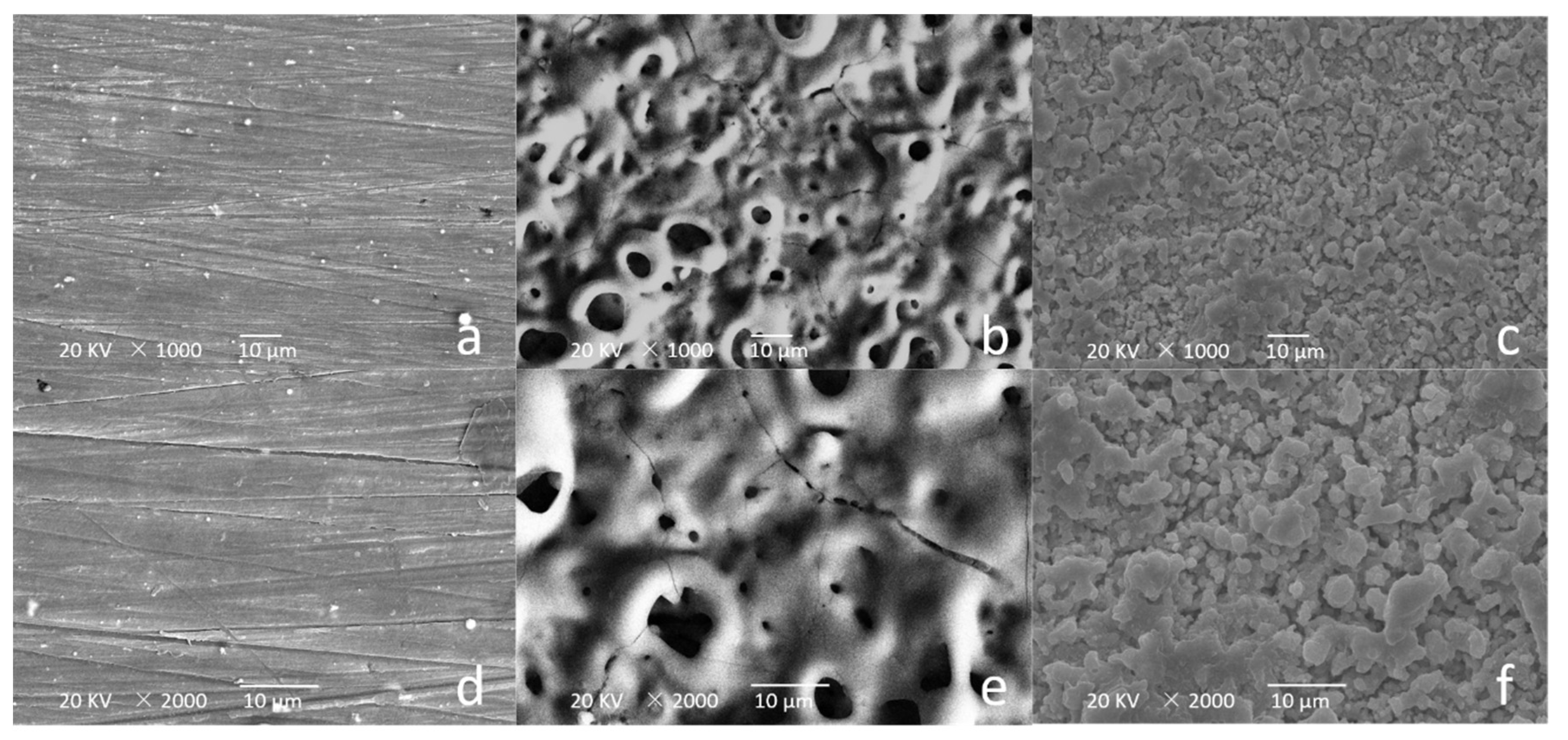
Figure 1. Scanning electron microscopy (SEM) images of (a,d) AZ31, (b,e) micro-arc oxide (MAO) + AZ31, and (c,f) aminated hydroxyethyl cellulose (AHEC) + MAO + AZ31.
Metals 11 00784 g001
Figure 2. Cross-sectional morphologies of (ac), MAO + AZ31 and (df) AHEC + MAO + AZ31.
Metals 11 00784 g002
Figure 3. X-ray diffraction (XRD) pattern of AZ31, MAO + AZ31, and AHEC + MAO + AZ31 specimens.
Metals 11 00784 g003
Figure 4. Fourier-transform infrared spectroscopy (FT-IR) spectrum of the AZ31, MAO + AZ31, AHEC + MAO + AZ31, and pure AHEC.
Metals 11 00784 g004
Figure 5. Energy-dispersive X-ray spectroscopy (EDS) determination of elemental distributions in (a) MAO + AZ31 and (b) AHEC + MAO + AZ31.
Metals 11 00784 g005
Figure 6. Potentiodynamic polarization curves for AZ31, MAO + AZ31, and AHEC + MAO + AZ31 in simulated body fluid (SBF) at room temperature.
Metals 11 00784 g006
Figure 7. Volume of hydrogen released per unit area of AZ31, MAO + AZ31, and AHEC + MAO + AZ31 samples immersed at 37 °C for 7 days.
Metals 11 00784 g007
Figure 8. Long-term soaking diagram of each group. The red arrows indicate corrosion points. Scale bar = 5 mm.
Metals 11 00784 g008
Figure 9. The cell viability of AZ31, MAO + AZ31 and AHEC + MAO + AZ31 specimens.
Metals 11 00784 g009
Figure 10. Inverted microscopy images (20×) of the MC3T3-E1 cells cultured with extracts from the (a) AZ31, (b) MAO + AZ31, (c) AHEC + MAO + AZ31 specimens, and (d) negative control group after 5 d.
Metals 11 00784 g010
Table 1. Coating thickness of AZ31 substrate (mean ± standard deviation).
SamplesMAOAHEC + MAO
Thickness (μm)23.52 ± 1.4227.83 ± 0.93
Table 2. EDS of samples (Round numbers).
Wt%CN0FNaPK
MAO + AZ3115333247162
AHEC + MAO + AZ31485381431
Table 3. Relative electrochemical parameters (mean ± standard deviation).
SamplesEcorr (V vs. Ag/AgCl)Icorr (A/cm2)
AZ31−1.526 ± 0.0021.557 ± 0.001 × 10⁻⁶
MAO + AZ31−1.405 ± 0.005 *1.121 ± 0.003 × 10⁻⁹ *
AHEC + MAO + AZ31−1.072 ± 0.004 *#2.401 ± 0.004 × 10⁻¹⁰ *#
* Compared with AZ31 magnesium alloy alone, the difference was statistically significant (p < 0.05). # Compared with MAO + AZ31 magnesium alloy, the difference of AHEC + MAO + AZ31 magnesium alloy was statistically significant (p < 0.05).
Publisher’s Note: MDPI stays neutral with regard to jurisdictional claims in published maps and institutional affiliations.