Article

High-Temperature Tensile Properties of Hastelloy X Produced by Laser Powder Bed Fusion with Different Heat Treatments

1
School of Materials and Chemistry, University of Shanghai for Science and Technology, Yangpu, Shanghai 200093, China
2
AECC Hunan Aviation Plant Research Institute, Zhuzhou 412002, China
3
School of Mechanical Engineering, University of Shanghai for Science and Technology, Shanghai 200093, China
4
School of Management, University of Shanghai for Science and Technology, Shanghai 200093, China
5
College of Sciences, Shanghai Institute of Technology, Shanghai 201418, China
6
Xi’an Hantang Analysis & Test Co., Ltd., Xi’an 710018, China
7
Center of Electron Microscopy, State Key Laboratory of Silicon Materials, School of Materials Science and Engineering, Zhejiang University, Hangzhou 310027, China
*
Author to whom correspondence should be addressed.
Metals 2022, 12(9), 1435; https://doi.org/10.3390/met12091435
Submission received: 21 July 2022 / Revised: 24 August 2022 / Accepted: 25 August 2022 / Published: 29 August 2022

Abstract

:
High temperature gradient and rapid solidification rate in the laser powder bed fusion (LPBF) process could result in the presence of columnar grains, which could cause poor high temperature tensile properties in the as-built LPBF Hastelloy X (HX) alloys. Heat treatment could effectively transform columnar grain into the equiaxed grain. However, carbides also are precipitated during heat treatment, which could lead to the reduction in ductility. In this study, we aimed to investigate the effect of carbide morphology and distribution on high-temperature tensile properties of LPBF HX alloys by using different heat treatment methods (the same dwell temperature, different cooling methods). The carbide morphology and distribution after furnace cooling, air cooling, and water quenching were characterized respectively, and were correlated with the high-temperature tensile properties. Scanning electron microscope (SEM) images for the fracture surface and cross-sectional area analysis found that the high-temperature tensile properties, especially the ductility, were affected by the carbide morphologies along grain boundaries.

1. Introduction

Laser powder bed fusion (LPBF) is a common additive manufacturing (AM) technology, which could build complex three-dimensional (3D) components with relatively high dimension accuracy directly from digital designs, layer by layer [1,2]. It has attracted great attention from different industry sectors due to its capability of net-shape forming and high accuracy [3,4]. Hastelloy-X (HX) is a solid solution strengthened nickel-base superalloy with good endurance hardness, creep toughness, and corrosion resistance [5]. Compared with the traditional manufacturing processes, the HX alloys formed by LPBF with reasonably relative density can achieve higher room temperature tensile properties, including strength and ductility [6,7,8]. Currently, LPBF HX alloys are used for many critical aerospace engine components, with the most famous one being the engine fuel nozzle [9].
HX could exhibit excellent properties, including high tensile and creep properties, and excellent corrosion resistance at the range of 540–1000 °C, which makes it very attractive and widely used for high-temperature components in aero engines and gas turbines [10,11,12]. The high-temperature performance is an important selection index for HX alloys used for aerospace engine components. In the rapid melting process of LPBF with the large temperature gradient, the grains will preferentially grow along the direction close to the temperature gradient [13]. Therefore, the initial grains formed on the first few layers could further grow into the orientation parallel with the building direction, which could lead to the formation of columnar grains [14]. The presence of larger columnar grains could lead to the strong mechanical property anisotropy of LPBF HX in the as-fabricated state [15]. During the tensile loading process, a strain incompatibility between columnar grains could occur, which makes cracks easy to initiate at the grain boundaries [16,17]. The initiated cracks further propagate along grain boundaries and could lead to the rapid fracture [18]. Therefore, it is commonly accepted that the existence of columnar grains could affect the tensile ductility of LPBF HX alloys, which include the high-temperature tensile properties. During the process of LPBF HX, solidification cracks could be easily formed due to the chemical segregation or excessive solidification gradient [19]. Furthermore, a small number of discontinuous carbides precipitated in the thermal cycle are found at the grain boundary and dendrite, and the crack preferentially extends along its propagation, which greatly reduces the high-temperature performance of the alloy [20].
The microstructure of as-built LPBF HX alloys is determined by the processing parameters, including laser power, scanning speed, hatch distance, and layer thickness, which determine energy inputs and thermal profiles [21]. With other processing parameters kept constant, high laser powers could create molten pools with large depths, which could cause powder splashing, and induce porosity and reduce the surface quality [22]. Low laser scan speed could also cause a deep molten pool with balling, which could be attributed to the low wettability and is easy to cause crack initiation [23]. Toyserkani et al. analyzed the effect of scanning speed on the microstructure and concluded that fine grains were obtained at high scanning speed (1300 mm/s) and coarse microstructure was obtained at low scanning speed (850 mm/s) [24]. Furthermore, improper processing parameters could result in the presence of defects in the LPBF HX alloys. Excessive laser energy input, which could be introduced by high laser power and low scanning speed, produced micro-cracks in LPBF HX alloys [25,26]. In the meantime, insufficient laser energy inputs, which could be attributed to the high scanning speed and low laser power, could lead to the formation of the lack of fusion (LOF) defects and poor mechanical properties [27].
Furthermore, the presence of carbide in LPBF HX alloys, along with their morphologies and distributions, could impact the high-temperature tensile properties [28,29]. Carbides are formed after many thermal cycles during the rapid solidification process [20]. In addition, the morphologies of carbides could be varied, which include the continuous and discontinuous morphologies [30]. The presence of continuous carbide could stabilize the grain boundary and prevent the excessive shear stress, which could lead to the strengthening of the grain boundary [31]. In the meantime, the precipitation of intergranular carbides can refine grains [31]. However, contradicting results could be found in the literature, with the presence of carbides identified on the fracture surface of HX alloys, which indicate that carbides are the preferable sites for crack initiation and could lead to accelerated crack propagation [32]. Despite the uncertain effects of carbides on the fracture behavior, it is certain that the control of carbide distribution and morphology is the key to determine the tensile properties of LPBF HX. Therefore, considerable research efforts have been made to control the precipitation of carbides. Heat treatment (HT), as one of the most effective approaches, could drastically change the morphologies and distribution of carbides [19].
Even though numerous studies have been carried out on the morphologies and distribution of carbides by using different heat treatment methods, it still remains uncertain how the carbides, particularly the ones with different morphologies and distributions, could affect the high-temperature tensile properties of LPBF HX alloys. In this work, we made an investigation into the effects of cooling rates on the distribution and morphology of carbides in LPBF HX alloys. Specifically, three different cooling methods from high-temperature heat treatments with the same dwell temperature were used, to evaluate the effects of cooling rates on the carbide formation. Furthermore, high-temperature tensile testing was carried out for the investigation into the effects of carbide morphologies on the high-temperature mechanical performance. It was found that the carbide morphologies, which were found determined by the cooling methods, are the key factor to determine the high-temperature tensile ductility of LPBF HX.

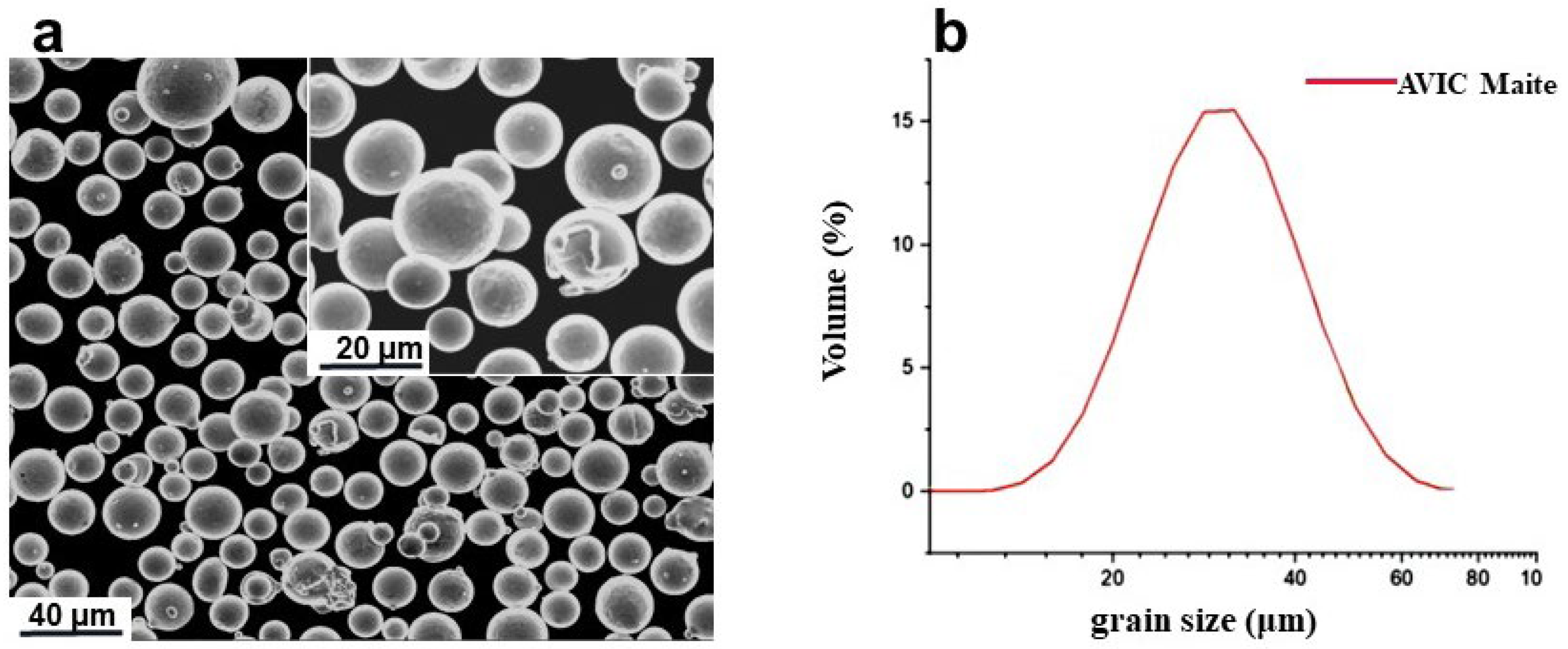
2. Experimental and Methods

Gas atomized pre-alloyed HX powders were obtained from AVIC Metal Powder Metallurgy Technology (Beijing) Co., Ltd. in China. The powder sizes have a spherical morphology as shown in Figure 1a and the powder sizes were 17.7 (D10), 30.5 (D50), and 58.3 μm (D90) (Figure 1b). The chemical composition of the HX powders and as-built HX samples are listed in Table 1. The present chemical compositions are within the allowable tolerance range reported in ASTM B435 [33]. All HX samples processed by LPBF were fabricated by the AmPro SP100 machine (Suzhou Ampro Laser Technology Co., Ltd, Suzhou, China) with the oxygen level maintained below 1000 ppm. The substrate was pre-heated to 80 °C. The processing parameters were laser power 170 W, scan speed 1060 mm/min, hatch distance 0.08 mm, and layer thickness 0.03 mm, which were obtained after the parameter optimization through the Dohelert matrix method, with the details of this method found in [34]. The porosity of 30 optical micrographs taken at 100× was analyzed by Image J (National Institutes of Health, Bethesda, MD, USA), which is commonly used for the porosity measurements of LPBF materials [35,36,37]. The as-fabricated samples were heat-treated at 1175 °C for 30 min followed by air cooling (AC), furnace cooling (FC), and water cooling (WQ), respectively. The cooling rates of the three cooling methods were measured to be about 72 °C/s (AC), 0.05 °C/s (FC), and 500 °C/s (WQ), respectively [27,38]. The details of these three heat treatments are shown in Table 2.
The cylindrical tensile samples were prepared according to the ASTM E21-20, with the gauge length 20 mm and gauge diameter 4 mm. The as-built and heat-treated HX alloys were tested at 855 °C by using a high-temperature tensile testing machine (INSTRON ETM205D, Boston, MA, USA). Before each tensile test, a heating rate of 10 °C/min and a dwell time of 30 min at the set temperature were used to homogenize the sample and environment temperature. The high-temperature tests were carried out at the rate of 0.015 stain/min up to the yield point, and then at a strain rate of 0.05 strain/min until the sample failure. The yield strength was calculated by extensometers. And the cross-head displacement was used to record displacement.
Microstructural characterization samples were ground with 220# to 3000# SiC papers, mechanically polished with Oxide Polishing Suspension (OP-S) (Xinke experimental supplies sales center, Shenyang, China), and then etched for about 10 s with a mixture of 49 vol.% HCl, 2 vol.% CuCl2 and 49 vol.% C2H6O. The Optical microscopy (OM) images were observed by a Zeiss Imager M2m optical microscope (Carl Zeiss AG, Jena, Germany). Scanning electron microscope (TESCAN XMH, SEM) (TESCAN, Brno, Czech Republic) with the working voltage of 10 kV, probe current of 15 nA, and work distance of 8–25 mm was used for the secondary electron image (SEI) acquisition. Energy dispersive spectroscopy (SmartEDX, EDS) (Carl Zeiss AG, Jena, Germany) was used to observe the distribution of carbides under the different heat-treatment conditions.

3. Results

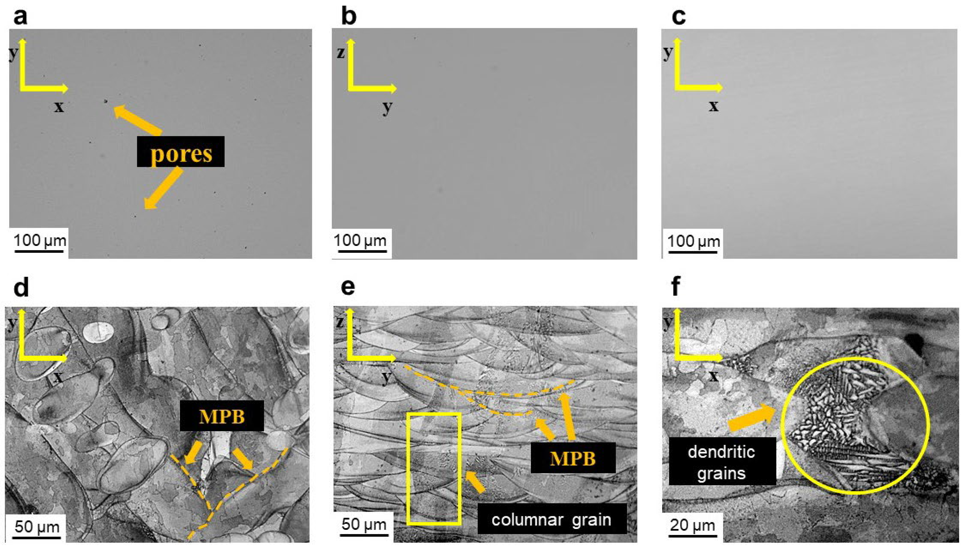
3.1. Microstructure Characterization of LPBF HX Alloys

With the parameter optimization carried out, the commonly seen defects such as pores could be effectively eliminated (compare Figure 2a with Figure 2b,c). The average size of pores measured by quantitative image analysis was 14.2 ± 12.7 μm (with the maximum identified defect with the size of 61.95 μm), and the porosity was measured as 0.23% [36]. In addition, the average length of micro-crack measured by Image J was 76.95 ± 35.4 μm, with the longest measured as 231.76 μm. Figure 2b,c shows some small pores existed in the microstructure, which are believed to have no influence on the tensile properties [35]. Furthermore, the porosity of them was measured as 0.01%, with the maximum defect size 2.3 μm. The microstructure characterization of as-fabricated LPBF HX samples after etching shows different molten pool shapes as indicated by the molten pool boundaries (MPB) (marked by yellow dash lines), on the x-y and z-y planes. On the x-y planes, LPBF HX exhibits crossed strip shapes with a width of 60 ± 10.4 μm (Figure 2d). In the meantime, the MPBs on the z-y plane could be characterized with arch-shape morphology (Figure 2e). The depth of the typical arch-shape melt pools is around 30 ± 8.7 μm and the width around 130 ± 52.4 μm in Figure 2e. In addition, the presence of columnar grains could be identified along the fabrication direction on the z-y planes, with their length across several neighboring layers as indicated by the yellow rectangle in Figure 2e. At the corresponding higher magnifications OM images (Figure 2f), we found that the molten pools consisted of very fine columnar grains with cellular and dendritic growth, which were formed due to the high-temperature gradient of the LPBF process. With strong temperature gradient, the grains preferentially grow in the direction closest to the temperature gradient [39]. Therefore, the grains grow epitaxially and form columnar grain structure after solidification. In addition, due to rapid directional solidification, cell substructures are formed inside the grains, accompanied by high dislocation densities [40].
The microstructure of LPBF HX samples after heat treatment (HT) at varied cooling methods is revealed in Figure 3. It can be found that the molten pool boundaries and dendritic grains disappeared after the HT process, which could be attributed to the fact that the HT could eliminate the inter-dendritic segregation of as-fabricated LPBF HX and lead to dendritic grains dissolution [9]. Furthermore, the recrystallization during the HT could be identified, revealing the transformation from columnar grains to equiaxed grains and the formation of twins (Figure 3a,c,e). The grain aspect ratio for as-built samples and heat-treated samples is shown in Figure 4, where the higher ratio represents the larger amounts of columnar grains, and the ratio of 1 would correspond to a complete circular shape. It could be seen that the aspect ratios of as-built samples, 1175-FC samples, 1175-AC samples, and 1175-WQ samples were 5.0 ± 0.4, 1.4 ± 0.2, 2.0 ± 0.1, and 2.4 ± 0.2, respectively. The result confirms that the columnar grains of LPBF HX alloys were eliminated and fully transformed into equiaxed grains after heat treatments. The average sizes of equiaxed grains were 149.8 ± 85.1 μm in the 1175-FC sample, 209.0 ± 103.5 µm in the 1175-AC sample, and 194.6 ± 132.6 µm in the 1175-WQ sample, respectively (Figure 3b,d,f). By comparing the grain sizes in HX samples treated with different heat treatment methods, it could be found that the increase in the cooling rate could lead to larger grains. And the size of grains was inhomogeneous after HT, which is mainly attributed to different internal stress at different positions of as-fabricated HX alloys. Furthermore, it could be found that carbides, which appear as thick layers in optical micrographs, precipitated at grain boundary after FC cooling from 30 min dwell at 1175 °C (Figure 3a). In contrast, we could hardly identify any carbides on the optical micrographs of samples after both air and water cooling (Figure 3c,e). It could be attributed to the fact the thinner carbides, which could be Molybdenum-rich carbides (M6C) and Chromium-rich carbides (M23C6) [41,42], were formed due to faster cooling rates and made not visible under optical microscopy.
Figure 5 shows the SEM micrographs of samples after different heat treatments. They further reveal the finer grains in LPBF HX with faster cooling rates (Figure 5a–c). The high-magnification images showed the continuous carbides along the grain boundaries, with some large carbide particles clustered at the triple junction locations (Figure 5d). With the increase in cooling rates, the carbides become much thinner and could only be identified on the grain boundaries, while are still in continuous morphology (Figure 5e,f). The presence of carbides at some grain boundaries in the sample with water quenching was not visible under SEM characterization (Figure 5f). In the meantime, the carbides are absent within grains in the water-quenched sample (Figure 5f). This is attributed to the fact that carbides with slow precipitation rate could be prevented from precipitation during rapid cooling [31]. Even though the precipitated carbides remain continuous after air cooling and water quenching, the faster cooling rates make them thinner.
The chemical compositions of carbides after furnace cooling were further characterized by energy dispersive spectroscopy (EDS), as shown in Figure 6. For FC samples, EDS measurement reveals that the element of carbon was marked in Figure 6b, and the content percentage was 14.19%. Besides, the composition of other chemical elements was 41.06 at.% Cr, 8.96 at.% Fe, 14.99 at.% Mo, 3.36 at.% O, and 2.02 at.% of all other remaining elements (as seen in Table 3). According to chemical composition, the EDS analysis shows the continuous presence of Cr-rich M23C6 carbides along the grain boundaries, after furnace cooling [43].

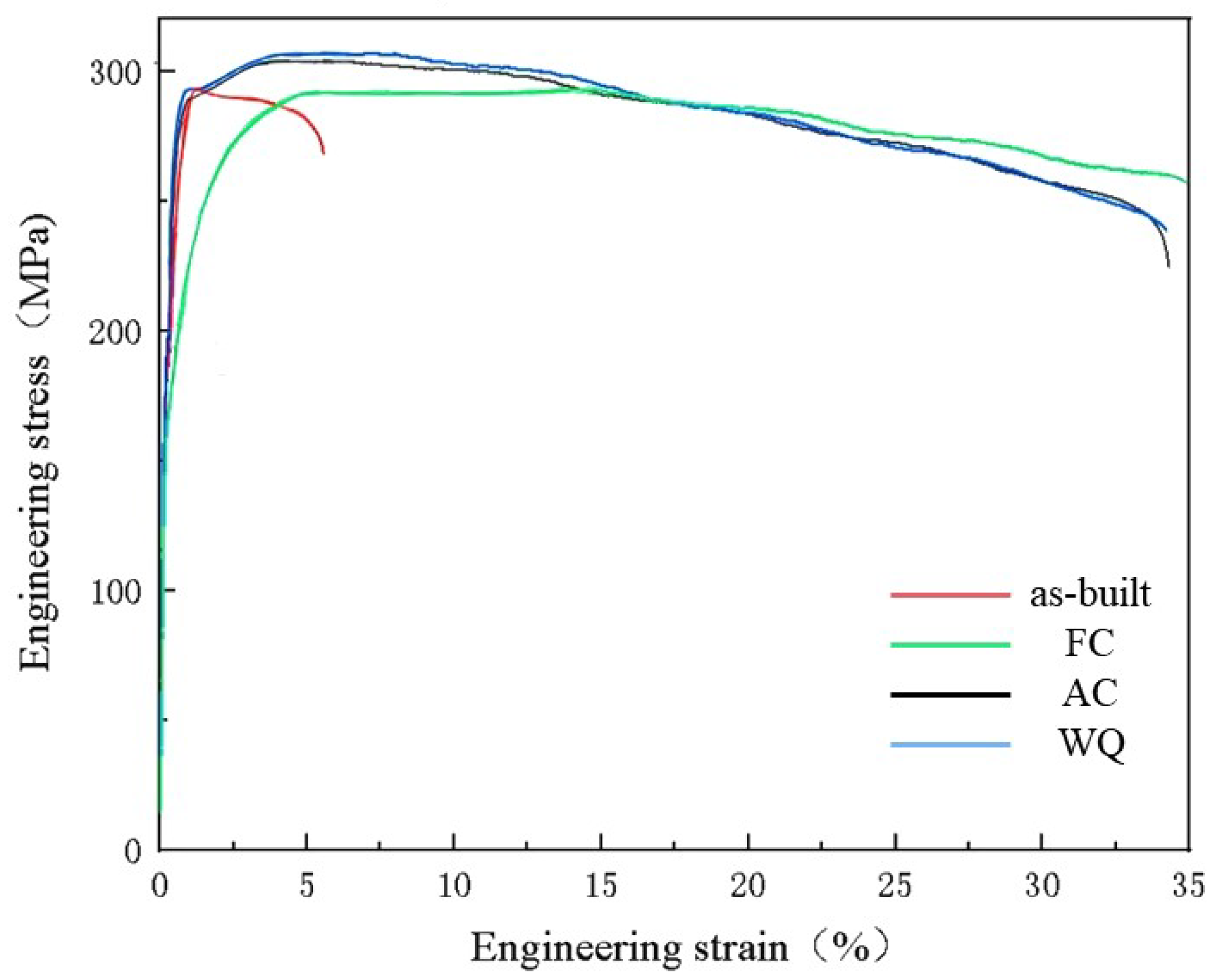
3.2. High-Temperature Tensile Properties

Figure 7 shows the high-temperature tensile performances at 855 °C for as-built LPBF HX alloys after different heat treatments. For the as-built samples, the ultimate tensile stress (UTS), yield stress (YS), and total elongation (EL) at 855 °C were 293.2 ± 1.1 MPa, 201.4 ± 0.9 MPa, and 6.5 ± 0.7%, respectively. As-built LPBF HX alloy has excellent tensile strength, because many low angles grain boundaries are formed during process of LPBF [44]. By comparing with the as-built samples, the YS of the FC samples decreased significantly, the UTS retained identical and the EL increased significantly. Specifically, the UTS, YS, and EL of 1175-FC samples were 293.5 ± 2.4 MPa, 176.1 ± 4.3 MPa, and 34.8 ± 0.5%. The UTS, YS, and EL of the 1175-AC sample were 304.3 ± 3.2 MPa, 196.7 ± 3.5 MPa, and 33.5 ± 0.8%, respectively. Both UTS and YS of 1175-AC samples were slightly higher than those of 1175-FC samples. For the WQ samples, the UTS and the YS increased with the further increase in cooling rate. The UTS, YS, and EL were 307.2 ± 0.8 MPa, 201.6 ± 4.1 MPa, and 33.0 ± 0.4%, respectively. All results of tensile test were shown in Table 3.

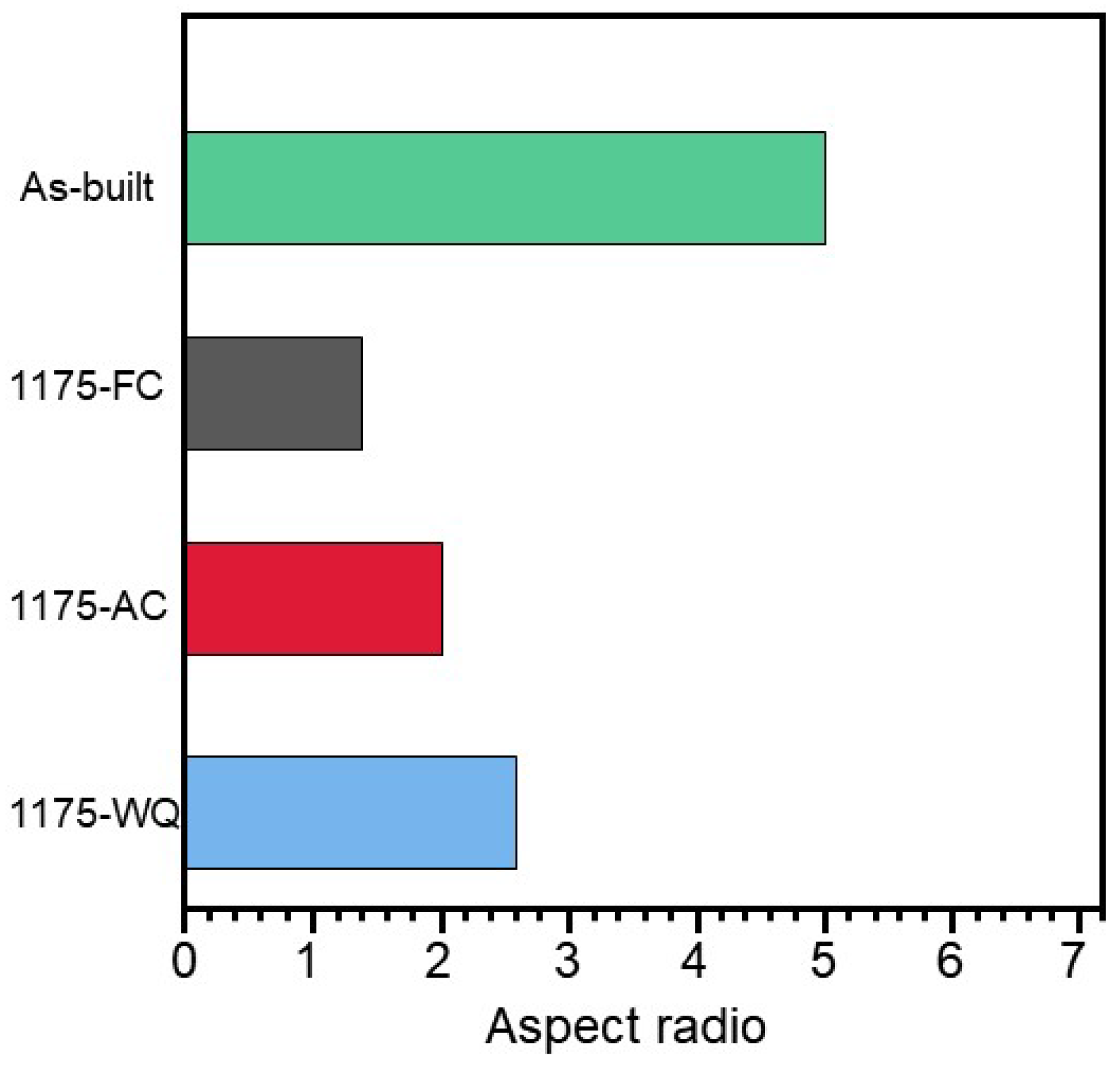
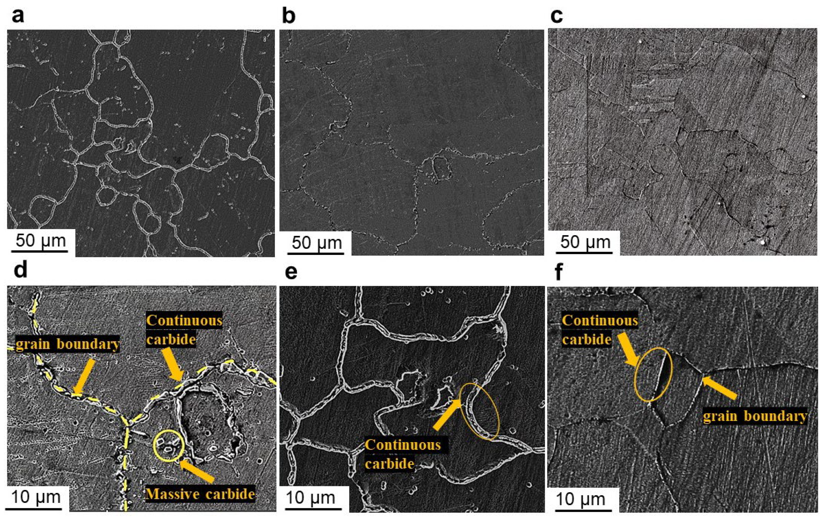
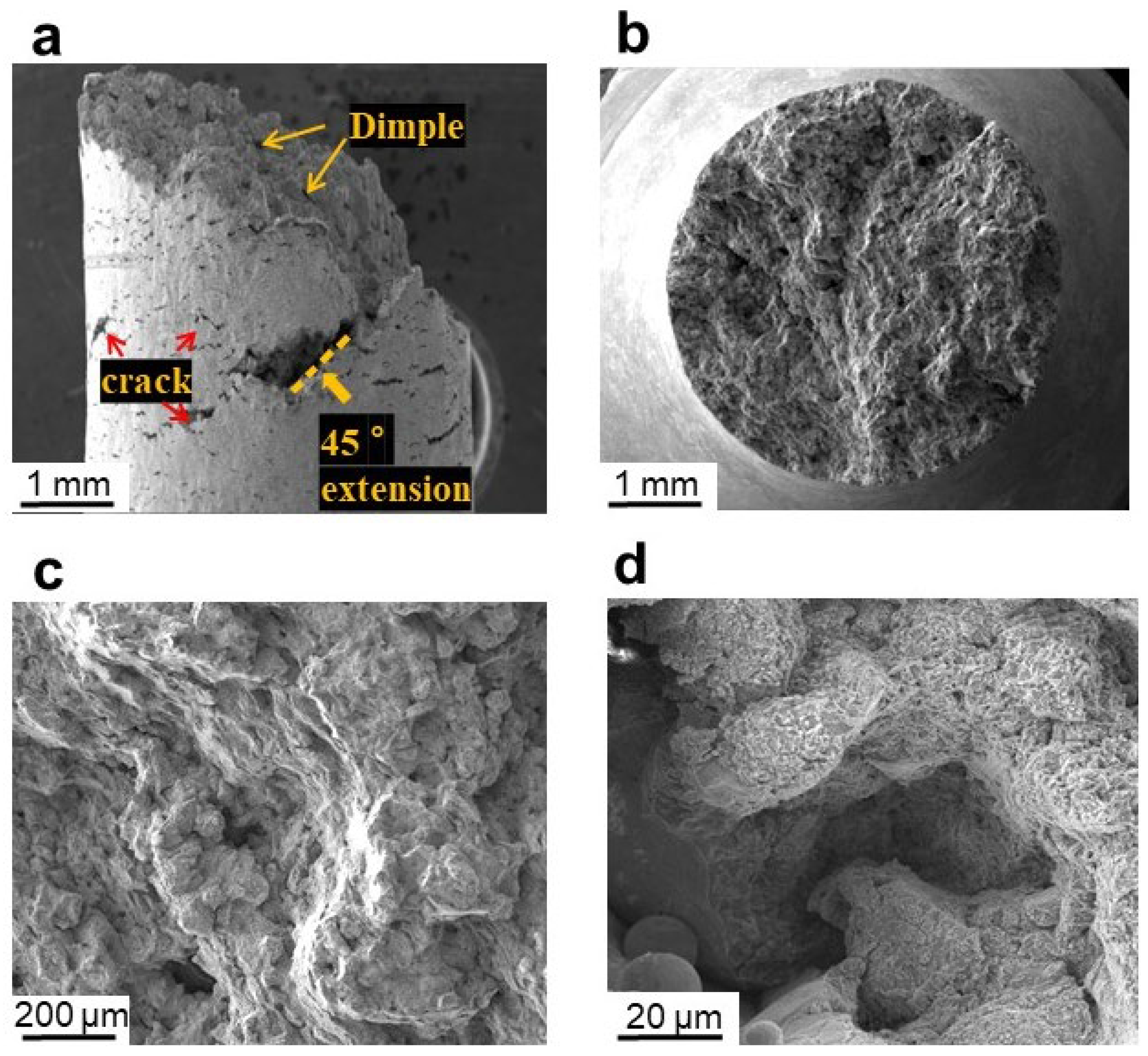
3.3. Fracture Surface Characterization

To further understand the failure mechanisms in samples after different cooling methods, the fracture surface characterizations were carried out. For 1175-FC samples, we could hardly identify any necking behavior on the fracture sample (Figure 8a). In the meantime, some small cracks (marked by red arrows) and a large crack (marked by dashed line) could be identified on the side surface of the failed sample, with the inclination angles of these cracks approximately 45° off the loading axis. This is attributed to the fact that the massive carbides in the microstructure of 1175-FC samples are used as the crack source during high-temperature tension, which leads to crack initiation and propagation.
Furthermore, the detailed characterization on the fracture surface of the failed sample shows the intergranular cracking, indicating the cracking through the brittle and continuous carbide phases on the grain boundaries (Figure 8c). Dimples with diameters of approximately 50 μm could be seen on the fracture surface. In the meantime, some defects, such as the lack-of-fusion defects in Figure 8d, was identified inside the depression, which could reduce the ductility as well.
The fracture surface characterization of the 1175-AC sample, along with the observation on the side surface, after tensile failure is shown in Figure 9. Compared with FC samples, the number of micro-cracks on the side of the fracture increased, while most of the cracks are perpendicular to the tensile direction (Figure 9a). Furthermore, the necking behavior in the 1175-AC sample is slightly more obvious as compared with that in 1175-FC sample. The fracture surface is composed of the fiber area in the center of the fracture and the shear lip area at the edge of the fracture (Figure 9b). Furthermore, the fracture surface showed the mixed mode of ductile fracture and cleavage (Figure 9c,d), indicating the failure mode mixed with inter-granular and trans-granular fracture. In the meantime, brittle fracture features could be observed in the ductile region. And the propagation of brittle crack is hindered by the ductile region.
The fracture surface characterization of the 1175-WQ sample, along with the observation on the side surface, after tensile failure is shown in Figure 10. The necking behavior of 1175-WQ sample is more obvious as compared with other samples, as shown in Figure 10a. Compared with 1175-AC sample, there are more cracks on the side surface of the failed sample. In the meantime, a large number of secondary cracks, along with some lack-of-fusion defects, could be identified on the fracture surface (Figure 10c,d). The presence of numerous secondary cracks could be potentially attributed to the absence of carbides on some grain boundaries due to the rapid cooling, which might favor the crack propagation as well. The presence of discontinuous carbides at the grain boundary will cause stress concentration, which is easy to result in crack initiation and propagation. Smooth facets on the fracture surface could be identified as well, which indicates the transgranular fracture mode.

4. Discussion

For as-built LPBF HX alloys, the poor high-temperature ductility was attributed to elongated columnar grains with fine dendrites penetrating into several neighboring layers in the as-fabricated state [45]. The formation of columnar grains are the consequence of large temperature gradient during the LPBF process, with their elongation directions along the LPBF building direction [40,46]. The presence of columnar grain could cause strain incompatibility, which was due to the differences in morphology and crystallography orientation between adjacent grains, and led to the initiation of cracks at the grain boundary [16,47]. In the meantime, the high residual stress, which is a strong indication of high dislocation density, is another cause to the low ductility along with the high strength at the testing temperature.
With the post solution treatments used, molten pool morphologies could be fully dissolved with the columnar grains transformed into the equiaxed grains, which is consistent with published studies [48]. Both microstructure changes are the consequence of the recrystallization during the solution treatments. The transition from columnar to equiaxed grains, which reduce the effective slip length, is considered as one of the factors contribute to the higher tensile ductility at high temperature [3]. In the meantime, the precipitation of carbides during the cooling process after high-temperature solution treatments, is another factor that could increase the high-temperature tensile properties. The carbides are mostly considered stable at the high service temperature of HX alloys, like 855 °C used in this study [49]. Continuous carbides could strengthen grain boundaries and hinder dislocation movement to improve the high-temperature yield strength and ductility [50]. In contrast, discontinuous carbides could be favored for crack initiation and propagation during the tensile loading process, and could lead to the reduced ductility.
By comparing the tensile results of high-temperature tensile samples, the high-temperature heat-treated samples have higher tensile ductility than the as-built state. In addition, by analyzing the fracture surface after different cooling methods, it could be convinced that the existence of carbides may affect the fracture behavior, consistent with that reported in the literature as well [16]. The continuous carbides at the grain boundary can hinder the dislocation movement and strengthen the grain boundary. The fracture surface of 1175-FC sample showed obvious grain deformation and a large number of small and deep dimples, with the fracture mode dominated by transcrystalline crack propagation. This indicates that the crack favors the propagation across the grain boundaries through the thick continuous carbides. In the meantime, transgranular crack propagation could be indicated on the fracture surfaces of both 1175-AC and 1175-WQ samples, which shows that continuous carbides with smaller thickness might have higher resistance to the crack propagation. This leads to the mix of both intergranular and transgranular fracture modes, which is further indicated by more micro-cracks and secondary cracks on the side and fracture surfaces of the samples with faster cooling rates.
However, the change in the fracture behavior is not related to any substantial variation in the tensile ductility. Further investigation by microstructure and carbide manipulations is needed for further improvement in the high-temperature mechanical properties, particularly the ductility. The ductility of LPBF HX in the current published works is relatively lower than that of conventional manufactured HX [1].

5. Conclusions

In this study, an investigation into the influence of different cooling rates on the microstructures of laser powder bed fusion Hastelloy X, particularly the carbide distribution and columnar grain evolution, was carried out. Furthermore, the high-temperature tensile properties of LPBF HX with different carbide morphologies and distributions were investigated, with the effects of carbides on the tensile properties proposed. The following conclusions can be summarized from this study:
1.
The high-temperature (1175 °C) solution heat treatments could homogenize the microstructure, eliminate the molten pool boundaries, and achieve the columnar to equiaxed grain transition.
2.
Slow cooling, such as furnace cooling (10 °C/min) used in this study, could lead to coarse carbide precipitation with continuous morphology along the grain boundaries. In contrast, rapid cooling (air cooling and water quenching) approach might lead to the formation of thin carbide layers.
3.
The chromium rich carbide (M23C6) is easily obtained at a low cooling temperature. The cooling rate of furnace cooling is slow, so M23C6 can be fully precipitated. The continuous carbide distribution at the grain boundary can play a strengthening role and hinder dislocations. Therefore, the high temperature plasticity of as-built LPBF HX alloy is greatly improved.

Author Contributions

Conceptualization, M.L. and K.Z.; methodology, M.L. and Q.Z.; validation, Q.Z. and Y.H.; formal analysis, W.Z., M.L. and K.Z.; resources, Y.H., W.Z., Y.L., K.Z., Y.W. and J.W.; writing—original draft preparation, M.L. and K.Z.; writing—review and editing, Y.H., W.Z. and K.Z.; visualization, M.L., Q.Z. and W.Z.; supervision, K.Z.; funding acquisition, K.Z., and Y.J. All authors have read and agreed to the published version of the manuscript.

Funding

This research was funded by the Shanghai Institute of Technology (ZQ2021-22), Shanghai Municipal Education Commission, the Sailing Program (20YF1431600) and internal funding from University of Shanghai for Science and Technology.

Institutional Review Board Statement

Not applicable.

Informed Consent Statement

Not applicable.

Data Availability Statement

The data presented in this study are available on request from the corresponding author.

Acknowledgments

The authors acknowledge financial support from the Shanghai Institute of Technology (ZQ2021-22), Shanghai Municipal Education Commission, the Sailing Program (20YF1431600), and internal funding from University of Shanghai for Science and Technology. The authors gratefully acknowledge the use of instruments at Advanced Materials Research Institute, and Yangtze Delta Analytical Characterization Platform is acknowledged for the scientific and technical assistance of SEM and EDS analysis. Minghao Liu wishes to appreciate the help from Qingsheng He for the sample fabrication, the help from Jie Liu for the data analysis, and the help from Jianwen Liu and Jing Zhu for the experimental test.

Conflicts of Interest

The authors declare no conflict of interest.

References

  1. Ghiaasiaan, R.; Muhammad, M.; Gradl, P.R.; Shao, S.; Shamsaei, N. Superior tensile properties of Hastelloy X enabled by additive manufacturing. Mater. Res. Lett. 2021, 9, 308–314. [Google Scholar] [CrossRef]
  2. Keshavarzkermani, A.; Esmaeilizadeh, R.; Ali, U.; Enrique, P.D.; Mahmoodkhani, Y.; Zhou, N.Y.; Bonakdar, A.; Toyserkani, E. Controlling mechanical properties of additively manufactured hastelloy X by altering solidification pattern during laser powder-bed fusion. Mater. Sci. Eng. A 2019, 762, 138081. [Google Scholar] [CrossRef]
  3. Zhang, M.; Zhang, B.; Wen, Y.; Qu, X. Research progress on selective laser melting processing for nickel-based superalloy. Int. J. Miner. Met. Mater. 2022, 29, 369–388. [Google Scholar] [CrossRef]
  4. Mertens, R.; Dadbakhsh, S.; Van Humbeeck, J.; Kruth, J.-P. Application of base plate preheating during selective laser melting. Procedia CIRP 2018, 74, 5–11. [Google Scholar] [CrossRef]
  5. Montero-Sistiaga, M.L.; Pourbabak, S.; Van Humbeeck, J.; Schryvers, D.; Vanmeensel, K. Microstructure and mechanical properties of Hastelloy X produced by HP-SLM (high power selective laser melting). Mater. Des. 2019, 165, 107598. [Google Scholar] [CrossRef]
  6. Wang, F. Mechanical property study on rapid additive layer manufacture Hastelloy® X alloy by selective laser melting technology. Int. J. Adv. Manuf. Technol. 2011, 58, 545–551. [Google Scholar] [CrossRef]
  7. Tomus, D.; Jarvis, T.; Wu, X.; Mei, J.; Rometsch, P.; Herny, E.; Rideau, J.-F.; Vaillant, S. Controlling the Microstructure of Hastelloy-X Components Manufactured by Selective Laser Melting. Phys. Procedia 2013, 41, 823–827. [Google Scholar] [CrossRef]
  8. Yin, Y.; Zhang, J.; Yang, S.; Liu, T.; Han, Q.; Zhang, Z.; Yang, H. Effect of microstructure on the electrochemical dissolution behaviour of Hastelloy® X superalloy processed by selective laser melting and heat treatments. Mater. Des. 2021, 206, 109828. [Google Scholar] [CrossRef]
  9. Jinoop, A.; Paul, C.; Kumar, J.G.; Anilkumar, V.; Singh, R.; Rao, S.; Bindra, K. Influence of heat treatment on the microstructure evolution and elevated temperature mechanical properties of Hastelloy-X processed by laser directed energy deposition. J. Alloy. Compd. 2021, 868, 159207. [Google Scholar] [CrossRef]
  10. Han, Q.; Gu, Y.; Setchi, R.; Lacan, F.; Johnston, R.; Evans, S.L.; Yang, S. Additive manufacturing of high-strength crack-free Ni-based Hastelloy X superalloy. Addit. Manuf. 2019, 30, 100919. [Google Scholar] [CrossRef]
  11. Saarimäki, J.; Lundberg, M.; Brodin, H.; Moverare, J. Dwell-fatigue crack propagation in additive manufactured Hastelloy X. Mater. Sci. Eng. A 2018, 722, 30–36. [Google Scholar] [CrossRef]
  12. Han, Q.; Gu, Y.; Soe, S.; Lacan, F.; Setchi, R. Effect of hot cracking on the mechanical properties of Hastelloy X superalloy fabricated by laser powder bed fusion additive manufacturing. Opt. Laser Technol. 2019, 124, 105984. [Google Scholar] [CrossRef]
  13. Rickenbacher, L.; Etter, T.; Hövel, S.; Wegener, K. High temperature material properties of IN738LC processed by selective laser melting (SLM) technology. Rapid Prototyp. J. 2013, 19, 282–290. [Google Scholar] [CrossRef]
  14. Pourbabak, S.; Montero-Sistiaga, M.L.; Schryvers, D.; Van Humbeeck, J.; Vanmeensel, K. Microscopic investigation of as built and hot isostatic pressed Hastelloy X processed by Selective Laser Melting. Mater. Charact. 2019, 153, 366–371. [Google Scholar] [CrossRef]
  15. Etter, T.; Kunze, K.; Geiger, F.; Meidani, H. Reduction in mechanical anisotropy through high temperature heat treatment of Hastelloy X processed by Selective Laser Melting (SLM). IOP Conf. Ser. Mater. Sci. Eng. 2015, 82, 012097. [Google Scholar] [CrossRef]
  16. Montero-Sistiaga, M.L.; Liu, Z.; Bautmans, L.; Nardone, S.; Ji, G.; Kruth, J.-P.; Van Humbeeck, J.; Vanmeensel, K. Effect of temperature on the microstructure and tensile properties of micro-crack free hastelloy X produced by selective laser melting. Addit. Manuf. 2019, 31, 100995. [Google Scholar] [CrossRef]
  17. Shi, Y.; Xiong, X.; Liu, Z.; Yang, Y.; Hou, J.; Wu, S.; Rao, J.H.; Zhang, K.; Huang, A. Mechanical Property Evaluation of a SLMed Martensitic Stainless Steel. Acta Met. Sin. (English Lett.) 2020, 33, 1466–1476. [Google Scholar] [CrossRef]
  18. Kitano, H.; Tsujii, M.; Kusano, M.; Yumoto, A.; Watanabe, M. Effect of plastic strain on the solidification cracking of Hastelloy-X in the selective laser melting process. Addit. Manuf. 2020, 37, 101742. [Google Scholar] [CrossRef]
  19. Wang, H.; Chen, L.; Dovgyy, B.; Xu, W.; Sha, A.; Li, X.; Tang, H.; Liu, Y.; Wu, H.; Pham, M.-S. Micro-cracking, microstructure and mechanical properties of Hastelloy-X alloy printed by laser powder bed fusion: As-built, annealed and hot-isostatic pressed. Addit. Manuf. 2021, 39, 101853. [Google Scholar] [CrossRef]
  20. Marchese, G.; Basile, G.; Bassini, E.; Aversa, A.; Lombardi, M.; Ugues, D.; Fino, P.; Biamino, S. Study of the Microstructure and Cracking Mechanisms of Hastelloy X Produced by Laser Powder Bed Fusion. Materials 2018, 11, 106. [Google Scholar] [CrossRef] [Green Version]
  21. Keller, C.; Mokhtari, M.; Vieille, B.; Briatta, H.; Bernard, P. Influence of a rescanning strategy with different laser powers on the microstructure and mechanical properties of Hastelloy X elaborated by powder bed fusion. Mater. Sci. Eng. A 2020, 803, 140474. [Google Scholar] [CrossRef]
  22. Wang, F.; Wu, X.H.; Clark, D. On direct laser deposited Hastelloy X: Dimension, surface finish, microstructure and mechanical properties. Mater. Sci. Technol. 2011, 27, 344–356. [Google Scholar] [CrossRef]
  23. Tian, Y.; Tomus, D.; Rometsch, P.; Wu, X. Influences of processing parameters on surface roughness of Hastelloy X produced by selective laser melting. Addit. Manuf. 2017, 13, 103–112. [Google Scholar] [CrossRef]
  24. Esmaeilizadeh, R.; Keshavarzkermani, A.; Ali, U.; Mahmoodkhani, Y.; Behravesh, B.; Jahed, H.; Bonakdar, A.; Toyserkani, E. Customizing mechanical properties of additively manufactured Hastelloy X parts by adjusting laser scanning speed. J. Alloys Compd. 2019, 812, 152097. [Google Scholar] [CrossRef]
  25. Harrison, N.J.; Todd, I.; Mumtaz, K. Reduction of micro-cracking in nickel superalloys processed by Selective Laser Melting: A fundamental alloy design approach. Acta Mater. 2015, 94, 59–68. [Google Scholar] [CrossRef]
  26. Han, Q.; Mertens, R.; Montero-Sistiaga, M.L.; Yang, S.; Setchi, R.; Vanmeensel, K.; Van Hooreweder, B.; Evans, S.L.; Fan, H. Laser powder bed fusion of Hastelloy X: Effects of hot isostatic pressing and the hot cracking mechanism. Mater. Sci. Eng. A 2018, 732, 228–239. [Google Scholar] [CrossRef]
  27. Kitano, H.; Kusano, M.; Tsujii, M.; Yumoto, A.; Watanabe, M. Process Parameter Optimization Framework for the Selective Laser Melting of Hastelloy X Alloy Considering Defects and Solidification Crack Occurrence. Crystals 2021, 11, 578. [Google Scholar] [CrossRef]
  28. Sanchez-Mata, O.; Wang, X.; Muñiz-Lerma, J.A.; Shandiz, M.A.; Gauvin, R.; Brochu, M. Fabrication of Crack-Free Nickel-Based Superalloy Considered Non-Weldable during Laser Powder Bed Fusion. Materials 2018, 11, 1288. [Google Scholar] [CrossRef]
  29. Han, Q.; Gu, Y.; Wang, L.; Feng, Q.; Gu, H.; Johnston, R.; Setchi, R. Effects of TiC content on microstructure and mechanical properties of nickel-based hastelloy X nanocomposites manufactured by selective laser melting. Mater. Sci. Eng. A 2020, 796, 140008. [Google Scholar] [CrossRef]
  30. Tang, Y.T.; Wilkinson, A.J.; Reed, R.C. Grain Boundary Serration in Nickel-Based Superalloy Inconel 600: Generation and Effects on Mechanical Behavior. Met. Mater. Trans. A 2018, 49, 4324–4342. [Google Scholar] [CrossRef] [Green Version]
  31. Marchese, G.; Bassini, E.; Aversa, A.; Lombardi, M.; Ugues, D.; Fino, P.; Biamino, S. Microstructural Evolution of Post-Processed Hastelloy X Alloy Fabricated by Laser Powder Bed Fusion. Materials 2019, 12, 486. [Google Scholar] [CrossRef] [PubMed]
  32. Tomus, D.; Rometsch, P.A.; Heilmaier, M.; Wu, X. Effect of minor alloying elements on crack-formation characteristics of Hastelloy-X manufactured by selective laser melting. Addit. Manuf. 2017, 16, 65–72. [Google Scholar] [CrossRef]
  33. Sah, I.; Kim, E.-S. High-temperature tensile behavior of diffusion-welded hastelloy X. J. Mech. Sci. Technol. 2022, 36, 1–10. [Google Scholar] [CrossRef]
  34. Chen, Z.; Wu, X.; Tomus, D.; Davies, C.H. Surface roughness of Selective Laser Melted Ti-6Al-4V alloy components. Addit. Manuf. 2018, 21, 91–103. [Google Scholar] [CrossRef]
  35. Bidulská, J.; Bidulský, R.; Grande, M.A.; Kvačkaj, T. Different Formation Routes of Pore Structure in Aluminum Powder Metallurgy Alloy. Materials 2019, 12, 3724. [Google Scholar] [CrossRef] [PubMed]
  36. Bidulský, R.; Bidulská, J.; Gobber, F.S.; Kvačkaj, T.; Petroušek, P.; Actis-Grande, M.; Weiss, K.-P.; Manfredi, D. Case Study of the Tensile Fracture Investigation of Additive Manufactured Austenitic Stainless Steels Treated at Cryogenic Conditions. Materials 2020, 13, 3328. [Google Scholar] [CrossRef]
  37. Beiss, P.; Dalgic, M. Structure property relationships in porous sintered steels. Mater. Chem. Phys. 2001, 67, 37–42. [Google Scholar] [CrossRef]
  38. Korotkov, V.A.; Anan’Ev, S.P.; Shekurov, A.V. Investigation of the effect of the cooling rate on the quality of the surface layer in plasma quenching. Weld. Int. 2013, 27, 407–410. [Google Scholar] [CrossRef]
  39. Keshavarzkermani, A.; Sadowski, M.; Ladani, L. Direct metal laser melting of Inconel 718: Process impact on grain formation and orientation. J. Alloys Compd. 2018, 736, 297–305. [Google Scholar] [CrossRef]
  40. Keshavarzkermani, A.; Esmaeilizadeh, R.; Enrique, P.D.; Asgari, H.; Zhou, N.Y.; Bonakdar, A.; Toyserkani, E. Static recrystallization impact on grain structure and mechanical properties of heat-treated Hastelloy X produced via laser powder-bed fusion. Mater. Charact. 2021, 173, 110969. [Google Scholar] [CrossRef]
  41. Tawancy, H.M. Long-term ageing characteristics of some commercial nickel-chromium-molybdenum alloys. J. Mater. Sci. 1981, 16, 2883–2889. [Google Scholar] [CrossRef]
  42. Kestens, L.; Jonas, J.J. ASM Handbook. Volume 14A: Metalworking: Bulk Forming. In ASM Handbook; Semiatin, L., Ed.; ASM International: Novelty, OH, USA, 2005; pp. 685–700. [Google Scholar]
  43. Hu, R.; Bai, G.; Li, J.; Zhang, J.; Zhang, T.; Fu, H. Precipitation behavior of grain boundary M23C6 and its effect on tensile properties of Ni–Cr–W based superalloy. Mater. Sci. Eng. A 2012, 548, 83–88. [Google Scholar] [CrossRef]
  44. Ni, X.; Kong, D.; Zhang, L.; Dong, C.; Song, J.; Wu, W. Effect of Process Parameters on the Mechanical Properties of Hastelloy X Alloy Fabricated by Selective Laser Melting. J. Mater. Eng. Perform. 2019, 28, 5533–5540. [Google Scholar] [CrossRef]
  45. Tomus, D.; Tian, Y.; Rometsch, P.A.; Heilmaier, M.; Wu, X. Influence of post heat treatments on anisotropy of mechanical behaviour and microstructure of Hastelloy-X parts produced by selective laser melting. Mater. Sci. Eng. A 2016, 667, 42–53. [Google Scholar] [CrossRef]
  46. Cloots, M.; Uggowitzer, P.J.; Wegener, K. Investigations on the microstructure and crack formation of IN738LC samples processed by selective laser melting using Gaussian and doughnut profiles. Mater. Des. 2016, 89, 770–784. [Google Scholar] [CrossRef]
  47. Nickel, A.; Barnett, D.; Prinz, F. Thermal stresses and deposition patterns in layered manufacturing. Mater. Sci. Eng. A 2001, 317, 59–64. [Google Scholar] [CrossRef]
  48. Sanchez-Mata, O.; Muñiz-Lerma, J.; Wang, X.; Atabay, S.; Shandiz, M.A.; Brochu, M. Microstructure and mechanical properties at room and elevated temperature of crack-free Hastelloy X fabricated by laser powder bed fusion. Mater. Sci. Eng. A 2020, 780, 139177. [Google Scholar] [CrossRef]
  49. Wang, D.; Zhang, J.; Lou, L. Formation and stability of nano-scaled M23C6 carbide in a directionally solidified Ni-base superalloy. Mater. Charact. 2009, 60, 1517–1521. [Google Scholar] [CrossRef]
  50. Belan, J. GCP and TCP Phases Presented in Nickel-base Superalloys. Mater. Today: Proc. 2016, 3, 936–941. [Google Scholar] [CrossRef]
Figure 1. (a) LPBF images of the HX powders used in this study; (b) particle size distribution diagram of LPBF-HX tensile samples.
Metals 12 01435 g001
Figure 2. OM images of as-built Hastelloy X (HX) sample: (a) pores in the samples before parameter optimization (as marked by yellow arrows); (b,c) near fully dense after parameter optimization along the y-z and x-y planes; (d,e) the melt pool boundaries (MPB) along the x-y and z-y planes; (f) cellular and columnar shapes of dendritic with molten pools.
Metals 12 01435 g002
Figure 3. Representative optical microscopy images of (a) 1175-FC sample, (c) 1175-AC sample, and (e) 1175-WQ sample. The equiaxed grain boundaries in (a,c,e) are traced by black-color lines. The histograms of equiaxed grains size are shown in (b,d,f).
Metals 12 01435 g003
Figure 4. Calculated grain aspect ratio for samples in different states: as-built, 1175-FC, 1175-AC, and 1175-WQ.
Metals 12 01435 g004
Figure 5. Microstructure characterization of HT samples by using low-magnification and high-magnification secondary electron images (SEM): (a,d) 1175-FC; (b,e) 1175-AC; (c,f) 1175-WQ.
Metals 12 01435 g005
Figure 6. EDS map scanning results after furnace cooling consist of: (a) Complete picture; (b) Carbon element; (c) Chromium element; (d) Molybdenum element; (e) Iron element; (f) Oxygen element.
Metals 12 01435 g006
Figure 7. High temperature (855 °C) tensile stress-strain curves of the as-built sample (marked by red solid line), furnace cooling (FC) sample (marked by green solid line), air cooling (AC) sample (marked by black solid line) and water quenching (WQ) sample (marked by blue solid lines).
Metals 12 01435 g007
Figure 8. Fracture surface characterization of tensile failed 1175-FC samples by using SEM: (a) the side surface of the fracture; (b) the front view of the fracture sample; (c,d) the morphology of fracture surface under different magnifications.
Metals 12 01435 g008
Figure 9. Fracture surface characterization of tensile failed 1175-AC samples by using SEM: (a) the side surface of the fracture sample; (b) the front view of the fracture sample; (c,d) the morphology of fracture surface under different magnifications.
Metals 12 01435 g009
Figure 10. Fracture surface characterization of tensile failed 1175-WQ samples by using secondary electron images (SEM): (a) the side of the fracture; (b) the front view of the fracture sample; (c,d) the morphology of fracture under different magnifications.
Metals 12 01435 g010
Table 1. Chemical composition of HX alloys.
Alloy (wt.%)NiCrFeMoCoCSiWAlO
Powder (%)Bal.21.519.279.141.520.0650.280.530.10.016
As-fabricatedBal.21.5319.319.221.540.080.290.550.10.03
Table 2. The heat treatments used in this study.
IDHT TemperatureDwell TimeCooling Mode
1175-FC1175 ℃30 minFurnace cooling
1175-AC1175 ℃30 minAir cooling
1175-WQ1175 ℃30 minWater quenching
Table 3. High-temperature tensile results.
SamplesUTS (MPa)YS (MPa)EL (%)
As-built293.2 ± 1.1201.4 ± 0.96.5 ± 0.7
FC293.5 ± 2.4176.1 ± 4.334.8 ± 0.5
AC304.3 ± 3.2196.7 ± 3.533.5 ± 0.8
WQ307.2 ± 0.8201.6 ± 4.133.0 ± 0.4
Publisher’s Note: MDPI stays neutral with regard to jurisdictional claims in published maps and institutional affiliations.

