Article

Influence of High-Temperature Deformation on the Dissolution of δ-Ferrite in Stainless Steels

by
Rahman Bajmalu Rostami
*,†,
Pedro de Souza Ciacco
,
Mauricio Claudio Viali Muñoz
,
Luis Fellipe Simoes
and
Calixto Isaac Garcia
Department of Mechanical Engineering and Materials Science, University of Pittsburgh, Pittsburgh, PA 15261, USA
*
Author to whom correspondence should be addressed.
These authors contributed equally to this work.
Metals 2024, 14(7), 783; https://doi.org/10.3390/met14070783
Submission received: 3 June 2024 / Revised: 28 June 2024 / Accepted: 29 June 2024 / Published: 3 July 2024

Abstract

:
The dissolution behavior of δ-ferrite in two commercial stainless steels, 15-5PH and M-154, was studied. In this work, a new approach combining hot deformation and additional post-treatment was investigated and compared with conventional annealing heat treatments for the dissolution of δ-ferrite. The results showed the acceleration in the dissolution of δ-ferrite using the new methodology. Samples from each steel were subjected to conventional annealing heat treatments at 1000 °C and 1150 °C, with soaking times of 1, 2, and 3 h. A second set of samples was subjected to hot compression experiments at 900 °C, under different strain rates, followed by post-processing heat treatments at 1000 °C and 1150 °C, while keeping the holding time constant for 10 min. Advanced microstructural characterization techniques such as Scanning Electron Microscopy (SEM), and Electron Probe Micro-Analysis (EPMA) were employed to investigate δ-ferrite dissolution in terms of changes in area fraction and chemical composition. The results indicated a strong correlation between the dissolution behavior of δ-ferrite and the processing parameters. In addition, thermodynamic calculations using Thermo-Calc software (version 2021.2.87071-368) were used to assess the diffusion of elements during the dissolution of δ-ferrite as a function of temperature and time.

1. Introduction

Stainless steels are used in a wide range of applications, from medical tools [1] to more demanding uses, such as the chemical, construction, aerospace, and automotive industries [2,3,4,5,6,7,8]. Based on the microstructure and materials properties, these steels can be classified as austenitic, ferritic, and martensitic stainless steel. In particular, martensitic stainless steel is a Fe-Cr-based medium-carbon steel with various industrial applications because of its reasonable corrosion resistance and good mechanical properties [9]. Another example of stainless steel is precipitation-hardened (PH), which is a type of low-carbon steel that, in addition to Ni and Cr, contains other substitutional elements including Ti, Co, Cu, Mo, and Al, offering a good combination of toughness and strength because of the presence of intermetallic precipitates uniformly distributed in the martensitic matrix [10]. Although these materials present remarkable properties and are widely used, the presence of δ-ferrite in the microstructure of stainless steels presents certain challenges, and its impact has been extensively investigated. For example, δ-ferrite degrades the properties of stainless steels by decreasing corrosion resistance, facilitating crack propagation between the matrix/δ-ferrite interface, and nucleating fracture sites during hot deformation [11,12,13,14,15]. Chandra et al. [16] reported a reduction in corrosion resistance of stainless steel 321 due to the presence of δ-ferrite, which transforms to the brittle and hard phase of sigma through aging heat treatments. Furthermore, a study of the combined influence of thermal aging and δ-ferrite content on the stress corrosion cracking (SCC) of cast austenitic stainless steels CF8A in a high-temperature water environment was performed by Lu et al. [17], highlighting the link between an increase in δ-ferrite content and SCC susceptibility. In another study, the causes of cracking during hot-working processes were investigated in martensitic AISI 416 stainless steel by Liu et al. [18]. The dissonance of deformation between the austenite and δ-ferrite during the hot-rolling was found to emerge cracks at the phase’s interface, consequently leading to failure. Another example by Alcantar-Mondragon et al. [19] involved the formation of δ-ferrite in the partially melted zones (PMZs) and heat-affected zones (HAZs) of submerged arc welding joints, which had an impact on the mechanical properties of martensitic stainless steel 12Cr–1Mo. A strong degradation in mechanical strength (70% in terms of nano-hardness) and an increase in pitting corrosion potential due to the formation of δ-ferrite and subsequently Cr-depleted areas were reported.
Because of the detrimental effects of δ-ferrite, many other studies have focused on the dissolution of this metastable phase. Traditionally, extended isothermal heat treatments have been used to lower the volume percentage of δ-ferrite [20,21,22,23]. For example, Fukomoto et al. [22] investigated δ-ferrite dissolution in SUS304 stainless steel in the temperature range of 1100 to 1200 °C. It was reported that the annealing temperature and the role of the microstructural condition, such as fine secondary dendrite arm spacing, had a significant impact on the dissolution rate of δ-ferrite. In conclusion, higher annealing temperatures provided energy for faster diffusion rates, shortening the diffusion distance. Similarly, Arh et al. [23] studied the δ-ferrite dissolution progress during the annealing process of SS2343 austenitic stainless steel at 1250, 1150, and 1050 °C with soaking times of 5 and 40 min. A similar behavior was observed in terms of increasing annealing time and temperature, consequently decreasing the δ-ferrite volume fraction.
This work presents and discusses the results of a novel approach that assesses the combined effects of thermo-mechanical processing and short post-annealing treatments on the kinetics of δ-ferrite dissolution.

2. Materials and Methods

Two commercially produced stainless steel alloys were investigated in this study. Table 1 presents the chemical composition of the steels.
These steels were selected from a larger number of alloys that have been extensively studied [24]. Steel 15-5PH showed the presence of δ-ferrite in the as-received condition, while steel M-154 did not originally exhibit δ-ferrite. Under these circumstances, re-engineering heat treatments were promoted to induce the formation of δ-ferrite on the M-154 steel.
The first step to re-engineering δ-ferrite was the melting temperature assessment of the M-154 steel. This calculation was performed with the aid of the commercially available J-Mat Pro v10 software. Based on this analysis, samples were sectioned and reheated to temperatures over 1400 °C followed by a slow cooling rate down to room temperature. Before reheating and to prevent oxidation during the process, the steel samples were encapsulated in quartz capsules under vacuum conditions. Then, the reheating process was executed using a 30 kW induction power system capable of reaching temperatures up to 1500 °C. The experiments were successful in creating δ-ferrite in steel M-154.
The re-engineered M-154 steel and the 15-5PH steel were then subjected to two different dissolution processes as follows:
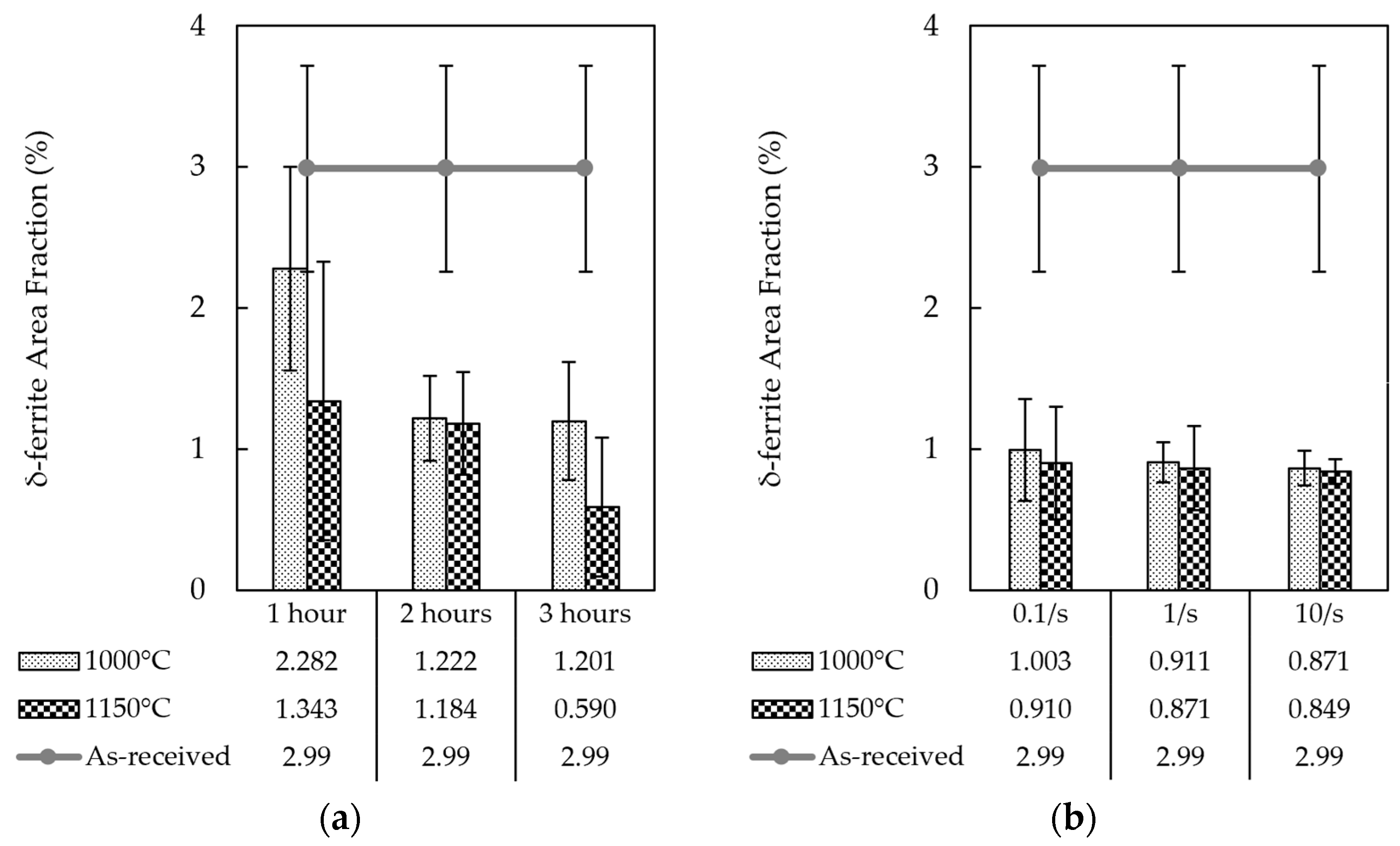
  • Conventional Isothermal Annealing Treatments: The heat treatments were performed at two different temperatures of 1000 and 1150 °C, while the soaking times were chosen as 1, 2, and 3 h, respectively. After each treatment, the samples were air-cooled to room temperature. A computer-controlled 3-Zone Tube Furnace (LINDBERG CC58434C/LINDBERG, Watertown, WI, USA) was used for these heat treatments.
  • Hot Compression Experiments: After heating the samples to the deformation temperature of 900 °C, a Universal Testing Machine (INSTRON 1000HDX/INSTRON, Norwood, MA, USA) equipped with tungsten carbide (WC) dies and an infrared furnace with four 1000 W lamps (ChambIR E4-D-01-A/ChambIR, Eden Prairie, MN, USA) was used to conduct compression tests under three distinct strain rates of 0.1, 1, and 10/s. The deformation strain was kept constant at 60% in all experiments. The post-process heat treatments were carried out at 1000 °C and 1150 °C for 10 min immediately after the deformation, followed by air cooling to room temperature. These annealing treatments were performed with the aid of a muffle furnace (Thermo Scientific Thermolyne F48028-80/Thermo Scientific, Asheville, NC, USA).
After conducting all physical experiments, standard metallographic procedures were applied for microstructural characterization. Following mounting and grinding, polishing was performed to further refine the surface, employing a 0.05 μm deagglomerated alumina solution with the aid of a Giga-S 0900 mechanical vibratory polisher (PACE TECHNOLOGIES, Tucson, AZ, USA) for 1 h. Murakami’s reagent (10 g of NaOH, 10 g of Potassium Ferricyanide, and 100 mL of DI Water/chemicals from SIGMA-ALDRICH, St. Louis, MO, USA) was used to etch the 15-5PH stainless steel, while M-154 was etched with Villela’s reagent (5 mL of HCl, 1 g of Picric Acid, and 100 mL of ethanol/chemicals from Fisher Chemicals, Fair Lawn, NJ, USA). Optical microscopy images were taken with the aid of a Zeiss Smartzoom digital optical microscope (Carl Zeiss Microscopy GmbH, Jena, Germany). Further processing of images was carried out using color thresholding in the free software ImageJ 1.53k to examine the area fraction and morphology of the δ-ferrite. A Zeiss SIGMA VP Scanning Electron Microscope (Carl Zeiss Microscopy GmbH, Oberkochen, Germany) equipped with Energy-Dispersive Spectroscopy (EDS) was applied to examine the microstructure of the samples. A 20 kV beam was used to verify the presence of δ-ferrite. Additionally, using EDS, a qualitative chemical analysis of the δ-ferrite was conducted for verification. The EPMA analysis was performed using a Jeol JXA-8530F Field Emission Probe Microanalyzer (EPMA)/JEOL Ltd., Tokyo, Japan for quantitative compositional examination of the δ-ferrite and matrix. The EPMA system was operated using a current of 100 nA and a 20 kV beam.
Additionally, a series of thermodynamic simulations were conducted using Thermo-Calc software version 2021.2.87071-368 equipped with the Diffusion (DICTRA) Add-on Module [25]. Using a database of 500 points acquired from compositional EPMA line scans and bulk composition of the two as-received alloys, 15-5PH and M-154, computations and predictions were employed. The dissolution time of the δ-ferrite phase during isothermal annealing was simulated and predicted under the assumption of a homogenized system.

3. Results and Discussion

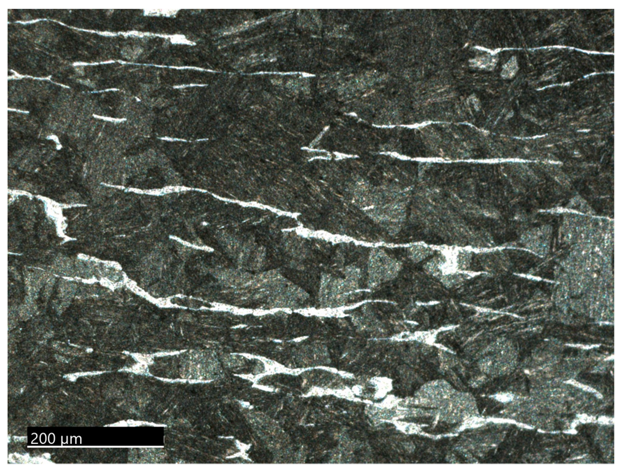
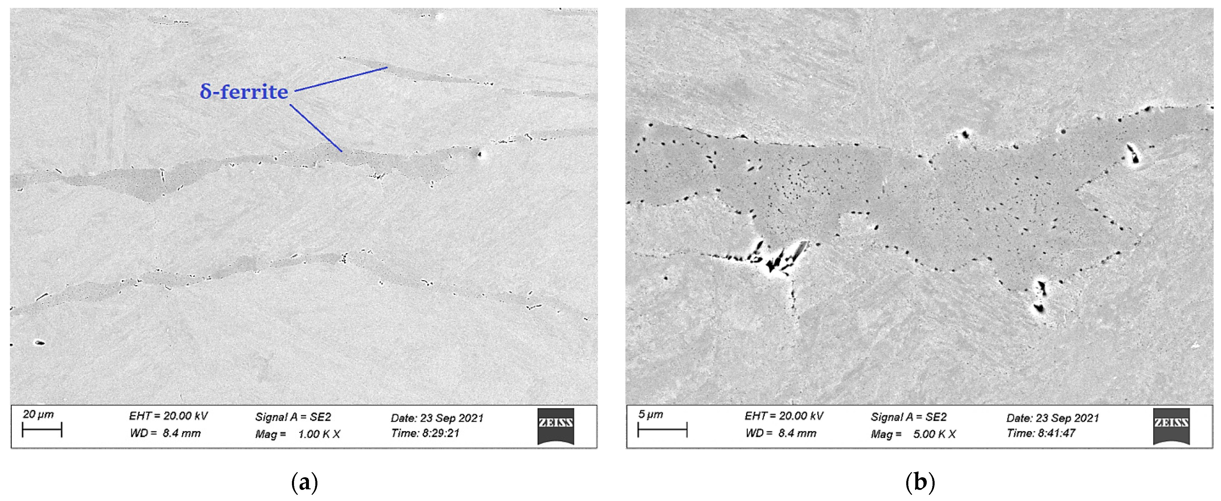
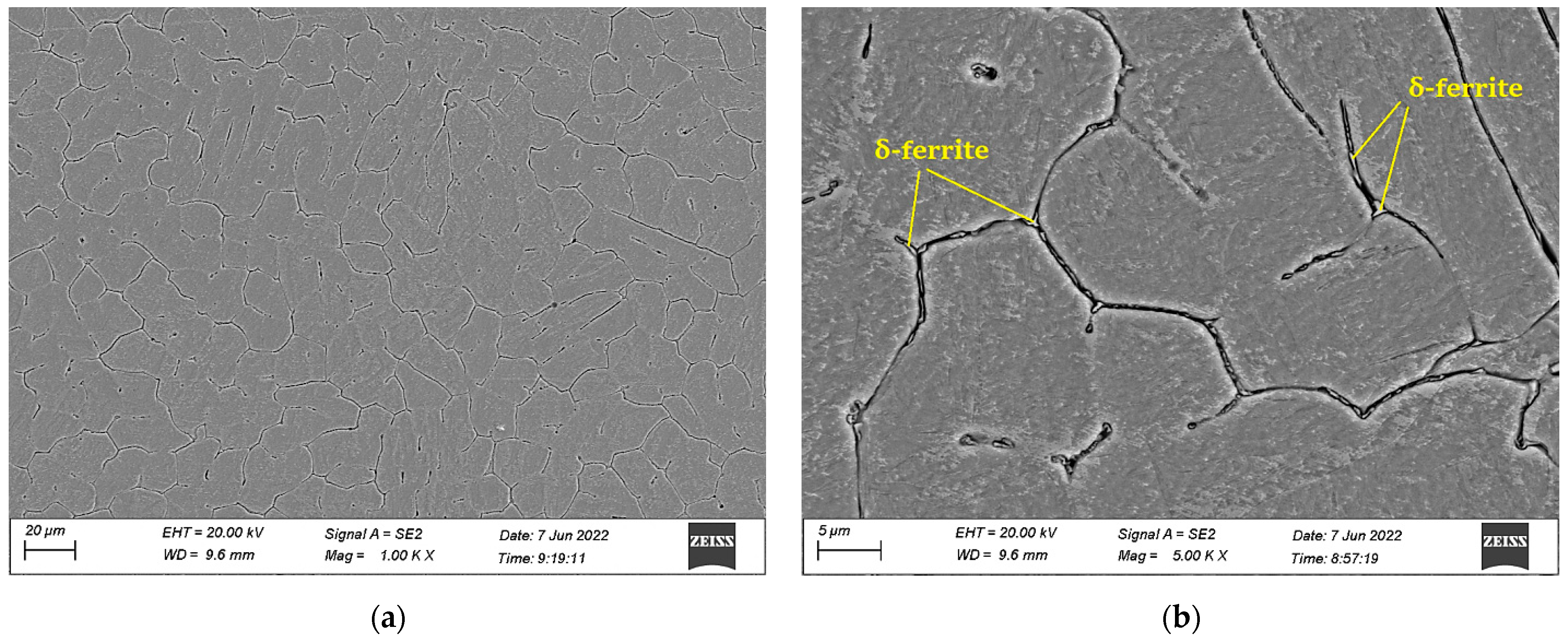
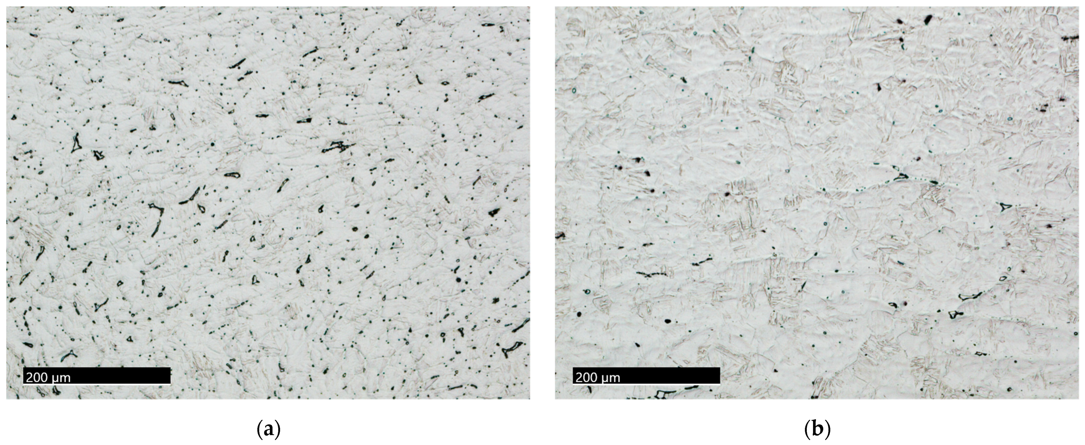
3.1. Microstructural Characterization

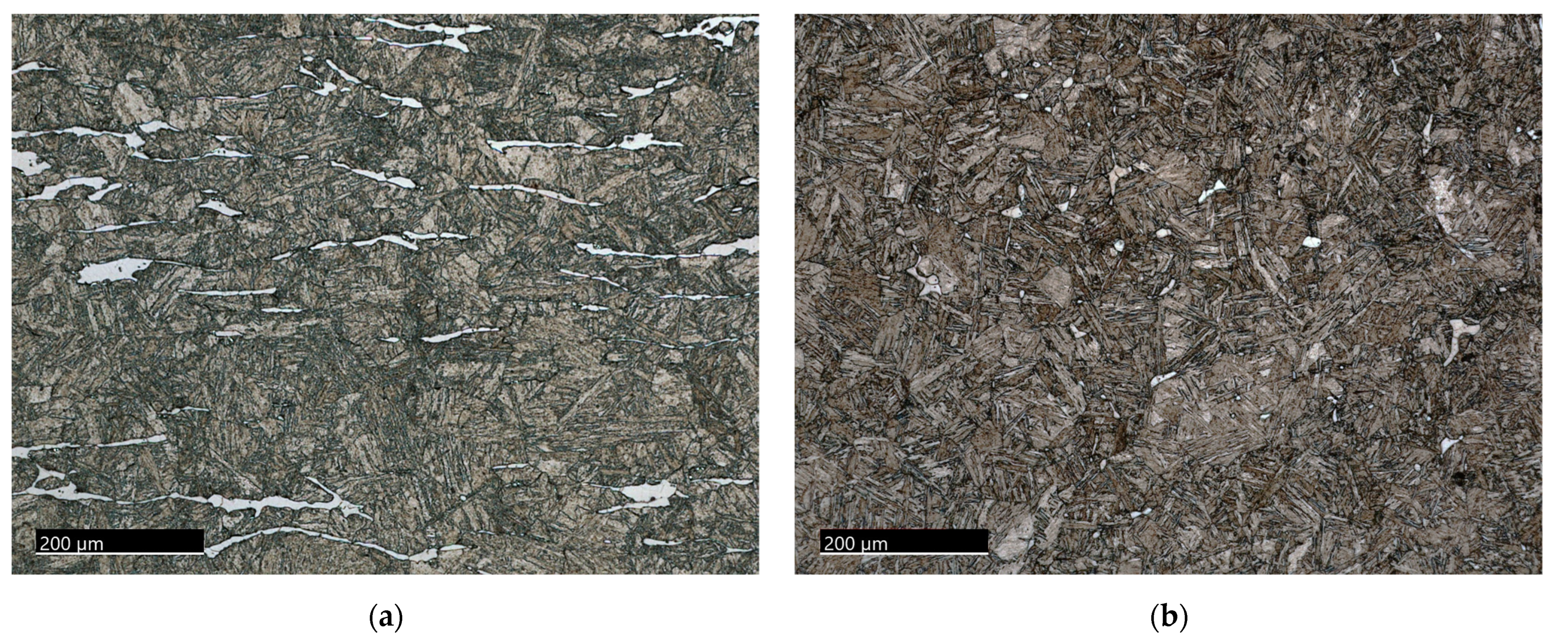
3.1.1. The 15-5PH Stainless Steel

Murakami’s reagent was used to reveal the presence of δ-ferrite in the 15-5 precipitation-hardened stainless steel, as shown in Figure 1. The δ-ferrite (white) phase is clearly observed as having the typical elongated morphology.
SEM analysis was utilized to explore the microstructure of the 15-5PH samples. The SEM micrographs are illustrated in Figure 2. This figure shows the presence of δ-ferrite (Figure 2a) precipitates within the δ-ferrite phase, and those at the interface matrix/δ-ferrite are shown in Figure 2b. Figure 3 shows a semi-quantitative map of the as-received sample obtained using the EPMA system. The results clearly illustrate the differences in the chemical composition of the δ-ferrite region, where the expected high concentration of chromium and the depletion of nickel can be seen. Copper (Cu) and niobium (Nb) carbides can also be observed in this figure.
An example of the variation in Cr across the matrix/δ-ferrite/matrix is illustrated in Figure 4. This behavior is in good agreement with the literature [26,27].
It is well-known that long isothermal treatments will change the morphological characteristics of δ-ferrite by the capillarity effect [28]. Consequently, the elongated δ-ferrite phase would change to a more thermodynamically stable round-shape morphology. Figure 5 shows the optical micrographs of the 15-5PH alloy subjected to annealing heat treatments at 1000 °C for 1 h (Figure 5a) and at 1150 °C for 3 h (Figure 5b), representing the change in morphology with temperature and time used in this study.
Figure 5 exhibits two major changes as follows: the decrease in the δ-ferrite volume fraction and the changes in its morphology. In comparison with the changes in the δ-ferrite during isothermal treatments, Figure 6 shows the results observed in the δ-ferrite after hot compression and post-processing at 1000 and 1150 °C and 10 min of holding time. The effect of hot compression and short-time post-processing clearly increases the kinetics of δ-ferrite dissolution.
Figure 7 compares the volume changes in δ-ferrite in the 15-5 PH steel using only isothermal treatments (Figure 7a) versus the effect of deformation processing parameters and post-processing (Figure 7b). The results presented in this figure show that the combined use of hot deformation and post-short isothermal treatments are more effective in increasing the dissolution kinetics of δ-ferrite compared with employing only long high-temperature isothermal treatments. During isothermal treatments, after 1 h at 1000 °C, the volume fraction of δ-ferrite decreased by about 24% from the as-received condition. Using the combination of hot deformation at a strain rate of 0.1/s and a short isothermal treatment (10 min) at the same 1000 °C decreased the volume fraction of δ-ferrite by 66%. Increasing the strain rate resulted in a faster dissolution rate of the δ-ferrite, as shown in Figure 7b. The increase in the dissolution kinetics can be attributed to the creation of intracrystalline defects during deformation, resulting in an increase in the diffusion rate of Cr and Ni at the matrix/δ-ferrite interphase during post-deformation treatments. These results are in good agreement with the work by Rezayat et al. [29].

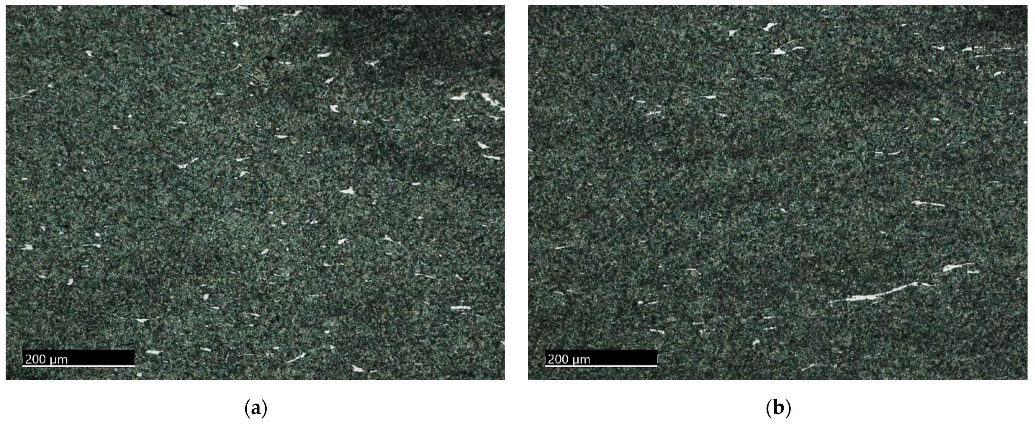
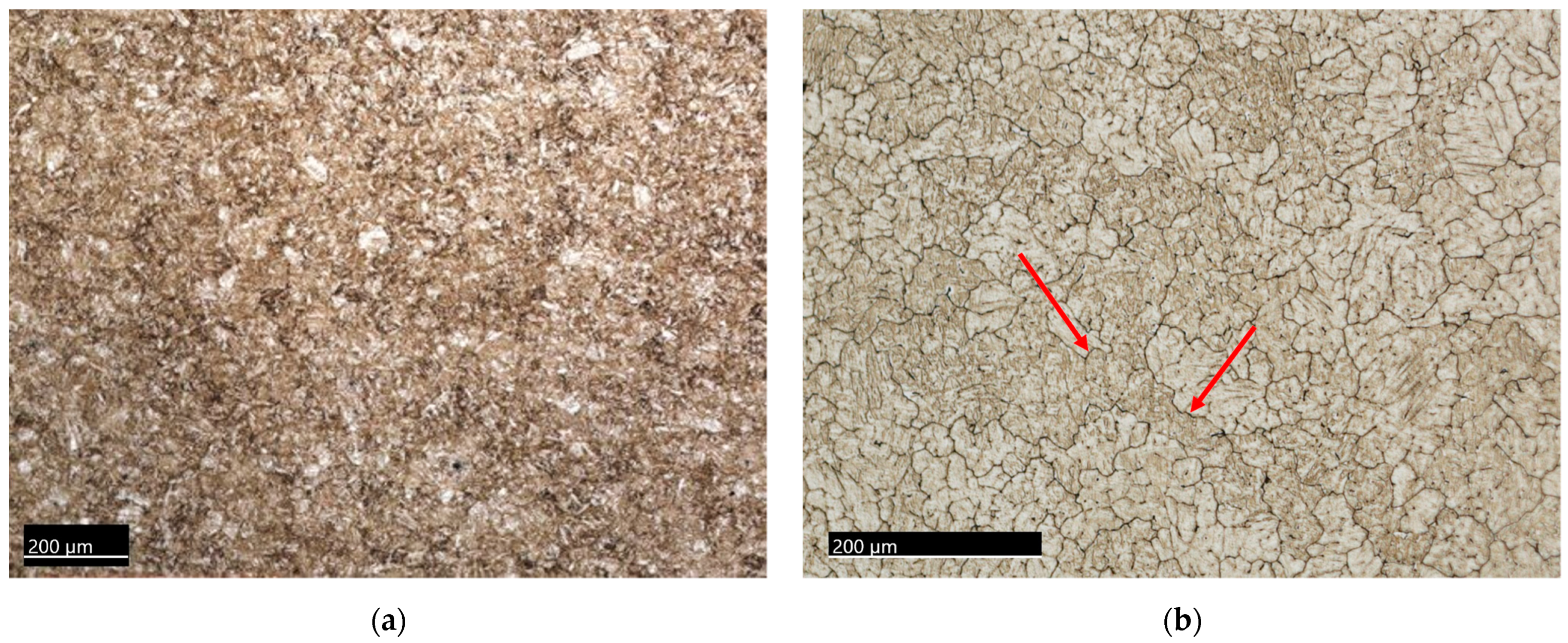
3.1.2. The M-154 Stainless Steel

A modified Villela’s reagent was used to reveal the microstructure of the M-154 martensitic stainless steel. Figure 8a shows the as-received condition with no δ-ferrite phase. Figure 8b shows the microstructure of the steel after the re-engineering treatment described in the experimental procedure section. The presence of δ-ferrite can be observed along the darkly etched prior austenite grain boundaries as they serve as favorable nucleation sites upon melting because of the higher amount of energy in these locations.
A more detailed examination of the microstructure of M-154 after the re-engineering treatment was performed with the aid of the SEM microscope, as illustrated in Figure 9. To verify the composition of this phase, EDS analysis was conducted, and the results are exhibited in Figure 10. As expected, the δ-ferrite is characterized by an enrichment in chromium content.
The changes in the δ-ferrite morphology after the treatments at 1000 °C for 1 h and at 1150 °C for 3 h for the M-154 samples are illustrated in Figure 11. After each treatment, the δ-ferrite continuity along the prior austenite grain boundaries transformed into smaller and round features. Additionally, a lower volume fraction of δ-ferrite could be observed after the 1150 °C treatment compared with the lower temperature treatment.
The microstructures of the M-154 steel after hot deformation at a strain rate of 10/s and post-annealing treatment at 1000 °C and 1150 °C are presented in Figure 12. As described in the previous section, the application of hot compression increased the number of intracrystalline defects/area, providing faster diffusion paths for the diffusion of Cr from the δ-ferrite to the matrix and hence increasing the kinetics of dissolution of the δ-ferrite. A comparison of the changes in δ-ferrite between isothermal treatments and combined hot deformation and short isothermal treatments is presented in Figure 13.
In general, the dissolution of the δ-ferrite phase accelerates at high temperatures because of the enhancement in chemical elements’ diffusion rates. In addition, chemical homogenization and, in particular, morphological change occur during the dissolution of the metastable δ-ferrite. As demonstrated in Figure 5 and Figure 11, the initial vermicular shape of δ-ferrite tends to disintegrate and spheroidize with an increase in annealing time and temperature. Subsequently, it causes the appearance of δ-ferrite in a larger number of individual spherical form islands, increasing the surface area between the matrix and δ-ferrite. This phenomenon leads to a reduction in the interfacial energy along their boundaries. Therefore, since δ-ferrite interfaces are typically considered crack initiation locations, such stereographical modifications are advantageous in this regard.
As shown in Figure 6 and Figure 12, applying the heat treatments as a post-processing step after mechanical deformation seems to represent another route to dissolve δ-ferrite. Applying the deformation on the stainless steel samples increases the strain on the grain boundaries and the subsequent application of heat treatment results in substantial decreases in the δ-ferrite phase fractions as the path to the favorable transformation of δ γ is eased [30].
The results of this investigation are in agreement with the knowledge that high-temperature isothermal treatments accelerate the dissolution of δ-ferrite in stainless steel systems. However, the major objective of the present work was to introduce the combined effect of high-temperature deformation parameters and short isothermal treatments on the dissolution behavior of δ-ferrite. The dissolution kinetics of δ-ferrite in the 15-5PH steel was accelerated as a function of deformation temperature, strain rate, and post-heat treatments (Figure 7), while in the M-154 steel, the effect of strain rate and post-heat treatments on the decrease in the content of δ-ferrite was not as pronounced when compared with the 15-5PH steel. A possible explanation for this behavior is likely a result of the synergistic effect of the matrix composition on the activity of Cr and the initial localized distribution of δ-ferrite during the re-engineering treatment. Therefore, temperature had a greater impact on dissolution than the strain rate (Figure 13). It is postulated that with longer annealing times during post-processing procedures, a more prominent influence of the deformation process could have been appreciated, as the 10-minute holding time appears insufficient to significantly enhance the discernible contrast for each strain rate.

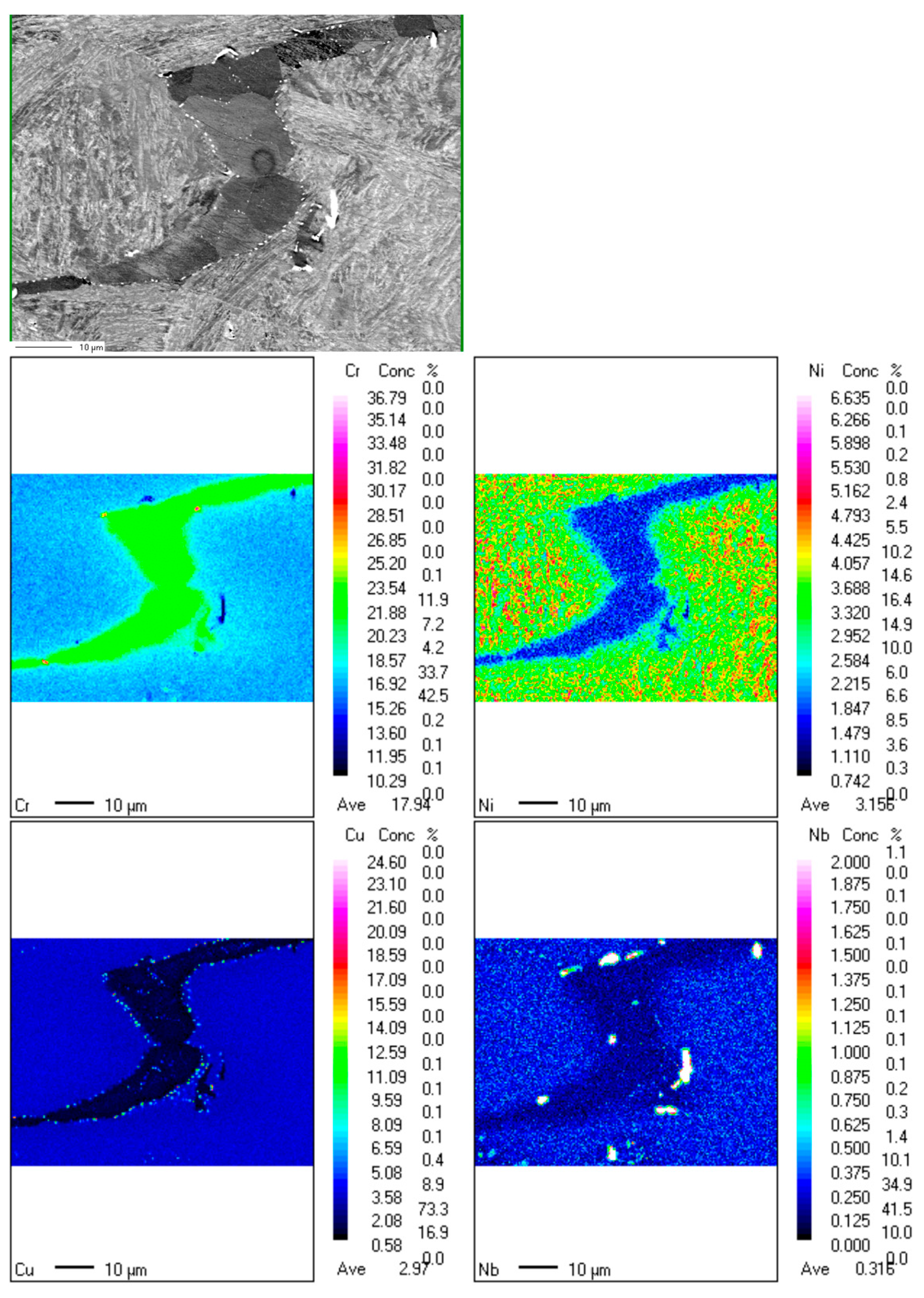
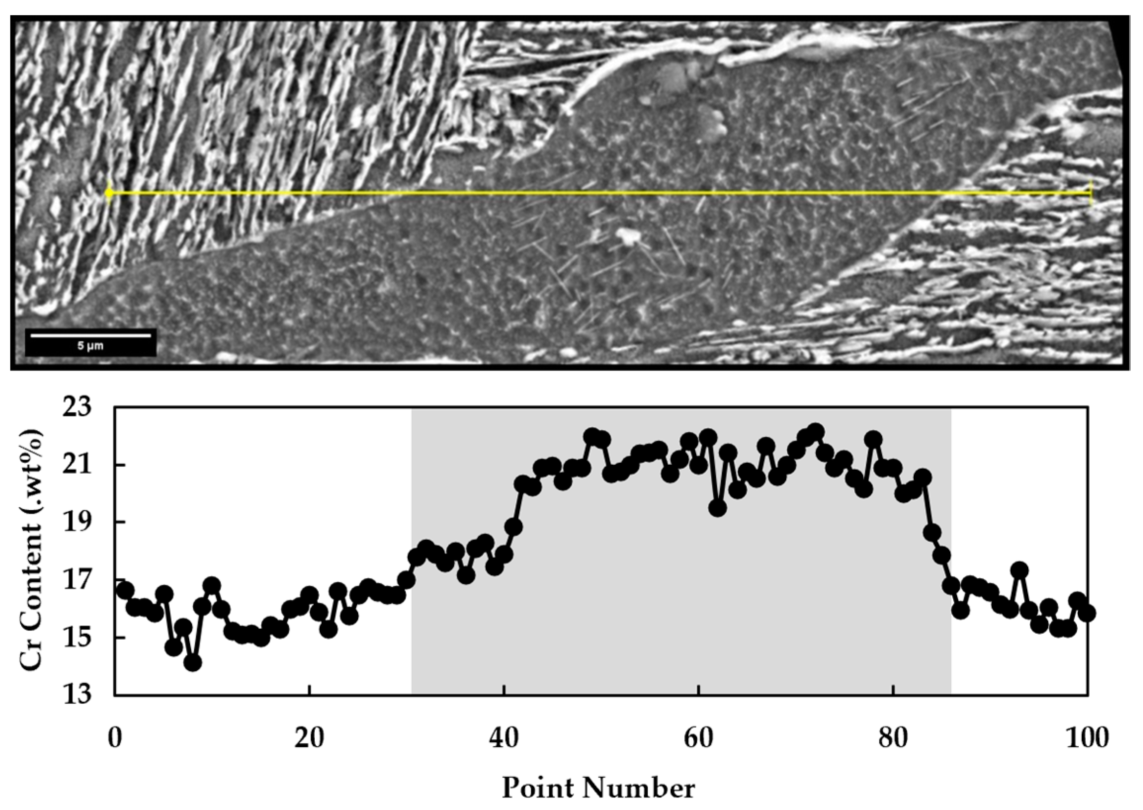
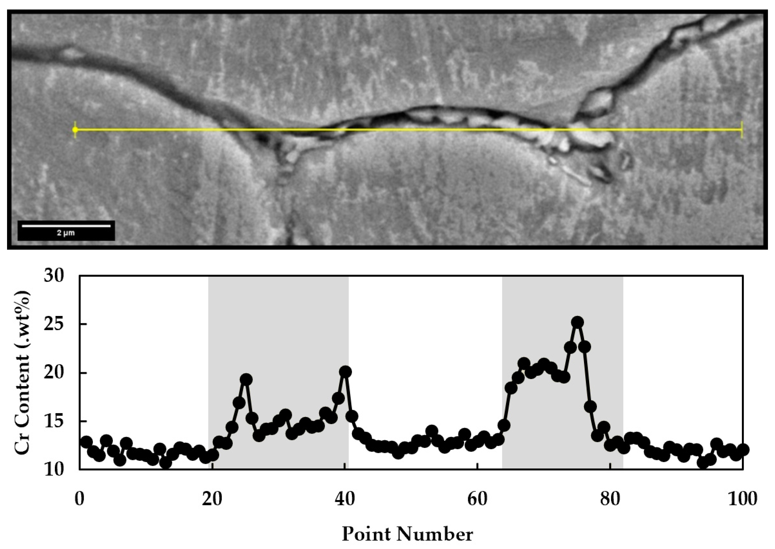
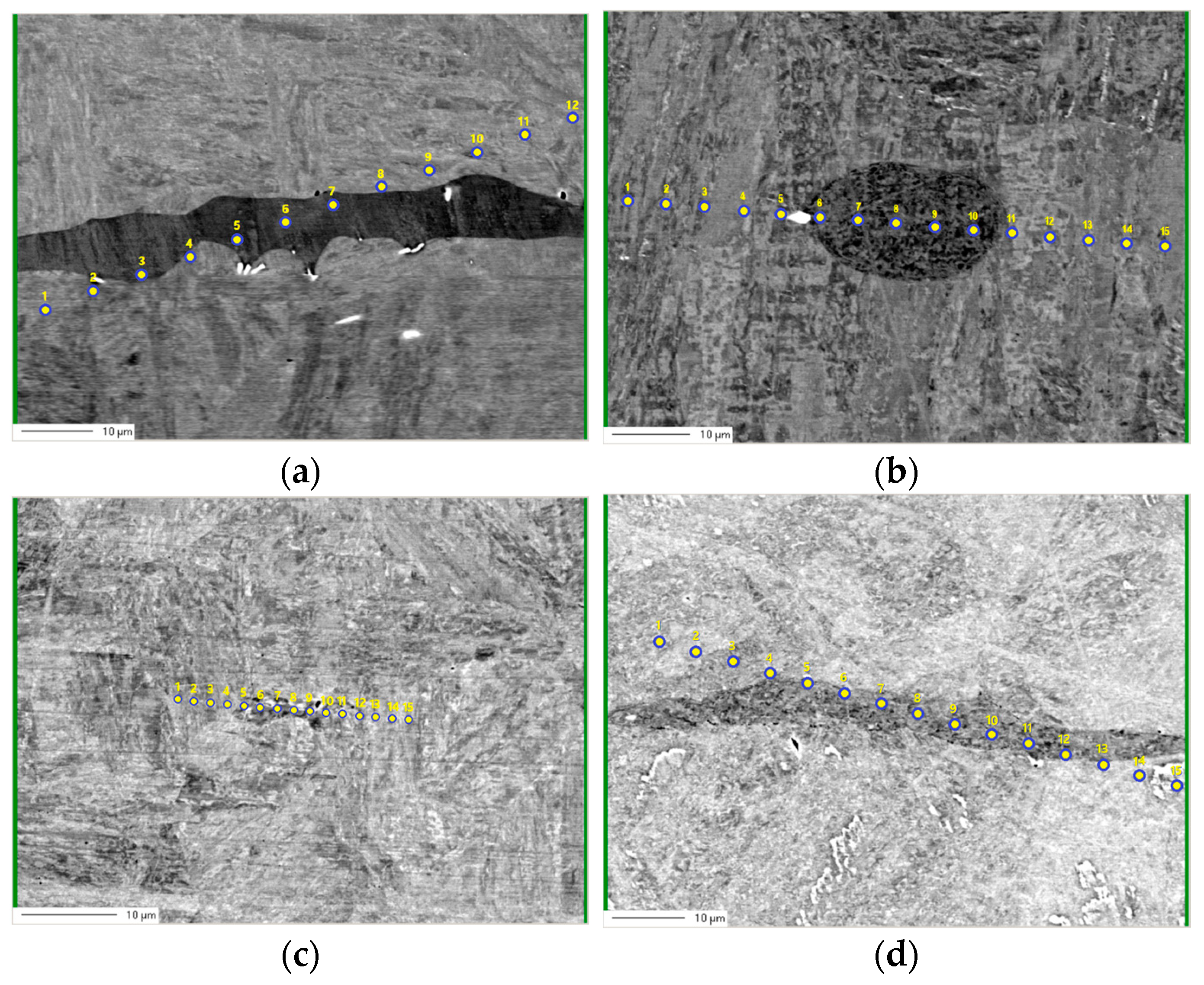
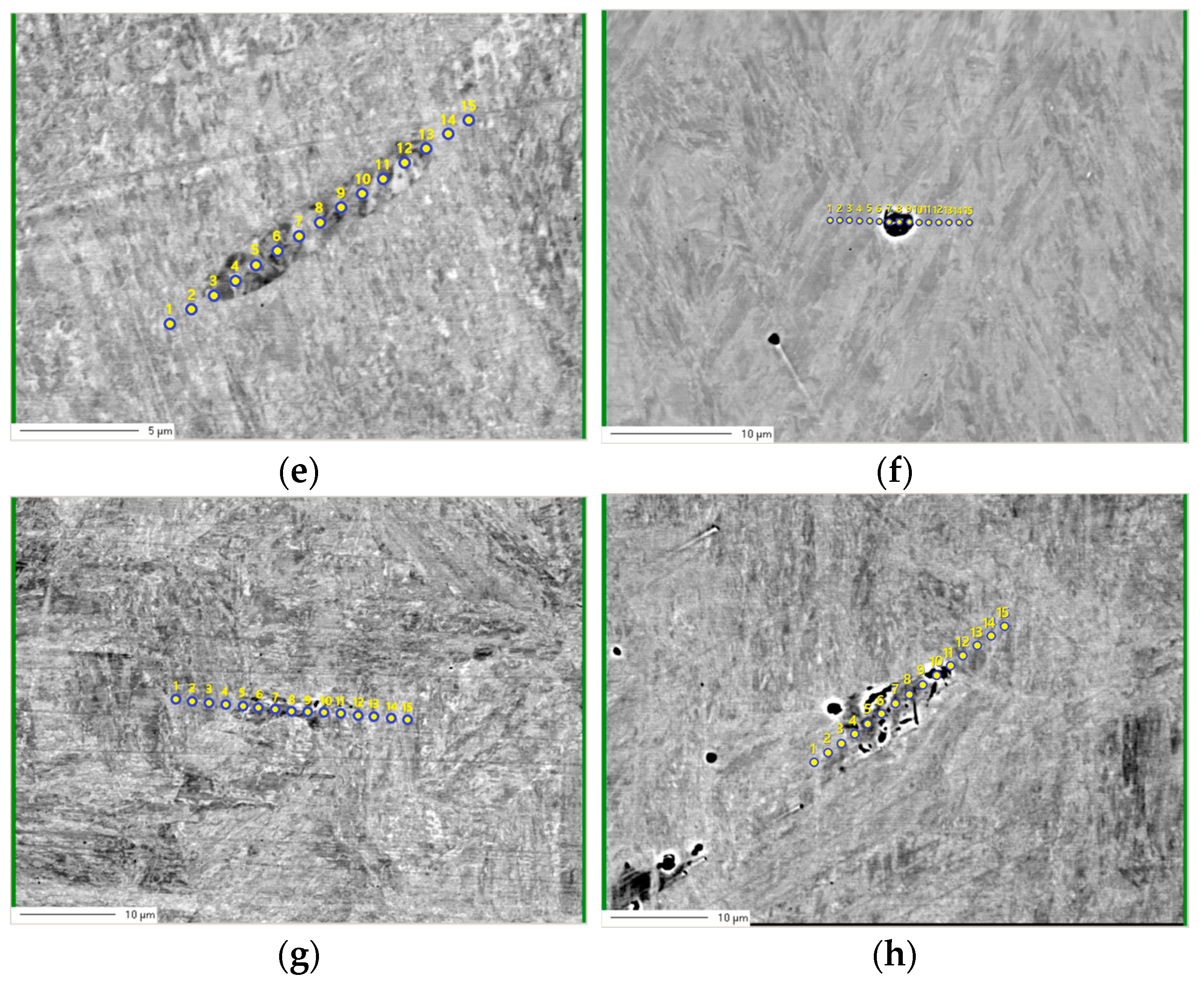
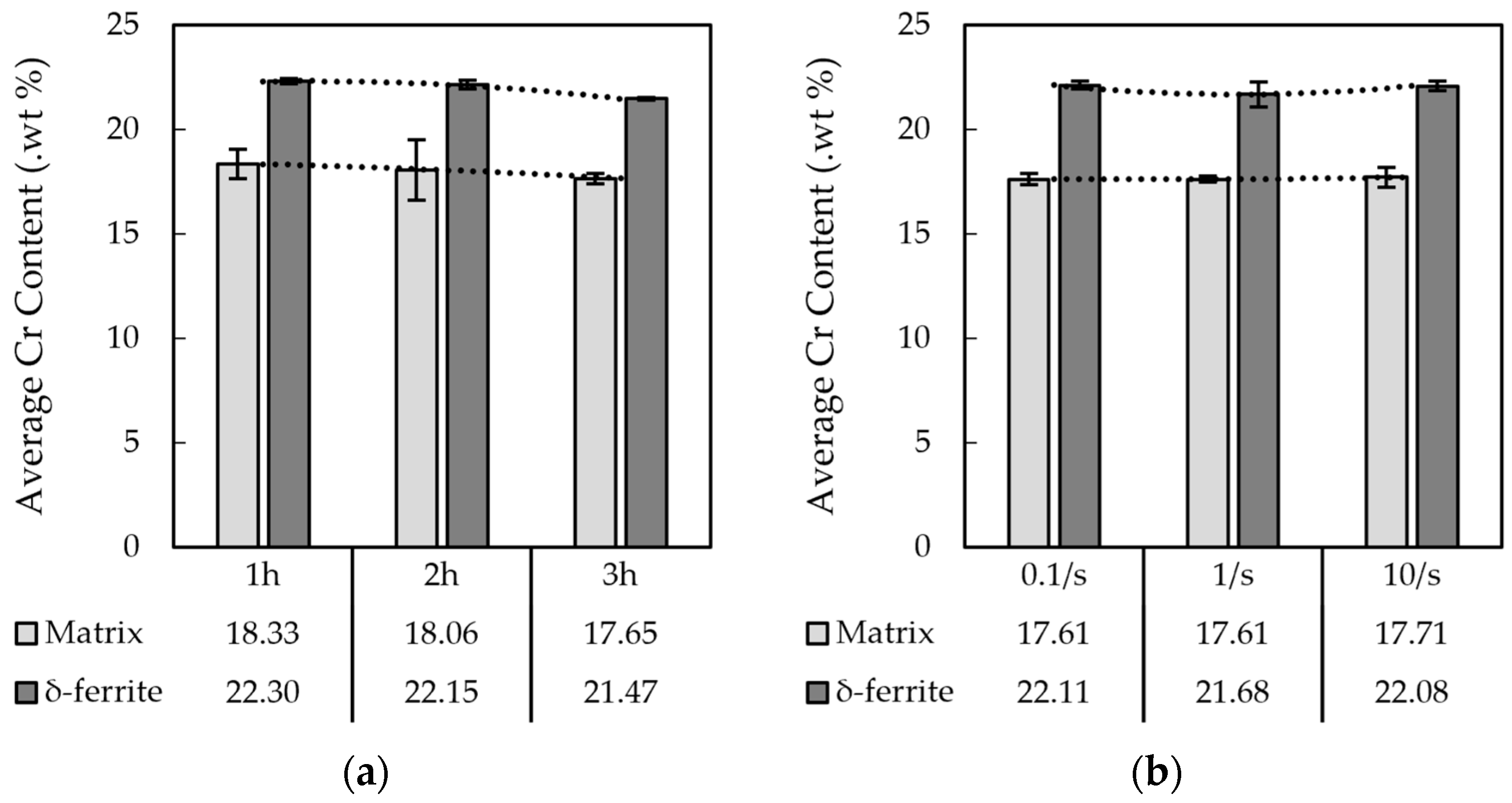
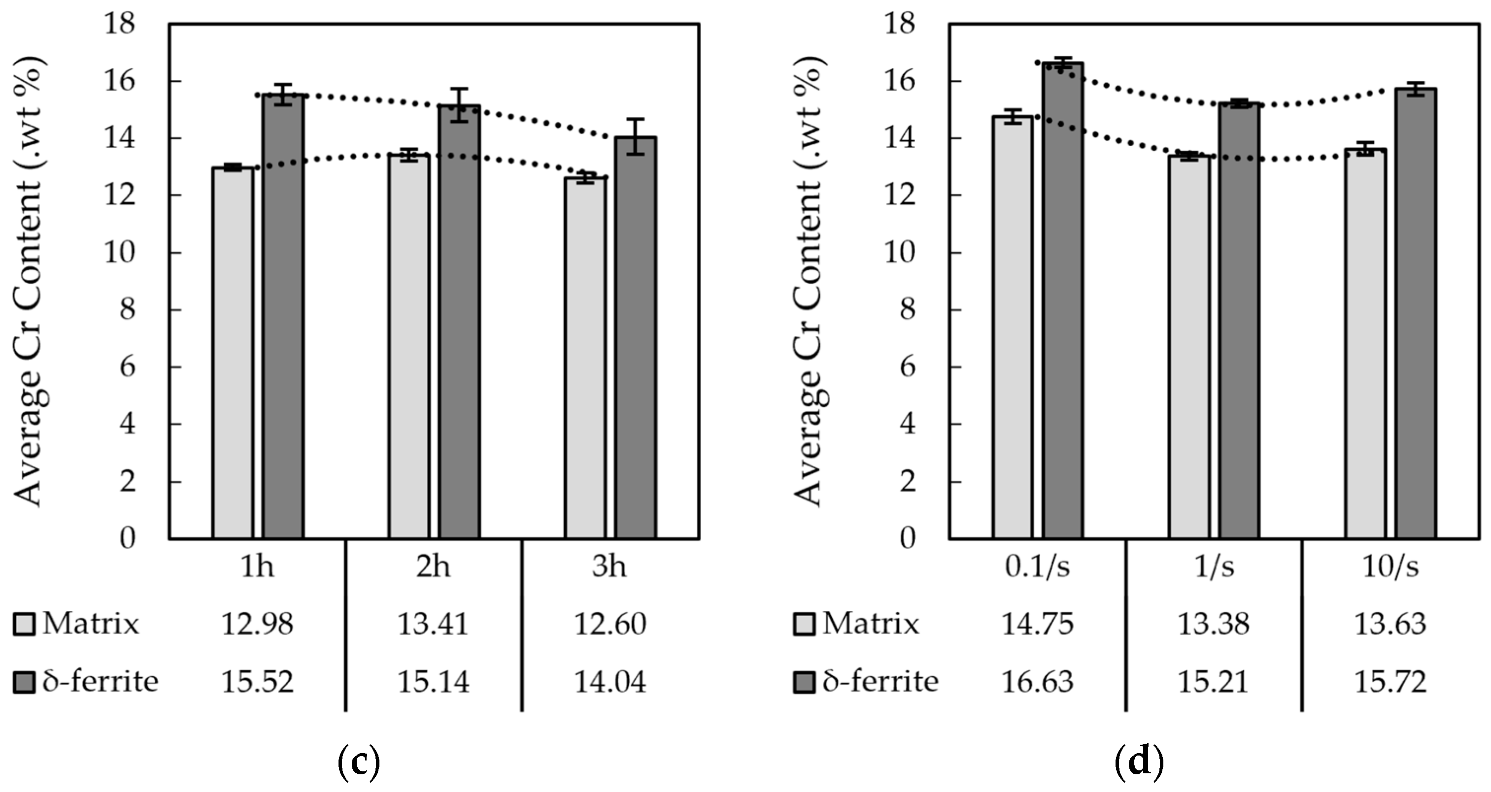
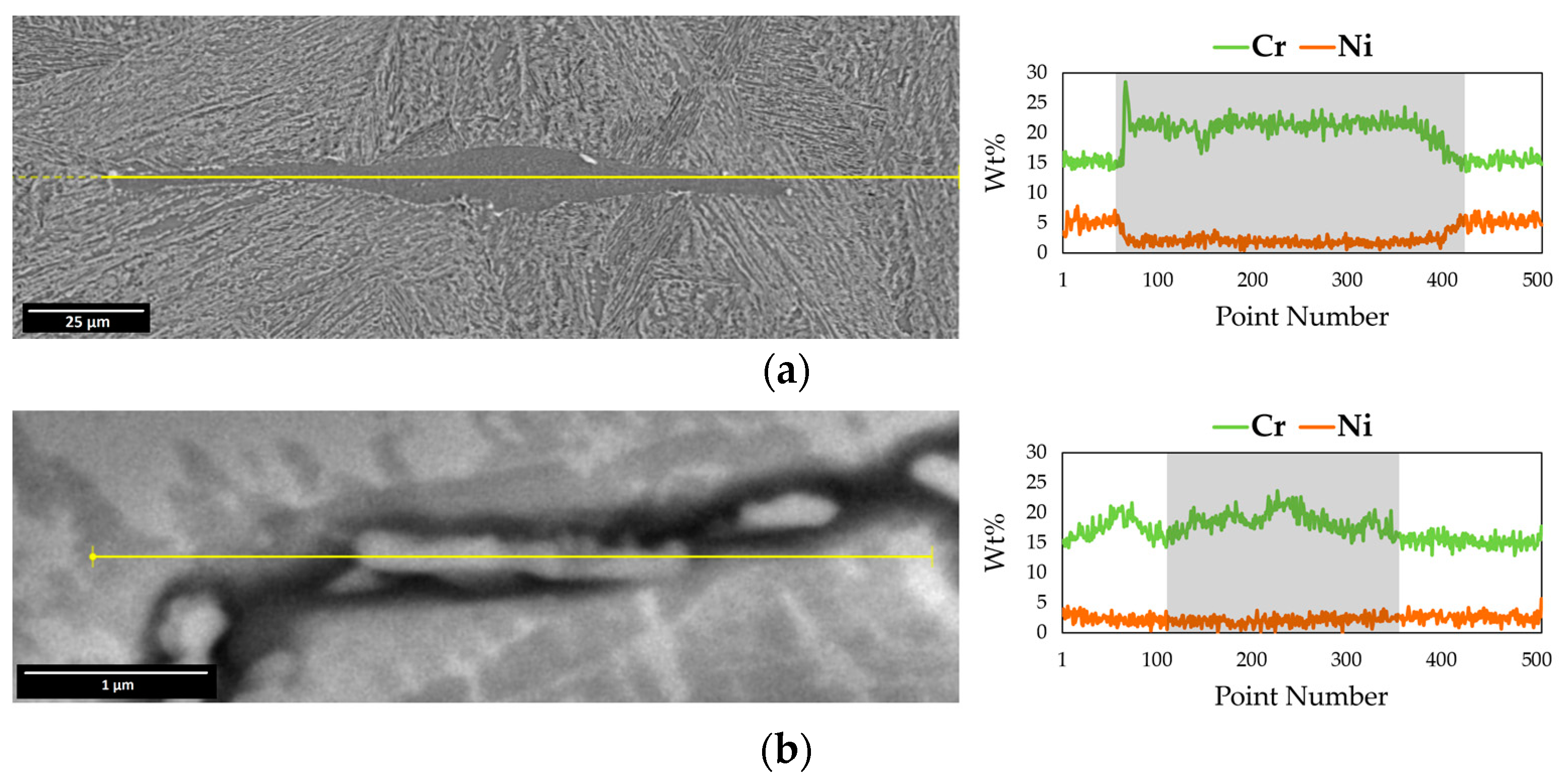
3.2. Electro Probe Micro Analysis (EPMA)

The EPMA technique was used to measure the changes in chemical composition quantitatively along the δ-ferrite phase and the matrix. As expected, the δ-ferrite phase presented higher concentrations of chromium and lower fractions of nickel for both materials. The variations in composition between the matrix and the δ-ferrite were obtained from all conducted experiments, both isothermal annealing and hot compression plus post-isothermal tests. Examples of the methodology used for EPMA data collection and composition line measurements are shown in Figure 14.
Figure 15 shows the comparison of Cr content for each annealed and compressed sample at 1150 °C. After conventional annealing experiments, a similar trend could be observed for the two studied materials: the system tends to chemically homogenize with the increase in the annealing temperature and/or time, promoting an approximation between the matrix and the δ-ferrite phase chemistries. Nonetheless, this chemical composition approximation took place at different rates and times, indicating an important contribution of the materials’ bulk compositions on the dissolution behavior of δ-ferrite. The compressed specimens showed a similar trend observed in the annealed specimens, but the chemical homogenization happened at much shorter times.
Because of the higher Cr solubility within the δ-ferrite, a notably higher Cr content is present in this phase, as well illustrated by microstructural characterization analysis. Thus, assuming the occurrence of complete dissolution, the chromium content homogeneity is expected to be observed between the stainless steel matrix and the δ-ferrite-containing regions. As the annealing temperature and time are raised, the δ-ferrite fraction decreases [31,32] and the chemical homogenization becomes more prominent. In other words, as the δ-ferrite fraction declines in the matrix, the chemical composition tends to take on comparable values both inside and outside the δ-ferrite area. Figure 15 illustrates this behavior.
Hence, the purpose of this work was not to dissolve and eliminate the δ-ferrite in the matrix completely by conducting either the conventional annealing or hot deformation but to compare the reduction in the δ-ferrite content in these different approaches. Moreover, similar behavior can be also noticed after the combination of hot deformation and a post-processing heat treatment compared with the conventional heat treatment.

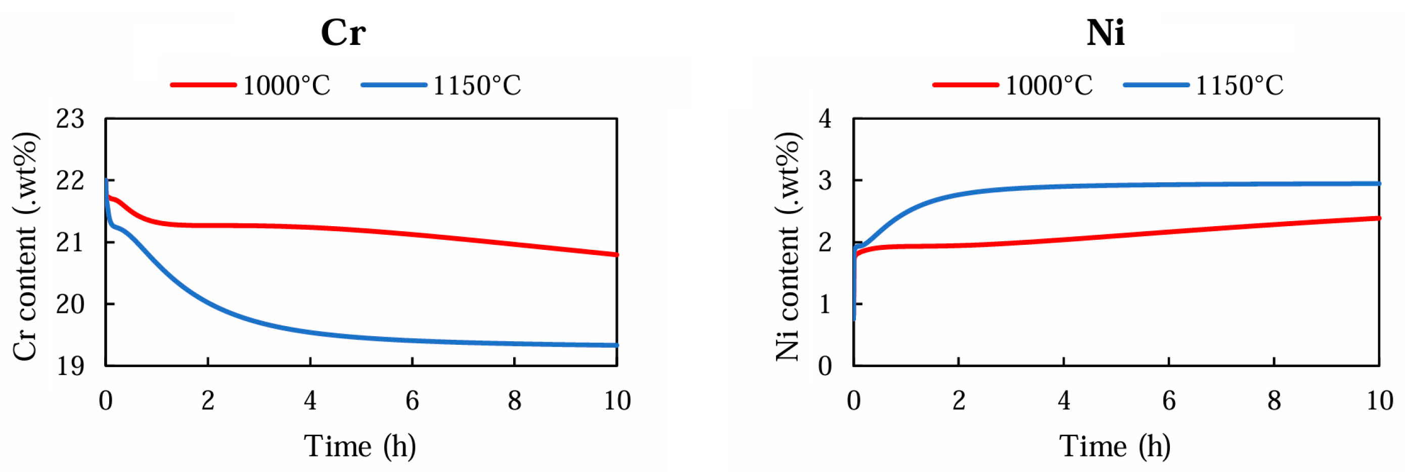
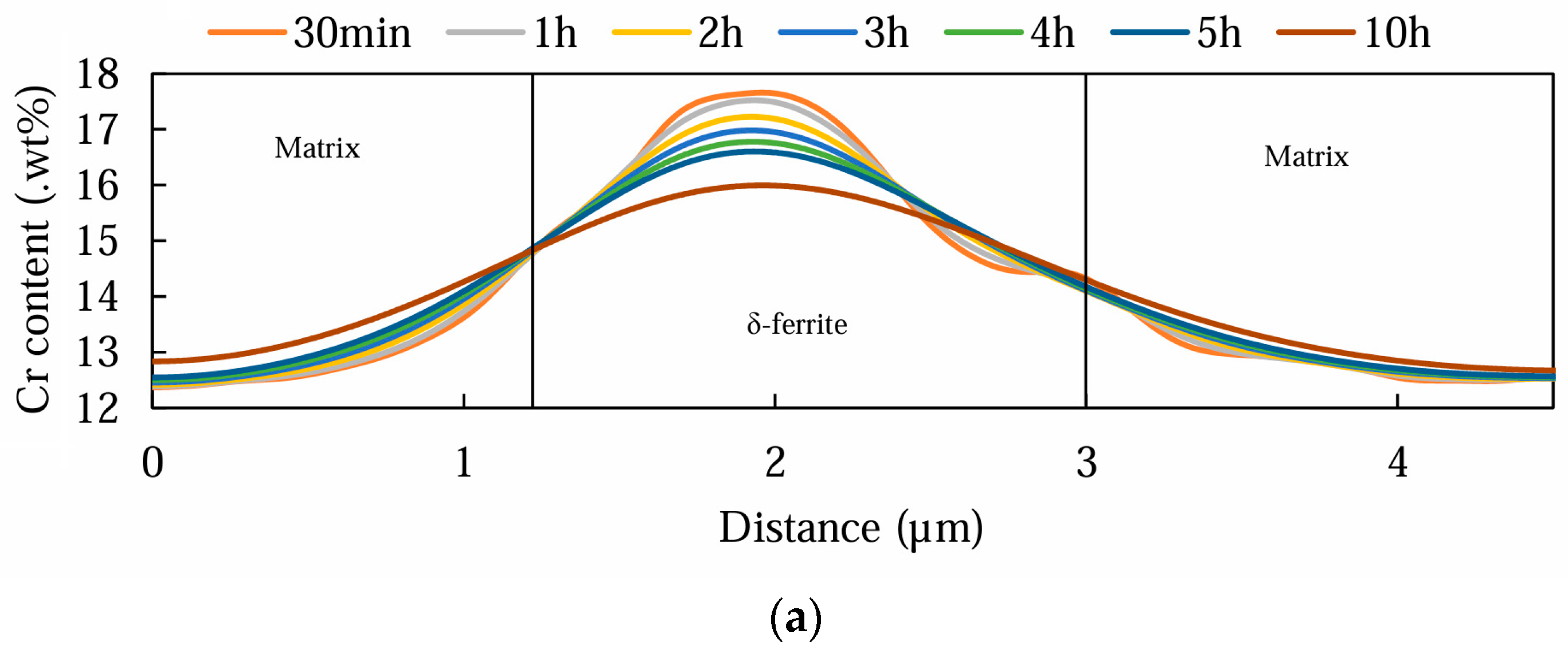
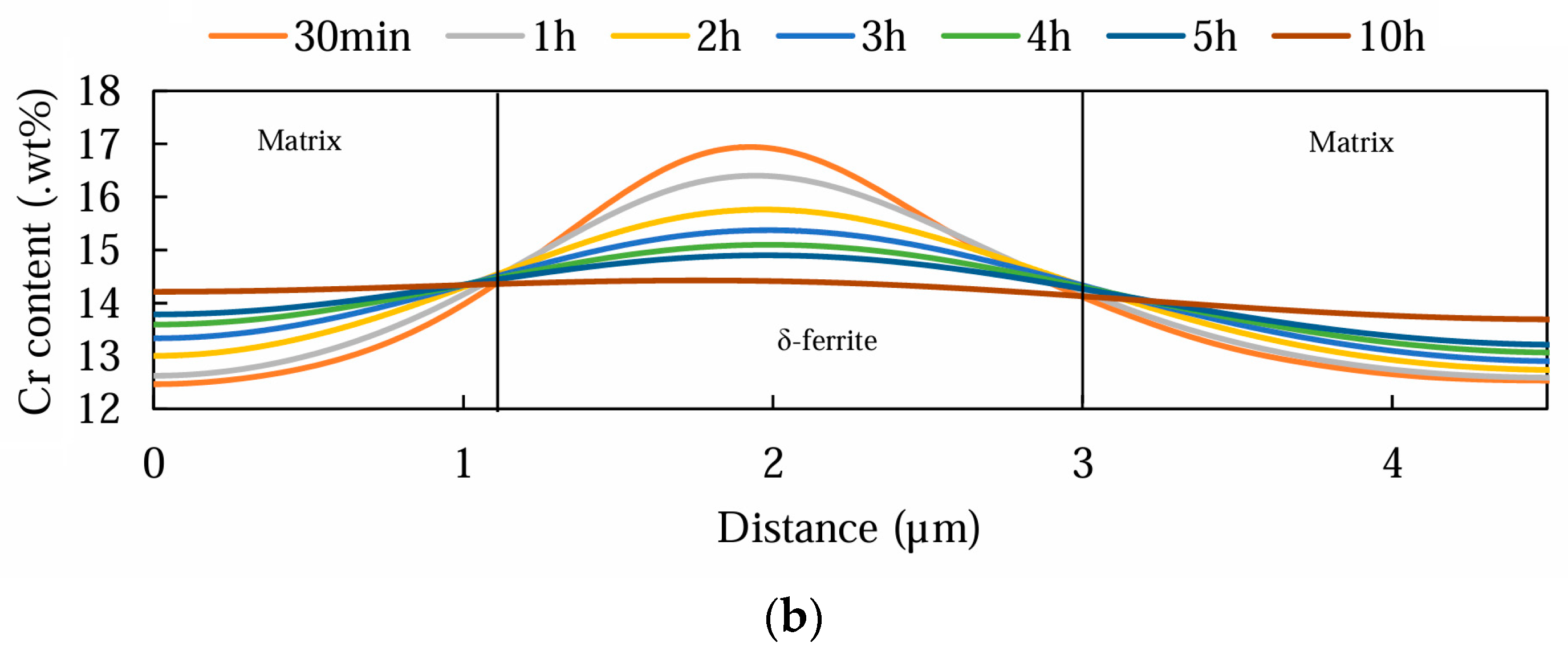
3.3. Thermodynamic Simulations

EDS compositional lines were acquired for each alloy as input data for the software. The line scans along with the percentage of Cr and Ni measured are illustrated in Figure 16.
The simulations were conducted under the assumption that δ-ferrite dissolution is a homogenization process, meaning that a simultaneous chemical accommodation occurs within each stainless steel matrix as the dissolution proceeds. All systems were examined considering the BCC matrix for both the 15-5PH and M-154 alloys. The two experimental temperatures, 1000 °C and 1150 °C, were set, and six distinct annealing times—30 min, 1 h, 2 h, 3 h, 4 h, 5 h, and 10 h—were chosen for the simulations. Figure 17 demonstrates the simulation results at 1000 and 1150 °C for the 15-5PH steel.
A comparison between the simulation results conducted at 1000 and 1150 °C in the 15-5PH material reveals two main observations. Firstly, as expected, increasing the simulation time leads to a reduction in the Cr content inside the δ-ferrite region in both suggested temperatures, implying the dissolution behavior. Hence, considering the higher temperature of 1150 °C, an immediate decrease in the Cr content can be observed as it seems to be homogenized after 3 h of annealing, confirming the faster dissolution behavior and reduction in the area fraction. Secondly, dissolution occurrence is also demonstrated by the fact that the distance corresponding to the δ-ferrite region shortens as the simulation temperature increases.
As evidenced by the EPMA analysis, the largest heterogeneity in the chemical composition can be expected at δ-ferrite areas because of the significant change in Ni and Cr content within those regions. At this region, the δ-ferrite grains were represented by a nickel depletion and chromium enrichment. Therefore, by fixing the simulation to take into account only the δ-ferrite areas, the amount of time needed to homogenize each of the materials fully was estimated. The Cr and Ni content variation during the time in the 15-5PH material is illustrated in Figure 18.
The DICTRA calculations indicate that homogenization can be obtained when the Ni content stabilizes after an increase while the Cr content levels stabilize after an initial decline.
In the simulation using 1000 °C, neither Cr nor Ni behaves uniformly by increasing the time, suggesting that these elements accommodate over periods greater than 10 h. In contrast, a plateau is observable after 6 h for the simulation at 1150 °C, which could be linked to δ-ferrite’s nearly complete dissolution as the microstructure is homogenized. The findings shown in Figure 18 can be confirmed by the EPMA measurements performed following the annealing heat treatments at 1150 °C (Figure 15a), where longer annealing times were noted to approximate the Cr content in the matrix and the δ-ferrite grains. This agrees with the reported EPMA results, with the selected annealing times for this project, of 1 h, not being sufficient to homogenize the Cr content fully, with a difference between the matrix and δ-ferrite still present after the treatment.
Considering the simulations that were performed for the M-154 stainless steel, as illustrated in Figure 19, a similar chemical homogenization behavior was identified. The Cr content seems to remain consistent between the δ-ferrite areas and the matrix with the increase in the simulation time. Furthermore, the process of chemical accommodation is more advantageous at higher temperatures, where the overestimation of time underlines the drop in Cr content within the δ-ferrite grains. Ten hours of annealing time in the simulation run at 1150 °C presents the least predictable deviation, most likely implying that at that point, δ-ferrite had completely dissolved. A similar pattern as that seen in Figure 15, following EPMA analysis, is also apparent in the simulation conducted at 1150 °C.
Figure 20 illustrates the DICTRA calculations regarding the Cr and Ni content variation in the δ-ferrite region for the M-154 material. Again, a specific distance was fixed and represented the δ-ferrite to simulate the complete dissolution time. Even after annealing for 10 h at 1000 °C, the accommodation of Ni and Cr was not fully achieved. On the contrary, when the simulation was run at 1150 °C, a tendency to reach constancy could be observed in the Cr content values through 10 h of annealing after dropping down at the first 4 h, which can be attributed to the shorter diffusion path offered by the energized system at higher temperature.
The thermodynamic calculations did not include the effect of strain or strain rate. The study of the effect of these variables would have been very interesting, but they fall outside of the scope of this study.

4. Conclusions

In the present study, the dissolution behavior of δ-ferrite in two types of stainless steel using conventional long isothermal annealing and hot deformation experiments was investigated. The introduction of hot deformation was very effective in increasing the dissolution of δ-ferrite in shorter times. The following results can be discerned: (1) Measurement of δ-ferrite’s area fraction and chemical composition revealed that applying a hot deformation followed by 10 min post-processing at a high temperature was effective as an alternative method to dissolve the metastable ferrite in a shorter time. (2) Increasing the annealing time and temperature enhances the diffusion of Ni and Cr elements, which subsequently promotes the dissolution process of δ-ferrite in the matrix. (3) A slight decrease in δ-ferrite’s area fraction can be observed by the increase in the strain rate, suggesting that under certain initial distributions of δ-ferrite, the dissolution process is affected by the working temperature more than the strain rate. (4) The simulation results conducted by Thermo-Calc software equipped with Diffusion Module DICTRA confirmed the influence of annealing time and temperature on the dissolution behavior of δ-ferrite.

Author Contributions

Conceptualization, C.I.G.; methodology, P.d.S.C., M.C.V.M. and L.F.S.; software, P.d.S.C.; validation, P.d.S.C. and M.C.V.M.; formal analysis, P.d.S.C.; investigation, P.d.S.C. and R.B.R.; resources, C.I.G.; data curation, P.d.S.C., M.C.V.M. and R.B.R.; writing—original draft preparation, R.B.R.; writing—review and editing, C.I.G.; visualization, R.B.R.; supervision, C.I.G.; project administration, C.I.G.; funding acquisition, C.I.G. All authors have read and agreed to the published version of the manuscript.

Funding

The authors gratefully acknowledge the generous financial support provided by the Forging Industry Educational and Research Foundation (FIERF) under Grant Number 716075.

Data Availability Statement

The original contributions presented in the study are included in the article, further inquiries can be directed to the corresponding author.

Conflicts of Interest

The authors declare no conflicts of interest.

References

  1. Winters, G.L.; Nutt, M.J. Stainless Steels for Medical and Surgical Applications; ASTM International: West Conshohocken, PA, USA, 2003; Volume 1438. [Google Scholar] [CrossRef]
  2. Baddoo, N. Stainless steel in construction: A review of research, applications, challenges and opportunities. J. Constr. Steel Res. 2008, 64, 1199–1206. [Google Scholar] [CrossRef]
  3. Emun, Y.; Zurob, H.; Kish, J. Corrosion Evaluation of Ferritic Stainless Steels for Automotive Exhaust Applications. Corrosion 2019, 75, 1276–1280. [Google Scholar] [CrossRef] [PubMed]
  4. Hamada, A.; Ghosh, S.; Ali, M.; Jaskari, M.; Järvenpää, A. Studying the strengthening mechanisms and mechanical properties of dissimilar laser-welded butt joints of medium-Mn stainless steel and automotive high-strength carbon steel. Mater. Sci. Eng. A 2022, 856, 143936. [Google Scholar] [CrossRef]
  5. Karabeyoglu, S.S.; Yaman, P. An Experimental Investigation of Martensitic Stainless Steel in Aircraft and Aerospace Industry for Thermal Wear Performance and Corrosion Potential. Pract. Metallogr. 2022, 59, 199–215. [Google Scholar] [CrossRef]
  6. Kumar, R.R.; Gupta, R.K.; Sarkar, A.; Prasad, M. Vacuum diffusion bonding of α-titanium alloy to stainless steel for aerospace applications: Interfacial microstructure and mechanical characteristics. Mater. Charact. 2022, 183, 111607. [Google Scholar] [CrossRef]
  7. Liu, T.; Zhu, X.; Chen, L.; Bi, H.; Lin, Y.; Long, J. Fatigue–creep behavior of two ferritic stainless steels in simulated automotive exhaust gas and argon. J. Mater. Sci. 2020, 55, 3684–3699. [Google Scholar] [CrossRef]
  8. Mishra, P.; Åkerfeldt, P.; Svahn, F.; Nilsson, E.; Forouzan, F.; Antti, M.-L. Microstructural characterization and mechanical properties of additively manufactured 21-6-9 stainless steel for aerospace applications. J. Mater. Res. Technol. 2023, 25, 1483–1494. [Google Scholar] [CrossRef]
  9. Bonagani, S.K.; Bathula, V.; Kain, V. Influence of tempering treatment on microstructure and pitting corrosion of 13 wt.% Cr martensitic stainless steel. Corros. Sci. 2018, 131, 340–354. [Google Scholar] [CrossRef]
  10. Ping, D.; Ohnuma, M.; Hirakawa, Y.; Kadoya, Y.; Hono, K. Microstructural evolution in 13Cr–8Ni–2.5 Mo–2Al martensitic precipitation-hardened stainless steel. Mater. Sci. Eng. A 2005, 394, 285–295. [Google Scholar] [CrossRef]
  11. Rho, B.S.; Hong, H.U.; Nam, S.W. The effect of δ-ferrite on fatigue cracks in 304L steels. Int. J. Fatigue 2000, 22, 683–690. [Google Scholar] [CrossRef]
  12. Du, D.; Wang, J.; Chen, K.; Zhang, L.; Andresen, P.L. Environmentally assisted cracking of forged 316LN stainless steel and its weld in high temperature water. Corros. Sci. 2019, 147, 69–80. [Google Scholar] [CrossRef]
  13. Cardoso, P.H.S.; Kwietniewski, C.; Porto, J.; Reguly, A.; Strohaecker, T.R. The influence of delta ferrite in the AISI 416 stainless steel hot workability. Mater. Sci. Eng. A 2003, 351, 1–8. [Google Scholar] [CrossRef]
  14. Bai, T.; Guan, K. Evaluation of stress corrosion cracking susceptibility of nanocrystallized stainless steel 304L welded joint by small punch test. Mater. Des. (1980–2015) 2013, 52, 849–860. [Google Scholar] [CrossRef]
  15. Mukherjee, M.; Pal, T.K. Role of microstructural constituents on surface crack formation during hot rolling of standard and low nickel austenitic stainless steels. Acta Metall. Sin. (Engl. Lett.) 2013, 26, 206–216. [Google Scholar] [CrossRef]
  16. Chandra, K.; Kumar, N.N.; Kain, V. Effect of retained δ-ferrite transforming to sigma phase on the hardness and corrosion resistance of stainless steel 321. Trans. Indian Inst. Met. 2022, 75, 959–966. [Google Scholar] [CrossRef]
  17. Lu, W.F.; Huang, J.Y.; Yung, T.Y.; Chen, T.C.; Tsai, K.C. Effects of thermal aging on the stress corrosion cracking behavior of cast stainless steel with different δ-ferrite levels in high temperature water environment. J. Nucl. Mater. 2022, 568, 153900. [Google Scholar] [CrossRef]
  18. Liu, B.B.; Zhang, H.; Xu, X.Y.; Tian, Q.R.; Fu, J.X. Analysis of Cracking Causes of AISI 416 Martensitic Stainless Steel During Hot Rolling. Trans. Indian Inst. Met. 2023, 76, 2643–2650. [Google Scholar] [CrossRef]
  19. Alcantar-Mondragón, N.; Reyes-Calderón, F.; García-García, V.; Garnica-González, P.; Estrada-Hernández, S. Thermal and diffusion conditions for delta ferrite formation in partially melted and heat-affected zones of multipass SAW joint performed in martensitic stainless steel 12Cr–1Mo. Results Mater. 2023, 20, 100489. [Google Scholar] [CrossRef]
  20. Cho, J.; Czerwinski, F.; Szpunar, J. The effect of reheating conditions and chemical composition on δ ferrite content in austenitic stainless steel slabs. J. Mater. Sci. 2000, 35, 1997–2003. [Google Scholar] [CrossRef]
  21. Zhang, Y.; Li, D.; Guan, L.; Cao, L.; Wang, W. Effect of Si on δ-Ferrite/γ-Austenite Transformation and M23C6 Precipitation During Solidification of X10CrAlSi18 Ferritic Heat-Resistant Stainless Steel. Steel Res. Int. 2024, 95, 2300528. [Google Scholar] [CrossRef]
  22. Fukumoto, S.; Iwasaki, Y.; Motomura, H.; Fukuda, Y. Dissolution behavior of δ-ferrite in continuously cast slabs of SUS304 during heat treatment. ISIJ Int. 2012, 52, 74–79. [Google Scholar] [CrossRef]
  23. Arh, B.; Tehovnik, F.; Vode, F. Transformation of the δ-Ferrite in SS2343 Austenitic Stainless Steel upon Annealing at 1050 °C, 1150 °C and 1250 °C. Metals 2021, 11, 935. [Google Scholar] [CrossRef]
  24. de Souza Ciacco, P. Study of the Formation and Dissolution of δ-Ferrite during High Temperature Forging Processing of Austenitic, Martensitic and PH Stainless Steels. Master’s Thesis, University of Pittsburgh, Pittsburgh, PA, USA, 2023. Available online: http://d-scholarship.pitt.edu/id/eprint/44265 (accessed on 13 June 2023).
  25. Borgenstam, A.; Höglund, L.; Ågren, J.; Engström, A. DICTRA, a tool for simulation of diffusional transformations in alloys. J. Phase Equilibria 2000, 21, 269–280. [Google Scholar] [CrossRef]
  26. Schäfer, L. Influence of delta ferrite and dendritic carbides on the impact and tensile properties of a martensitic chromium steel. J. Nucl. Mater. 1998, 258, 1336–1339. [Google Scholar] [CrossRef]
  27. Kasana, S.S.; Pandey, O. Influence of Chemical Composition and Thermo-Mechanical Processing on the Formation of Delta Ferrite in 10.7 CrMoVNbN Steel. Trans. Indian Inst. Met. 2020, 73, 2491–2502. [Google Scholar] [CrossRef]
  28. Ravibharath, R.; Muthupandi, V.; Bala Srinivasan, P.; Devakumaran, K. Characterization of solidification cracking in 304HCu austenitic stainless steel welds. Trans. Indian Inst. Met. 2020, 73, 2345–2353. [Google Scholar] [CrossRef]
  29. Rezayat, M.; Mirzadeh, H.; Namdar, M.; Parsa, M.H. Unraveling the effect of thermomechanical treatment on the dissolution of delta ferrite in austenitic stainless steels. Metall. Mater. Trans. A 2016, 47, 641–648. [Google Scholar] [CrossRef]
  30. Ghosh, S.K.; Mahata, D.; Roychaudhuri, R.; Mondal, R. Effect of rolling deformation and solution treatment on microstructure and mechanical properties of a cast duplex stainless steel. Bull. Mater. Sci. 2012, 35, 839–846. [Google Scholar] [CrossRef]
  31. Shaikh, H.; Khatak, H.S.; Seshadri, S.K.; Gnanamoorthy, J.B.; Rodriguez, P. Effect of ferrite transformation on the tensile and stress corrosion properties of type 316 L stainless steel weld metal thermally aged at 873 K. Metall. Mater. Trans. A 1995, 26, 1859–1868. [Google Scholar] [CrossRef]
  32. Cui, C.; Gao, X.; Su, G.; Gao, C.; Liu, Z.; Misra, R.D. Effect of thermal treatment on the evolution of delta ferrite in 11Cr–3Co–2.3 W steel. Mater. Sci. Technol. 2018, 34, 2087–2096. [Google Scholar] [CrossRef]
Figure 1. Optical microscopy showing the as-received 15-5PH steel.
Metals 14 00783 g001
Figure 2. SEM micrographs of the 15-5PH stainless steel microstructure: (a) δ-ferrite and (b) the presence of precipitation.
Metals 14 00783 g002
Figure 3. Map analysis of the δ-ferrite region in the 15-5PH stainless steel via EPMA. Reprinted from Ref. [24].
Metals 14 00783 g003
Figure 4. The EDS line-scan analysis (yellow line) performed on the 15-5PH as-received material; the shaded area represents the δ-ferrite region. Reprinted from Ref. [24].
Metals 14 00783 g004
Figure 5. Optical microscopy of the 15-5PH steel after annealing at (a) 1000 °C for 1 h and (b) 1150 °C for 3 h.
Metals 14 00783 g005
Figure 6. Optical microscopy of the 15-5PH steel after hot compression and post-processing annealing for 10 min at (a) 1000 °C and (b) 1150 °C. The strain rate was fixed at 10/s.
Metals 14 00783 g006
Figure 7. Area fraction measurements showing the amount of δ-ferrite measured after each experiment conducted on the 15-5PH stainless steel: (a) the conventional method and (b) the hot deformation process. Reprinted from Ref. [24].
Metals 14 00783 g007
Figure 8. Optical microscopy showing the M-154 stainless steel in (a) the as-received condition, reprinted from Ref. [24]. (b) Red arrows indicate the presence of δ-ferrite along grain boundaries after re-engineering heat treatments.
Metals 14 00783 g008
Figure 9. SEM micrographs of M-154 stainless steel (a) after re-engineering and (b) the formation of δ-ferrite.
Metals 14 00783 g009
Figure 10. EDS line-scan analysis (yellow line) showing the increase in Cr in the δ-ferrite observed in M-154 after re-engineering; the shaded areas represent the δ-ferrite regions. Reprinted from Ref. [24].
Metals 14 00783 g010
Figure 11. Optical microscopy of the M-154 steel after annealing at (a) 1000 °C for 1 h and (b) 1150 °C for 3 h.
Metals 14 00783 g011
Figure 12. Optical microscopy of the M-154 steel after hot compression and post-processing annealing for 10 min at (a) 1000 °C and (b) 1150 °C. The strain rate was fixed at 10/s.
Metals 14 00783 g012
Figure 13. Area fraction measurements showing the amount of δ-ferrite measured after each experiment conducted on the M-154 stainless steel: (a) the conventional method and (b) the hot deformation process. Reprinted from Ref. [24].
Metals 14 00783 g013
Figure 14. Examples of EPMA measurements: (a) 15-5PH at 1000 °C for 1 h; (b) 15-5PH at 1150 °C for 3 h; (c) 15-5PH at 1000 °C with 0.1/s strain rate; (d) 15-5PH at 1150 °C with 10/s strain rate; (e) M-154 at 1000 °C for 1 h; (f) M-154 at 1150 °C for 3 h; (g) M-154 at 1000 °C with 0.1/s strain rate; and (h) M-154 at 1150 °C with 10/s strain rate. Reprinted from Ref. [24].
Metals 14 00783 g014aMetals 14 00783 g014b
Figure 15. Examples showing EPMA measurements: (a) 15-5PH annealed at 1150 °C; (b) 15-5PH hot compression at 1150 °C; (c) M-154 annealed at 1150 °C; and (d) M-154 hot compression at 1150 °C. Reprinted from Ref. [24].
Metals 14 00783 g015aMetals 14 00783 g015b
Figure 16. EDS compositional line-scan (yellow line) as the input data for the (a) 15-5PH and (b) M-154 materials. Reprinted from Ref. [24].
Metals 14 00783 g016
Figure 17. DICTRA simulations showing the Cr content evolution after different annealing times for 15-5PH at (a) 1000 °C and (b) 1150 °C. Reprinted from Ref. [24].
Metals 14 00783 g017
Figure 18. DICTRA prediction of the changes in the Cr and Ni contents as a function of time in the δ-ferrite location for the 15-5PH stainless steel. Reprinted from Ref. [24].
Metals 14 00783 g018
Figure 19. DICTRA simulations showing the Cr content evolution after different annealing times for the M-154 alloy at (a) 1000 °C and (b) 1150 °C. Reprinted from Ref. [24].
Metals 14 00783 g019aMetals 14 00783 g019b
Figure 20. DICTRA prediction of the changes in the Cr and Ni contents as a function of time in the δ-ferrite location for the M-154 stainless steel. Reprinted from Ref. [24].
Metals 14 00783 g020
Table 1. Chemical composition (wt.%) of as-received samples.
GradeCMnSiCrNiMoCuNbV
15-5PH0.040.540.2215.464.060.203.800.250.06
M-1540.130.720.2011.502.801.720.17-0.3
Disclaimer/Publisher’s Note: The statements, opinions and data contained in all publications are solely those of the individual author(s) and contributor(s) and not of MDPI and/or the editor(s). MDPI and/or the editor(s) disclaim responsibility for any injury to people or property resulting from any ideas, methods, instructions or products referred to in the content.

