Communication

Phase Mapping Using a Combination of Multi-Functional Scanning Electron Microscopy Detectors and Imaging Modes

Department of Mechanical and Energy Engineering, Southern University of Science and Technology, 1088 Xueyuan Avenue, Shenzhen 518055, China
*
Authors to whom correspondence should be addressed.
Metals 2024, 14(8), 899; https://doi.org/10.3390/met14080899
Submission received: 3 April 2024 / Revised: 2 July 2024 / Accepted: 20 July 2024 / Published: 7 August 2024
(This article belongs to the Special Issue State-of-Art: Metals Failure Analysis)

Abstract

:
Microstructure degradation and phase transformations are critical concerns in nickel-based superalloys during thermal exposure. Understanding the phase transformation mechanism requires the detailed mapping of the distribution of each phase at different degradation stages and in various precipitation sizes. However, differentiating between phases in large areas, typically on the scale of millimeters and often relying on scanning electron microscopy (SEM) techniques, has traditionally been a challenging task. In this study, we present a novel and efficient phase mapping method that leverages multiple imaging detectors and modes in SEM. This approach allows for the relatively rapid and explicit differentiation and mapping of the distribution of various phases, including MC, M23C6, γ′, and η phases, as demonstrated in a typical superalloy subjected to aging experiments at 800 °C.

1. Introduction

Nickel-based superalloys are extensively utilized in extreme service conditions owing to their remarkable properties, including high strength and high corrosion resistance. These exceptional attributes primarily arise from a substantial volume fraction of alloying elements such as Cr, Mo, Ti, and C. However, the incorporation of these elements inevitably leads to the formation of complex phases, including carbides and intermetallic compounds, alongside the major strengthening phase γ′-Ni3Al [1]. During service and thermal exposure processes, these phases undergo degradation and may transform into other phases. A specific phase of concern is the η-Ni3Ti phase, which is believed to transform from γ′-Ni3Al [2,3]. A fully grown η phase is generally considered unfavorable for high-temperature properties as it depletes γ′ and increases the local strain concentration [4,5]. Therefore, these phase transformations are important as both the original degraded phases and newly transformed phases will greatly affect the properties of alloys. Understanding the η phase nucleation and growth mechanism and controlling the precipitation of the η phase are of both scientific and industrial significance. Recently, it was found that the degradation of MC carbides is closely related to the precipitation of the η phase, following the transformation reaction MC + γ → M23C6 + η [6,7]. However, despite long-standing documentation of the degradation of MC carbides, the detailed transformation mechanisms associated with η formation remain unclear. For instance, the precipitation sequences of the transformed phases and the specific relationships between η, MC, and γ′ have rarely been reported [8,9]. Understanding these relationships and mechanisms is critical for controlling η precipitation in superalloys.
One of the challenges in studying these transformations is the difficulty in observing initial precipitates of small size (~nm) and distinguishing each phase in large areas. While traditional transmission electron microscopy (TEM) can explicitly identify every phase, its application is limited to small areas. Although modern SEM techniques offer high resolution in the nanometer range, they face challenges in providing sufficient phase contrast to accurately map different phase distributions. This is especially true when dealing with very similar compositions, as exemplified by the difficulty in distinguishing γ′-Ni3(Al,Ti) and η-Ni3Ti in normal SEM images [9,10]. The compositions of these phases are closely related, making their distinction challenging even with the successful implementation of backscatter electron detectors. Addressing these challenges is crucial for advancing our understanding of the intricate phase transformations in nickel-based superalloys during thermal exposure, such as the η formation process, and ultimately for controlling detrimental phase precipitations. Using the advanced detection system in newly developed SEM shows a potential way to improve the contrast of the second phase in metals [11].
In the current study, we developed a relatively rapid and user-friendly approach to identify and map different phases in SEM by combining various images captured from different detectors and imaging modes, demonstrating the feasibility of achieving excellent phase contrast in advanced SEM. The advantage of this method is its capability to distinguish different phases with high resolution over large areas without requiring difficult sample preparation, compared to other characterization methods. In the current research, using this method, the η phase was distinguished from MC and γ′ phases, providing a precise configuration of the phase distribution in a quasi-3D manner. This will facilitate our understanding of the degradation process of MC carbides and the associated η phase formation mechanism. This method and technique are also applicable to other phase systems and are likely to be useful in resolving questions such as phase transformation mechanisms on multiple scales.

2. Materials and Experimental Methods

The nickel-based superalloy Waspaloy was chosen for this study. The alloy has a measured chemical composition of 3.1% Ti, 1.6% Al, 19.5% Cr, 13.5% Co, 4.3% Mo, 0.07% C, 0.05% Zr, and the balance Ni and was aged at 800 °C for 10,000 h. The specimens were gradually ground, polished, and culminated with 0.05 µm colloidal silica. Electrolytic etching (10% phosphoric acid at 3–5 V for 3–5 s to remove the γ matrix) was used for microstructure observation by using a field emission gun scanning electron microscope (SEM) equipped with a trinity detection system (FEI Apreo 2). The specific usage of different detectors and imaging modes in SEM is the core issue of the current research, which will be explained in detail in the Section 3. An electron probe X-ray micro-analyzer (SHIMADZU EPMA-1720, Shimadzu, Kyoto, Japan) was used for elemental composition detection for phases. The FIB (Focused Ion Beam, FEI Helios NanoLab 600i, Thermo Fisher Scientific, Waltham, MA, USA) lift-out technique was used to extract specific sites of aged samples for further TEM analyses (FEI-Talos F200X, Thermo Fisher Scientific, Waltham, MA, USA).

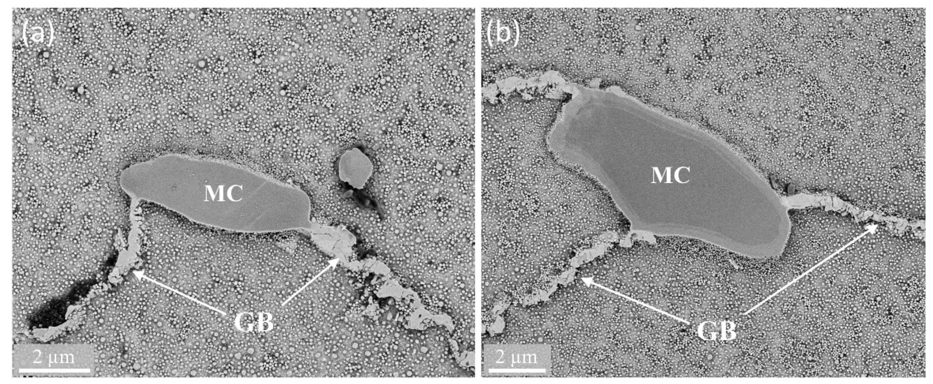
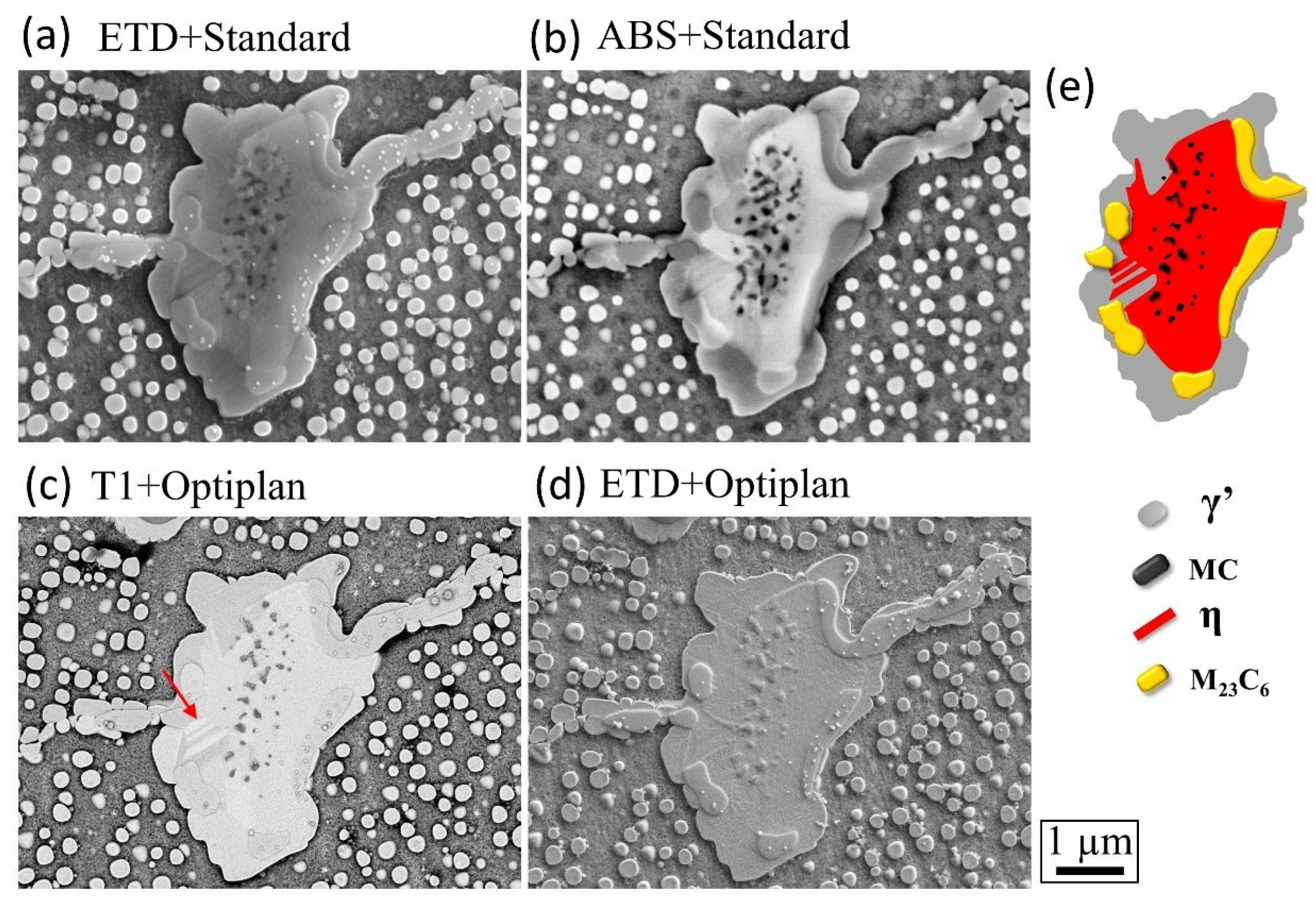
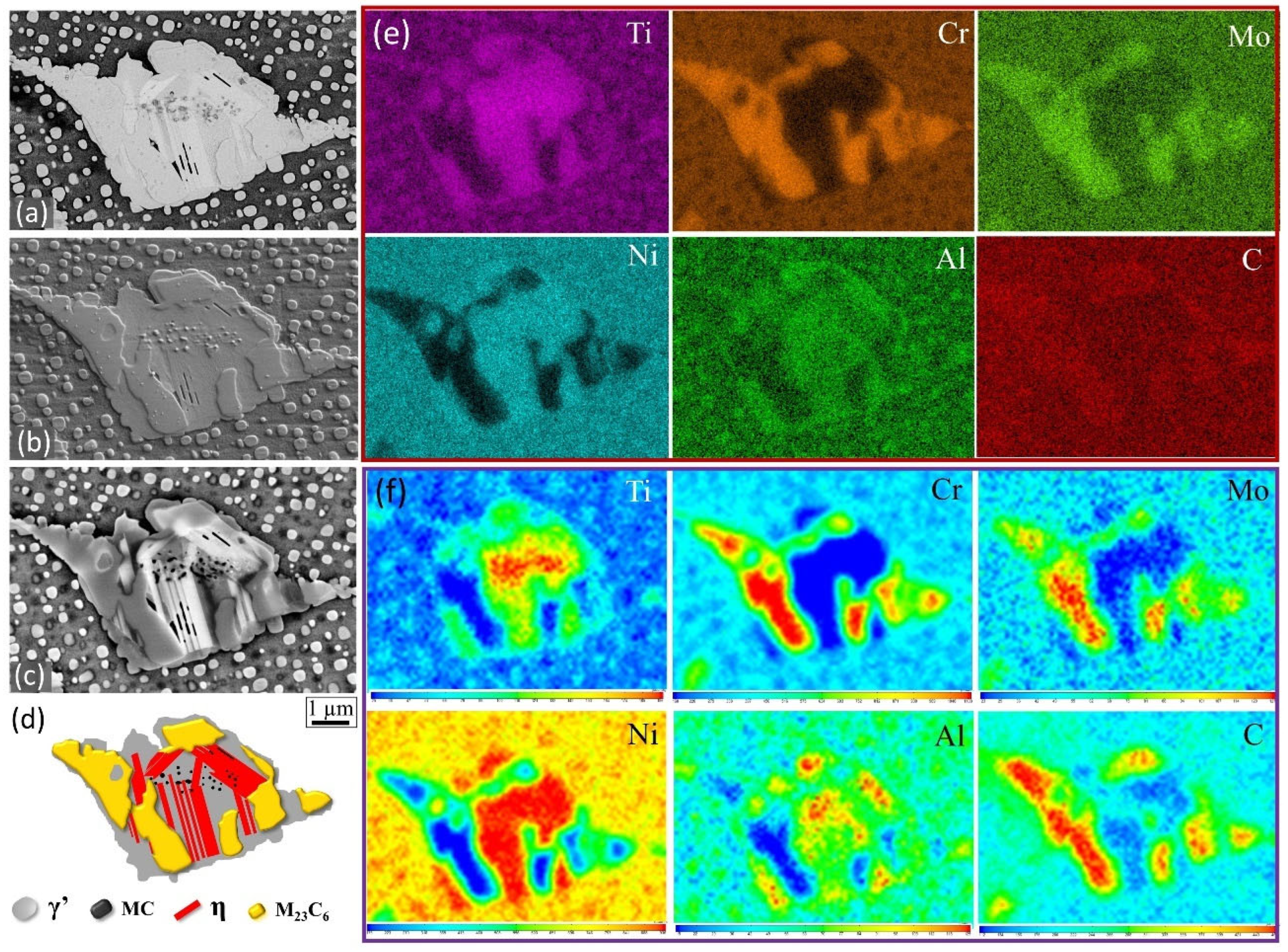
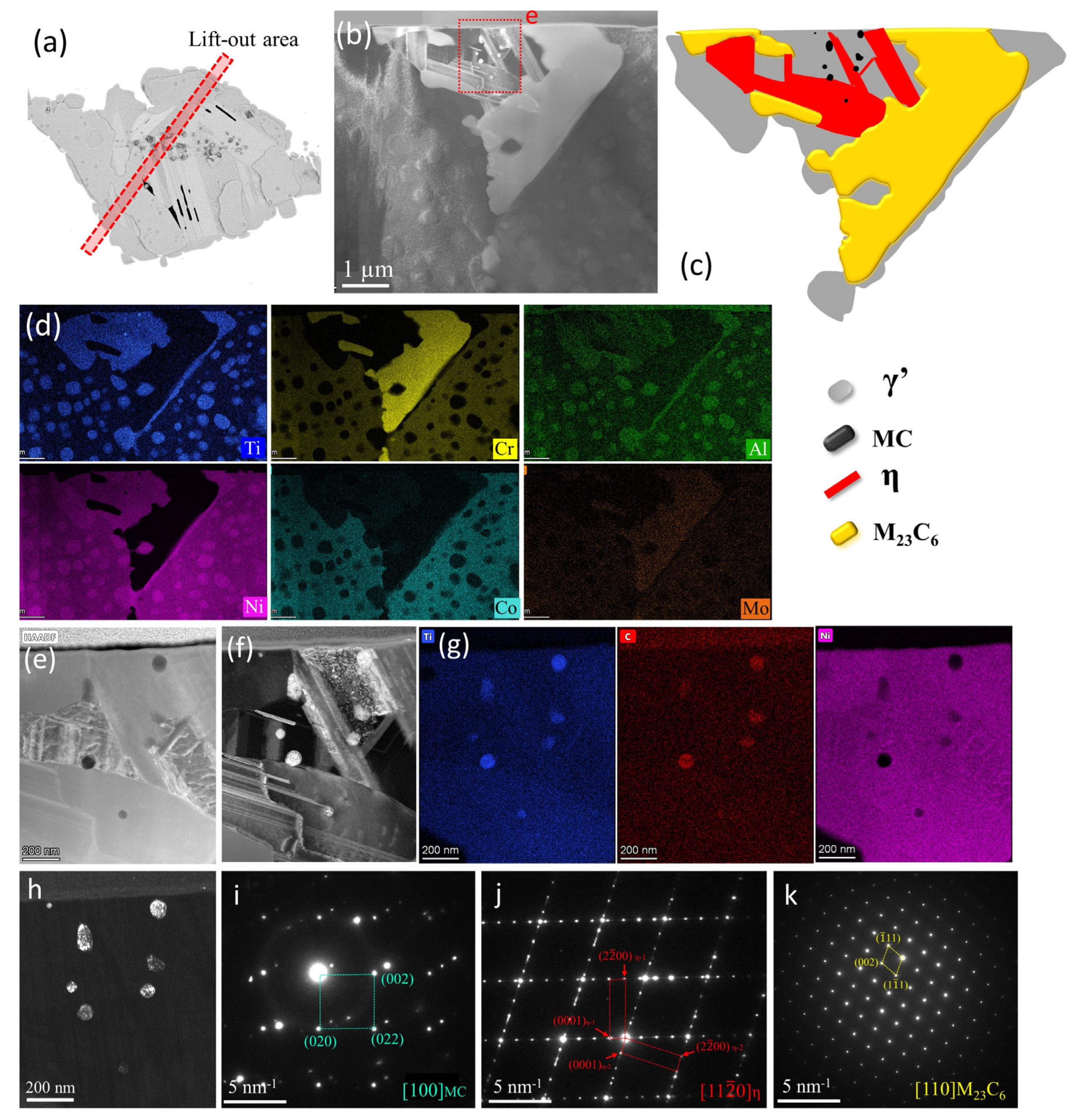
3. Results and Discussion

Figure 1 presents a typical grain boundary structure containing primary MC carbides after heat treatment, before the aging experiment. In the heat-treated sample, grain boundaries exhibit continuous M23C6 particles and occasional blocky MC carbides, with other MC carbides residing within the grains.
In Figure 2 and Figure 3, we illustrate two instances of phase mapping through image combinations. Specifically, these examples showcase the decomposition of MC carbides on grain boundaries with the subsequent formation of newly transformed γ′, η, and M23C6 carbides after 10,000 h of aging at 800 °C. Long-term thermal exposure induces major changes in the microstructure, notably the degradation of MC carbides and the formation of new phases such as η. Figure 2a displays a typical secondary electron image taken using the Everhart–Thornley Detector (ETD) in “Standard” mode (“ETD + Standard”). This image exhibits poor contrast, with discrete black dots in the central area representing mainly MC carbides (confirmation of the MC phase will be provided in Figure 4). Characterization via the Angular Backscattered (ABS) Detector in “Standard” mode (“ABS + Standard”) improves the contrast of MC, as shown in Figure 2b. Additionally, the η phase with a whitish contrast can be roughly distinguished, although with lower resolution compared to Figure 2c. While the backscatter imaging technique shown in Figure 2b has been successful in phase contrast imaging, it usually requires a high-energy electron beam (high voltage and high beam current, in this case, 20 kV/3.2 nA), leading to lower resolution. Figure 2c presents a similar backscatter image but utilizes the in-lens detector (T1) in “Optiplan” mode (“T1 + Optiplan”) with low voltage and current (2 kV/50 pA). This detector and mode offer decent phase contrast with high resolution. The fine structure of the η phase, indicated by the red arrow in Figure 2c, is now visible compared to the same area in Figure 2b. It is important to note that the phase contrast between η and γ′ can vary between the “T1 + Optiplan” image (the top part in Figure 2c) and the “ABS + Standard” image (the bottom part in Figure 2b). Figure 2d displays another “Optiplan” image using the ETD detector (“ETD + Optiplan”), providing a much better contrast for M23C6 due to the subtle difference in height between these phases. Combining images from different detectors and modes, and leveraging the distinct contrast of each image, allows for the exact phase distributions of all phases in the reaction area to be obtained and schematically shown in Figure 2e.
Figure 3 presents another example closely resembling the one in Figure 2. Additionally, SEM-EDS mapping for the area was conducted and is illustrated in Figure 3e. EDS point or mapping is frequently employed to ascertain composition and serves as a supplementary technique for phase identification. In this case, particles rich in Cr and Mo can be confidently identified as M23C6-type carbides and the surrounding γ′ particles rich in Ti and Al. However, due to the resolution limitation, distinguishing between the distribution of η and γ′ is challenging since they share similar compositions. It is also difficult to identify the MC phase via EDS when the size of MC is small (in nm), as seen in Figure 2 and Figure 3a–d. For the purpose of comparison, EPMA mapping, which is more sensitive to chemical composition than EDS, was conducted for the same area and is displayed in Figure 3f. It exhibits a similar composition distribution to EDS mapping and demonstrates higher sensitivity, such as clearly higher Ti content in the MC particles area. Nevertheless, similar limitations observed in EDS, such as the difficulty of mapping the η phase distribution, also apply to EPMA.
To validate the proposed phases shown in Figure 3 (and also indirectly in Figure 2), particularly the η phase and small MC dots, we conducted a site-targeted FIB lift-out on the region in Figure 3a. This was followed by detailed characterization using TEM and STEM (EDS), as depicted in Figure 4. The precise location of the lift-out site is indicated in Figure 4a, which is extracted from Figure 3a. The overview image of the lifted FIB specimen is shown in Figure 4b, and all of the phases in the core area of Figure 4b are schematically presented in Figure 4c, combining information such as chemical composition obtained through STEM-EDS mapping in Figure 4d and the diffraction patterns of each phase (Figure 4i–h). Special attention was given to the small MC dots, as displayed in Figure 4e–h, which correspond to the boxed area in Figure 4b. The EDS mapping in Figure 4g, the selected area diffraction pattern (SADP) in Figure 4i, and the corresponding dark field image in Figure 4h confirm that the small dot-like particles are M(Ti)C-type carbides. These MC carbides align well with the black dot particles observed in Figure 4a (and Figure 3a). Similarly, the η-Ni3Ti phase and M(Cr)23C6 carbides are confirmed by the SADP (Figure 4j and Figure 4h, respectively) and the EDS mapping (Figure 4d). It is worth noting that there are two sets of η phases with a rotation angle of 70° around the zone axis of [11 2 ¯ 0]. Overall, the phase distributions in the cross-section plane (schematically shown in Figure 4c) correspond well to the phase distributions in the horizontal plane (schematically shown in Figure 3d). This validates the accurate phase identification and distribution based on the contrast in images as shown in Figure 2e and Figure 3d.
There are several techniques available for phase identification, such as EBSD (electron backscatter diffraction) [12,13], SEM-EDS [14], STEM-EDS [15], and TEM-PED (Precession Electron Diffraction) [16]. Traditionally and theoretically, phase mapping or identification can be achieved using EBSD for bulk samples. However, even with advanced EBSD, it would still be challenging to distinguish the small particles in the size of nanometers (MC particles herein). Additionally, the EBSD scanning process usually takes a long time, especially with smaller step sizes for small particles [17]. Furthermore, preparing samples for EBSD requires higher quality and more time compared to normal sample preparation for SEM observation. Also, it might be possible to identify phases through EDS point analyses with quantitative data in SEM. However, as evidenced in Figure 2e,f, obtaining a clear view of the phase distributions through element mapping is extremely challenging due to resolution limitations. Moreover, element mapping is typically a time-consuming process, even with modern EDS detectors in SEM. TEM (PED) [9] and STEM (EDS) have been proven to be powerful tools for phase identification and mapping with high resolution. However, sample preparation for TEM, either using conventional methods (such as twin-jet polishing) or FIB lift-out, is time consuming. Additionally, the observation area is limited in the TEM specimen. Compared to these aforementioned techniques, the method demonstrated herein is relatively fast and reliable for phase mapping. It involves merely combining a few SEM images with different contrasts for the same area. The advantage of this method lies in the balance of probing small phases (in nanometers) and large searchable areas (in millimeters) in SEM, with relatively easy sample preparation and image acquisition. A brief summary of the comparisons between the method proposed herein and other phase mapping methods is listed in Table 1.

4. Conclusions

A method that combines the varying contrasts from different imaging detectors and modes was developed, enabling relatively rapid and precise identification and mapping of phase distributions, particularly for phases such as MC, M23C6, γ, and η. The method reaches a balance of probing small phases (in nanometers) and exploring large areas (in millimeters) in SEM, with relatively easy sample preparation and image acquisition. The method is applicable to other phase and alloy systems and is significant in revealing phase transformation processes and mechanisms.

Author Contributions

G.L.: conceptualization, writing—review and editing, data analysis, methodology, investigation, and funding acquisition. Y.Z.: conceptualization, formal analysis, supervision, project administration, and writing—review and editing; S.W.: conceptualization, formal analysis, funding acquisition, supervision, project administration, and writing—review and editing. All authors have read and agreed to the published version of the manuscript.

Funding

This research was funded by the Shenzhen Science and Technology Program (Grant No. RCBS20210706092409014), the Guangdong Basic and Applied Basic Research Foundation (Grant No. 2022A1515110637) and the Shenzhen Science and Technology Innovation Commission through awards JCYJ20210324104414040 and 20220815150609002.

Data Availability Statement

The raw data supporting the conclusions of this article will be made available by the authors on request.

Conflicts of Interest

The authors declare that they have no known competing financial interests or personal relationships that could have appeared to have influenced the work reported in this paper.

References

  1. Pedroso, A.F.V.; Sousa, V.F.C.; Sebbe, N.P.V.; Silva, F.J.G.; Campilho, R.D.S.G.; Sales-Contini, R.C.M.; Jesus, A.M.P. A Comprehensive Review on the Conventional and Non-Conventional Machining and Tool-Wear Mechanisms of INCONEL®. Metals 2023, 13, 585. [Google Scholar] [CrossRef]
  2. Liu, G.; Kong, L.; Xiao, X.; Birosca, S. Microstructure evolution and phase transformation in a nickel-based superalloy with varying Ti/Al ratios: Part 2—Phase transformation. Mater. Sci. Eng. A 2022, 831, 142228. [Google Scholar] [CrossRef]
  3. Sazerat, M.; Nait-Ali, A.; Cervellon, A.; Lopez-Galilea, I.; Burlot, G.; Gillet, S.; Eyidi, D.; Joulain, A.; Villechaise, P.; Weber, S.; et al. High temperature microstructure stability of Waspaloy produced by Wire Arc Additive Manufacturing. J. Alloys Compd. 2023, 966, 171626. [Google Scholar] [CrossRef]
  4. Chu, D.J.; Park, C.; Lee, J.; Jung, W.S. Effect of Ti/Al Ratio on Precipitation Behavior during Aging of Ni-Cr-Co-Based Superalloys. Metals 2023, 13, 1959. [Google Scholar] [CrossRef]
  5. Detrois, M.; Jablonski, P.D.; Hawk, J.A. The effect of η phase precipitates on the creep behavior of alloy 263 and variants. Mater. Sci. Eng. A 2021, 799, 140337. [Google Scholar] [CrossRef]
  6. Lvov, G.; Levit, V.I.; Kaufman, M.J. Mechanism of primary MC carbide decomposition in Ni-base superalloys. Metall. Mater. Trans. A 2004, 35, 1669–1679. [Google Scholar] [CrossRef]
  7. Ge, H.; Liu, G.; Zheng, S.; Yang, Y.; Liu, K.; Ma, X. Dislocation climbing dominated decomposition and fracture of carbides in a Ni-based superalloy. Acta Mater. 2023, 246, 118669. [Google Scholar] [CrossRef]
  8. Liu, G.; Xiao, X.; Véron, M.; Birosca, S. The nucleation and growth of η phase in nickel-based superalloy during long-term thermal exposure. Acta Mater. 2020, 185, 493–506. [Google Scholar] [CrossRef]
  9. Hou, K.; Ou, M.; Xing, W.; Ma, G.; Hao, X.; Wang, M.; Ma, Y. The formation of η-Ni3Ti phase microstructure in a cast nickel-based superalloy with high Ti/Al ratio. J. Mater. Res. Technol. 2024, 29, 764–778. [Google Scholar] [CrossRef]
  10. Choi, B.G.; Kim, I.S.; Kim, D.H.; Jo, C.Y. Temperature dependence of MC decomposition behavior in Ni-base superalloy GTD 111. Mater. Sci. Eng. A 2008, 478, 329–335. [Google Scholar] [CrossRef]
  11. BGeorgin, B.M.; Viswanathan, G.B.; Welk, B.A.; Kloenne, Z.T.; Fraser, H.L. Optimizing image contrast of second phases in metal alloys. Ultramicroscopy 2021, 228, 113346. [Google Scholar] [CrossRef] [PubMed]
  12. Carneiro, Í.; Simões, S. Recent Advances in EBSD Characterization of Metals. Metals 2020, 10, 1097. [Google Scholar] [CrossRef]
  13. Sharma, J.; Nicolaÿ, A.; De Graef, M.; Bozzolo, N. Phase discrimination between δ and η phases in the new nickel-based superalloy VDM Alloy 780 using EBSD. Mater. Charact. 2021, 176, 111105. [Google Scholar] [CrossRef]
  14. Münch, B.; Martin, L.; Leemann, A. Segmentation of elemental EDS maps by means of multiple clustering combined with phase identification. J. Microsc. 2015, 260, 411–426. [Google Scholar] [CrossRef] [PubMed]
  15. Parish, C.M.; Brewer, L.N. Multivariate statistics applications in phase analysis of STEM-EDS spectrum images. Ultramicroscopy 2010, 110, 134–143. [Google Scholar] [CrossRef] [PubMed]
  16. Rauch, E.; Véron, M. Automated crystal orientation and phase mapping in TEM. Mater. Charact. 2014, 98, 1–9. [Google Scholar] [CrossRef]
  17. Jiang, J.; Britton, T.B.; Wilkinson, A.J. Measurement of geometrically necessary dislocation density with high resolution electron backscatter diffraction: Effects of detector binning and step size. Ultramicroscopy 2013, 125, 1–9. [Google Scholar] [CrossRef] [PubMed]
Figure 1. Example of bulk MC carbides on the grain boundary (GB) after heat treatment (before the aging experiment).(a) MC on the GB; (b) MC on the GBs junction.
Metals 14 00899 g001
Figure 2. Example 1 of phase mapping via image combination from different detectors and imaging modes. “ETD”, “ABS”, and “T1” are different detectors. “Standard” and “Optiplan” are different imaging modes. The specific meaning of these detectors and modes is explained in the manuscript. (ad) Images from different combinations; (e) schematic of the phase mapping. The red arrow in (c) indicates the plate-like η phases.
Metals 14 00899 g002
Figure 3. Another example of phase mapping via image combination from different detectors and imaging modes. (a) “T1 + Optiplan”; (b) “ETD + Optiplan”; (c) “ABS + Standard”; (d) schematic of phase mapping; (e) SEM-EDS elemental mapping; (f) EPMA elemental mapping.
Metals 14 00899 g003
Figure 4. Phase identification in Figure 3 via FIB lift-out and TEM characterization: (a) lift-out area as indicated by the box area; (b) image of the FIB specimen; (c) schematic of the phase mapping in (b); (d) TEM-EDS mapping of the interested area in (b); (eg) images of MC carbides and representative elemental mapping in the box area of (b); (h) dark field image of dot-like MC carbides; (ik) diffraction patterns of MC, η, and M23C6, respectively.
Metals 14 00899 g004
Table 1. Comparison between the method proposed herein and other phase mapping methods.
Phase Mapping MethodsAdvantagesDisadvantagesNote
Imaging with different detectors and modesRelatively quick and accurate Proposed herein
EBSDHigh accuracy for large-size phasesHigh demand of sample surface; time consuming for scanning; limited resolutionWidely used for accurate phase mapping for bulk alloys [8]
SEM-EDS Time consuming for scanning; limited accuracyCommon method for auxiliary phase identification and mapping for bulk alloys
STEM-EDSHigh accuracyDifficulties in specimen preparation; time consuming for scanning; limited research areasCommon method for auxiliary phase identification and mapping for TEM foils
TEM-PEDHigh accuracyDifficulties in specimen preparation; time consuming for scanning, limited research areasWidely used for accurate phase for TEM foils with nanometer resolution [9]
Disclaimer/Publisher’s Note: The statements, opinions and data contained in all publications are solely those of the individual author(s) and contributor(s) and not of MDPI and/or the editor(s). MDPI and/or the editor(s) disclaim responsibility for any injury to people or property resulting from any ideas, methods, instructions or products referred to in the content.

