Article

Heavy Metal Behavior in Lichen-Mine Waste Interactions at an Abandoned Mine Site in Southwest Japan

1
Graduate School of Science and Engineering, Ehime University, Bunkyo-cho 2-5, Matsuyama, Ehime 790-8577, Japan
2
Cyclotron Research Center, Iwate Medical University, Tomegamori 348-58, Takizawa, Iwate 020-0603, Japan
*
Author to whom correspondence should be addressed.
Metals 2015, 5(3), 1591-1608; https://doi.org/10.3390/met5031591
Submission received: 13 July 2015 / Revised: 26 August 2015 / Accepted: 28 August 2015 / Published: 2 September 2015
(This article belongs to the Special Issue Hydrometallurgy)

Abstract

:
The lichen, Stereocaulon exutum Nylander, occurring in a contaminated abandoned mine site was investigated to clarify (1) the behavior of heavy metals and As during the slag weathering processes mediated by the lichen; and (2) the distribution of these elements in the lichen thallus on slag. The heavy metals and As in the slag are dissolved from their original phases during the weathering process by lichen substances (organic acids) and hypha penetration, in addition to non-biological weathering. The dissolved elements are absorbed into the lichen thallus. Some of these dissolved elements are distributed in the cells of the hyphae. The others are distributed on the surface of the hyphae as formless particles and show lateral distribution inside the cortex of the thallus. The Cu and Zn concentrations in the thalli are positively correlated with the concentrations in the corresponding substrata and a positive intercept in the regression curve obtained using a linear function. These chemical characteristics make this lichen a good biomarker for Cu and Zn contamination of the substrata of the lichen. Therefore, the present study supposes that Stereocaulon exutum has a possible practical application in biomonitoring or risk assessment of heavy metal pollution at abandoned mine sites.

Graphical Abstract

1. Introduction

Lichen covers more than 6% of the land surface of the earth [1]. Lichen is found in various areas, including extreme environments such as tropical forests, deserts, alpine regions, polar regions, and urban areas, and even in highly polluted areas [2,3]. Therefore, as organisms that indicate the environmental conditions of their habitats, lichens could have broad utility as bioindicators and biomarkers throughout the world.
The interactions of lichens with their substrata have been investigated. In addition to abiotic weathering, the weathering of rocks is accelerated by lichens [4,5,6]. Biological weathering by lichens occurs through two main processes [7,8]. One is a chemical process involving lichen substances called lichen acids. Lichen acids are organic acids that affect the chemical decomposition of minerals. The other is a physical process of penetration and expansion of lichen hyphae. The mechanical fragmentation by hyphae increases the surface area of the rocks and minerals and accelerates the chemical decomposition.
Lichens have been well studied as a biomonitoring tool for air pollution [9,10,11,12,13]. The lichens absorb mineral nutrients and trace elements, including metals from dry and wet atmospheric deposition, due to the lack of a protective cuticle and a vascular root system [14,15]. Therefore, some lichens are sensitive to airborne pollutants [16]. Some characteristics of lichens that could be used as bioindicators and biomarkers for heavy metal pollution of their substrata have been revealed in recent studies. Several lichens absorb and/or accumulate heavy metals from the substrata [17,18]. Osyczka and Rola (2013) found that the relationships between the Zn and Cd contents in Cladonia rei Schaer. thalli and in the host substrate through specific non-linear regression models could be described by a power function [19].
However, the interactions between lichens and the corresponding substrata have not been discussed comprehensively in relation to the behavior of heavy metals during the weathering process and the heavy metal concentrations. Thus, there is insufficient information on the availability of lichens as bioindicators or biomarkers for heavy metal pollution of their substrata. Therefore, this study has clarified (1) the behavior of Cu, Zn, As, and Pb during the slag weathering processes mediated by Stereocaulon exutum Nyl., and (2) the distribution of these elements in S. exutum thalli growing on slag. Finally, the present study evaluated the possible practical application of the lichen for environmental monitoring in terms of heavy metal and As pollution.

2. Materials and Methods

2.1. Study Area

This study was conducted at an abandoned mine site in Eastern Okayama, Southwest Japan, at an altitude of approximately 250 m above sea level (Figure 1). The site is located in the temperate zone with an average annual temperature of 14.1 °C (the seasonal averages are 2.8 °C in January and 25.8 °C in July) and a total annual precipitation of 1200 mm (the seasonal totals were 22.0 mm in January and 299 mm in August) in 2014 [20].
Figure 1. A location map of study area (indicated by a black dot).
Metals 05 01591 g001
Mainly Cu had been smelted at the abandoned mine site more than 50 years ago. The host rocks for the ore deposits are mainly rhyolite, andesite, sandstone, mudstone, and phreatomagmatic breccia [21]. These host rocks contain pyrite and chalcopyrite with quartz and calcite veins.
The surface of the waste dump consists primarily of tailings composed of phreatomagmatic breccia and rhyolite, and fractured slag overlies solidified, coherent slag [21]. The tailing and fractured slag have been kept on-site, in part, by wooden boards. However, these boards are now decaying and are no longer effective [21].
The soil and the stream water that flows through the waste dump are contaminated by Zn and/or As because the slag and tailings leach from the waste dump and accumulate on erosion control dams and sandbanks. The pH of the stream is nearly neutral (7.2–7.8), but the water contains Zn and As in excess of the environmental water quality standards determined by the Ministry of Health, Labor and Welfare, Japan [22,23]. The soil in the waste dump also contains As in excess of Japan’s environmental quality standards set by the Ministry of the Environment [22,23].
The slag is one of the potential pollutants in this area and contains high levels of heavy metals [21]. S. exutum can grow even on the slag, and it could affect the slag weathering. Therefore, this fruticose lichen was chosen as the object of this study.

2.2. Sampling Methods

The 18 lichens, 12 slags, seven tailings, and four rocks were randomly sampled from the slag and tailing dump, outcrops, and sandbanks at the abandoned mine site from 21 April 2011 to 28 September 2013. The lichen samples were taken with the corresponding substrata and packed into plastic bags that have a zipper. The slag, tailing, and rock samples were packed into plastic bags or put into cartons. The lichen samples for SEM-EDS analysis were stored in cool and dark place. The lichen samples for PIXE analysis were stored in a desiccator after cleaning by hyper-pure water.

2.3. Analytical Methods

The lichens were identified by morphological and biochemical classification using color reactions with the following three solutions: 10% aqueous potassium hydroxide [KOH], saturated aqueous calcium hypochlorite [Ca(OCl)2], and 5% alcoholic p-phenylenediamine solutions.
Rocks and slag were sampled at random, and their structures were observed using a scanning electron microscope (SEM). Then, the substrata were investigated by X-ray powder diffraction (XRD) analysis, and a scanning electron microscope equipped with an energy-dispersive X-ray spectrometer (SEM-EDS) was used to identify the primary and subsidiary phases and to clarify the behavior of heavy metals and As during the slag weathering process.
Eight pulverized slags and four pulverized rocks were analyzed by XRD to identify their mineral components. XRD analyses were performed on an Ultima IV (Rigaku, Tokyo, Japan) spectrometer housed at Ehime University (Ehime, Japan) using Cu Kα (λ = 1.54056 Å) radiation. The diffraction patterns for non-constant azimuth analysis were collected using an accelerating voltage of 40 kV, a specimen current of 40 mA, an analytical speed of 2°/min, and an analytical range of 5°–70°.
To investigate the distribution of potentially toxic elements in the slag, the slag and lichen samples were imaged and analyzed by SEM-EDS on a JSM-6510LV (JEOL, Tokyo, Japan) and X-Max 50 (Oxford Instruments, Tokyo, Japan) detector with INCA software (Oxford Instruments, Tokyo, Japan) at Ehime University. The slag samples were embedded in resin and prepared as polished thin sections that were then carbon-coated. The glass-phase content in the slag was estimated by counting the intersection points of a 200 μm mesh overlaid on back-scattered electron images. The SEM was operated with an accelerating voltage of 15 kV and a beam current of 0.8 nA. A counting time of 50 s was used for quantitative analysis, and count times of >1 h were required for element mapping. Enstatite (MgSiO3), K-feldspar (KAlSi3O8), and anorthite (CaAl2Si2O8) (Japan Electron Optics Laboratory Mineral Standard Samples for Electron Probe Micro Analyzer, Tokyo, Japan) were analyzed to confirm the analytical precision and accuracy. The following standards were used: NaAlSi2O6 for Na, Mg2SiO4 for Mg, Al2O3 for Al, CaSiO3 for Si, FeS2 for S, KBr for K, Ti for Ti, Cr for Cr, Mn for Mn, FeS2 for Fe in matte drops, Fe2O3 for Fe in magnetite, Fe2SiO4 for Fe in fayalite and glass, Co for Co, Cu for Cu, ZnO-glass for Zn, InAs for As, Ag for Ag, and Sn for Sn. The Co standard was also used to optimize the quantification of the analyses. The L characteristic X-ray lines were used for the Cu, Zn, As, Ag, and Sn quantification, and the K lines were used for other elements.
The heavy metal concentrations of the substrata, including seven slags, eight tailings, and seven host rocks, were determined by energy dispersive X-ray fluorescence (ED-XRF) spectrometry using an Epsilon5 instrument (PANalytical, Almelo, The Netherlands) at Ehime University. Pressed powder pellet samples were used for the XRF analysis. The samples were powdered to a grain size of <1 μm in a tungsten carbide vibrating sample mill (Sample Mill model TI-100, HEIKO, Tokyo, Japan) and an agate mortar at Ehime University. C13H14O6 was used as a binder (20 wt. % in the pressed powder pellets) and homogenized with the powdered samples by shaking for 1 h. The XRF analyses were conducted at an excitation voltage of 100 kV. The following standards were used as secondary targets for irradiation with the relevant excited X-rays for the individual elements: Al for Na and Mg; Ti for Al–Sc; Ge for Ti–Ga; Mo for Ge–Y, Tl–Ra, and Ac–U; and Al2O3 for other elements, including lanthanides. The X-ray source was a Gd anode. A counting time of 300 s was repeated three times on a Ge detector for each secondary target, and elemental concentrations were determined by the fundamental parameter method. Geological standards, including JSd-1, JSd-2, JSd-3, JSl-1, Jlk-1, and JMn-1 (Geological Survey of Japan Referenced Materials) [24,25], were analyzed to confirm the analytical precision (relative standard deviations are 1.86% Fe2O3, 11.3% Cu, 3.09% Zn, 10.9% As, 45.5% Sn, and 53.2% Pb).
The heavy metal concentrations of the lichens were determined by particle-induced X-ray emission (PIXE) performed at the Nishina Memorial Cyclotron Center established by the Japan Radioisotope Association. The analytical conditions followed Sera et al. (1992) [26]. A small cyclotron provides a 2.9 MeV-proton beam on the target after passing through a beam collimator of graphite. The maximum beam intensity on the target is approximately 40 nA for a beam spot diameter of 2 mm and 80 nA for a diameter of 6 mm. Elements from Na to U are detected by two ORTEC Si (Li) detectors. The elements heavier than Ca are detected by the first detector, which has a 0.025 mm-thick Be window and a 6 mm active diameter, with X-rays with an energy resolution of 154 eV at 5.9 keV and a 300- to 500-μm-thick Mylar absorber inserted between the target and the detector. The other low atomic number elements are detected by the second detector, which has a 0.008 mm Be window and a 4 mm active diameter, a resolution of 157 eV, and a small graphite aperture without an absorber. The upper 1–5 mm portion of the lichen thallus was cut out from the substrata to remove the effects of the trapped substratum fragments by the hypha structure and rinsed with ultrapure water. The rinsed samples were dried at 80 °C for 24 h in an oven. The dried samples (30 mg) were digested with 1 mL of HNO3 and heated in a microwave (150 W). The analytical accuracy and precision of the analyses were verified using the NIES CRM No. 1 environmental sample.

2.4. Statistical Methods

The distributional normality of the Cu, Zn, As, and Pb concentrations in S. exutum and the corresponding substrata was verified by the Shapiro-Wilk normality test. The correlation of the scatter plots was verified by Spearman’s rank-correlation coefficient. All statistical analyses were performed with EZR (Saitama Medical Center, Jichi Medical University, Saitama, Japan), a graphical user interface for R (The R Foundation for Statistical Computing, Vienna, Austria). More precisely, it is a modified version of R Commander designed to add statistical functions frequently used in biostatistics [27].

3. Results

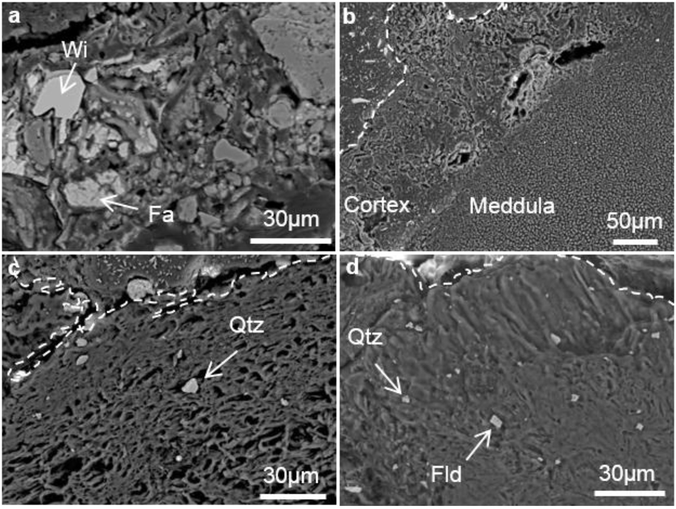
3.1. Microstructure of the Hyphae of Stereocaulon exutum Thallus and the Effects of the Lichen on Slag Weathering

The hyphae of the thallus form loosely-complicated structures and adhere to each other near the interface with the slag. The hyphae that compose the medulla of the thallus form lines in the direction of growth. Hyphae that compose the cortex of the upper thallus form complicated structures, and these different hyphae partially adhere to each other.
Stereocaulon exutum has both physical and chemical effects on the slag during the weathering process. The surface of the weathered slag under the lichen is penetrated and fractured by the lichen hyphae during the physical process (Figure 2a–c). The hyphae penetrate and expand into the crack of the slag and along the mineral interfaces. Etch-pits occur on the weathered willemite and matte drops under the lichen during the chemical process (Figure 2e).
Figure 2. Back-scattered electron images of (a) lichen-slag interface; (b) slag fragments caught by hyphae structure; (c) hyphae penetrating crack of slag; (e) etch-pits of pseudomorph of willemite; and (d) a secondary electron image of slag fragments caught by hyphae structure. W-Wi: weathered willemite; Wi: willemite; Fa: fayalite.
Metals 05 01591 g002
Figure 3. Back-scattered electron images of thallus containing fragments. Distances from substrata increase from (a) to (d) every 2 mm. (a) is the section where 100 μm high from slag; (b) and (c) are the section of thallus; (d) is the section of apothecium. The broken lines indicate thallus-resin interfaces. Wi: willemite; Fa: fayalite; Qtz: quartz; Fld: feldspar.
Metals 05 01591 g003
The slag fragments in the thallus show differences in the distance from the substrata. The lower portion of the thallus contains fragments of the fractured slag (Figure 2b,d). The amounts of the slag fragments in the upper portion (more than 1 mm from the substratum) are less comparable with the portion near the interface between the lichen and the slag. The fragments trapped in the upper portion consist of mainly quartz and plagioclase (Figure 3).

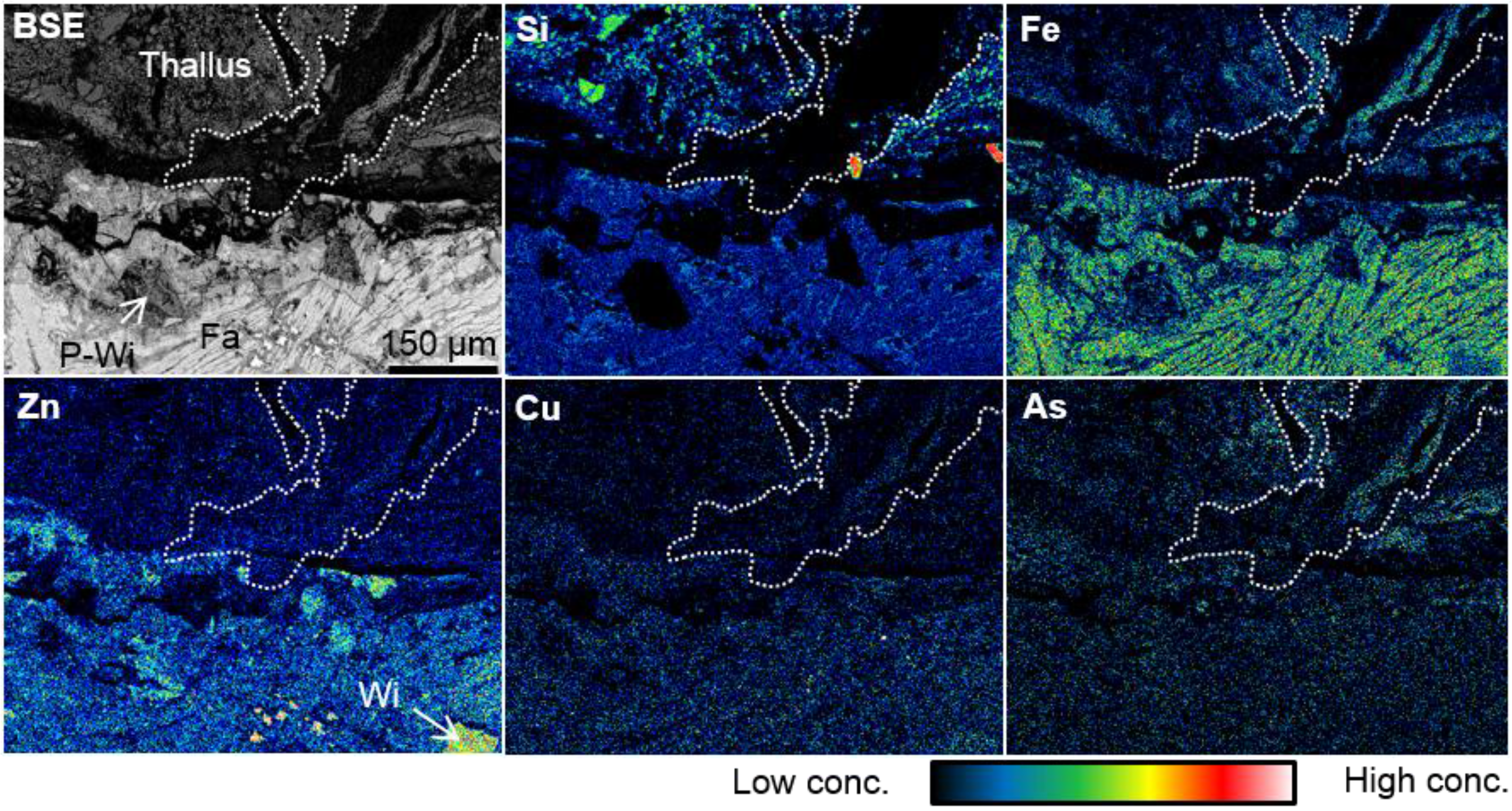
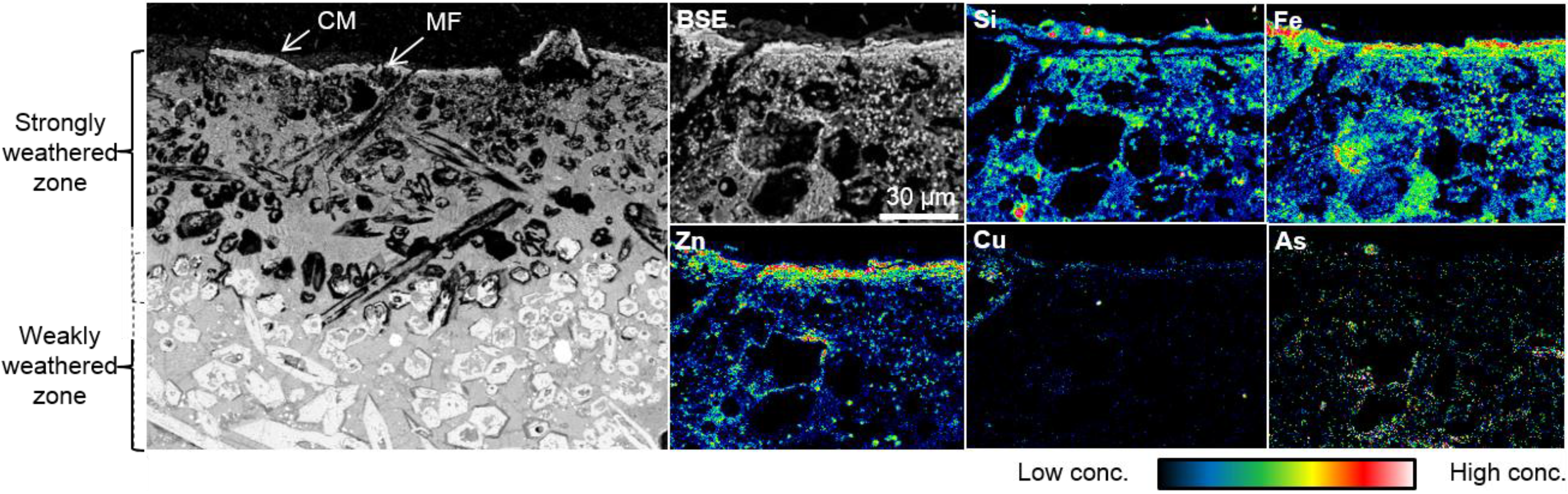
3.2. Heavy Metal Distribution in the Weathered Slag

The slag is composed mainly of willemite, fayalite, and/or magnetite with a silicate glassy matrix and contains matte drops, which are Cu-, Zn-, Pb-metals, -alloys, and -sulfides. Willemite is the main host of Zn, and fayalite is the main host of Fe. Matte drops are the main hosts of heavy metals, S, and As.
The heavy metals and As are dissolved from the original phases during the weathering process. The outermost portion in the weathered zone comprises mainly clay minerals. The willemite and matte drops are converted to Fe-hydroxides during the weathering process. The heavy metal (except for Fe and As) concentrations in the weathered phases are lower than in the original phases.
Figure 4. Back-scattered electron images and the elemental maps of weathered slag obtained by SEM-EDS. MF: Metal film-like structure; CM: Clay minerals.
Metals 05 01591 g004
Figure 5. A back-scattered electron image and the elemental maps of a lichen-slag interface obtained by SEM-EDS. The broken lines indicate thallus. Fa: fayalite; P-Wi: pseudomorph of willemite.
Metals 05 01591 g005
Heavy metals and As in the willemite-rich slag are partially concentrated inside the clay minerals and form a film-like structure (Figure 4). However, during the lichen-mediated weathering process, the heavy metals and As do not form the structure under the lichen thallus but rather tend to be concentrated in the lichen thalli (Figure 5).

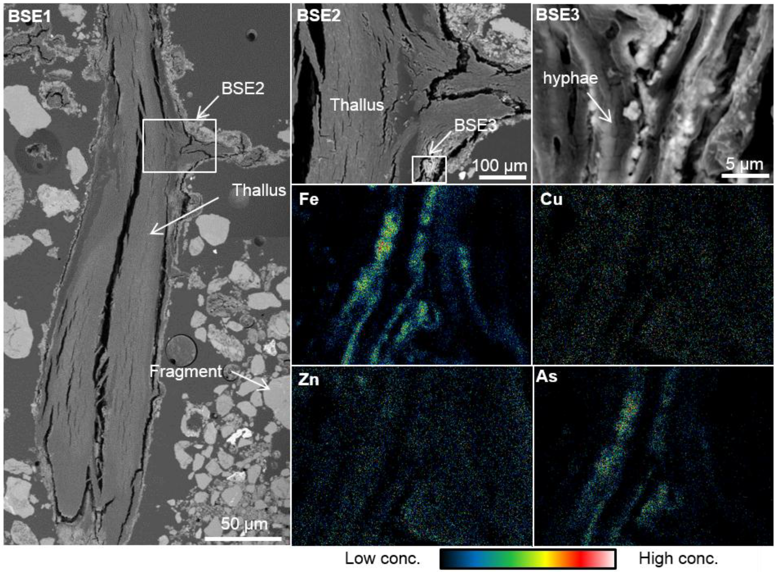
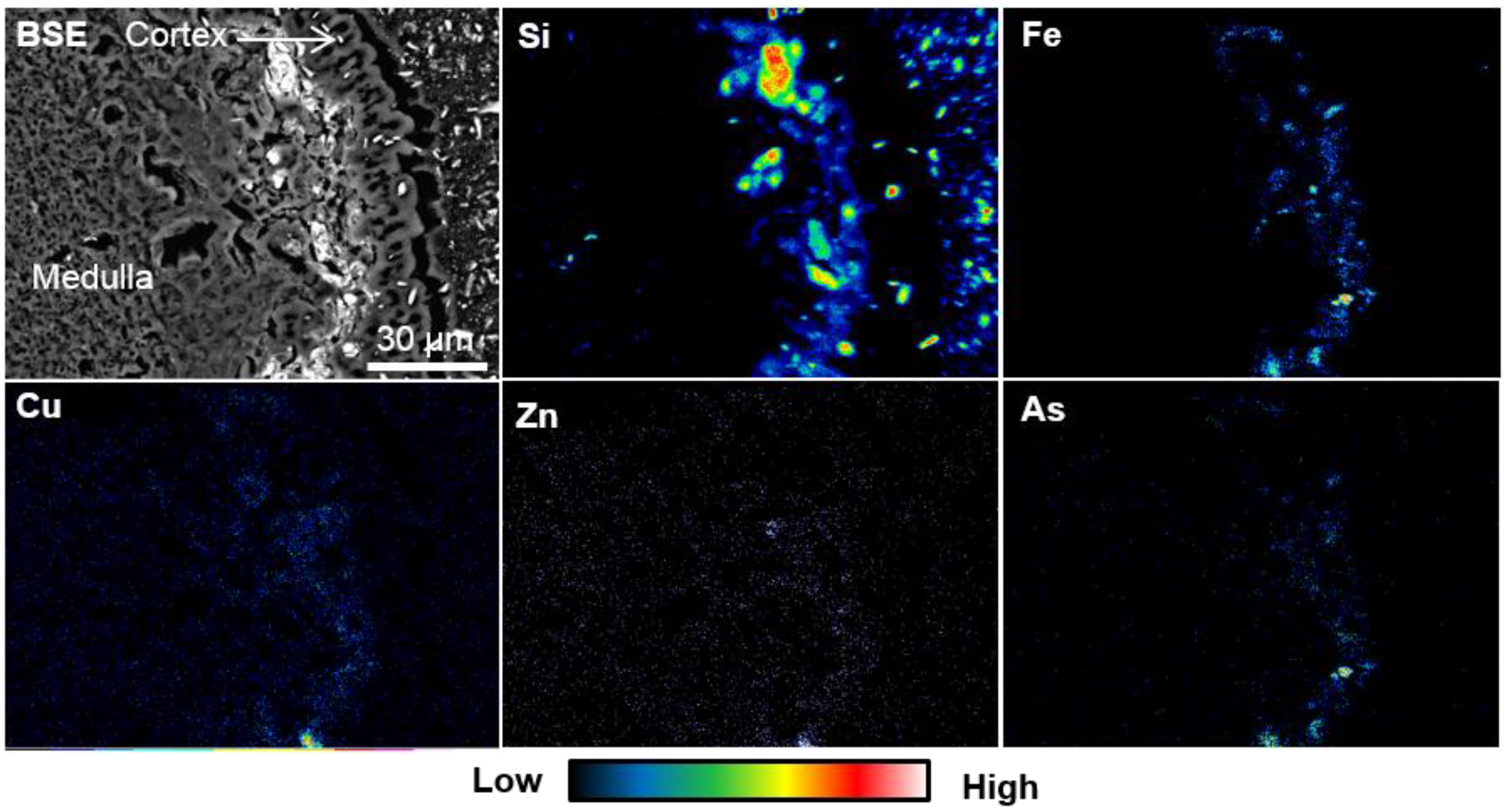
3.3. Elemental Distribution in the S. exutum Thallus

The thallus contains heavy metals and As. Fe and As are especially contained on the surface of the hyphae in the thallus (Figure 6). Formless particles occur on this portion. The hyphae contain Fe, Cu, Zn, and As in the cells of their rhizome (Figure 7). Several elements, including Fe, Cu, Zn, and As, are contained inside the cortex of the thallus and show lateral distribution (Figure 8). Formless particles occur in this portion. The concentrations of these elements in the phyllocladium were not found.
Figure 6. Back-scattered electron images and the elemental maps of hyphae inside the cortex of the thallus. The white box in BSE2 indicates the area of BSE3 and the mapping analysis area.
Metals 05 01591 g006
Figure 7. A back-scattered electron image and the elemental maps of the lichen hyphae.
Metals 05 01591 g007
Figure 8. A back-scattered electron image and the elemental maps of the lichen thallus.
Metals 05 01591 g008

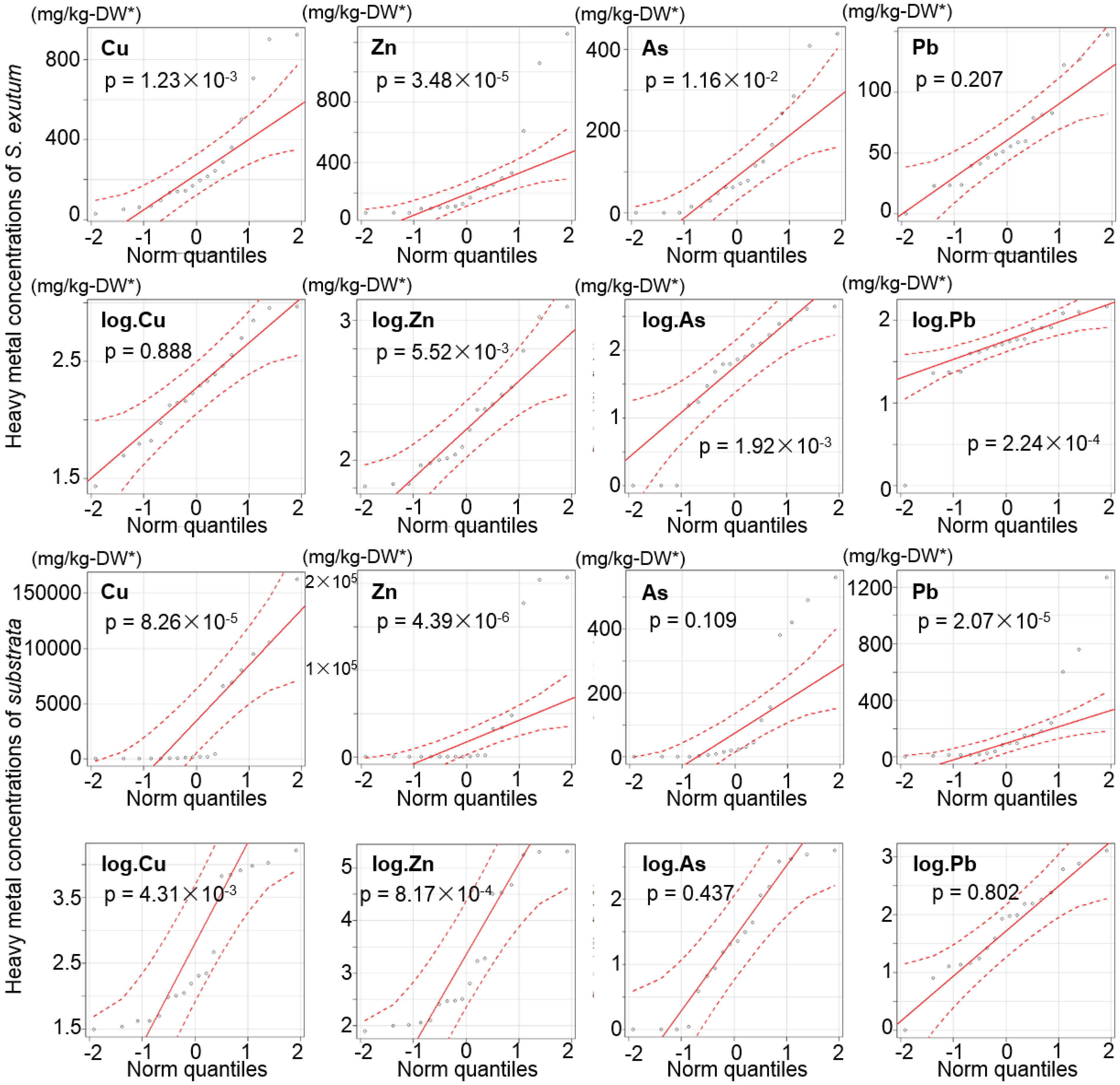
3.4. Heavy Metals and As Concentrations of Lichens and the Corresponding Substrata

The heavy metal and As concentrations of Stereocaulon exutum on slag and rocks, including tailings, gravel, and the host rock, are 33.6–1250 mg/kg-DW Zn, 6.51–923 mg/kg-DW Cu, 12.0–147 mg/kg-DW Pb, and ND-438 mg/kg-DW As. The heavy metal and As concentrations of the substrata are as follows: slag contains 20.6%–3.27%-DW Zn, 1.62%–0.66%-DW Cu, ND–758 mg/kg-DW Pb, and 31.3–561 mg/kg-DW As; the tailings contain 640–101 mg/kg-DW Zn, 154–34.2 mg/kg-DW Cu, 12.7–1280 mg/kg-DW Pb, and ND–23.0 mg/kg-DW As; the outcrop rocks contain 79.1–1940 mg/kg-DW Zn, 96.6–461 mg/kg-DW Cu, 8.08–97.1 mg/kg-DW Pb, and 1.12–490 mg/kg-DW As.
On determination of the correlations between heavy metal concentrations in lichens and the corresponding substrata, the distributional normality was verified by the Shapiro-Wilk normality test (Table 1). As no significant normal distribution of the scattered plots was shown in corresponding concentrations of Cu, Zn, As, and Pb in the lichen and the corresponding substrata (Table 1, Figure 9), the correlation was verified by Spearman’s rank-correlation coefficient. The results of the test show that the Cu and Zn concentrations in S. exutum thalli exhibit positive correlations with those in the corresponding substrata under the 5% significance level but not As and Pb concentrations (Table 2, Figure 10). The formulas of the regression curves have positive X-intercepts (Figure 10).
Table 1. p-values of heavy metal concentrations in lichens and the corresponding substrata obtained by Shapiro-Wilk normality test.
Elementsp-Values
S. exutumSubstrata
Cu1.23 × 10−38.26 × 10⁻⁵
Zn3.48 × 10−54.39 × 10⁻⁶
As1.16 × 10−20.109
Pb0.2072.07 × 10−5
log.Cu0.8884.31 × 10−3
log.Zn5.52 × 10−38.17 × 10⁻⁴
log.As1.92 × 10−30.437
log.Pb2.24 × 10−40.802
Figure 9. QQ plots of heavy metal concentrations of lichens and the corresponding substrata and the logarithmic data. The p-values were obtained by Shapiro-Wilk normality test. n = 18.
Metals 05 01591 g009
Table 2. Spearman’s correlation rho and p-values of the scattered plots of Cu, Zn, As, and Pb concentrations in the lichens and the corresponding substrata. n = 18.
Elementsrsp-Values
Cu0.6187.43 × 10−3
Zn0.8291.28 × 10−5
As0.4210.119
Pb0.3360.188
Figure 10. Scatter diagrams show Cu and Zn concentration of S. exutum and the corresponding substrata with linear functions after logarithmic transformation. Note: significance level = 5%. n = sample number.
Metals 05 01591 g010

4. Discussion

4.1. Effects of S. exutum on Heavy Metals and As Behavior in the Slag Weathering Process

In addition to abiological weathering, Stereocaulon exutum affects and accelerates the weathering of the original phases, and the effect can be attributed to both physical and chemical processes. The physical effects are caused by hyphal penetration and expansion, and the chemical effects are caused by lichen substances.
Fragmentation and disaggregation of the lithic surface below the lichens have been well known and investigated in previous studies [4,5]. Various types of rocks, including calcareous rock, siliceous rock, and sedimentary rock, can be weathered physically by the hyphal penetration [7,8,28,29,30,31]. The slag that is composed of mainly silicate phases is also fractured by the hyphae even in the coherent portion.
The fragments of the substrata of lichens are incorporated into the thallus and coated by extracellular polymers and secondary minerals [4,5]. The fragments of the weathered slag are observed mainly in the lower 1–5 mm portion of the thallus, closer to the substratum. However, the trapped fragments in the upper than 5 mm portion of the thallus consist of mainly quartz and feldspar. These results indicate that the fragments are not transported to the upper portion of the thallus. Thus, the origin of the fragments trapped in the upper thallus could not be the substrata but atmospheric deposition. The trapped slag fragments consist of the Fe hydroxides containing Zn as weathered secondary minerals. However, no extracellular polymers were found in this study.
In addition to penetration of hyphae, substrata are mechanically disrupted by expansion, contraction, freezing, and thawing of the lichen thallus [5]. The hyphae penetrate into cracks of the willemite and matte drops in the slag and along the interface of these phases. The lichen thallus has an abundant water storage capacity [32]. Therefore, the slag is physically disaggregated through wetting and drying of the hyphae, and chemical weathering could be accelerated by the water supply.
Lichens affect not only physical weathering but also chemical weathering mostly by lichen compounds [6]. The main chemical dissolution processes of minerals by lichens are generated by respiratory CO2, the excretion of oxalic acid, and the chemical action of lichen compounds [33]. Mineral etching is a frequent feature of weathering at the rock-lichen interface [6]. The etch-pits in the weathered willemite and matte drops are evidence of the chemical weathering of these phases in this study. The excretion of various organic compounds can effectively dissolve the original phases and chelate metallic cations [4,5,6]. Several lichen compounds such as depsides and depsidones are slightly soluble and have the ability to chelate in conjunction with the presence of polar groups such as OH, CHO, and COOH [34]. Although the dissolution and decomposition of rock-forming minerals by lichen acids has been demonstrated in laboratory experiments [35,36], the dissolution and decomposition processes have not been conclusively proven in previous field studies [37,38].
S. exutum contains the depsidone (lobaric acid) in the thallus and could affect the dissolution and decomposition of slag phases. Although the present study also provides no scientific evidence to clarify the processes, the results show the evidence of heavy metal and As dissolution from the original phases by acids. Willemite and matte drops, which are the main hosts of heavy metals and As, are transformed to Fe hydroxide during the weathering process. The heavy metal and As concentrations of weathered phases in the weathered portion of the slag are lower than the heavy metal and As concentrations in the unweathered portion [21]. The lower heavy metal and As concentrations in the weathered phases and the occurrence of etch-pits in the pseudomorphs of willemite and matte drops in only the lichen-mediated weathered portion are evidence of the effect of lichens on decomposing of slag phases and dissolving of heavy metals from the original phases. Although this study could not clarify whether the S. exutum or other microbes are mostly effective in dissolving and decomposing slag phases by excretion of acids, the results indicate the lichen affects biochemical weathering of smelting slag in terms of heavy metal and As dissolution from the original phases for the first time.

4.2. The Accumulation and the Behavior of Heavy Metals in the Lichen Thallus

The heavy metal uptake ability and the accumulation capacity of lichens has been well demonstrated in various previous studies [19,39]. Several lichens, such as Cladonia spp. and Stereocaulon spp. that grow near Cu smelters and Cu-polluted sites contain high levels of heavy metals [40,41,42]. S. exutum growing on the tailing and slag dump at this study site also contains high concentrations of Cu, Zn, As, and Pb.
The heavy metals accumulated by S. exutum through the slag weathering process are contained in the lichen thallus. The kinetics and thermodynamics of ion exchange, such as binding constants and charge balances, have been well demonstrated in previous studies [43]. Cation uptake occurs extracellularly in lichens [44]. Nieboer and Richardson (1980) supposed that the affinity of ions for the exchange sites varies in an order corresponding to monovalent Class A < divalent Class A < borderline divalent < divalent Class B [45]. They categorized ions as follows: Class A ions are composed of alkaline metals, alkaline-earth metals, Al, Y, lanthanoid, and actinoid; borderline ions are composed of Sc, Ti, V, Cr, Mn, Fe, Co, Ni, Cu2+, Zn, Ga, As, Cd, In, Sn, Sb, and Pb2+; and Class B ions are composed of Cu1+, Rh, Pd, Ag, Ir, Pt, Au, Hg, Tl, Pb4+, and Bi [45]. The distribution of the cations has been estimated as follows four fractions into an intercellular and surface fraction, ion exchange site fraction, intracellular fraction, and residual fraction [46].
The SEM observations and elemental mapping analysis of the lichen thalli clarified at least two distributions of the absorbed elements in this study. Some of the elements are distributed throughout cells or the exchange sites of the cell wall of the hyphae in the thallus and show no trend. The others are distributed on the surface of the hyphae as unformed materials and show lateral distribution inside the cortex. As the elements do not concentrate in the phyllocladium, the fungal component positively absorbs the elements rather than the algal partner. Although this study could not clarify the form of the ions that are absorbed in the lichen thallus, the elements in the cell may indicate the results of absorption of the ions into the cytoplasm from external solutions or through positively and negatively charged anionic binding sites. The distribution of Fe and As on the surface of the hyphae as unformed materials could show the results of elemental precipitation or making compounds during the evaporation of external solutions. The elements in the external solutions may be affected by lichen substances during the processes of precipitation and chemical combination. As the carboxyl group can easily form complexes with Cu and Zn [47], these ions could make some compounds with lichen substances or some enzymes that are excreted by S. exutum. While the Cu and Zn could be affected by the lichen substances, Fe and As could be affected by inorganic precipitation and adsorption. The Fe and As in the thallus tend to be more concentrated on the surface of the hyphae of the thallus as formless particles in the study samples. As arsenic is easily adsorbed by Fe hydroxides [48], it is possible that the As and Fe absorbed by lichens may deposit on the surface of the hypha structure as Fe hydroxides.
The SEM observations and PIXE analysis indicate that the upper thallus contains heavy metals and As not as slag fragments but rather as ions and/or compounds. The hyphae of S. exutum are composed of filamentous fungi with a diameter of generally 1–10 μm. The filamentous fungi of ascomycete commonly have a septum with a stoma in the hyphae, and protoplasmic streaming occurs between the cells of the fungi [49,50]. Therefore, the dissolved elements from the weathered slag phases are taken into the cells of the lichen thallus and are transported to the upper portion.
While biominerals such as moolooite found by Purvis (1984) have not been found in this study [51], formless particles rich in Fe and As have been found on the surface of the hyphae in the thallus. In addition, Fe, Cu, Zn, and As are distributed throughout the cells of the hyphae. These results support the possibility of different mechanisms of Fe and As absorption and accumulation by lichens, compared with previous studies.

4.3. The Correlation of Cu and Zn Concentrations in the Lichen and Substrata

The relationship of heavy metal concentrations between lichens and the corresponding substrata has been demonstrated in previous studies [19,41]. Copper concentrations of Cladonia subconistea and C. humilis growing on Cu-hyperaccumulator moss Scopelophila cataractae at a Cu-polluted site are much higher than the copper concentrations of control samples growing on soil [41]. Osyczka and Rola (2013) [19] demonstrated the relationship of Zn and Cd concentrations between C. rei thalli and the host substrata using specific non-linear regression models described by a power function. In this study, the concentrations of Cu and Zn in lichens after logarithmic transformation show a positive correlation with their concentrations in the corresponding substrata that are obtained by linear regression models described by a linear function as well as the result of Osyczka and Rola (2013) [19], while the lichen species and the type of their substrata are different. The regression model clarified an impressive ability of S. exutum to accumulate Cu and Zn because the model showed the positive intercept. Furthermore, the relationship between the concentrations in the lichen thalli and the corresponding substrata shows no peak in the scatter diagram, which also supports the characterization of S. exutum as a Cu and Zn accumulator.
The dominant source of heavy metals should be estimated on the discussion of the relationships of the heavy metal content between lichens and the corresponding substrata. The fragments of the weathered slag have not been transported to the upper portion, and the analyzed portion is the portion higher than 1–5 mm from the substratum. Although the fragments that are contained in the analyzed portion are composed of mainly quartz and feldspar as material from atmospheric depositions, the Cu and Zn concentrations of the lichen show a positive correlation with the concentrations of their corresponding substrata. Therefore, the Cu and Zn concentrations in the lichen reflect the Cu and Zn contents absorbed by the lichen from the corresponding substratum and not the fragments of the substratum. Accordingly, the S. exutum appears to absorb mainly the heavy metals from the substrata below the lichen thallus at the study site.

5. Conclusions

Stereocaulon exutum affects slag weathering in biological processes by lichen substances (organic acids) and hyphae penetration and could accelerate non-biological weathering. Heavy metal and As in the slag are dissolved from the silicate, sulfide, and metal phases during the weathering process. The heavy metals form metal film-like structures in the non-lichen-mediated weathering portion of the slag but not in the lichen-mediated weathered portion. The elements are absorbed into the lichen and contained within the thallus. Some of the elements are distributed in the cells of the hyphae. The others are distributed on the surface of the hyphae as formless particles and show lateral distribution inside the cortex of the thallus.
S. exutum shows other interesting results related to the chemical properties. The Cu and Zn concentrations in the thalli are positively correlated with the concentrations in the corresponding substrata and the positive intercept of the regression curve obtained by a linear function. These chemical characteristics make this lichen a good biomarker for Cu and Zn contamination of the substrata of the lichen, making lichen-solid interactions very important and interesting to the field of environmental studies. Therefore, the present study supposes that S. exutum has a possible practical application to biomonitoring or risk assessment of heavy metal pollution at abandoned mine sites.

Acknowledgments

We are grateful to A. Kawamata (Ehime prefectural science museum, Japan) for help with identifications of lichens. The authors are grateful to H. Ohfuji (Ehime University, Japan) for help with analysis by SEM-EDS. We thank H. Akamatsu (Ehime University, Japan) for help with making thin sections. We thank the two anonymous referees for their perceptive comments and recommendations. This study was partially supported by JSPS KAKENHI Grant Number 15J04570 and grant from Fukada Geological Institute (Fukada Grant-in-Aid, 2013).

Author Contributions

Y. Sueoka mainly performed the present study and wrote the paper. M. Sakakibara provided advice and recommendations on analytical methods and polishing of the paper. K. Sera performed the PIXE analysis.

Conflicts of Interest

The authors declare no conflict of interest.

References

  1. Purvis, O.W. Chapter Three: Lichens and industrial pollution. In Ecology of Industrial Pollution; Batty, L.C., Hallberg, K.B., Eds.; Cambridge University Press: Cambridge, UK, 2010; pp. 41–69. [Google Scholar]
  2. Edwards, H.G.M.; Moody, C.D.; Villar, S.E.J.; Wynn-Williams, D.D. Raman spectroscopic detection of key biomarkers of cyanobacteria and lichen symbiosis in extreme Antarctic habitats: Evaluation for Mars Lander missions. Icarus 2005, 174, 560–571. [Google Scholar] [CrossRef]
  3. Bačkor, M.; Loppi, S. Interactions of lichens with heavy metals. Biol. Plant. 2009, 53, 214–222. [Google Scholar] [CrossRef]
  4. Adamo, P.; Violante, P. Weathering of rocks and neogenesis of minerals associated with lichen activity. Appl. Clay Sci. 2000, 16, 229–256. [Google Scholar] [CrossRef]
  5. Chen, J.; Blume, H.P.; Beyer, L. Weathering of rocks induced by lichen colonization—A review. Catena 2000, 39, 121–146. [Google Scholar] [CrossRef]
  6. Wilson, M.J.; Jones, D. Lichen weathering of minerals: Implications for pedogenesis. Geol. Soc. Lond. Spec. Publ. 2012, 11, 5–12. [Google Scholar] [CrossRef]
  7. Banfield, J.F.; Baker, W.W.; Welch, S.A.; Taunton, A. Biological impact on mineral dissolution: Application of the lichen model to understanding mineral weathering in the rhizosphere. Proc. Natl. Acad. Sci. USA 1999, 96, 3404–3411. [Google Scholar] [CrossRef] [PubMed]
  8. Favero-Longo, S.E.; Gazzano, C.; Girlanda, M.; Castelli, D.; Tretiach, M.; Baiocchi, C.; Piervittori, R. Physical and chemical deterioration of silicate and carbonate rocks by meristematic microcolonial fungi and endolothic lichens (Chaetothyriomycetidae). Geomicrobiol. J. 2011, 28, 732–744. [Google Scholar] [CrossRef]
  9. Conti, M.E.; Cecchetti, G. Biological monitoring: Lichens as bioindicators of air pollution assessment—A review. Environ. Pollut. 2001, 114, 471–492. [Google Scholar] [CrossRef]
  10. Golubev, A.V.; Golubeva, V.N.; Krylov, N.G.; Kuznetsova, V.F.; Mavrin, S.V.; Aleinikov, A.Y.; Hoppes, W.G.; Surano, K.A. On monitoring anthropogenic airborne uranium concentrations and 235U/238U isotopic ratio by Lichen—Bio-indicator technique. J. Environ. Radioact. 2005, 84, 333–342. [Google Scholar] [CrossRef] [PubMed]
  11. Ohmura, Y.; Kawachi, M.; Ohtara, K.; Sugiyama, K. Long-term monitoring of Parmotrema tinctrumand qualitative changes of air pollution in Shimizu Ward, Shizuoka City, Japan. J. Jpn. Soc. Atmos. Environ. 2008, 43, 47–54. [Google Scholar]
  12. Pinho, P.; Augusto, S.; Martins-Loução, M.A.; Pereira, M.J.; Soarea, A.; Máguas, C.; Branquinho, C. Causes of change in nitrophytic and oligotrophic lichen species in a Mediterranean climate: Impact of land cover and atmospheric pollutants. Environ. Pollut. 2008, 154, 380–389. [Google Scholar] [CrossRef] [PubMed]
  13. Guidotti, M.; Stella, D.; Dominici, C.; Blasi, G.; Owczarek, M.; Vitali, M.; Protano, C. Monitoring of traffic-related pollution in a province of central Italy with translocated lichen Pseudovernia furfuracea. Bulll. Environ. Contam. Toxicol. 2009, 83, 852–858. [Google Scholar] [CrossRef] [PubMed]
  14. Čeburnis, D.; Steinnes, E. Conifer needles as biomonitors of atmospheric heavy metal deposition: Comparison with mosses and precipitation, role of the canopy. Atmos. Environ. 2000, 34, 4265–4271. [Google Scholar] [CrossRef]
  15. Loppi, S.; Pirintsos, S.A. Epiphytic lichens as sentinels for heavy metal pollution at forest ecosystems (central Italy). Environ. Pollut. 2003, 121, 327–332. [Google Scholar] [CrossRef]
  16. Nash, T.H. Sensitivity of lichens to sulfur dioxide. Am. Biol. Lichenol. Soc. 2012, 76, 333–339. [Google Scholar]
  17. Purvis, O.W.; Pawlik-Skowronska, B.; Cressey, G.; Jones, G.C.; Kearsley, A.; Spratt, J. Mineral phases and element composition of the copper hyperaccumulator lichens Lecanora polytropa. Mineral. Mag. 2008, 72, 607–616. [Google Scholar] [CrossRef]
  18. Purvis, O.W.; Bennett, J.P.; Spratt, J. Copper localization, elemental content, and thallus colour in the copper hyperaccumulator lichen Lecanora. sierrae from California. Lichenologist 2011, 43, 165–173. [Google Scholar] [CrossRef]
  19. Osyczka, P.; Rola, K. Response of the lichen Cladonia. rei Schaer. To strong heavy metal contamination of the substrate. Environ. Sci. Pollut. Res. Int. 2013, 20, 5076–5084. [Google Scholar] [CrossRef] [PubMed]
  20. Homepage of the Japan Meteorological Agency. Available online: http://www.data.jma.go.jp/gmd/risk/obsdl/index.php (accessed on 30 July 2015).
  21. Sueoka, Y.; Sakakibara, M. Primary phases and natural weathering of smelting slag at an abandoned mine site in southwest Japan. Minerals 2013, 3, 412–426. [Google Scholar] [CrossRef]
  22. Homepage of the Ministry of the environment, Japan. Available online: https://www.env.go.jp/kijun/dojou.html (accessed on 31 August 2015).
  23. Sakakibara, M.; Ohmori, Y.; Ha, N.T.H.; Sano, S.; Sera, K. Phytoremediation of heavy metal-contaminated water and sediment by Eleocharis. acicularis. Clean Soil Air Water 2011, 39, 735–741. [Google Scholar] [CrossRef]
  24. Imai, N.; Terashima, S.; Itoh, S.; Ando, A. Compilation of analytical data on nine GSJ geochemical reference samples, “Sedimentary Rock Series”. Geostand. Newsl. 1996, 20, 165–216. [Google Scholar] [CrossRef]
  25. Imai, N.; Terashima, S.; Itoh, S.; Ando, A. Compilation of analytical data for five GSJ geochemical reference samples: The “Instrumental Analysis Series”. Geostand. Newsl. 1998, 23, 223–250. [Google Scholar] [CrossRef]
  26. Sera, K.; Yanagisawa, T.; Tsunoda, H.; Futatsugawa, S.; Hatakeyama, S.; Saitoh, S.; Suzuki, S.; Orihara, H. Bio-PIXE at the Takizawa Facility (Bio-PIXE with a baby cyclotron). Int. J. PIXE 1992, 2, 325–330. [Google Scholar] [CrossRef]
  27. Kanda, Y. Investigation of the freely available easy-to-use software “EZR” for medical statistics. Bone Marrow Transplant. 2013, 48, 452–458. [Google Scholar] [CrossRef] [PubMed]
  28. Friedmann, E.I. Endolithic microorganisms in the Antarctic cold desert. Science 1982, 215, 1045–1053. [Google Scholar] [CrossRef] [PubMed]
  29. Sanders, W.B.; Ascaso, C.; Wierzchos, J. Physical interactions of two rhizomorph-forming lichens with their rock substrate. Bot. Acta 1994, 107, 432–439. [Google Scholar] [CrossRef]
  30. Wierzchos, J.; Ascaso, C. Application of back-scattered electron imaging to the study of the lichen-rock interface. J. Microsc. 1994, 175, 54–59. [Google Scholar] [CrossRef]
  31. Wierzchos, J.; Ascaso, C. Morphological and chemical features of bioweathered granitic biotite induced by lichen activity. Clays Clay Miner. 1996, 44, 652–657. [Google Scholar] [CrossRef]
  32. Creveld, M.C. Epilithic Lichen Communities in the Alpine Zone of Southern Norway; J. Gantner Verlag KG: Vaduz, Liechtenstein, 1981; pp. 48–55. [Google Scholar]
  33. Syers, J.K.; Iskandar, I.K. Pedogenetic significance of lichens. In The Lichens; Ahmadjian, V., Hale, M.E., Eds.; Academic Press: New York, NY, USA, 1973; pp. 225–248. [Google Scholar]
  34. Iskandar, I.K.; Syers, J.K. Solubility of lichen compounds in water: Pedogenetic implications. Lichenologist 1971, 5, 45–50. [Google Scholar] [CrossRef]
  35. Iskandar, I.K.; Syers, J.K. Metal-complex formation by lichen compounds. J. Soil Sci. 1972, 23, 255–265. [Google Scholar] [CrossRef]
  36. Ascaso, C.; Galvan, J. Studies on the pedogenetic action of lichen acids. Pedobiologia 1976, 16, 321–331. [Google Scholar]
  37. Jones, D.; Wilson, M.J. Chemical activity of lichens on mineral surfaces—A review. Int. Biodeterior. 1985, 21, 99–104. [Google Scholar]
  38. Wilson, M.J. Interactions between lichens and rocks. Cryptogam. Bot. 1995, 5, 299–305. [Google Scholar]
  39. Goyal, R.; Seaward, M.R. Metal uptake in terricolous lichens: Metal localization within the thallus. New Phytol. 1981, 89, 631–645. [Google Scholar] [CrossRef]
  40. Nieboer, E.; Ahmed, H.M.; Puckett, K.J.; Richardson, D.H.S. Heavy metal content of lichens in relation to distance from a nickel smelter in Sudbury, Ontario. Lichenologist 1972, 5, 292–304. [Google Scholar] [CrossRef]
  41. Nakajima, H.; Fujimoto, K.; Yoshitani, A.; Yamamoto, Y.; Sakurai, H.; Itoh, K. Effect of copper stress on cup lichens Cladonia. humilis and C. subconistea growing on copper-hyperaccumulating moss Scopelophila. cataractae. Ecotoxicol. Environ. Saf. 2012, 84, 341–346. [Google Scholar] [CrossRef] [PubMed]
  42. Nakajima, H.; Yamamoto, Y.; Yoshitani, A.; Itoh, K. Effect of metal stress on photosynthetic pigments in the Cu-hyperaccumulating lichens Cladonia. humilis and Stereocaulon. japonicum growing in Cu-polluted sites in Japan. Ecotoxicol. Environ. Saf. 2013, 97, 154–159. [Google Scholar] [CrossRef] [PubMed]
  43. Nieboer, E.; Richardoson, D.H.S. Lichens as monitors of atmospheric depositions. In Atmospheric Pollutants in Natural Waters; Eisenreich, S.J., Ed.; Ann Arbor Science Publisher: Ann Arbor, MI, USA, 1981; pp. 339–388. [Google Scholar]
  44. Nieboer, E.; Richardoson, D.H.S.; Tomassini, F.D. Mineral uptake and release by lichens: An review. Bryologist 1978, 81, 226–246. [Google Scholar] [CrossRef]
  45. Nieboer, E.; Richardoson, D.H.S. The replacement of the nondescript term “heavy metals” by a biologically and chemically significant classification of mental ions. Environ. Pollut. 1980, 1, 3–26. [Google Scholar] [CrossRef]
  46. Brown, D.H.; Beckett, R.P. Uptake and effect of cations on lichen metabolism. Lichenologist 1984, 16, 173–188. [Google Scholar] [CrossRef]
  47. Shimada, T.; Shimizu, T.; Uehara, N. Aggregation property of thermo-sensitive copolymers having polyamino groups and carboxyl groups. Bunseki Kagaku 2002, 51, 689–695. [Google Scholar] [CrossRef]
  48. Sato, T.; Fukushi, K.; Yonada, T. Environmental behavior and management of hazardous inorganic anions in nature. J. MMIJ 2007, 123, 132–144. [Google Scholar] [CrossRef]
  49. Yaguchi, T. Classification and examination of fungi. Jpn. J. Food Microbiol. 2010, 27, 47–55. [Google Scholar] [CrossRef]
  50. Fahselt, D.; Madazia, S. Scanning electron microscopy of invasive fungi in lichens. Bryologist 2012, 104, 24–39. [Google Scholar] [CrossRef]
  51. Purvis, O.W. The occurrence of copper oxalate in lichens grown on copper sulphide-bearing rocks in Scandinavia. Lichenologist 1984, 16, 197–204. [Google Scholar] [CrossRef]

