Article

Exogenous L-Glutathione Improves Vitrification Outcomes in Murine Preimplantation Embryos

by
Nor-Shahida Abdul Rahman
1,2,
Nor-Ashikin Mohamed Noor Khan
1,*,
Zolkapli Eshak
3,
Mimi-Sophia Sarbandi
1,4,
Aqila-Akmal Mohammad Kamal
1,
Mastura Abd Malek
1,
Fathiah Abdullah
1,4,
Maizaton Atmadini Abdullah
2 and
Fezah Othman
2
1
Maternofetal and Embryo Research Group (MatE), Faculty of Medicine, Universiti Teknologi MARA, Selangor Branch, Sungai Buloh Campus, Sungai Buloh 47000, Selangor, Malaysia
2
Faculty of Medicine and Health Sciences, Universiti Putra Malaysia, Serdang 43400, Selangor, Malaysia
3
Faculty of Pharmacy, Universiti Teknologi MARA, Selangor Branch, Puncak Alam Campus, Bandar Puncak Alam 42300, Selangor, Malaysia
4
Faculty of Applied Sciences, Universiti Teknologi MARA, Perak Branch, Tapah Campus, Tapah Road 35400, Perak, Malaysia
*
Author to whom correspondence should be addressed.
Antioxidants 2022, 11(11), 2100; https://doi.org/10.3390/antiox11112100
Submission received: 7 September 2022 / Revised: 4 October 2022 / Accepted: 19 October 2022 / Published: 25 October 2022
(This article belongs to the Special Issue The 10th Anniversary of Antioxidants: Past, Present and Future)

Abstract

:
Vitrification is an important tool to store surplus embryos in assisted reproductive technology (ART). However, vitrification increases oxidative damage and results in decreased viability. Studies have reported that L-glutathione (GSH) supplementation improves the preimplantation development of murine embryos. Glutathione constitutes the major non-protein sulphydryl compound in mammalian cells, which confers protection against oxidative damage. However, the effect of GSH supplementation on embryonic vitrification outcomes has yet to be reported. This study aims to determine whether GSH supplementation in culture media improves in vitro culture and vitrification outcomes, as observed through embryo morphology and preimplantation development. Female BALB/c mice aged 6–8 weeks were superovulated through an intraperitoneal injection of 10 IU of pregnant mare serum gonadotrophin (PMSG), followed by 10 IU of human chorionic gonadotrophin (hCG) 48 h later. The mated mice were euthanized by cervical dislocation 48 h after hCG to harvest embryos. Two-cell embryos were randomly assigned to be cultured in either Group 1 (GSH-free medium), Group 2 (GSH-free medium with vitrification), Group 3 (0.01 mM GSH-supplemented medium), or Group 4 (0.01 mM GSH-supplemented medium with vitrification). Non-vitrified (Groups 1 and 3) and vitrified (Groups 2 and 4) embryos were observed for morphological quality and preimplantation development at 24, 48, 72, and 96 h. In the non-vitrified groups, there were significant increases in the number of Grade-1 blastocysts in GSH cultures (p < 0.05). Similarly, in the vitrified groups, GSH supplementation was also seen to significantly increase blastocyst formation. Exogenous GSH supplementation resulted in a significant increase in intracellular GSH, a release of cytochrome c from mitochondria, and a parallel decrease in intracellular reactive oxygen species (ROS) levels in vitrified eight-cell embryos (p < 0.05). GSH supplementation was shown to upregulate Bcl2 expression and downregulate Bax expression in the vitrified preimplantation embryo group. The action of exogenous GSH was concomitant with an increase in the relative abundance of Gpx1 and Sod1. In conclusion, our study demonstrated the novel use and practical applicability of GSH supplementation for improving embryonic cryotolerance via a decrease in ROS levels and the inhibition of apoptotic events by improvement in oxidative status.

1. Introduction

Vitrification involves short exposures to high concentrations of cryoprotectants, with extreme cooling and warming rates. It has the unique physical feature of preserving living cells in a glass-like state without the formation of ice crystals. The technique has been successfully applied to humans, as well as to several livestock species. Vitrification is preferred due to its fast, simple, and cost-effective technique in comparison with conventional slow freezing. However, reduced embryonic viability because of vitrification remains an unsolved problem. It has altered gamete and embryo physiology [1,2,3,4], the subcellular architecture of embryos [5,6] and gene expression [7,8,9,10,11], hence affecting developmental competence and the overall health and quality of embryos [12].
Despite the growing success of vitrification procedures, studies are still underway to examine its negative impact on cell membranes and mitochondrial bioenergetic processes triggered by oxidative stress (OS). Oxidative stress is known to impair enzyme activation, cause mitochondrial dysfunction, deplete adenosine triphosphate (ATP), affect apoptotic mechanisms, and fragment DNA, leading to developmental retardation [13,14].
In healthy cells, cytochrome c is located in the mitochondrial intermembrane spaces, where it is part of the electron transport chain. Several pro-apoptotic stimuli induce permeabilization of the mitochondrial outer membrane and promote the mobilization of cytochrome c [15]. In the intrinsic apoptotic pathway, pro-apoptotic proteins, such as Bax and Bak, translocate to the mitochondria, resulting in mitochondrial membrane permeabilization [16]. The pro-apoptotic gene, Bax, and antiapoptotic gene, Bcl2, play important roles in regulating apoptosis. The Bcl2 family is subdivided into two major groups: pro-apoptotic (Bax) and anti-apoptotic (Bcl2). Bax promotes cell death through permeabilization of the mitochondrial outer membrane in response to different cellular stresses. In contrast, Bcl2 prevents apoptosis by inhibiting the activity of Bax. This, in turn, provides a route for the release of intermembrane space proteins, as well as cytochrome c into the cytosol. Once in the cytosol, cytochrome c mediates the allosteric activation of apoptosis-protease-activating factor 1 (Apaf-1), which is required for the proteolytic maturation of caspase-9 and caspase-3. Activated caspases ultimately lead to apoptotic cell dismantling [17].
Early studies reported that the OS induced by vitrification can suppress embryonic defense capacity against reactive oxygen species (ROS) [18]. Several studies have reported that cryopreservation techniques, either slow freezing or vitrification, are associated with an increase in ROS. In the cryopreservation of two-cell murine embryos, production of hydrogen peroxide (H2O2) was observed, with slow freezing imposing a greater negative effect compared to vitrification [19]. In porcine oocytes, increases in H2O2 levels [20,21], as well as a concomitant decrease in GSH [20], following cryopreservation has been reported. This documented increase in OS associated with cryopreservation subsequently manifests as perturbations in metabolic function and gene expression patterns [22,23,24,25,26].
The high lipid contents of oocytes and embryos render them particularly susceptible to OS during cryopreservation [27,28]. The production of large amounts of ROS alters the balance between oxidation–reduction reactions and impairs the intracellular antioxidant system [29]. An imbalance in the oxidation system significantly reduces cell viability [30]. Reactive oxygen species may impact the development of embryos through cellular signaling and apoptotic regulation [31]. Cryogenic damage was also reported to greatly affect mitochondrial DNA (mtDNA) and nuclear DNA, as well as reduce cellular ATP content and other genome-related structures [29]. A safe and practical strategy of reinforcing embryos against ROS damage is, therefore, required.
The production of ROS has been reported to be counterbalanced by antioxidants, such as vitamins C and E, as well as by enzymes, such as catalase (CAT), superoxide dismutase (SOD), and glutathione peroxidase (GPx), that convert ROS to less-damaging molecules [14]. Studies reported that β-mercaptoethanol, cysteamine, cystine, cysteine, N-acetyl-L-cysteine (NAC), and SOD could be used to protect in-vitro-produced bovine embryos against OS [32]. Other studies have also reported the protective effects of antioxidants on mitochondrial activity and nuclear maturation, leading to better pre- and postimplantation embryonic development [33,34].
Glutathione (GSH) is a tripeptide (L-γ-glutamyl-L-cysteinyl-glycine) with multiple functions in living organisms [35,36]. As a carrier of an active thiol group in the form of a cysteine residue, it acts as an antioxidant either directly through interaction with reactive oxygen and nitrogen species (ROS and RNS) or indirectly through its role as a cofactor for a range of enzymes [37,38]. Several studies have reported that intracellular levels of both GSH and ROS are major determinants of preimplantation embryonic development [39,40,41]. After fertilization, GSH plays a role in the formation of the male pronucleus and is crucial for early embryogenesis up to the blastocyst stage. Previous studies have demonstrated that oocyte exposure to GSH improves postfertilization development to the blastocyst stage [42,43,44].
The supplementation of GSH in culture medium was reported to improve embryo development and quality by increasing intracellular GSH and, thereby, decreasing intracellular oxidative activity during preimplantation development [45]. In addition, GSH supplementation improved blastocyst formation and decreased the level of DNA damage by exhibiting an extensive relocation of mitochondria to the inner oocyte cytoplasm [46]. Despite reports on the benefits of GSH supplementation on preimplantation embryo development, its usefulness in improving vitrification outcomes has not been explored.
We postulate that the supplementation of culture media with GSH is effective in reducing OS, resulting in better in vitro culture (IVC) and vitrification outcomes, as observed through preimplantation development. The objectives of this study are (1) to assess the effect of GSH supplementation on the reduction of ROS generation in eight-cell embryos and (2) to determine the effect of GSH supplementation on the apoptosis- and oxidative-stress-related gene expressions of non-vitrified and vitrified preimplantation embryos.

2. Materials and Methods

2.1. Chemicals and Reagents

Pregnant mare serum gonadotropin (PMSG) and human chorionic gonadotropin (hCG) were purchased from Folligon, Intervet, Rahway, NJ, USA. Embryo manipulation tools were purchased from LabIVF (Kuala Lumpur, Malaysia). L-glutathione and other experimental reagents were purchased from Sigma Aldrich, St. Louis, MO, USA.

2.2. Animal Husbandry and Maintenance

Female BALB/c mice of 6–8 weeks of age were used as embryo donors. Male mice of the same strain aged 8–10 weeks were used to set up natural mating. Mice were purchased from the Laboratory Animal Facility and Management (LAFAM) Unit, Universiti Teknologi MARA. They were housed in polyurethane cages. The temperature of the holding room was maintained at 27 °C. The mice were exposed to a 12 h light:dark cycle (12:12) and provided with food pellets and water ad libitum.

2.3. Experimental Design

A total of 337 embryos from female BALB/c mice were used in the experiment. Two-cell embryos were harvested from superovulated female mice after PMSG and hCG stimulation (Figure 1). The superovulated mice were then mated with fertile male mice. Female mice with copulation plugs were euthanized on day 5 after PMSG, and two-cell embryos were collected. The two-cell embryos collected were cultured either in the presence, or absence of GSH until they reached the eight-cell stage. At the eight-cell stage, embryos were randomly assigned to either non-vitrified or vitrified groups. The treatment, therefore, comprised four groups, namely Group 1 (GSH-free medium without vitrification), Group 2 (GSH-free medium with vitrification), Group 3 (0.01 mM GSH-supplemented medium without vitrification), and Group 4 (0.01 mM GSH-supplemented medium with vitrification).
After vitrification, the recovered eight-cell embryos were further cultured either in the presence or absence of GSH for another 12 or 96 h. After the 12 h culture, representative eight-cell embryos were subjected to quantification of intracellular ROS and GSH, as well as cytochrome c (Section 2.8, Section 2.9 and Section 2.10). At 96 h, the number of blastocysts was counted (development observation). After development observation was noted, the blastocysts were subjected to a gene expression study (Section 2.11). In this study, several concentrations of GSH (1.0, 0.5, and 0.01 mM) were tested. The concentration of 0.01 mM GSH was chosen, as it resulted in the best morphology and in vitro development (unpublished data).

2.4. Superovulation, Embryo Collection, and In Vitro Culture

Female mice were superovulated via an intraperitoneal injection of 10 IU pregnant mare serum gonadotropin (PMSG; Folligon Intervet, Rahway, NJ, USA), followed by 10 IU human chorionic gonadotropin (hCG; Folligon Intervet, Rahway, NJ, USA) 48 h later. Each female was cohabited with a single fertile male of the same strain after hCG injection.
The presence of a vaginal plug was used to confirm copulation. Mice with vaginal plugs were euthanized by cervical dislocation after 48 h of hCG injection. Embryos at the two-cell stage were flushed out of excised oviducts by the expiration of M2 medium (Sigma-Aldrich, St. Louis, MO, USA, Cat. No: M7167) through a 32-gauge hypodermic needle attached to a 1.0 mL syringe. The embryos were rinsed in M2 medium before being transferred into 50 μL droplets of either GSH-free or GSH-supplemented M16 medium (Sigma-Aldrich, St. Louis, MO, USA, Cat. No:M7292) overlaid with paraffin oil (Irvine Scientific, Santa Ana, CA, USA). They were incubated in a humidified incubator at 37 ℃ with 5% CO2 and 5% O2 until they reached the eight-cell stage. The morphological appearance and quality of the eight-cell embryos were then categorized according to Cuevas Saiz et al. [47]. Embryos in Grades I and II categories were selected to undergo culture in vitro with or without vitrification until the blastocyst stage.

2.5. Morphological Assessment and Grading of Two-Cell Embryos

The morphology of two-cell embryos was assessed according to Cuevas Saiz et al. [47]. Embryos were categorized as either normal or abnormal based on morphology. Normal morphology was defined as embryos having equal, rounded blastomeres absent of cell fragmentation. Meanwhile, abnormal morphology was denoted by blastomeres of unequal sizes and the presence of fragmentation. The embryos were then divided into four groups according to the experimental design (Figure 1).

2.6. Vitrification Warming of Eight-Cell Embryos

The vitrification protocol used in this study was adapted from Kasai et al. [48] with modifications. The eight-cell embryos were chosen based on the recommendation by Zhang et al. [49]. The eight-cell-stage embryos from IVC were exposed to EFS40 vitrification solution containing M2 medium with 40% v/v ethylene glycol, 18% w/v Ficoll 70, and 0.3 M sucrose for five minutes and then were loaded into an insemination straw and immediately plunged into liquid nitrogen (LN2). For warming, embryos were removed from LN2 and quickly exposed to a TS1 medium solution consisting of 0.75 M PBI-sucrose, 40% (v/v) ethylene glycol, 18% w/v Ficoll 70, and 0.3 M sucrose. Recovered embryos were washed in M2 medium and observed with an inverted microscope (Leica, Wetzlar, Germany: Leica DM IL LED).

2.7. In Vitro Culture and Preimplantation Embryo Developmental Assessment

Embryos were cultured in M16 medium with or without GSH to observe morphology and further preimplantation development. To evaluate morphological quality and preimplantation development, embryos from the two-cell to blastocyst stages were observed. Pre-compacted embryos were classified based on Cuevas Saiz et al. [47]. Embryos at two-cell, four-cell, and eight-cell stages that had equal sizes and numbers of blastomeres absent of fragmentation were defined as morphologically normal. Morula and blastocyst stages were classified based on the percentage of blastomere fragmentation: ≤10%, >10% to 20%, >25% to 35%, and >35%. Fragmentation was defined as the presence of cellular debris of blastomeric origin formed from portions of cytoplasm delimited by a cell membrane [50]. Fragmentation and normal morphology were manually scored at both times. Embryos were considered surviving if they had ≥50% cells intact immediately after warming and were considered intact and surviving if 100% of the blastomeres survived. Preimplantation development was calculated as the percentage of embryos reaching each developmental stage over the total number of embryos cultured. Observations were carried out at 24, 48, 72, and 96 h.

2.8. Measurement of Intracellular GSH

A total of 50 embryos at the eight-cell stage from Group 1, Group 2, Group 3, and Group 4 were observed for intracellular GSH assays. They were washed twice with 10 µM/L 4-chloromethyl-6,8-difluoro-7-hydroxycoumarin (Cell Tracker Blue CMF2HC, Molecular Probes, CA, USA). After one hour of in vitro culture, the vitrified embryos were placed in M2 medium containing 10 µm/L 4-chloromethyl-6,8-difluoro-7-hydroxycoumarin (Cell Tracker Blue CMF2HC, Molecular Probes, CA, USA) for 20 min at 37 °C. After incubation, embryos were washed in 0.1% PVA-D-PBS and mounted with antifade medium (ProLong Gold Antifading Agent; Molecular Probes, Life Technologies, California, USA). Fluorescent emissions were captured from 50 embryos using a Confocal Laser Scanning Microscope (Leica CLSM, Wetzlar, Germany) with a UV filter. Throughout the experiment, the embryos were manipulated under low light to minimize environmental influences. The fluorescent intensities of the images were quantified using LAS AF Lite version 2.6 by mean gray values of fluorescence. Background fluorescent values were subtracted from the final values before analyzing the statistical difference among the groups.

2.9. Measurement of Intracellular ROS

A total of 50 embryos at the eight-cell stage from Group 1, Group 2, Group 3, and Group 4 were used to measure the intracellular ROS levels using 2′, 7′-dichlorofluoresceindiacetate fluorescence assays. Embryos were fixed with 4% paraformaldehyde overnight as previously described [51]. The embryos were washed twice with phosphate-buffered saline (PBS) and then incubated for 30 min in 1 mL of 10 µM 2′, 7′-dichlorofluorescein diacetate fluorescent probe (DCHFDA, Molecular Probes, Life Technologies, CA, USA) at 37 °C. After washing with 1% PBS-BSA, embryos were mounted with antifade medium (ProLong Gold Antifading Agent; Molecular Probes, Life Technologies, CA, USA). Fluorescent emissions from the embryos were acquired using a Confocal Laser Scanning Microscope (Leica CLSM) with a UV filter at 460 nm to quantify the fluorescent signal intensities (pixels). The fluorescent intensities of images were quantified using LAS AF Lite version 2.6 (Leica Microsystem CMS GmbH, Wetzlar, Germany) by mean gray values of fluorescence. Background fluorescent values were subtracted from the final values before analyzing the statistical difference among the groups.

2.10. Measurement of Cytochrome C Activity

The activity of cytochrome c in the eight-cell stage (n = 50 in each group) was assessed using fluorescent probes according to the manufacturer’s instructions. The embryos were fixed with 4% paraformaldehyde overnight, permeabilized with 0.1% Triton X-100 (Sigma Aldrich, St. Louis, MO, USA) for 10 min, and blocked with 1% BSA (Sigma Aldrich, St. Louis, MO, USA) for one hour at room temperature. The embryos were then incubated with primary antibody, which was a cytochrome c monoclonal antibody (Thermo Fisher Scientific), at 2 µg/mL in 0.1% BSA, incubated for three hours at room temperature, and then labelled with goat anti-mouse IgG (H + L) Superclonal alexa fluor 488 conjugate as a secondary antibody (Thermo Fisher Scientific) at a dilution of 1:2000 for 45 min at room temperature. Images were captured at 40X magnification using a Confocal Laser Scanning Microscope (Leica, CLSM), and the quantification of fluorescent intensities was carried out using image analysis software (LAS AF Lite version 2.6, Leica Microsystem CMS GmbH, Wetzlar, Germany) by mean gray values of fluorescence. Background fluorescent values were subtracted from the final values before analyzing the statistical difference among the groups.

2.11. Reverse Transcription and Microfluidic qPCR

The quantification of apoptosis-inducing factors (Bax and Bcl2) and oxidative-stress-related genes (Gpx1 and Sod1) in embryos subjected and not subjected to vitrification procedures was carried out with a BioMark HD system (Fluidigm Microfluidic Technology, San Francisco, CA, USA). In this study, blastocysts obtained from in vitro culture of non-vitrified and vitrified groups were used. Preliminary screening was performed to obtain an adequate RNA concentration of A260/A 280 (1.8–2.0) from 30 blastocysts for each biological replicate. A total of 120 blastocysts were used as an RNA source in the gene expression analysis. RT-qPCR Kits (Macherey-Nagel, Düren Germany, Cat. No.740902.50) were used. Complementary DNA (cDNA) synthesis was performed using Reverse Transcription Master Mix from Fluidigm® according to the manufacturer’s protocol with random primers in a final volume of 5 µL containing 6 ng of total RNA. The cDNA samples were diluted by adding 20 µL of low-TE buffer (10 mM Tris; 0.1 mM EDTA; pH = 8.0 (TEKNOVA)) and stored at −20 °C. A total of 1.25 µL of each diluted cDNA was used for multiplex pre-amplification with Fluidigm® PreAmp Master Mix at 19 cycles. An amount of 1 µL of pre-amplification target genes related to the apoptosis pathway was contained in a total volume of 5 µL reaction sample. Data were normalized to the expression level of the reference gene (Gapdh) in each sample. The primer sequences are listed in Table 1. The cycle threshold (Ct) value was defined as the number of PCR cycles in which the fluorescence signal exceeded the detection threshold value. The 2−ΔΔCq method was used to calculate the fold change in the expression [52]. The control was set to 100%, and the experimental and control samples were compared [53].

2.12. Statistical Analyses

Statistical analyses were performed using SPSS software for Windows, version 23 (Statistical Package for Social Sciences, Armonk, NY, USA). Graphs were illustrated using GraphPad Prism 8 for Windows, version 8.0.1 (GraphPad Software., La Jolla CA, USA). Two-cell embryos and blastocysts were scored based on qualitative assessment. Statistical differences in morphology among groups were determined using the chi-squared test, with significance determined at p < 0.05. Fluorescence intensity in the ROS analysis was determined using one-way ANOVA, followed by Tukey’s multiple comparison tests. Data were expressed as means ± standard error for the mean (SEM). For gene expression analysis, values for delta-delta CT (ΔΔCT) were obtained from Fluidigm Real-time PCR analysis software. All the results were statistically significant at a level of p < 0.05.

3. Results and Discussion

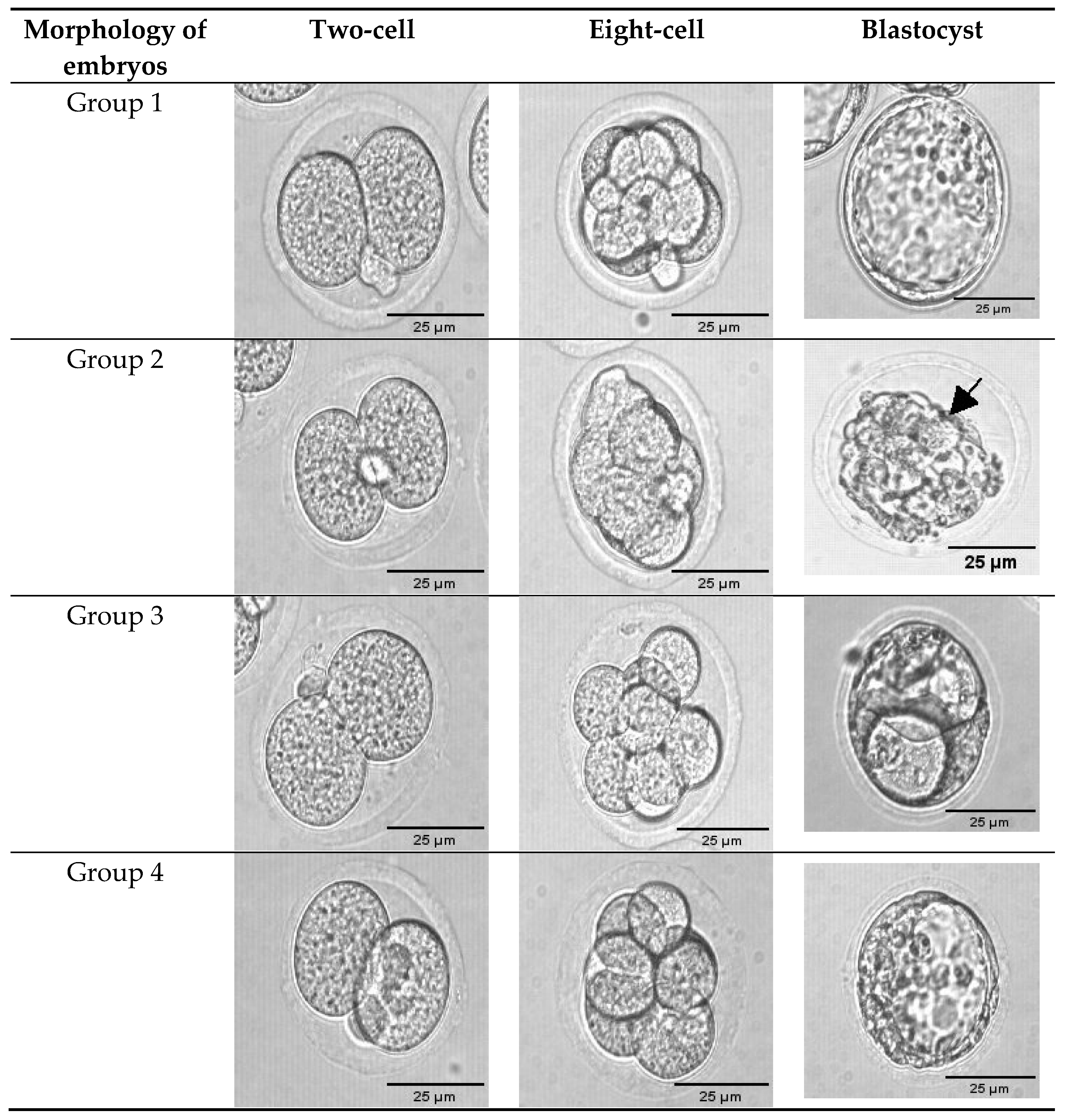
3.1. Exogenous GSH Supplementation Improved Preimplantation Embryo Morphology

The most widely used method of embryo selection is the visual assessment of embryo morphology. Numerous parameters can be evaluated at various developmental stages, providing valuable information about embryonic quality [54]. Embryos can be graded based on the morphology of their pronuclei on day 1 after fertilization [55,56], the number and shape of blastomeres and the degree of fragmentation on days 2 or 3 [13,57,58], or the morphology of the blastocyst on day 5 [24,59].
Images of embryos with or without GSH treatment were morphologically scored using the scoring system of Cuevas Saiz et al. [47]. Each embryo was quantitatively classified according to the size of the blastomere and embryonic fragmentation, as shown in Figure 2. The degree of fragmentation was initially described as the embryonic volume occupied by anucleate cytoplasmic fragments and is given as a percentage [60]. Glutathione supplementation was seen to significantly increase (p < 0.05) the number of Grade-1 blastocysts from 69% to 80% (Groups 1 and 3, respectively). Similarly, GSH supplementation also significantly increased postvitrification blastocyst formation from 63% to 81% (Groups 2 and 4, respectively) (Table 2). Similar to our findings, prior research has found that an increasing proportion of oocytes develop in GSH-supplemented maturation medium after IVF reaches the blastocyst stage [61,62].
It has been reported that a correlation exists between embryo morphology and viability [63,64]. The size and shape of blastomeres, the presence of extruded cells or fragmentation, compaction, the color of embryos, and the developmental stage reached at a particular point after fertilization are all significant parameters in embryo morphology assessment [65]. A previous study showed a correlation between oocyte cytoplasmic morphology and ICSI-induced fertilization rate and embryo quality. Significantly lower fertilization, embryo cleavage, and embryo quality have been observed in oocytes with cytoplasmic inclusions compared to oocytes with normal cytoplasm [66,67]. Fragmentation has also been linked to an increased risk of chromosomal abnormalities [68,69], leading to reduced blastocyst formation and implantation rates [70]. Viability is determined by embryo quality. Rapidly dividing embryos are more viable and have greater developmental potential than slowly dividing embryos [71,72].

3.2. Exogenous GSH Supplementation Enhanced Preimplantation Embryo Development

The effect of GSH supplementation on embryo development is shown in Table 2. The addition of GSH resulted in significantly higher numbers of embryos reaching the blastocyst stage. The number of blastocysts was significantly higher in Group 1 (GSH-free medium) compared to Group 3 (GSH-supplemented medium) (69% vs. 80% respectively). A previous study also demonstrated that embryos cultured in GSH-supplemented medium showed better development compared to embryos cultured in GSH-free medium [73]. In addition, oocyte exposure to GSH has improved postfertilization development to the blastocyst stage [27,46,73,74,75]. Supplementation of GSH in culture medium was reported to improve embryo development and quality by increasing intracellular GSH and decreasing intracellular oxidative activity during preimplantation development [75]. Another study reported that GSH improved blastocyst formation and decreased DNA damage by exhibiting the extensive relocation of mitochondria to the inner oocyte cytoplasm [46].
Our findings showed that GSH improved blastocyst development of vitrified embryos from 63.0% (Group 2) to 81.0% (Group 4) (p < 0.05) (Table 2). As discussed earlier, most studies have investigated the impact of GSH supplementation on development in vitro. There have been no published data on the impact of GSH supplementation on the cryotolerance of vitrified embryos. Further research on the possible mechanism of GSH-enhanced cryotolerance in vitrified embryos is, therefore, warranted.

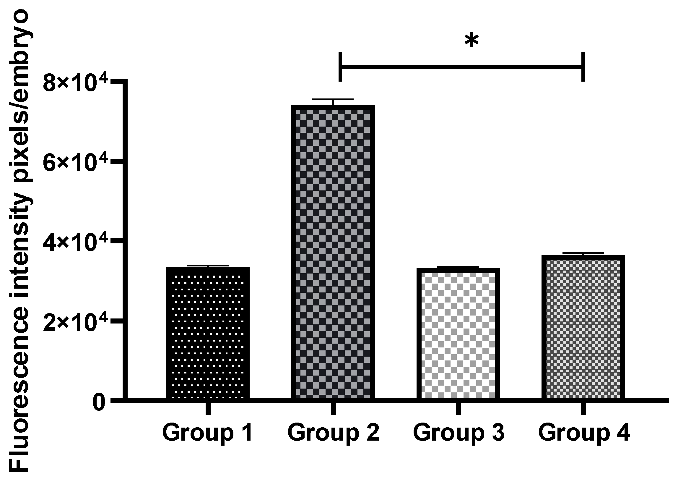
3.3. Exogenous GSH Supplementation Reduced Oxidative Stress in Vitrified Preimplantation Embryos

The intracellular ROS levels were examined to investigate whether OS could be reduced by GSH supplementation. The intensities of fluorescence generated by oxidized DCHFDA in eight-cell embryos are shown for all the groups (Figure 3). In non-vitrified groups (Groups 1 and 3), there was no significant difference in embryonic OS levels. However, in vitrified groups (Groups 2 and 4), GSH treatment significantly reduced ROS levels (p < 0.05) (Figure 4).
Previous studies have shown that elevated levels of ROS induced by vitrification are a possible cause of low preimplantation development rates [76,77,78]. In mammalian oocytes, vitrification has been reported to damage the endogenous antioxidant system [14] and mitochondria [79]. Mitochondria contain multiple electron carriers that can produce ROS. Damage to the organelle can result in an imbalance between the production and scavenging of ROS, resulting in net ROS production. As a result, ROS is partially responsible for the apoptosis or developmental arrest in embryos associated with cryopreservation. Under normal conditions, embryos have a defense mechanism against ROS in the form of antioxidant enzymes, including SOD and CAT. However, there is a possibility that embryo freezing disrupts these protective mechanisms [6].
An increase in ROS production in mammalian embryos has been shown to cause developmental arrests [80], embryonic fragmentation, or programmed cell death [45,81]. ROS-induced programmed cell death in embryos was attributed to changes in mitochondrial function, leading to higher production of ROS [82]. The addition of antioxidants was reported to prevent embryonic OS and apoptosis [83]. Therefore, we proposed that supplementation of GSH during vitrification was able to reduce OS by regenerating the GSH balance in an embryo. Glutathione supplementation prevents lipid peroxidation by removing excessive ROS in media [84]. It was reported that GSH improved oocyte fertilization by reducing the number of disulfide bonds in the zona pellucida, rendering them less rigid [27].

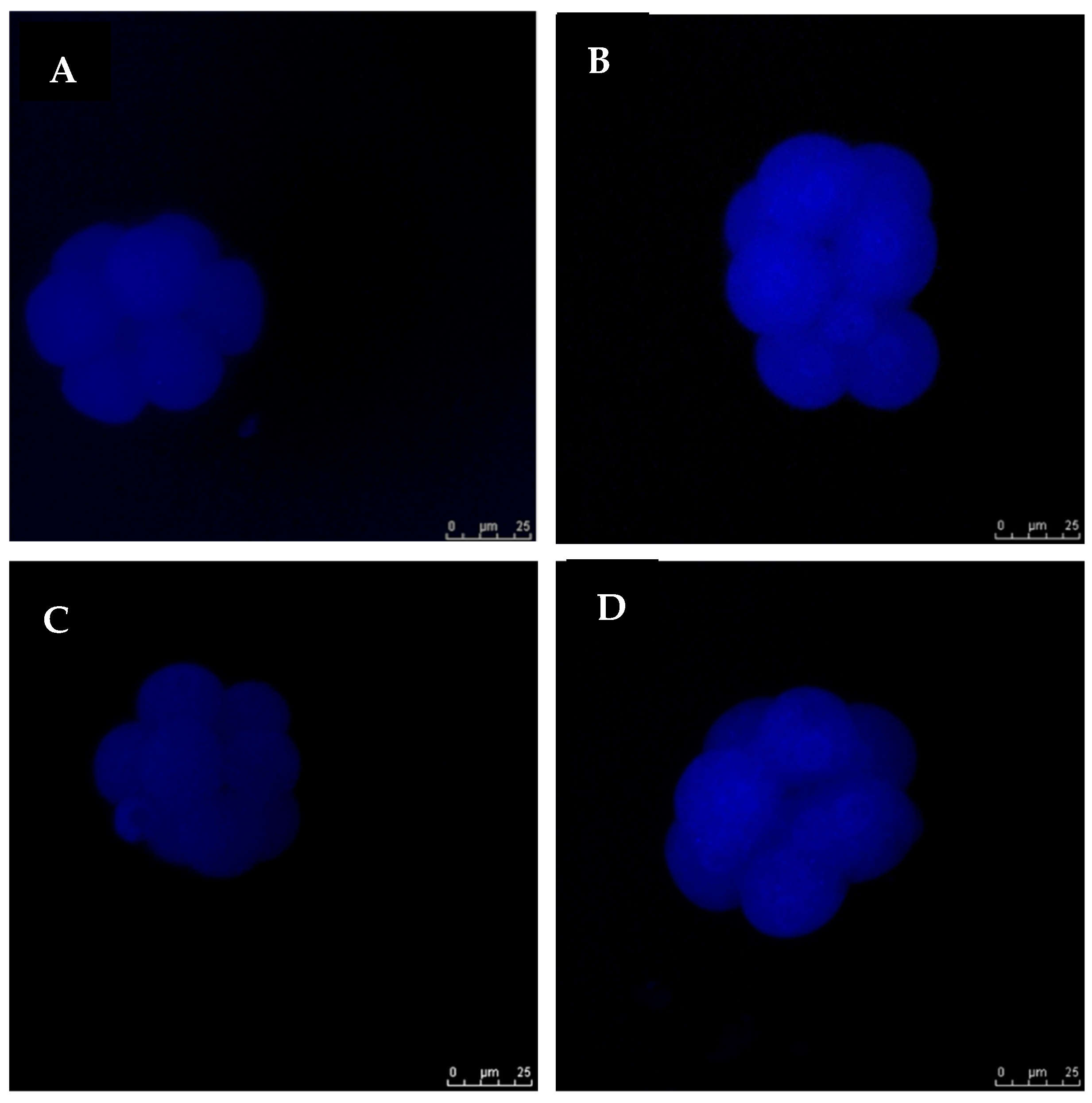
3.4. Exogenous GSH Supplementation Increased Endogenous GSH Levels in Vitrified Preimplantation Embryos

The intracellular levels of GSH in eight-cell embryos from all the groups were examined. Figure 5 shows images of embryos stained with 4-chloromethyl-6,8-difluoro-7-hydroxycoumarin. No remarkable difference in intracellular GSH levels was noted between non-vitrified embryos in Groups 1 and 2. However, the intracellular GSH level in vitrified embryos was significantly higher (p < 0.05) in Group 4 compared to Group 3 (Figure 6). The addition of GSH into culture media was, therefore, able to reduce ROS levels and increase the viability of vitrified embryos.
Exogenous GSH supplementation during in vitro culture has been reported to act as a defense mechanism against ROS by improving the intracellular GSH system [75,84]. An earlier study stated that the supplementation of 1 mM GSH to maturation medium stimulated intracellular GSH and maintained the redox balance in bovine IVF embryos [75]. However, further research is required to determine the efficiency in which specific embryonic stages acquire and use exogenous GSH from the culture medium. It is known that endogenous GSH in an embryo is essential for its protection against OS and other forms of cellular injury [84,85]. The degradation of GSH occurs exclusively in the extracellular space and on the surfaces of cells that express γ-glutamyl transpeptidase (GGT). The enzyme may transfer the γ-glutamyl moiety of GSH to amino acids and peptides. Frequently, cells take up the products of GSH hydrolysis as individual amino acids or as dipeptides. The balance between GSH production, consumption, and transportation determines the intra- and extracellular GSH levels [36]. The molecular mechanism by which GSH protects embryos from damage inflicted by vitrification, however, remains to be elucidated.

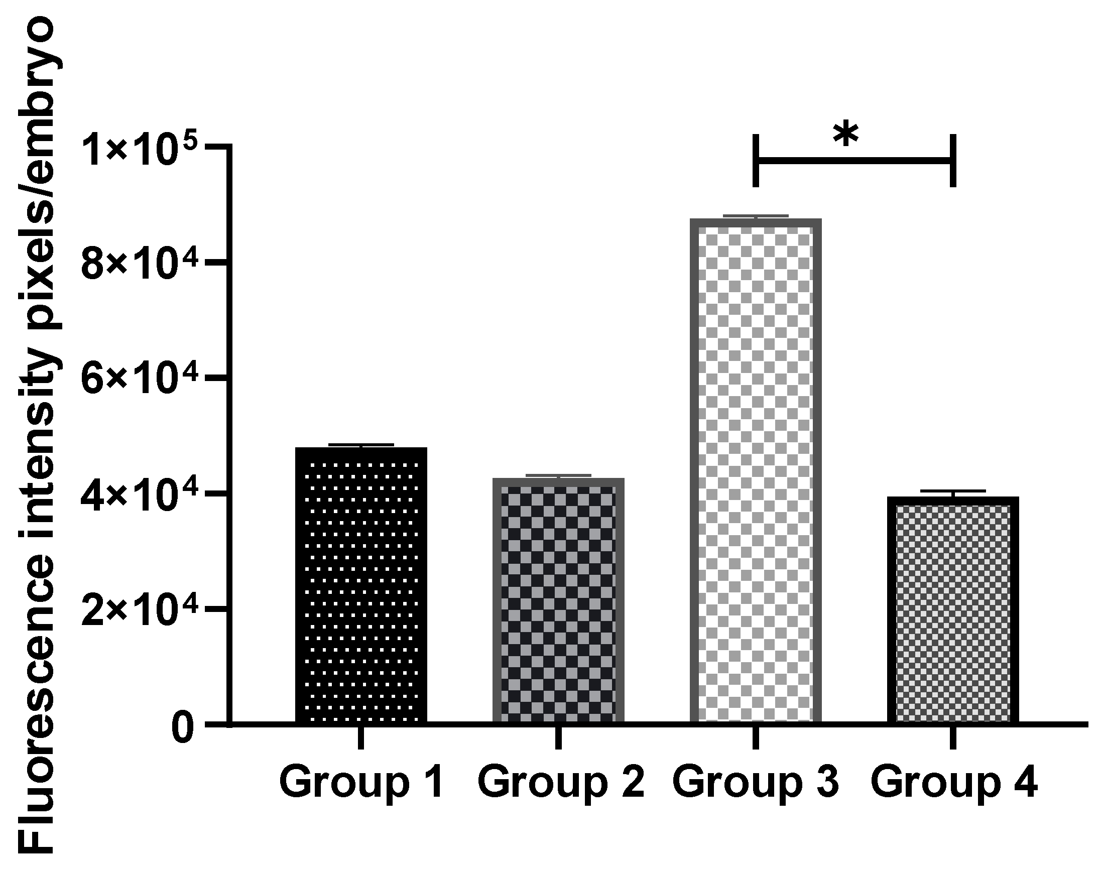
3.5. Exogenous GSH Supplementation Decreased Cytochrome C Expression in Vitrified Preimplantation Embryos

The expression of cytochrome c in non-vitrified and vitrified eight-cell stage embryos cultured with and without 0.01 mM GSH supplementation was analyzed using immunocytochemical staining (Figure 7). No significant difference in cytochrome c expression was observed between the non-vitrified embryos in Groups 1 and 2. However, 0.01 mM GSH-supplemented medium (Group 4) exhibited a significantly lower (p < 0.05) expression of cytochrome c compared to Group 3 (Figure 8).
The processes of vitrification and thawing have been reported to damage the endogenous antioxidant system [21] and mitochondrial distribution in mouse embryos and to effect embryonic development [86].
Mitochondria are organelles for ATP production that are important for controlling cell growth, dynamic response, signaling, and apoptosis in mammalian cells. Damage caused by excessive ROS results in the release of cytochrome c into the cytoplasm, which is one of the messengers of mitochondria-dependent apoptotic pathways [44]. Oxidative stress causes the release of cytochrome c and other apoptogenic factors from mitochondria, which then activates apoptosis [87].
In this study, 0.01 mM GSH supplementation in the culture medium significantly decreased the expression of cytochrome c in vitrified eight-cell embryos, suggesting that GSH supplementation improved mitochondrial function and cryotolerance. Similar to our results, previous studies reported that the addition of GSH to mouse embryo culture medium reduced ROS in embryos and increased the percentage of blastocysts [88]. Similar results were obtained in porcine [42] and bovine [75] models.
Therefore, we concluded that the addition of GSH to the culture medium could suppress ROS in the culture medium and reduce the amount of ROS transported into embryos. Thus, exogenous GSH supplementation could serve as a defense against ROS during vitrification through the maintenance of intracellular GSH.

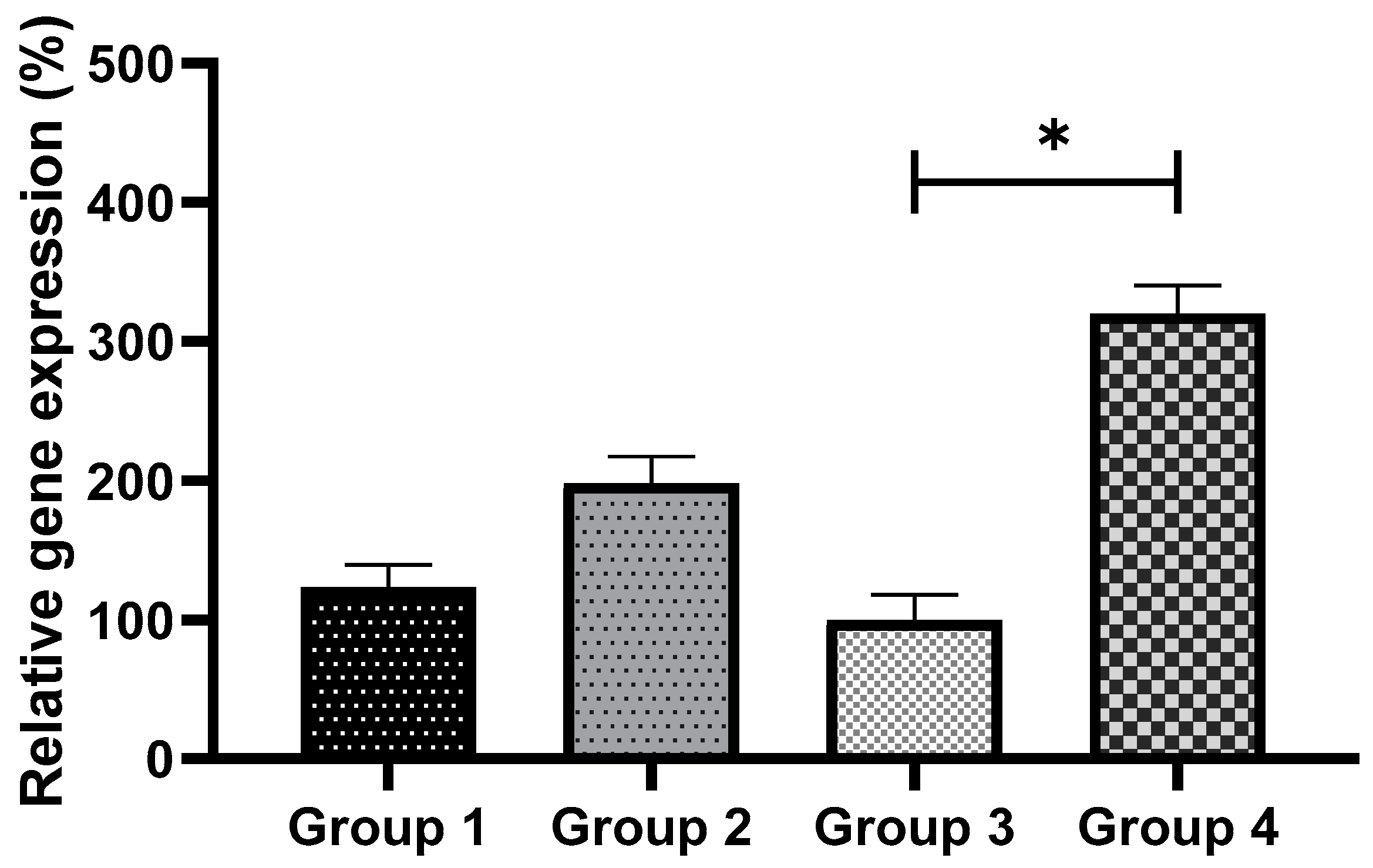
3.6. Exogenous GSH Supplementation Upregulated the Expression of Bcl2 Gene and Downregulated the Expression of Bax Gene in Vitrified Preimplantation Embryo

The expressions of Bax and Bcl2 (apoptosis-related genes) were analyzed for all the groups, as shown in Figure 9; Figure 10, respectively. No significant differences were observed in the expression of Bax across the non-vitrified groups. However, in the vitrified groups, GSH supplementation (Group 3) showed a significantly lower (p < 0.05) Bax expression compared to Group 4 (Figure 9). In tandem, Bcl2 expression was found to be significantly higher (p < 0.05) in Group 4 compared to Group 3 (Figure 10). The results from the Bax and Bcl2 expressions suggest that GSH supplementation reduced apoptosis in vitrified embryos.
The Bcl2 family has been shown to regulate mitochondrial outer membrane permeabilization and can be either pro-apoptotic (genes such as Bax) or anti-apoptotic (genes such as Bcl2) in the mitochondrion-mediated apoptosis pathway [21]. Anti-apoptotic genes, such as Bcl2, prevent apoptosis by inhibiting the release of cytochrome c from mitochondria. It has been shown that vitrification increases the mRNA levels of Bax in mouse oocytes [89] and bovine oocytes [90].
In our study, we measured the expressions of apoptosis-related genes via RT-qPCR, demonstrating that vitrification significantly increased the Bax mRNA level, contributing to the induction of apoptosis in vitrified mouse embryos, in agreement with reports in bovine [91,92], porcine [93], murine, and oocyte [94] studies. In addition, our results showed that GSH supplementation significantly decreased the Bax mRNA level and increased the Bcl2 mRNA level, illustrating that GSH supplementation in culture medium inhibited mitochondrion-mediated apoptosis by regulating the expression of the pro- (Bax) and anti-apoptotic (Bcl2) genes of the Bcl2 family, which indicated a high developmental rate of the embryos (Table 2). These results are consistent with previous research by Circu and Aw [95], which showed that supplementing with GSH upregulated the anti-apoptotic Bcl2 gene, increasing the levels of GSH in cells and indicating rapid embryonic development.
This finding was supported by the cytochrome c expression results, which showed that culture medium supplemented with 0.01 mM GSH had a significantly lower cytochrome c level and that reduced apoptosis resulted in a greater number of Grade-1 embryos and improved preimplantation development.

3.7. Exogenous GSH Supplementation Upregulated the Expression of Gpx1 and Sod1 Genes in Vitrified Preimplantation Embryo

The relative transcript abundances of glutathione peroxidase 1 (Gpx1) and superoxide dismutase 1 (Sod1) produced in response to GSH supplementation in culture medium with and without vitrification are shown in Figure 11 and Figure 12, respectively. There were no significant differences in the expression levels of Gpx1 and Sod1 in non-vitrified groups (Group 1 vs Group 2). In contrast, 0.01 mM GSH supplementation in the culture medium resulted in a significant (p < 0.05) upregulation of Sod1 in vitrified preimplantation embryos (Group 3 vs Group 4) (Figure 12). Similarly, a previous study reported that Sod1 gene expression was associated with the superior quality of in-vitro-produced murine embryos [96].
With regard to relative Gpx1 transcript abundance in vitrified preimplantation embryos, a significant (p < 0.05) decrease in Gpx1 expression was observed in Group 3 embryos. The present study confirms a previous report that cryopreservation decreased antioxidant enzyme expression in murine two-cell embryos [97]. Cryopreservation induced the downregulation of Gpx1 and Sod1 expressions, indicating that cryopreservation may alter the gene expressions of oocytes and embryos [98].
The Gpx1-encoded glutathione peroxidase-1 protein is essential for apoptosis. It helps in hydrogen peroxide catabolism and cellular redox balance. Glutathione peroxidase (GPx) is essential for the equilibrium between the two-step enzymatic antioxidant reaction that prevents damage to tissues and cells, which involves the transfer of SOD from superoxide anion to hydrogen peroxide, as well as GPx from hydrogen peroxide to water [99]. Glutathione peroxidase 1 plays the primary protective role in managing oxidative injury and death from ROS, as indicated by evidence derived from Gpx1 knockout mice [100]. In accordance with our findings, GSH supplementation in vitrified embryos (Group 4) resulted in significant (p < 0.05) increases in the expressions of Gpx1 and Sod1.
Therefore, these studies demonstrated the importance of the Gpx1 and Sod1 genes in preimplantation embryo cryopreservation. GSH is the predominant antioxidant in all cell types [101], and it is not surprising that the glutathione cycle is required for freeze–thaw tolerance. These studies proved the significance of exogenous GSH in reducing freeze–thaw stress and improving cryotolerance. However, the exact mechanism by which the supplementation of GSH to the culture medium increased the quality of the vitrified embryos is still unknown. Based on the present results, the GSH-mediated increase in Sod1 and Gpx1 transcript abundances is suggested to help in the balance and maintenance of cellular redox. This would result in improved embryonic cryotolerance.

4. Conclusions

In conclusion, the supplementation of 0.01 mM L-glutathione (GSH) in culture media increased intracellular GSH levels, as well as reduced mitochondrial dysfunction, ROS levels, and apoptosis. These mechanisms may contribute to improved preimplantation development in both non-vitrified and vitrified embryos. Our findings cast a new light on the novel use of GSH as a simple yet effective means to improve vitrification outcomes.

Author Contributions

Conceptualization, N.-S.A.R. and N.-A.M.N.K.; methodology, N.-S.A.R. and N.-A.M.N.K.; software, Z.E.; validation, N.-A.M.N.K., M.A.A. and F.O.; formal analysis, N.-S.A.R. and N.-A.M.N.K.; investigation, N.-S.A.R.; resources, N.-A.M.N.K.; data curation, N.-S.A.R.; writing—original draft preparation, N.-A.M.N.K. and N.-S.A.R.; writing—review and editing, N.-S.A.R., N.-A.M.N.K., M.-S.S., M.A.M., A.-A.M.K. and F.A., Z.E., F.O., M.A.A.; visualization, N.-S.A.R.; supervision, N.-A.M.N.K., M.A.A., F.O. and Z.E.; project administration, N.-A.M.N.K.; funding acquisition, N.-A.M.N.K. All authors have read and agreed to the published version of the manuscript.

Funding

The study was financially supported by the Ministry of Higher Education, Malaysia (FRGS 5/3 (454/2019) and FRGS-RACER 5/3 (088/2019). The APC was funded by the Faculty of Medicine, Universiti Teknologi MARA, Malaysia.

Institutional Review Board Statement

This study was carried out in accordance with the Universiti Teknologi MARA guidelines for the care and use of animals. All experimental procedures were approved by the Institutional Animal Care and Use Committee (ACUC) (ACUC-01/14, UiTM).

Informed Consent Statement

Not applicable.

Data Availability Statement

All data is contained within the article.

Acknowledgments

The authors would like to thank laboratory assistant Abu Thalhah Abdul Aziz for his technical assistance in this work.

Conflicts of Interest

The authors declare no conflict of interest.

References

  1. Fuller, B.; Paynter, S. Fundamentals of cryobiology in reproductive medicine. Reprod. Biomed. Online 2004, 9, 680–691. [Google Scholar] [CrossRef]
  2. Larman, M.G.; Minasi, M.G.; Rienzi, L.; Gardner, D.K. Maintenance of the meiotic spindle during vitrification in human and mouse oocytes. Reprod. Biomed. Online 2007, 15, 692–700. [Google Scholar] [CrossRef]
  3. Dalcin, L.; Silva, R.C.; Paulini, F.; Silva, B.D.; Neves, J.P.; Lucci, C.M. Cytoskeleton structure, pattern of mitochondrial activity and ultrastructure of frozen or vitrified sheep embryos. Cryobiology 2013, 67, 137–145. [Google Scholar] [CrossRef] [PubMed] [Green Version]
  4. Somoskoi, B.; Martino, N.A.; Cardone, R.A.; Lacalandra, G.M.; Dell’Aquila, M.E.; Cseh, S. Different chromatin and energy/redox responses of mouse morulae and blastocysts to slow freezing and vitrification. Reprod. Biol. Endocrinol. 2015, 13, 22. [Google Scholar] [CrossRef] [PubMed] [Green Version]
  5. Dasiman, R.; Rahman, N.S.A.; Othman, S.; Mustafa, M.F.; Yusoff, N.J.M.; Jusoff, W.H.W.; Khan, N.A.M.N. Cytoskeletal alterations in different developmental stages of in vivo cryopreserved preimplantation murine embryos. Med. Sci. Monit. Basic Res. 2013, 19, 258–266. [Google Scholar] [CrossRef] [PubMed] [Green Version]
  6. Oikonomou, Z.; Chatzimeletiou, K.; Sioga, A.; Oikonomou, L.; Tarlatzis, B.C.; Kolibianakis, E. Effects of vitrification on blastomere viability and cytoskeletal integrity in mouse embryos. Zygote 2017, 25, 75–84. [Google Scholar] [CrossRef]
  7. Tachataki, M.; Winston, R.M.; Taylor, D.M. Quantitative RT–PCR reveals tuberous sclerosis gene, TSC2, mRNA degradation following cryopreservation in the human preimplantation embryo. MHR. Basic Sci. Reprod. Med. 2003, 9, 593–601. [Google Scholar] [CrossRef] [Green Version]
  8. Larman, M.G.; Katz-Jaffe, M.G.; McCallie, B.; Filipovits, J.A.; Gardner, D.K. Analysis of global gene expression following mouse blastocyst cryopreservation. Hum. Reprod. 2011, 26, 2672–2680. [Google Scholar] [CrossRef] [Green Version]
  9. Stinshoff, H.; Wilkening, S.; Hanstedt, A.; Brüning, K.; Wrenzycki, C. Cryopreservation affects the quality of in vitro produced bovine embryos at the molecular level. Theriogenology 2011, 76, 1433–1441. [Google Scholar] [CrossRef]
  10. Monzo, C.; Haouzi, D.; Roman, K.; Assou, S.; Dechaud, H.; Hamamah, S. Slow freezing and vitrification differentially modify the gene expression profile of human metaphase II oocytes. Hum. Reprod. 2012, 27, 2160–2168. [Google Scholar] [CrossRef]
  11. Sahraei, S.S.; Shahhoseini, M.; Movaghar, B. Vitrification Has an Effect like Culture on Gene Expression and Histone Modification In Mouse Embryos. Cryo Lett. 2018, 39, 102–112. [Google Scholar]
  12. Harvey, A.J.; Kind, K.L.; Thompson, J.G. REDOX regulation of early embryo development. Reproduction 2002, 123, 479–486. [Google Scholar] [CrossRef] [PubMed]
  13. Ajduk, A.; Zernicka-Goetz, M. Advances in embryo selection methods. Biol. Rep. 2012, 4, 11. [Google Scholar] [CrossRef] [PubMed]
  14. Takahashi, M. Oxidative stress and redox regulation on in vitro development of mammalian embryos. J. Reprod. Dev. 2012, 58, 1–9. [Google Scholar] [CrossRef] [PubMed] [Green Version]
  15. Garrido, C.; Galluzzi, L.; Brunet, M.; Puig, P.E.; Didelot, C.; Kroemer, G. Mechanisms of cytochrome c release from mitochondria. Cell Death Differ. 2006, 13, 1423–1433. [Google Scholar] [CrossRef] [Green Version]
  16. Gross, A.; McDonnell, J.M.; Korsmeyer, S.J. BCL-2 family members and the mitochondria in apoptosis. Genes Dev. 1999, 13, 1899–1911. [Google Scholar] [CrossRef] [Green Version]
  17. Bratton, S.B.; Salvesen, G.S. Regulation of the Apaf-1–caspase-9 apoptosome. J. Cell Sci. 2010, 123, 3209–3214. [Google Scholar] [CrossRef] [Green Version]
  18. Trapphoff, T.; Heiligentag, M.; Simon, J.; Staubach, N.; Seidel, T.; Otte, K.; Eichenlaub-Ritter, U. Improved cryotolerance and developmental potential of in vitro and in vivo matured mouse oocytes by supplementing with a glutathione donor prior to vitrification. Mol. Hum. Reprod. 2016, 22, 867–881. [Google Scholar]
  19. Lane, M.; Maybach, J.M.; Gardner, D.K. Addition of ascorbate during cryopreservation stimulates subsequent embryo development. Hum Reprod. 2002, 17, 2686–2693. [Google Scholar] [CrossRef] [Green Version]
  20. Somfai, T.; Ozawa, M.; Noguchi, J.; Kaneko, H.; Kuriani Karja, N.W.; Farhudin, M.; Dinnyés, A.; Nagai, T.; Kikuchi, K. Developmental competence of in vitro-fertilized porcine oocytes after in vitro maturation and solid surface vitrification: Effect of cryopreservation on oocyte antioxidative system and cell cycle stage. Cryobiology 2007, 55, 115–126. [Google Scholar] [CrossRef]
  21. Gupta, M.K.; Uhm, S.J.; Lee, H.T. Effect of vitrification and beta-mercaptoethanol on reactive oxygen species activity and in vitro development of oocytes vitrified before or after in vitro fertilization. Fertil. Steril. 2010, 93, 2602–2607. [Google Scholar] [CrossRef] [PubMed]
  22. Boonkusol, D.; Gal, A.B.; Bodo, S.; Gorhony, B.; Kitiyanant, Y.; Dinnyes, A. Gene expression profiles and in vitro development following vitrification of pronuclear and 8-cell stage mouse embryos. Mol. Reprod. Dev. 2006, 73, 700–708. [Google Scholar] [CrossRef] [PubMed]
  23. Mamo, S.; Bodo, S.; Kobolak, J.; Polgar, Z.; Tolgyesi, G.; Dinnyes, A. Gene expression profiles of vitrified in vivo derived 8-cell stage mouse embryos detected by high density oligonucleotide microarrays. Mol. Reprod. Dev. 2006, 73, 1380–1392. [Google Scholar] [CrossRef] [PubMed]
  24. Balaban, B.; Urman, B.; Sertac, A.; Alatas, C.; Aksoy, S.; Mercan, R. Blastocyst quality affects the success of blastocyst-stage embryo transfer. Fertil. Steril. 2000, 74, 282–287. [Google Scholar] [CrossRef]
  25. Succu, S.; Bebbere, D.; Bogliolo, L.; Ariu, F.; Fois, S.; Leoni, G.G.; Berlinguer, F.; Naitana, S.; Ledda, S. Vitrification of in vitro matured ovine oocytes affects in vitro pre-implantation development and mRNA abundance. Mol. Reprod. Dev. 2007, 75, 538–546. [Google Scholar] [CrossRef]
  26. Bartolac, L.K.; Lowe, J.L.; Koustas, G.; Grupen, C.G.; Sjöblom, C. Vitrification, not cryoprotectant exposure, alters the expression of developmentally important genes in in vitro produced porcine blastocysts. Cryobiology 2018, 80, 70–76. [Google Scholar] [CrossRef]
  27. Hosseini, S.M.; Forouzanfar, M.; Hajian, M.; Asgari, V.; Abedi, P.; Hosseini, L.; Nasr-Esfahani, M.H. Antioxidant supplementation of culture medium during embryo development and/or after vitrification-warming; Which is the most important? J. Assist. Reprod. Genet. 2009, 26, 355–364. [Google Scholar] [CrossRef] [Green Version]
  28. Dehghani-Mohammadabadi, M.; Salehi, M.; Farifteh, F.; Nematollahi, S.; Arefian, E.; Hajjarizadeh, A.; Parivar, K.; Nourmohammadi, Z. Melatonin modulates the expression of BCL-xl and improve the development of vitrified embryos obtained by IVF in mice. J. Assist. Reprod. Genet. 2014, 31, 453–461. [Google Scholar] [CrossRef] [Green Version]
  29. Kopeika, J.; Thornhill, A.; Khalaf, Y. The effect of cryopreservation on the genome of gametes and embryos: Principles of cryobiology and critical appraisal of the evidence. Hum. Reprod. Update 2015, 21, 209–227. [Google Scholar] [CrossRef] [Green Version]
  30. Menezo, Y.J.R.; Silvestris, E.; Dale, B.; Elder, K. Oxidative stress and alterations in DNA methylation: Two sides of the same coin in reproduction. Reprod. Biomed. Online 2016, 33, 668–683. [Google Scholar] [CrossRef] [Green Version]
  31. Agarwal, A.; Majzoub, A. Role of Antioxidants in Assisted Reproductive Techniques. World J. Men’s Health 2017, 35, 77–93. [Google Scholar] [CrossRef] [PubMed]
  32. Sakatani, M.; Kobayashi, S.; Takahashi, M. Effects of Heat Shock on In Vitro Development and Intracellular Oxidative State of Bovine Preimplantation Embryos. Mol. Reprod. Dev. 2004, 67, 77–82. [Google Scholar] [CrossRef] [PubMed]
  33. De Matos, D.G.; Furnus, C.C.; Moses, D.F.; Martinez, A.G.; Matkovic, M. Stimulation of glutathione synthesis of in vitro matured bovine oocytes and its effect on embryo development and freezability. Mol. Reprod. Dev. 1996, 45, 451–457. [Google Scholar] [CrossRef]
  34. Bavister, B.D.; Squirrell, J.M. Mitochondrial distribution and function in oocytes and early embryos. Hum. Reprod. 2000, 15, 189–198. [Google Scholar] [CrossRef]
  35. Diaz Vivancos, P.; Wolff, T.; Markovic, J.; Pallardo, F.V.; Foyer, C.H. A nuclear glutathione cycle within the cell cycle. Biochem. J. 2010, 431, 169–178. [Google Scholar] [CrossRef] [PubMed] [Green Version]
  36. Lushchak, V.I. Glutathione homeostasis and functions: Potential targets for medical interventions. J. Amino Acids 2012, 2012, 736837. [Google Scholar] [CrossRef] [Green Version]
  37. Duan, S.; Chen, C. S-nitrosylation/denitrosylation and apoptosis of immune cells. Cell. Mol. Immunol. 2007, 4, 353–358. [Google Scholar]
  38. Cooper, A.J.; Pinto, J.T.; Callery, P.S. Reversible and irreversible protein glutathionylation: Biological and clinical aspects. Expert Opin. Drug Metab. Toxicol. 2011, 7, 891–910. [Google Scholar] [CrossRef] [Green Version]
  39. Zofia, L. The role of glutathione in mammalian gametes. Reprod. Biol. 2005, 5, 5–17. [Google Scholar]
  40. Hansen, J.M.; Harris, C. Glutathione during embryonic development. Biochim. Biophys. Acta 2015, 1850, 1527–1542. [Google Scholar] [CrossRef]
  41. Yao, T.; Asayama, Y. Human preimplantation embryo culture media: Past, present, and future. J. Mamm. Ova Res. 2016, 33, 17–34. [Google Scholar] [CrossRef]
  42. Ozawa, M.; Hirabayashi, M.; Kanai, Y. Developmental competence and oxidative state of mouse zygotes heat-stressed maternally or in vitro. Reproduction 2002, 124, 683–689. [Google Scholar] [CrossRef] [PubMed]
  43. Yuzawa, K.; Sawada, T.; Hossain, M.S.; Takagi, Y.; Hamano, K.; Tsujii, H. Effect of glutathione on the development of rat embryos following microinsemination. Reprod. Med. Biol. 2009, 8, 11–17. [Google Scholar] [CrossRef] [PubMed] [Green Version]
  44. Sun, X.; Zhang, H.; Zhang, Y.; Yang, Q.; Zhao, S. Caspase-dependent mitochondrial apoptotic pathway is involved in astilbin-mediated cytotoxicity in breast carcinoma cells. Oncol. Rep. 2018, 40, 2278–2286. [Google Scholar] [CrossRef] [PubMed]
  45. Guérin, P.; El Mouatassim, S.; Ménézo, Y. Oxidative stress and protection against reactive oxygen species in the pre-implantation embryo and its surroundings. Hum. Reprod. Update 2001, 7, 175–189. [Google Scholar] [CrossRef] [PubMed]
  46. Li, F.; Cui, L.; Yu, D.; Hao, H.; Liu, Y.; Zhao, X.; Du, W. Exogenous glutathione improves intracellular glutathione synthesis via the γ-glutamyl cycle in bovine zygotes and cleavage embryos. J. Cell. Physiol. 2019, 234, 7384–7394. [Google Scholar] [CrossRef]
  47. Cuevas Saiz, I.; Gatell, M.C.P.; Vargas, M.C.; Mendive, A.D.; Enedáguila, N.R.; Solanes, M.M.; Canal, C.B.; López, J.T.; Bonet, A.B.; de Mendoza, M.V.H. The Embryology Interest Group: Updating ASEBIR’s morphological scoring system for early embryos, morulae and blastocysts. Med. Reprod. Y Embriol. Clínica 2018, 5, 42–54. [Google Scholar]
  48. Kasai, M.; Komi, J.H.; Takakamo, A.; Tsudera, H.; Sakurai, T.; Machida, T. A simple method for mouse embryo cryopreservation in a low toxicity vitrification solution, without appreciable loss of viability. Reproduction 1990, 89, 91–97. [Google Scholar] [CrossRef]
  49. Zhang, J.; Cui, J.; Ling, X.; Li, X.; Peng, Y.; Guo, X.; Heng, B.C.; Tong, G.Q. Vitrification of mouse embryos at 2-cell, 4-cell and 8-cell stages by cryotop method. J. Assist. Reprod. Genet. 2009, 26, 621–628. [Google Scholar] [CrossRef] [Green Version]
  50. Johansson, M.; Hardarson, T.; Lundin, K. There Is a Cutoff Limit in Diameter Between a Blastomere and a Small Anucleate Fragment. J. Assist. Reprod. Genet. 2003, 20, 309–313. [Google Scholar] [CrossRef]
  51. Hsieh, C.-Y.; Chen, C.-L.; Yang, K.-C.; Ma, C.-T.; Choi, P.-C.; Lin, C.-F. Detection of Reactive Oxygen Species During the Cell Cycle Under Normal Culture Conditions Using a Modified Fixed-Sample Staining Method. J. Immunoass. Immunochem. 2014, 36, 149–161. [Google Scholar] [CrossRef] [PubMed]
  52. Livak, K.J.; Schmittgen, T.D. Analysis of relative gene expression data using real-time quantitative PCR and the 2−ΔΔCT method. Methods 2001, 25, 402–408. [Google Scholar] [CrossRef] [PubMed]
  53. Zatecka, E.; Ded, L.; Elzeinova, F.; Kubatova, A.; Dorosh, A.; Margaryan, H.; Dostalova, P.; Korenkova, V.; Hoskova, K.; Peknicova, J. Effect of zearalenone on reproductive parameters and expression of selected testicular genes in mice. Reprod. Toxicol. 2014, 45, 20–30. [Google Scholar] [CrossRef] [PubMed]
  54. Ebner, T.; Moser, M.; Sommergruber, M.; Tews, G. Selection based on morphological assessment of oocytes and embryos at different stages of preimplantation development: A review. Hum. Reprod. Update 2003, 9, 251–262. [Google Scholar] [CrossRef]
  55. Montag, M.; Van Der Ven, H. Evaluation of pronuclear morphology as the only selection criterion for further embryo culture and transfer: Results of a prospective multicentre study. Hum. Reprod. 2001, 16, 2384–2389. [Google Scholar] [CrossRef]
  56. Tesarik, J.; Greco, E. The probability of abnormal preimplantation development can be predicted by a single static observation on pronuclear stage morphology. Hum. Reprod. 1999, 14, 1318–1323. [Google Scholar] [CrossRef] [Green Version]
  57. van Royen, E.; Mangelschots, K.; de Neubourg, D.; Valkenburg, M.; van de Meerssche, M.; Ryckaert, G.; Eestermans, W.; Gerris, J. Characterization of a top quality embryo, a step towards single-embryo transfer. Hum. Reprod. 1999, 14, 2345–2349. [Google Scholar] [CrossRef] [Green Version]
  58. Ziebe, S.; Petersen, K.; Lindenberg, S.; Andersen, A.G.; Gabrielsen, A. Embryo morphology or cleavage stage: How to select the best embryos for transfer after in-vitro fertilization. Hum. Reprod. 1997, 12, 1545–1549. [Google Scholar] [CrossRef]
  59. Gardner, D.K.; Phil, D.; Lane, M.; Stevens, J.; Schlenker, T.; Schoolcraft, W.B. Blastocyst score affects implantation and pregnancy outcome: Towards a single blastocyst transfer. Fertil. Steril. 2000, 73, 1155–1158. [Google Scholar] [CrossRef]
  60. Halvaei, I.; Khalili, M.A.; Esfandiari, N.; Safari, S.; Talebi, A.R.; Miglietta, S.; Nottola, S.A. Ultrastructure of cytoplasmic fragments in human cleavage stage embryos. J. Assist. Reprod. Genet. 2016, 33, 1677–1684. [Google Scholar] [CrossRef] [Green Version]
  61. Lian, H.; Gao, Y.; Jiao, G.; Sun, M.; Wu, X.; Wang, T.; Tan, J. Antioxidant supplementation overcomes the deleterious effects of maternal restraint stress-induced oxidative stress on mouse oocytes. Reproduction 2013, 146, 559–568. [Google Scholar] [CrossRef] [PubMed] [Green Version]
  62. Gunawan, M.; Nuriza, N.; Kaiin, E.M.; Sjahfirdi, L. The effect of glutathione antioxidant addition in maturation medium on the morphology of Garut sheep (Ovis aries) oocytes after vitrification. J. Phys. Conf. Ser. 2021, 1725, 012062. [Google Scholar] [CrossRef]
  63. Chappel, S. The Role of Mitochondria from Mature Oocyte to Viable Blastocyst. Obstet. Gynecol. Int. 2013, 2013, 183024. [Google Scholar] [CrossRef] [Green Version]
  64. Rienzi, L.; Gracia, C.; Maggiulli, R.; LaBarbera, A.R.; Kaser, D.J.; Ubaldi, F.M.; Racowsky, C. Oocyte, embryo and blastocyst cryopreservation in ART: Systematic review and meta-analysis comparing slow freezing versus vitrification to produce evidence for the development of global guidance. Hum. Reprod. Update 2017, 23, 139–155. [Google Scholar] [CrossRef] [PubMed] [Green Version]
  65. Jahromi, H.; Dehnavi, H. Morphological and morphometric study of early-cleavage mice embryos resulting from in vitro fertilization at different cleavage stages after vitrification. Iran. J. Vet. Res. 2016, 17, 55–58. [Google Scholar]
  66. Xia, P. Intracytoplasmic sperm injection: Correlation of oocyte grade based on polar body, perivitelline space and cytoplasmic inclusions with fertilization rate and embryo quality. Hum. Reprod. 1997, 12, 1750–1755. [Google Scholar] [CrossRef] [PubMed]
  67. Bartolacci, A.; Intra, G.; Coticchio, G.; Dell’Aquila, M.; Patria, G.; Borini, A. Does morphological assessment predict oocyte developmental competence? A systematic review and proposed score. J. Assist. Reprod. Genet. 2022, 39, 3–17. [Google Scholar] [CrossRef]
  68. Magli, M.C.; Gianaroli, L.; Ferraretti, A.P. Chromosomal abnormalities in embryos. Mol. Cell. Endocrinol. 2001, 183, S29–S34. [Google Scholar] [CrossRef]
  69. Maurer, M.; Ebner, T.; Puchner, M.; Mayer, R.B.; Shebl, O.; Oppelt, P.; Duba, H.C. Chromosomal Aneuploidies and Early Embryonic Developmental Arrest. Int. J. Fertil. Steril. 2015, 9, 346–353. [Google Scholar]
  70. Esfandiari, N.; Burjaq, H.; Gotlieb, L.; Casper, R.F. Brown oocytes: Implications for assisted reproductive technology. Fertil. Steril. 2006, 86, 1522–1525. [Google Scholar] [CrossRef]
  71. Barnett, D.K.; Bavister, B.D. What is the relationship between the metabolism of preimplantation embryos and their developmental competence? Mol. Reprod. Dev. 1996, 43, 105–133. [Google Scholar] [CrossRef]
  72. Phan, V.; Littman, E.; Harris, D.; La, A. Correlation between embryo morphology and development and chromosomal complement. Asian Pac. J. Reprod. 2014, 3, 85–89. [Google Scholar] [CrossRef]
  73. Khalili, M.A.; Anvari, M. The effect of in vitro culture on cleavage rates and morphology of the in vivo-developed embryos in mice. Iran. J. Reprod. Med. 2007, 5, 17–22. [Google Scholar]
  74. Dinara, S.; Sengoku, K.; Tamate, K.; Horikawa, M.; Ishikawa, M. Effects of supplementation with free radical scavengers on the survival and fertilization rates of mouse cryopreserved oocytes. Hum. Reprod. 2001, 16, 1976–1981. [Google Scholar] [CrossRef] [PubMed] [Green Version]
  75. Sun, W.J.; Pang, Y.W.; Liu, Y.; Hao, H.S.; Zhao, X.M.; Qin, T.; Du, W.H. Exogenous glutathione supplementation in culture medium improves the bovine embryo development after in vitro fertilization. Theriogenology 2015, 84, 716–723. [Google Scholar] [CrossRef]
  76. Mukaida, T.; Wada, S.; Takahashi, K.; Pedro, P.B.; An, T.Z.; Kasai, M. Vitrification of human embryos based on the assessment of suitable conditions for 8-cell mouse embryos. Hum. Reprod. 1998, 13, 2874–2879. [Google Scholar] [CrossRef] [Green Version]
  77. Zhou, G.; Zeng, Y.; Guo, J.; Meng, Q.; Meng, Q.; Jia, G.; Zhu, S. Vitrification transiently alters Oct-4, Bcl2 and P53 expression in mouse morulae but does not affect embryo development in vitro. Cryobiology 2016, 73, 120–125. [Google Scholar] [CrossRef]
  78. Chaves, D.F.; Corbin, E.; Almiñana, C.; Locatelli, Y.; Souza-Fabjan, J.M.G.; Bhat, M.H.; Mermillod, P. Vitrification of immature and in vitro matured bovine cumulus-oocyte complexes: Effects on oocyte structure and embryo development. Livest. Sci. 2017, 199, 50–56. [Google Scholar] [CrossRef]
  79. Tait, S.W.G.; Green, D.R. Mitochondrial regulation of cell death. Cold Spring Harb. Perspect. Biol. 2013, 5, a008706. [Google Scholar] [CrossRef] [Green Version]
  80. Kitagawa, Y.; Suzuki, K.; Yoneda, A.; Watanabe, T. Effects of oxygen concentration and antioxidants on the in vitro developmental ability, production of reactive oxygen species (ROS), and DNA fragmentation in porcine embryos. Theriogenology 2004, 62, 1186–1197. [Google Scholar] [CrossRef]
  81. Martín-romero, F.J.; Miguel-lasobras, E.M.; Domínguez-arroyo, J.A.; González-Carrera, E.; Alvarez, I.S. Contribution of culture media to oxidative stress and its effect on human oocytes. Reprod. Biomed. Online 2008, 17, 652–661. [Google Scholar] [CrossRef]
  82. Dennery, P.A. Effects of oxidative stress on embryonic development. Birth Defects Res. Part C Embryo Today Rev. 2007, 81, 155–162. [Google Scholar] [CrossRef] [PubMed]
  83. Singh, C.K.; Kumar, A.; Hitchcock, D.B.; Fan, D.; Goodwin, R.; Lavoie, H.A.; Singh, U.S. Resveratrol prevents embryonic oxidative stress and apoptosis associated with diabetic embryopathy and improves glucose and lipid profile of diabetic dam. Mol. Nutr. Food Res. 2011, 55, 1186–1196. [Google Scholar] [CrossRef] [PubMed] [Green Version]
  84. Truong, T.T.; Soh, Y.M.; Gardner, D.K. Antioxidants improve mouse preimplantation embryo development and viability. Hum. Reprod. 2016, 31, 1445–1454. [Google Scholar] [CrossRef] [PubMed] [Green Version]
  85. Moawad, A.R.; Tan, S.L.; Taketo, T. Beneficial effects of glutathione supplementation during vitrification of mouse oocytes at the germinal vesicle stage on their preimplantation development following maturation and fertilization in vitro. Cryobiology 2017, 76, 98–103. [Google Scholar] [CrossRef] [PubMed]
  86. Lei, T.; Guo, N.; Liu, J.-Q.; Tan, M.-H.; Li, Y.-F. Vitrification of in vitro matured oocytes: Effects on meiotic spindle configuration and mitochondrial function. Int. J. Clin. Exp. Pathol. 2014, 7, 1159–1165. [Google Scholar]
  87. Mahfouz, M.; Kummerow, F. Vitamin C or Vitamin B6 supplementation prevent the oxidative stress and decrease of prostacyclin generation in homocysteinemic rats. Int. J. Biochem. Cell Biol. 2004, 36, 1919–1932. [Google Scholar] [CrossRef]
  88. Gardiner, C.S.; Reed, D.J. Status of Glutathione during Oxidant-Induced Oxidative Stress in the Preimplantation Mouse Embryo1. Biol. Reprod. 1994, 51, 1307–1314. [Google Scholar] [CrossRef] [Green Version]
  89. Jang, W.; Lee, S.; Choi, H.; Lim, J.; Heo, Y.; Cui, X.; Kim, N. Vitrification of immature-mouse oocytes by the modified-cut standard straw method. Cell Biol. Int. 2013, 38, 164–171. [Google Scholar] [CrossRef]
  90. Chen, J.-Y.; Li, X.-X.; Xu, Y.-K.; Wu, H.; Zheng, J.-J.; Yu, X.-L. Developmental competence and gene expression of immature oocytes following liquid helium vitrification in bovine. Cryobiology 2014, 69, 428–433. [Google Scholar] [CrossRef]
  91. Kuzmany, A.; Havlicek, V.; Wrenzycki, C.; Wilkening, S.; Brem, G.; Besenfelder, U. Expression of mRNA, before and after freezing, in bovine blastocysts cultured under different conditions. Theriogenology 2011, 75, 482–494. [Google Scholar] [CrossRef] [PubMed]
  92. Madrid Gaviria, S.; López Herrera, A.; Urrego, R.; Restrepo Betancur, G.; Echeverri Zuluaga, J.J. Effect of resveratrol on vitrified in vitro produced bovine embryos: Recovering the initial quality. Cryobiology 2019, 89, 42–50. [Google Scholar] [CrossRef] [PubMed]
  93. Pereira, B.A.; Zangeronimo, M.G.; Castillo-Martín, M.; Gadani, B.; Chaves, B.R.; Rodríguez-Gil, J.E.; Yeste, M. Supplementing maturation medium with insulin growth factor I and vitrification-warming solutions with reduced glutathione enhances survival rates and development ability of in vitro matured vitrified-warmed pig oocytes. Front. Physiol. 2019, 9, 1894. [Google Scholar] [CrossRef] [PubMed] [Green Version]
  94. Li, Z.; Gu, R.; Lu, X.; Zhao, S.; Feng, Y.; Sun, Y. Preincubation with glutathione ethyl ester improves the developmental competence of vitrified mouse oocytes. J. Assist. Reprod. Genet. 2018, 35, 1169–1178. [Google Scholar] [CrossRef] [PubMed]
  95. Circu, M.L.; Aw, T.Y. Glutathione and modulation of cell apoptosis. Biochim. Et Biophys. Acta 2012, 1823, 1767–1777. [Google Scholar] [CrossRef] [PubMed] [Green Version]
  96. Wang, F.; Tian, X.; Zhang, L.; Tan, D.; Reiter, R.J.; Liu, G. Melatonin promotes the in vitro development of pronuclear embryos and increases the efficiency of blastocyst implantation in murine. J. Pineal Res. 2013, 55, 267–274. [Google Scholar] [CrossRef]
  97. Gao, C.; Han, H.-B.; Tian, X.-Z.; Tan, D.-X.; Wang, L.; Zhou, G.-B.; Zhu, S.-E.; Liu, G.-S. Melatonin promotes embryonic development and reduces reactive oxygen species in vitrified mouse 2-cell embryos. J. Pineal Res. 2011, 52, 305–311. [Google Scholar] [CrossRef]
  98. Castillo-Martín, M.; Yeste, M.; Soler, A.; Morató, R.; Bonet, S. Addition of L-ascorbic acid to culture and vitrification media of IVF porcine blastocysts improves survival and reduces HSPA1A levels of vitrified embryos. Reprod. Fertil. Dev. 2015, 27, 1115–1123. [Google Scholar] [CrossRef]
  99. Zarbakhsh, S. Effect of antioxidants on preimplantation embryo development in vitro: A review. Zygote 2021, 29, 179–193. [Google Scholar] [CrossRef]
  100. Roy, P.; Lei, X.G. Knockout of SOD1 or GPX1 led to decreased bone remodeling in young adult female mice. FASEB J. 2007, 21, A114. [Google Scholar] [CrossRef]
  101. Ribas, V.; García-Ruiz, C.; Fernández-Checa, J.C. Glutathione and mitochondria. Front. Pharmacol. 2014, 5, 151. [Google Scholar] [CrossRef] [PubMed]
Figure 1. Diagrammatical representation of the study. Group 1 (GSH-free medium), Group 2 (GSH-free medium with vitrification), Group 3 (0.01 mM GSH-supplemented medium), and Group 4 (0.01 mM GSH-supplemented medium with vitrification). Abbreviation: IVC; in vitro culture.
Antioxidants 11 02100 g001
Figure 2. Morphologies of two-cell in vitro cultures to eight-cell embryos that underwent vitrification and without vitrification were observed until development to blastocyst stage from Groups 1–4. Arrows show fragmented embryos. Group 1 (GSH-free medium); Group 2 (GSH-free medium with vitrification); Group 3 (0.01 mM GSH-supplemented medium), Group 4 (0.01 mM GSH-supplemented medium with vitrification). The scale bar for the images represents 25 µm.
Antioxidants 11 02100 g002
Figure 3. Fluorescent photomicrograph of embryos showing intracellular ROS contents. Images are viewed under 400× magnification. (A): Group 1 (GSH-free medium); (B): Group 2 (GSH-free medium with vitrification); (C): Group 3 (0.01 mM GSH-supplemented medium), (D): Group 4 (0.01 mM GSH-supplemented medium with vitrification). The scale bar for the images represents 25 µm.
Antioxidants 11 02100 g003
Figure 4. Comparison of intracellular ROS levels in non-vitrified and vitrified embryos. Data are expressed as the means ± SEM. * indicates p < 0.05. Data were analyzed using one-way ANOVA with post hoc Tukey multiple comparison tests. Group 1 (GSH-free medium); Group 2 (GSH-free medium with vitrification); Group 3 (0.01 mM GSH-supplemented medium), Group 4 (0.01 mM GSH-supplemented medium with vitrification).
Antioxidants 11 02100 g004
Figure 5. Fluorescent photomicrograph of embryos showing intracellular GSH contents. Images are viewed under 40× magnification. (A): Embryo from Group 1 (GSH-free medium); (B): embryo from Group 2 (GSH-supplemented medium); (C): embryo from Group 3 (GSH-free medium with vitrification); (D): embryo from Group 4 (GSH-supplemented medium with vitrification).
Antioxidants 11 02100 g005
Figure 6. Comparison of intracellular GSH levels between non-vitrified and vitrified embryos. Data are expressed as significant differences between the means ± SEM. * indicates p < 0.05. Group 1 (GSH-free medium); Group 2 (GSH-free medium with vitrification); Group 3 (0.01 mM GSH-supplemented medium); Group 4 (0.01 mM GSH-supplemented medium with vitrification).
Antioxidants 11 02100 g006
Figure 7. Fluorescent photomicrograph of embryos showing cytochrome c expression. Images are viewed under 40X magnification. (A): Embryo from Group 1 (GSH-free medium); (B): embryo from Group 2 (GSH-supplemented medium); (C): embryo from Group 3 (GSH-free medium with vitrification); (D): embryo from Group 4 (GSH-supplemented medium with vitrification).
Antioxidants 11 02100 g007
Figure 8. Comparison of cytochrome c expression between non-vitrified and vitrified embryos. Data are expressed as significant differences between the means ± SEM. * indicates p < 0.05. Group 1 (GSH-free medium); Group 2 (GSH-supplemented medium); Group 3 (GSH-free medium with vitrification); Group 4 (GSH-supplemented medium with vitrification).
Antioxidants 11 02100 g008
Figure 9. The effect of GSH supplementation on pro-apoptotic Bax gene expression in non-vitrified and vitrified groups. Data are expressed as the means ± SEM. * p < 0.05. Group 1 (GSH-free medium); Group 2 (GSH-supplemented medium); Group 3 (GSH-free medium with vitrification); Group 4 (GSH-supplemented medium with vitrification).
Antioxidants 11 02100 g009
Figure 10. The effect of GSH supplementation on Bcl2 gene expression in non-vitrified and vitrified groups. Data are expressed as the means ± SEM. * p < 0.05. Group 1 (GSH-free medium); Group 2 (GSH-supplemented medium); Group 3 (GSH-free medium with vitrification); Group 4 (GSH-supplemented medium with vitrification).
Antioxidants 11 02100 g010
Figure 11. The effect of GSH supplementation on Gpx1 gene expression in non-vitrified and vitrified groups. Data are expressed as the means ± SEM. * indicates p < 0.05. Group 1 (GSH-free medium); Group 2 (GSH-supplemented medium); Group 3 (GSH-free medium with vitrification); Group 4 (GSH-supplemented medium with vitrification).
Antioxidants 11 02100 g011
Figure 12. The effect of GSH supplementation on Sod1 gene expression in non-vitrified and vitrified groups. Data are expressed as the means ± SEM. * p < 0.05. Group 1 (GSH-free medium); Group 2 (GSH-supplemented medium); Group 3 (GSH-free medium with vitrification); Group 4 (GSH-supplemented medium with vitrification).
Antioxidants 11 02100 g012
Table 1. Primer sequences of the target genes used for quantitative reverse transcription polymerase chain reaction.
GenePrimer Sequence (5′–3′) ForwardPrimer Sequence (3′–5′) ReverseAccession Number
GapdhCAAGGTCATCCCAGAGCTGAACAGATCCACGACGGACACANM_001289726.N
BaxATGTGTGTGGAGAGCGTCAACTGATCAGCTCGGGCACTTTANM_007527.N
Bcl2GCGTGGTTGCCCTCTTCTAGATGCCGGTTCAGGTACTCANM_009741.N
Gpx1TCGGTTTCCCGTGCAATCAGTCGGACGTACTTGAGGGAANM_008160.N
Sod1CTCACTCTCAGGAGAGCATTCCTTCCACCTTTGCCCAAGTCANM_011434.N
Abbreviations: Gapdh, glyceraldehyde-3-phosphate dehydrogenase; Bax, Bcl2-associated X apoptosis regulator; Gpx1, glutathione peroxidase 1; Sod1, superoxide dismutase 1.
Table 2. Effect of GSH supplementation on the development of blastocysts.
GroupsNo. of Two-Cell
Embryos (%)
No. of Eight-Cell
Embryos (%)
No. of Blastocysts (%)
n(n ± SD) (n ± SD)
Group 176 (100%)21.00 ± 3.49 (83%) 17.67 ± 3.06 (69%) a
Group 260 (100%)15.00 ± 2.86 (75%)12.67 ± 1.89 (63%) a
Group 3111 (100%)31.67± 5.14 (86%) 29.67 ± 0.62 (80%) b
Group 490 (100%)26.67± 2.78 (89%) 24.33 ± 1.43 (81%) c
Each categorical variable is described as a percentage, with between-group differences tested by chi-squared test (p < 0.05). Different superscripts within the same column (no. of blastocysts) represent significant differences (p < 0.05).
Publisher’s Note: MDPI stays neutral with regard to jurisdictional claims in published maps and institutional affiliations.

