Article

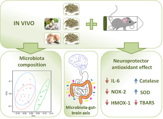
Agaricus Mushroom-Enriched Diets Modulate the Microbiota-Gut-Brain Axis and Reduce Brain Oxidative Stress in Mice

by
Josune García-Sanmartín
1,†,
Miriam Bobadilla
1,†,
Eduardo Mirpuri
1,
Vanessa Grifoll
2,
Margarita Pérez-Clavijo
2 and
Alfredo Martínez
1,*
1
Oncology Area, Center for Biomedical Research of La Rioja (CIBIR), 26006 Logroño, Spain
2
Centro Tecnológico de Investigación del Champiñón de La Rioja (CTICH), 26560 Autol, Spain
*
Author to whom correspondence should be addressed.
These authors contributed equally to this work.
Antioxidants 2022, 11(4), 695; https://doi.org/10.3390/antiox11040695
Submission received: 10 March 2022 / Revised: 28 March 2022 / Accepted: 29 March 2022 / Published: 31 March 2022

Abstract

:
Neurodegenerative diseases pose a major problem for developed countries, and stress has been identified as one of the main risk factors in the development of these disorders. Here, we have examined the protective properties against brain oxidative stress of two diets supplemented with 5% (w/w) of Agaricus bisporus (white button mushroom) or Agaricus bisporus brunnescens (Portobello mushroom) in mice. These diets did not modify the weight gain of the animals when compared to those fed with a regular diet, even after feeding on them for 15 weeks. The long-term modification of the microbiota after 12 weeks on the diets was investigated. At the phylum level, there was a large increase of Verrucomicrobia and a reduction of Cyanobacteria associated with the mushroom diets. No changes were observed in the Firmicutes/Bacteroidetes ratio, whose stability is a marker for a healthy diet. At the family level, three groups presented significant variations. These included Akkermansiaceae and Tannerellaceae, which significantly increased with both diets; and Prevotellaceae, which significantly decreased with both diets. These bacteria participate in the generation of microbiota-derived short-chain fatty acids (SCFAs) and provide a link between the microbiota and the brain. Mice subjected to restraint stress showed an upregulation of Il-6, Nox-2, and Hmox-1 expression; a reduction in the enzymatic activities of catalase and superoxide dismutase; and an increase in lipid peroxidation in their brains. All these parameters were significantly prevented by feeding for 3 weeks on the Agaricus-supplemented diets. In summary, the supplementation of a healthy diet with Agaricus mushrooms may significantly contribute to prevent neurodegenerative diseases in the general population.

Graphical Abstract

1. Introduction

The incidence of neurodegenerative diseases (NDs) is currently rising due to the increasing lifespan of the world’s population [1]. NDs encompass a heterogeneous group of chronic disorders that include Alzheimer’s disease (AD) and others dementias, such as Huntington’s disease, Parkinson’s disease [2], multiple sclerosis, human prion, and motoneuron diseases [3]. Following data from the World Health Organization, the number of people with dementia in 2015 was 46.8 million worldwide but is expected to rise to 131.5 million by 2050 [4]. Of these, AD is the most common ND, accounting for 60–70% of cases, which represents nearly 45 million people [5,6]. NDs are characterized by the progressive loss of vulnerable neurons in specific regions of the brain [7]. Heretofore, all these diseases represent the fourth cause of global disease burden in developed countries and lack an effective treatment [8].
AD is characterized by the loss of synapses and cortical neurons, which are due to the accumulation of amyloid-beta peptide and hyperphosphorylated Tau protein in the brain [9]. Aging, diabetes mellitus, and oxidative stress are known as the main risk factors of developing NDs [5,10], and oxidative stress is usually considered the more dangerous [11] due to the heavy dependence of the brain on a constant oxygen supply [12]. In contrast, cognitive and physical activities and adherence to the Mediterranean diet are associated with a decreased risk of developing AD [13].
Nowadays, growing evidence supports an association between lifestyle habits and a delay in AD occurrence [14]. In that regard, a healthy diet containing some drinks, such as wine and other fruit juices, and prebiotic foods, such as mushrooms or vegetables, exhibits positive properties with antioxidative and anti-inflammatory attributes [15,16,17,18]. Today, a new paradigm is gaining great support, namely the connection among diet, changes in the gut microbiota, and modifications on brain physiopathology [19,20]. This is currently known as the microbiota-gut-brain axis [21,22,23].
It has been shown that edible mushrooms modulate the gut microbiota towards a healthier composition [24]. The industrial cultivation of mushrooms of the Agaricus genus, including A. bisporus (white button mushroom) and A. bisporus brunnescens (Portobello or cremini mushroom), is an important staple of the economy for the region of La Rioja in Northern Spain [25]. The beneficial effects of this mushroom genus have been known for long, especially for A. blazei [26]. In the case of the white button mushrooms, several studies have established that their consumption induces a healthier microbiota [27], a better immunological function [28], and a reduction of obesity and fatty liver development [29]. No references were found for the potential relationship of Portobello mushrooms with the microbiota or for the connection of either white button or Portobello mushrooms with the microbiota-gut-brain axis.
Therefore, the aim of this study was to investigate the impact of Agaricus mushroom-enriched diets on the microbiota-gut-brain axis. To accomplish this, we followed two specific goals: to establish the influence of such diets in long-term microbiota composition, and the impact of the diets on the brain’s oxidative status.

2. Materials and Methods

2.1. Mushroom-Enriched Diets

Mushrooms (white button and Portobello) were produced at the Centro Tecnológico de Investigación del Champiñón de La Rioja (CTICH), following local industry standard cultivation methods. Clean carpophores were cut into small pieces, freeze-dried, and ground into a dry powder that was sent to Research Diets (New Brunswick, NJ, USA). Pellets for three different diets were generated under the supervision of professional nutritionists: (i) a regular basic diet containing 15% fat (D11112201, Open Standard Diet, Research Diets), (ii) a basic diet supplemented with 5% (w/w) freeze-dried white button mushroom powder, and (iii) a basic diet supplemented with 5% (w/w) freeze-dried Portobello mushroom powder. Nutritional composition of the three diets is shown in Table 1.

2.2. Experimental Animals, Feces Collection, and DNA Extraction

All procedures involving animals were carried out in accordance with the European Communities Council Directive (2010/63/EU) and Spanish legislation (RD53/2013) on animal experiments and with approval from the ethical committee on animal welfare of our institution (Órgano Encargado del Bienestar Animal del Centro de Investigación Biomédica de La Rioja, OEBA-CIBIR, procedure number AMR14).
In order to investigate the long-term impact of the diets on mouse microbiota, six-week-old C57BL/6J male mice (n = 30) (Charles River) were randomly divided into 3 groups and fed with the diets for 15 weeks. Mice were housed under standard conditions at a temperature of 22 °C (±1 °C) and a 12-h light/dark cycle with free access to food and water. Males were selected to avoid interferences with the estrous cycle. Individual mouse weights and cage food consumption were recorded weekly. All animals were housed in the same room under specific pathogen-free (SPF) conditions and maintained by the same personnel in order to normalize their microbiota. After 12 weeks, fresh fecal contents were collected from each animal and weighed. The DNA was subsequently extracted from fecal microbiota using the DNeasy Blood & Tissue Kit (Qiagen, Venlo, The Netherlands). DNA purity and concentration were determined by a Nanodrop spectrophotometer (ND-1000; Thermo Fisher Scientific, Waltham, MA, USA).

2.3. Bacterial 16S rDNA Massive Sequencing and Sequence Postprocessing

Samples were amplified for the 16S rDNA hypervariable region V3–V4, as previously described [30], following the protocol 16S Metagenomic Sequencing Library Preparation (Illumina, INC, San Diego, CA, USA) using a MiSeq ultra-sequencer (Illumina) and a 3430-cycle paired-end read run. Internal controls included the gut and soil microbiome standards (MSA-1006 and MSA-3001, ATCC, Manassas, VA, USA). The quality of the initial reads was evaluated with FastQC v0.11.9 [31]. The bioinformatics analysis was carried out using the different applications of protocol Qiime2 [32], as described [33]. Specifically, we used version qiime2-2021.4, installed through a Conda environment in platform Ubuntu Linux 20.04.2 LTS. For taxonomic assignation, we used the SILVA database in its latest version, 132 [34].
Alpha- and beta-diversity phylogenetic indexes were calculated. From these data, bacterial groups with differential abundance among diets were identified (ANCOM test) [35], and principal component graphs were drawn.
Metagenomic sequencing raw data have been deposited at [36].

2.4. Restraint Stress Model

In order to study the effects of the diets on brain’s oxidative stress parameters, additional mice (n = 30) (Charles River) were used for this assay. Mice were randomly divided into six experimental groups (three diets, exposed or not to stress, n = 5 per group) and fed with the diets for 3 weeks. After that time, animals were subjected to an acute model of stress by immobilization, as previously described [17,18]. Briefly, mice were placed inside 50-mL conical tubes, with adequate ventilation and with no access to food or water for 6 h. Due to the corticosterone circadian rhythm, restraint stress was applied at the same time of the day (9:00 a.m.) in all experiments.
Upon finishing the stress, animals were immediately sacrificed. The whole brain was divided into two equal halves by a sagittal section. One side was used for RNA extraction and the other one for an antioxidant enzyme analysis (see below).

2.5. RNA Extraction and qRT-PCR

Total RNA was isolated from mouse brains and purified as described [17]. Briefly, total RNA was isolated using Trizol reagent (Invitrogen, Waltham, MA, USA), and the resulting RNA (5 µg) was reverse-transcribed using the Superscript III First-Strand Synthesis System for RT-PCR (Invitrogen). cDNA was amplified using SYBR Green PCR Master Mix (Applied Biosystems, Foster City, CA, USA) in a real-time PCR (7300 Real-Time PCR System, Applied Biosystems). For each transcript, a specific calibration curve was included to analyze the expression of Nox-2, Hmox-1, Il-6, Tnfα, and Nrf2. As a housekeeping gene, Gapdh was used. Specific primers are shown in Table 2.

2.6. TBARS, SOD, and Catalase Activity

For the determination of the oxidative stress parameters and antioxidant components in the brain, frozen tissues were homogenized (RIPA buffer, Thermo Scientific) supplemented with Complete and Phospho STOP (Roche, Basel, Switzerland) protease and phosphatase inhibitors), as described [18]. The activities of superoxide dismutase (SOD) and catalase were studied with commercially available kits (CA061 and CA063, respectively, Canvax, Córdoba, Spain). Lipid peroxidation was determined using a commercial TBARS assay kit (CA995, Canvax). The final products were detected with a microplate reader (POLARstar Omega, BMG Labtech, Ortenberg, Germany) at 530/590 nm (for catalase and TBARS) or 450 nm for SOD.

2.7. Statistical Analysis

For comparisons, the data distribution was checked for normality (Shapiro–Wilk test) and homoscedasticity (Levene test). When datasets were normally distributed with equal variances, one-way or two-way ANOVA was used. A nonparametric Kruskal-Wallis test was used for not-normal data. The significance level was set at p < 0.05.

3. Results

3.1. Mushroom-Enriched Diets Do Not Modify Mouse Body Weight in the Long Term

The weights of the animals fed with the three experimental diets were followed for 15 weeks. No significant differences were found in the weights of the animals subjected to the different diets (Figure 1A). Food consumption was constant during the whole period, with no significant differences among the diets (Figure 1B). At 11 weeks, mice had to be transferred to larger cages due to their increasing size. This change induced a transitory diminution in food consumption that was corrected by the following week (Figure 1B).

3.2. Mushroom-Enriched Diets Induced a More Beneficial Microbiota Composition

The microbiota of mice that were exposed to the experimental diets for 12 weeks were identified by massive sequencing and biostatistical analysis. At the phylum level, there was a large increase of Verrucomicrobia associated with the mushroom diets (p = 0.014; p = 0.001 for white button- and Portobello-enriched diets compared to the control, respectively), which was more important in mice that took the Portobello-enriched diet (Figure 2A,C). In addition, the frequency of the phylum Cyanobacteria significantly diminished with the diets (p = 0.033; p = 0.021, respectively) (Figure 2D). These differences translated into clearly separated data point groups in a principal component analysis for the supplemented diets vs. the basal one (Figure 2B). The Firmicutes/Bacteroidetes ratio has been identified as a potential clinical marker for dysbiosis [37], so it was also calculated (Figure 2E). No significant differences were found between the mushroom diets and the control (p = 0.584 and p = 0.391, respectively).
At the family level, three groups presented significant variations. These included Akkermansiaceae (Figure 3A), Tannerellaceae (Figure 3B), and Prevotellaceae (Figure 3C). The Akkermansiaceae family belongs to the Verrucomicrobia phylum. In parallel with the phylum data, the counts of Akkermansiaceae in basic diet-fed mice were very low but became significantly higher in animals that were fed with the white button diet (p = 0.014) or with the Portobello diet (p = 0.001) (Figure 3A). The pattern of the Tannerellaceae family (phylum Bacteroidetes) frequency was similar to that of Akkermansiaceae, with low counts for the basic diet and significant increases for both mushroom-enriched diets (p = 0.002 and p = 0.003, respectively) (Figure 3B). On the contrary, the Prevotellaceae family (also from the Bacteroidetes phylum) displayed a significant count reduction associated with the mushroom diets (p = 0.043 for both) (Figure 3C).

3.3. Mushroom-Enriched Diets Reduced the Expression of Stress-Related Genes

The brains of mice that were exposed to the diets for 3 weeks were analyzed by qRT-PCR for several genes related to stress management (Figure 4). The expression of Il-6, Nox-2, and Hmox-1 was significantly upregulated by stress (p = 0.036, p = 0.036, and p = 0.014, respectively), and that elevation was partially or totally prevented by the mushroom diets. When measuring Il-6 expression, both mushroom diets intensely reduced stress-mediated Il-6 overexpression (p = 0.016 for white button and p = 0.001 for Portobello) (Figure 4A). In the case of Nox-2, the Portobello diet reduced the expression to values similar to the unstressed controls (p = 0.037 versus the stressed control), but the white button diet did not have any significant effect (Figure 4C). For Hmox-1, both diets reduced its overexpression, but the white button diet was significantly more effective (p = 0.001) than the Portobello one (p = 0.027) (Figure 4D). The diets had no significant effects on the expression of Tnfα or Nrf2 (Figure 4B,E).

3.4. Mushroom-Enriched Diets Normalized Stress-Induced Changes in Enzyme Activities and Lipid Peroxidation

Catalase and superoxide dismutase (SOD) activities were significantly reduced by stress (p = 0.036, p = 0.027, respectively) (Figure 5A,B). On the other hand, lipid peroxidation (TBARS), measured as the MDA levels, was significantly elevated by stress (p = 0.042) (Figure 5C). Both mushroom diets were able to elevate catalase and SOD enzymatic activities (Figure 5A,B) and to reduce lipid peroxidation (Figure 5C) to levels comparable with the unstressed brain. In the absence of stress, neither diet had a significant effect on these parameters (Figure 5).

4. Discussion

In this study, we have found that mushroom-enriched diets containing two variants of the Agaricus bisporus species exert a positive effect on microbiota composition, and they strongly contribute to reduce oxidative stress in the brain. These results suggest that supplementing a healthy diet with Agaricus mushrooms may be a beneficial habit for the general population.
We used metagenomics to understand the evolution of the microbiota in the gut of mice subjected to the diets, but a combination of different omics (metatranscriptomics, metaproteomics, and metabolomics, in addition to metagenomics) would provide a more detailed description of microbiota–host interactions [38]. This multiomics approach may strengthen our results and validate what pathways link microbiota modulation, oxidative stress, mitochondria physiology, and neurodegeneration.
Mushrooms, as well as natural products and medicinal plants, have important effects on the gut microbiota composition and, through it, modulate inflammation, cancer, and other diseases [39,40]. The modulation of the microbiota seems to be due to the prebiotic potential of mushroom components, including polysaccharides, terpenoids, and other bioactive compounds [41,42]. Our present data indicate that Agaricus mushrooms also have a positive influence in the composition of the microbiota, and through it, they contribute to reducing brain oxidative stress.
We also showed that supplementing a healthy diet with up to 5% of Agaricus mushrooms does not have an effect on mouse weight, even after 15 weeks. A previous study showed that A. bisporus can reduce obesity and accumulation of fat in the liver when experimental animals are exposed to a high-fat diet [29]. Therefore, the combination of a healthy diet and the intake of Agaricus mushrooms seems to be a good habit.
We demonstrated that the Agaricus-supplemented diets were able to modulate the microbiota towards a healthier composition. Although the study of microbiota variations and their association with different diseases is still in its infancy, several parameters are rising as potential clinical markers, including the Firmicutes/Bacteroidetes ratio [37]. This ratio seems to be critical to maintaining a healthy intestinal homeostasis, and both its increase and its reduction are regarded as dysbiosis, with the former associated with obesity and the latter with inflammatory bowel disease and cancer [43,44]. Interestingly, exposure to a mushroom-enriched diet did not modify this parameter, thus indicating that such diets are healthy for the consumers.
In addition, we found a very significant increase of the Verrucomicrobia phylum in those animals that consumed the mushroom diets. This phylum is considered a marker for a healthy microbiota, and the reduction of its frequency is related to dysbiosis, including children with irritable bowel syndrome [45]. Therefore, the increase we found after feeding the mice with the mushroom-enriched diets suggests a positive effect. We also found a significant reduction in the frequency of phylum Cyanobacteria associated with the diets. This phylum has been found to increase in patients with colon adenomas [46], so their reduction with the Agaricus-enriched diets seems to be a positive finding. Furthermore, some Cyanobacteria found in the environment may produce cyanotoxins, which are highly neurotoxic [47]. Whether some Cyanobacteria from the gut microbiota can generate such toxins is unknown.
When studying the frequency of different families of bacteria, three families showed a significant modulation by the diets. These included Akkermansiaceae, Tannerellaceae, and Prevotellaceae. Members of the Akkermansiaceae family are considered promising probiotics [48]; there is an inverse correlation between Akkermansia and obesity [49], and decreased metabolic parameters (insulin resistance, insulinemia, and cholesterol) were associated with this bacterial family in a clinical trial [50]. The positive effects of Akkermansia are due to several reasons, including the stimulation of mucosal microbial networks and the improvement of the intestinal barrier function, providing crucial host immunological responses. Several studies have demonstrated the possible involvement of Akkermansia reduction in the development of intestinal and metabolic disorders. Indeed, its levels inversely correlate to inflammatory bowel disease, obesity, and diabetes. Conversely, the therapeutic administration of Akkermansia decreases the development of such diseases [51,52].
The Tannerellaceae family includes anaerobic, Gram-negative bacterial species that have been implicated in periodontal diseases and are associated with esophageal cancer [53]. On the other hand, the presence of this group of bacteria in the gut seems to have a beneficial effect. For instance, in a mouse model of collagen-induced arthritis, Tannerellaceae were less abundant in the diseased animals than in the healthy ones [54]. In addition, Tannerellaceae species seem to be influenced by the amount of gluten in the food. For instance, Tannerellaceae increased in the microbiome of mice fed a gluten-containing diet, whereas Akkermansiaceae species increased in the intestinal microbiomes of mice fed a gluten-free diet [55]. In summary, the increase in the frequency of the Tannerellaceae family found in our mushroom-fed mice seems to be a positive trait for their microbiota composition.
Changes in the levels of bacteria belonging to the Prevotellaceae family can have different consequences. For instance, the levels of this bacterial group decrease in colorectal cancer models [56,57] but increase in children with irritable bowel syndrome [45], indicating that other parameters related to the specific disease may influence the effects of the bacteria. In our case, the mushroom diets induced a clear decrease in the members of this bacterial family. Given the positive effect of the diets on the overall brain stress markers, we could infer that the decrease in the number of Prevotellaceae bacteria may be a positive event for the modulation of the microbiota-gut-brain axis.
Oxidative stress is recognized as a very significant contributor to the pathogenesis of many devastating NDs [58]. In particular, mitochondrial dysfunction leads to the aberrant production of reactive oxygen species (ROS), which are capable of oxidizing lipids and proteins, ultimately causing cell death [59]. To model this problem in rodents, acute restraint stress has been commonly used, since it stimulates several cellular events, resulting in enhanced ROS production in the brain [60]. It has been shown that the extracellular release of ROS triggers inflammatory processes [61] that, finally, enhance the local production of cytokines IL-1β, IL-6, and TNFα [62]. NOX-2 is known for generating superoxide molecules under oxidative stress-mediated circumstances, and HMOX-1 acts as an oxidative stress-induced heat shock protein [63]. In our results, the expression of both genes (Nox-2 and Hmox-1) increased in the brains of stressed mice, which agrees with previous reports [17,18,64]. The fact that the Agaricus-supplemented diets brought these levels to normal clearly indicates the powerful antioxidant activity of these supplements.
Based on our previous experience [17,18], we were expecting an increase in Tnfα and a reduction in Nrf2 levels as a response to restraint stress. Although there were trends in that direction, no significant differences were observed in this study. Perhaps the stress was not strong or long enough to fully modulate these markers.
Furthermore, restraint stress precipitates many neurochemical, hormonal, and behavioral abnormalities that are associated with an imbalance of the brain’s intracellular redox state. Many authors have shown that restraint stress enhances lipid peroxidation and decreases the antioxidant enzyme activities in rodent brains [17,60]. SOD and catalase are the best-known antioxidant enzymes in the brain [65], and we found that the Agaricus-supplemented diets significantly increased the stress-reduced enzymatic activity of both proteins to levels undistinguishable from the non-stressed animals. On the other hand, lipid peroxidation is one of the main consequences of excessive brain ROS levels. MDA is one of the end products of such an activity and is commonly used as a marker of oxidative stress [66]. In this line, we found a significant increase of MDA in the brains of stressed mice that was prevented by the diets. All these data demonstrate the strong protective effect of the Agaricus-supplemented diets on brain stress and point to their beneficial effect on the diet.
The beneficial effects of the gut bacteria on the brain seem to be mediated, at least partially, by the microbiota-derived short-chain fatty acids (SCFAs). These are monocarboxylic acids with 2–5 carbons that are the products of the anaerobic fermentation of indigestible polysaccharides, such as dietary fiber and resistant starch [67]. Acetate, propionate, and butyrate are the main SCFAs created in the human gut [68]. After their formation, SCFAs are taken by colonocytes through monocarboxylate transporters [69]. These SCFAs are not metabolized in the colonocytes but are transmitted through the portal circulation into the liver, where they are used as an energy substrate for hepatocytes [70]. However, an important fraction of the SCFAs reaches systemic circulation and the brain. Akkermansia and Prevotella have been shown to participate in the generation of propionate and butyrate [71].
α-L-rhamnosidases are enzymes able to release aglycones and glucose by cleaving the terminal α-L-rhamnoses present in glycosylated phenolic molecules [72]. Since the human intestine possesses no rhamnosidase activity [73], the vast majority of these rhamnose glycosides remains unabsorbed and reaches the colon. The ability to transform specific rhamnose glycosides, such as hesperidin and rutin, has been demonstrated in some probiotic bacteria, including Tannerellaceae [74]. In the human body, L-rhamnose is converted into propionate, which has anti-inflammatory and antioxidant effects [75]. Of course, an excessive production of Tannerella-induced propionate, such as that generated during gingival infections, may induce NDs such as AD [76].
The interactions among the microbiota, the bowel, and the brain are complex. The bacteria can send biochemical signals directly to the brain (SCFAs) or to the gut (Toll-like receptors), which will communicate with the brain via spinal and vagal visceral afferent pathways. On the other hand, the brain would send sympathetic and parasympathetic inputs to the gut and, from there, influence the microbiota through the motility and secretion of bacteria-controlling peptides. However, nowadays, given the very complex relationships, there seems to be a tripartite conversation among the three components rather than a simple bidirectional process [22].

5. Conclusions

In summary, diets supplemented with either white button or Portobello mushrooms showed great potential in reducing oxidative stress markers in the brain, and they seem to do this through a modulation of the microbiota towards a healthier composition. The combination of physical activity [77] and a healthy diet supplemented with Agaricus mushrooms may significantly contribute to the cognitive health of the general population.

Author Contributions

Conceptualization, M.P.-C. and A.M.; methodology, J.G.-S., M.B., E.M. and V.G.; validation, J.G.-S., M.B., E.M. and V.G.; formal analysis, J.G.-S., M.B. and A.M.; investigation, J.G.-S., M.B., E.M. and V.G.; writing, A.M.; supervision, E.M., M.P.-C. and A.M.; and funding acquisition, E.M., M.P.-C. and A.M. All authors have read and agreed to the published version of the manuscript.

Funding

This research was funded by ADER (Agencia de Desarrollo Económico de La Rioja) (2017-I-IDD-00067), by the Agriculture Counsel of La Rioja, European Association for Innovation Operative Groups (24P/18), and co-funded by the European Regional Development Fund (FEDER).

Institutional Review Board Statement

All procedures involving animals were carried out in accordance with the European Communities Council Directive (2010/63/EU) and Spanish legislation (RD53/2013) on animal experiments and with approval from the ethical committee on animal welfare of our institution (Órgano Encargado del Bienestar Animal del Centro de Investigación Biomédica de La Rioja, OEBA-CIBIR, procedure number AMR14).

Informed Consent Statement

Not applicable.

Data Availability Statement

Metagenomic sequencing raw data was deposited at http://www.ncbi.nlm.nih.gov/bioproject/814431 (accessed on 1 March 2022).

Acknowledgments

We gratefully acknowledge María de Toro Hernando (Genomics & Bioinformatics Core Facility, CIBIR) for her valuable help in interpreting the massive sequencing data.

Conflicts of Interest

The authors declare no conflict of interests. The funders had no role in the design of the study; in the collection, analysis, or interpretation of the data; in the writing of the manuscript; or in the decision to publish the results.

References

  1. Heemels, M.T. Neurodegenerative diseases. Nature 2016, 539, 179. [Google Scholar] [CrossRef] [PubMed] [Green Version]
  2. Dorsey, E.R.; Sherer, T.; Okun, M.S.; Bloem, B.R. The Emerging Evidence of the Parkinson Pandemic. J. Parkinson’s Dis. 2018, 8, S3–S8. [Google Scholar] [CrossRef] [PubMed] [Green Version]
  3. Villain, N.; Dubois, B. Alzheimer’s Disease Including Focal Presentations. Semin. Neurol. 2019, 39, 213–226. [Google Scholar] [CrossRef] [PubMed]
  4. Prince, M.J.; Wu, F.; Guo, Y.; Gutierrez Robledo, L.M.; O’Donnell, M.; Sullivan, R.; Yusuf, S. The burden of disease in older people and implications for health policy and practice. Lancet 2015, 385, 549–562. [Google Scholar] [CrossRef]
  5. Reale, M.; Costantini, E.; Jagarlapoodi, S.; Khan, H.; Belwal, T.; Cichelli, A. Relationship of Wine Consumption with Alzheimer’s Disease. Nutrients 2020, 12, 206. [Google Scholar] [CrossRef] [PubMed] [Green Version]
  6. Anand, R.; Gill, K.D.; Mahdi, A.A. Therapeutics of Alzheimer’s disease: Past, present and future. Neuropharmacology 2014, 76 Pt A, 27–50. [Google Scholar] [CrossRef] [PubMed]
  7. Dugger, B.N.; Perl, D.P.; Carlson, G.A. Neurodegenerative Disease Transmission and Transgenesis in Mice. Cold Spring Harb. Perspect. Biol. 2017, 9, a023549. [Google Scholar] [CrossRef] [Green Version]
  8. Cirmi, S.; Ferlazzo, N.; Lombardo, G.E.; Ventura-Spagnolo, E.; Gangemi, S.; Calapai, G.; Navarra, M. Neurodegenerative Diseases: Might Citrus Flavonoids Play a Protective Role? Molecules 2016, 21, 1312. [Google Scholar] [CrossRef] [PubMed] [Green Version]
  9. Scheltens, P.; Blennow, K.; Breteler, M.M.; de Strooper, B.; Frisoni, G.B.; Salloway, S.; Van der Flier, W.M. Alzheimer’s disease. Lancet 2016, 388, 505–517. [Google Scholar] [CrossRef]
  10. Kumar, A.; Singh, A.; Ekavali. A review on Alzheimer’s disease pathophysiology and its management: An update. Pharmacol. Rep. 2015, 67, 195–203. [Google Scholar] [CrossRef]
  11. Youssef, P.; Chami, B.; Lim, J.; Middleton, T.; Sutherland, G.T.; Witting, P.K. Evidence supporting oxidative stress in a moderately affected area of the brain in Alzheimer’s disease. Sci. Rep. 2018, 8, 11553. [Google Scholar] [CrossRef] [PubMed] [Green Version]
  12. Valko, M.; Leibfritz, D.; Moncol, J.; Cronin, M.T.; Mazur, M.; Telser, J. Free radicals and antioxidants in normal physiological functions and human disease. Int. J. Biochem. Cell Biol. 2007, 39, 44–84. [Google Scholar] [CrossRef] [PubMed]
  13. Reale, M.; Kamal, M.A.; Velluto, L.; Gambi, D.; Di, N.M.; Greig, N.H. Relationship between inflammatory mediators, Abeta levels and ApoE genotype in Alzheimer disease. Curr. Alzheimer Res. 2012, 9, 447–457. [Google Scholar] [CrossRef] [PubMed]
  14. Caruana, M.; Cauchi, R.; Vassallo, N. Putative Role of Red Wine Polyphenols against Brain Pathology in Alzheimer’s and Parkinson’s Disease. Front. Nutr. 2016, 3, 31. [Google Scholar] [CrossRef] [Green Version]
  15. Rai, S.N.; Mishra, D.; Singh, P.; Vamanu, E.; Singh, M.P. Therapeutic applications of mushrooms and their biomolecules along with a glimpse of in silico approach in neurodegenerative diseases. Biomed. Pharmacother. 2021, 137, 111377. [Google Scholar] [CrossRef] [PubMed]
  16. Moreno-Arribas, M.V.; Bartolome, B.; Penalvo, J.L.; Perez-Matute, P.; Motilva, M.J. Relationship between Wine Consumption, Diet and Microbiome Modulation in Alzheimer’s Disease. Nutrients 2020, 12, 3082. [Google Scholar] [CrossRef]
  17. Bobadilla, M.; García-Sanmartín, J.; Martínez, A. Natural Food Supplements Reduce Oxidative Stress in Primary Neurons and in the Mouse Brain, Suggesting Applications in the Prevention of Neurodegenerative Diseases. Antioxidants 2021, 10, 46. [Google Scholar] [CrossRef]
  18. Bobadilla, M.; Hernández, C.; Ayala, M.; Alonso, I.; Iglesias, A.; García-Sanmartín, J.; Mirpuri, E.; Barriobero, J.I.; Martínez, A. A Grape Juice Supplemented with Natural Grape Extracts Is Well Accepted by Consumers and Reduces Brain Oxidative Stress. Antioxidants 2021, 10, 677. [Google Scholar] [CrossRef] [PubMed]
  19. Mohajeri, M.H.; La, F.G.; Steinert, R.E.; Weber, P. Relationship between the gut microbiome and brain function. Nutr. Rev. 2018, 76, 481–496. [Google Scholar] [CrossRef] [PubMed]
  20. Tan, L.Y.; Yeo, X.Y.; Bae, H.G.; Lee, D.P.S.; Ho, R.C.; Kim, J.E.; Jo, D.G.; Jung, S. Association of Gut Microbiome Dysbiosis with Neurodegeneration: Can Gut Microbe-Modifying Diet Prevent or Alleviate the Symptoms of Neurodegenerative Diseases? Life 2021, 11, 698. [Google Scholar] [CrossRef]
  21. Margolis, K.G.; Cryan, J.F.; Mayer, E.A. The Microbiota-Gut-Brain Axis: From Motility to Mood. Gastroenterology 2021, 160, 1486–1501. [Google Scholar] [CrossRef] [PubMed]
  22. Gershon, M.D.; Margolis, K.G. The gut, its microbiome, and the brain: Connections and communications. J. Clin. Investig. 2021, 131, e143768. [Google Scholar] [CrossRef] [PubMed]
  23. Doifode, T.; Giridharan, V.V.; Generoso, J.S.; Bhatti, G.; Collodel, A.; Schulz, P.E.; Forlenza, O.V.; Barichello, T. The impact of the microbiota-gut-brain axis on Alzheimer’s disease pathophysiology. Pharmacol. Res. 2021, 164, 105314. [Google Scholar] [CrossRef] [PubMed]
  24. Vamanu, E.; Dinu, L.D.; Pelinescu, D.R.; Gatea, F. Therapeutic Properties of Edible Mushrooms and Herbal Teas in Gut Microbiota Modulation. Microorganisms 2021, 9, 1262. [Google Scholar] [CrossRef]
  25. Mushroom Production in La Rioja. Available online: https://larioja.org/larioja-client/cm/agricultura/images?idMmedia=600508 (accessed on 1 March 2022).
  26. Hetland, G.; Tangen, J.M.; Mahmood, F.; Mirlashari, M.R.; Nissen-Meyer, L.S.H.; Nentwich, I.; Therkelsen, S.P.; Tjonnfjord, G.E.; Johnson, E. Antitumor, Anti-Inflammatory and Antiallergic Effects of Agaricus blazei Mushroom Extract and the Related Medicinal Basidiomycetes Mushrooms, Hericium erinaceus and Grifola frondosa: A Review of Preclinical and Clinical Studies. Nutrients 2020, 12, 1339. [Google Scholar] [CrossRef] [PubMed]
  27. Hess, J.; Wang, Q.; Gould, T.; Slavin, J. Impact of Agaricus bisporus Mushroom Consumption on Gut Health Markers in Healthy Adults. Nutrients 2018, 10, 1402. [Google Scholar] [CrossRef] [PubMed] [Green Version]
  28. Solano-Aguilar, G.I.; Jang, S.; Lakshman, S.; Gupta, R.; Beshah, E.; Sikaroodi, M.; Vinyard, B.; Molokin, A.; Gillevet, P.M.; Urban, J.F. The Effect of Dietary Mushroom Agaricus bisporus on Intestinal Microbiota Composition and Host Immunological Function. Nutrients 2018, 10, 1721. [Google Scholar] [CrossRef] [Green Version]
  29. Iniguez, M.; Pérez-Matute, P.; Villanueva-Millán, M.J.; Recio-Fernández, E.; Roncero-Ramos, I.; Pérez-Clavijo, M.; Oteo, J.A. Agaricus bisporus supplementation reduces high-fat diet-induced body weight gain and fatty liver development. J. Physiol. Biochem. 2018, 74, 635–646. [Google Scholar] [CrossRef]
  30. Martínez-Herrero, S.; Larrayoz, I.M.; Narro-Iñiguez, J.; Villanueva-Millán, M.J.; Recio-Fernández, E.; Pérez-Matute, P.; Oteo, J.A.; Martínez, A. Lack of Adrenomedullin Results in Microbiota Changes and Aggravates Azoxymethane and Dextran Sulfate Sodium-Induced Colitis in Mice. Front. Physiol. 2016, 7, 595. [Google Scholar] [CrossRef] [Green Version]
  31. Initial Readquality. Available online: http://www.bioinformatics.babraham.ac.uk/projects/fastqc/ (accessed on 1 March 2022).
  32. Qiime2 Protocol. Available online: https://qiime2.org/ (accessed on 1 March 2022).
  33. Estaki, M.; Jiang, L.; Bokulich, N.A.; McDonald, D.; González, A.; Kosciolek, T.; Martino, C.; Zhu, Q.; Birmingham, A.; Vázquez-Baeza, Y.; et al. QIIME 2 Enables Comprehensive End-to-End Analysis of Diverse Microbiome Data and Comparative Studies with Publicly Available Data. Curr. Protoc. Bioinform. 2020, 70, e100. [Google Scholar] [CrossRef]
  34. Silva Database. Available online: https://www.arb-silva.de/ (accessed on 1 March 2022).
  35. Mandal, S.; van, T.W.; White, R.A.; Eggesbo, M.; Knight, R.; Peddada, S.D. Analysis of composition of microbiomes: A novel method for studying microbial composition. Microb. Ecol. Health Dis. 2015, 26, 27663. [Google Scholar] [CrossRef] [Green Version]
  36. Bioproject 814431. Available online: http://www.ncbi.nlm.nih.gov/bioproject/814431 (accessed on 1 March 2022).
  37. Di, P.F. Gut Microbiota Parameters Potentially Useful in Clinical Perspective. Microorganisms 2021, 9, 2402. [Google Scholar] [CrossRef]
  38. Tilocca, B.; Pieroni, L.; Soggiu, A.; Britti, D.; Bonizzi, L.; Roncada, P.; Greco, V. Gut-Brain Axis and Neurodegeneration: State-of-the-Art of Meta-Omics Sciences for Microbiota Characterization. Int. J. Mol. Sci. 2020, 21, 4045. [Google Scholar] [CrossRef]
  39. Cheung, M.K.; Yue, G.G.L.; Chiu, P.W.Y.; Lau, C.B.S. A Review of the Effects of Natural Compounds, Medicinal Plants, and Mushrooms on the Gut Microbiota in Colitis and Cancer. Front. Pharmacol. 2020, 11, 744. [Google Scholar] [CrossRef]
  40. Lyu, M.; Wang, Y.F.; Fan, G.W.; Wang, X.Y.; Xu, S.Y.; Zhu, Y. Balancing Herbal Medicine and Functional Food for Prevention and Treatment of Cardiometabolic Diseases through Modulating Gut Microbiota. Front. Microbiol. 2017, 8, 2146. [Google Scholar] [CrossRef] [PubMed] [Green Version]
  41. Liu, X.; Yu, Z.; Jia, W.; Wu, Y.; Wu, D.; Zhang, H.; Liu, Z.; Yang, Y.; Zhang, J.; Liu, Y.; et al. A Review on Linking the Medicinal Functions of Mushroom Prebiotics with Gut Microbiota. Int. J. Med. Mushrooms 2020, 22, 943–951. [Google Scholar] [CrossRef] [PubMed]
  42. Ruthes, A.C.; Cantu-Jungles, T.M.; Cordeiro, L.M.C.; Iacomini, M. Prebiotic potential of mushroom d-glucans: Implications of physicochemical properties and structural features. Carbohydr. Polym. 2021, 262, 117940. [Google Scholar] [CrossRef] [PubMed]
  43. Stojanov, S.; Berlec, A.; Strukelj, B. The Influence of Probiotics on the Firmicutes/Bacteroidetes Ratio in the Treatment of Obesity and Inflammatory Bowel disease. Microorganisms 2020, 8, 1715. [Google Scholar] [CrossRef] [PubMed]
  44. Patrizz, A.; Dono, A.; Zorofchian, S.; Hines, G.; Takayasu, T.; Husein, N.; Otani, Y.; Arévalo, O.; Choi, H.A.; Savarraj, J.; et al. Glioma and temozolomide induced alterations in gut microbiome. Sci. Rep. 2020, 10, 21002. [Google Scholar] [CrossRef]
  45. Rigsbee, L.; Agans, R.; Shankar, V.; Kenche, H.; Khamis, H.J.; Michail, S.; Paliy, O. Quantitative profiling of gut microbiota of children with diarrhea-predominant irritable bowel syndrome. Am. J. Gastroenterol. 2012, 107, 1740–1751. [Google Scholar] [CrossRef]
  46. Lu, Y.; Chen, J.; Zheng, J.; Hu, G.; Wang, J.; Huang, C.; Lou, L.; Wang, X.; Zeng, Y. Mucosal adherent bacterial dysbiosis in patients with colorectal adenomas. Sci. Rep. 2016, 6, 26337. [Google Scholar] [CrossRef]
  47. Sini, P.; Dang, T.B.C.; Fais, M.; Galioto, M.; Padedda, B.M.; Luglie, A.; Iaccarino, C.; Crosio, C. Cyanobacteria, Cyanotoxins, and Neurodegenerative Diseases: Dangerous Liaisons. Int. J. Mol. Sci. 2021, 22, 8726. [Google Scholar] [CrossRef] [PubMed]
  48. Zhang, T.; Li, Q.; Cheng, L.; Buch, H.; Zhang, F. Akkermansia muciniphila is a promising probiotic. Microb. Biotechnol. 2019, 12, 1109–1125. [Google Scholar] [CrossRef] [PubMed] [Green Version]
  49. Everard, A.; Belzer, C.; Geurts, L.; Ouwerkerk, J.P.; Druart, C.; Bindels, L.B.; Guiot, Y.; Derrien, M.; Muccioli, G.G.; Delzenne, N.M.; et al. Cross-talk between Akkermansia muciniphila and intestinal epithelium controls diet-induced obesity. Proc. Natl. Acad. Sci. USA 2013, 110, 9066–9071. [Google Scholar] [CrossRef] [PubMed] [Green Version]
  50. Depommier, C.; Everard, A.; Druart, C.; Plovier, H.; van, H.M.; Vieira-Silva, S.; Falony, G.; Raes, J.; Maiter, D.; Delzenne, N.M.; et al. Supplementation with Akkermansia muciniphila in overweight and obese human volunteers: A proof-of-concept exploratory study. Nat. Med. 2019, 25, 1096–1103. [Google Scholar] [CrossRef]
  51. Macchione, I.G.; Lopetuso, L.R.; Ianiro, G.; Napoli, M.; Gibiino, G.; Rizzatti, G.; Petito, V.; Gasbarrini, A.; Scaldaferri, F. Akkermansia muciniphila: Key player in metabolic and gastrointestinal disorders. Eur. Rev. Med. Pharmacol. Sci. 2019, 23, 8075–8083. [Google Scholar] [CrossRef]
  52. Wang, L.; Tang, L.; Feng, Y.; Zhao, S.; Han, M.; Zhang, C.; Yuan, G.; Zhu, J.; Cao, S.; Wu, Q.; et al. A purified membrane protein from Akkermansia muciniphila or the pasteurised bacterium blunts colitis associated tumourigenesis by modulation of CD8(+) T cells in mice. Gut 2020, 69, 1988–1997. [Google Scholar] [CrossRef] [Green Version]
  53. Malinowski, B.; Wesierska, A.; Zalewska, K.; Sokolowska, M.M.; Bursiewicz, W.; Socha, M.; Ozorowski, M.; Pawlak-Osinska, K.; Wicinski, M. The role of Tannerella forsythia and Porphyromonas gingivalis in pathogenesis of esophageal cancer. Infect. Agents Cancer 2019, 14, 3. [Google Scholar] [CrossRef] [Green Version]
  54. Wang, B.; He, Y.; Tang, J.; Ou, Q.; Lin, J. Alteration of the gut microbiota in tumor necrosis factor-alpha antagonist-treated collagen-induced arthritis mice. Int. J. Rheum. Dis. 2020, 23, 472–479. [Google Scholar] [CrossRef]
  55. Marietta, E.V.; Gomez, A.M.; Yeoman, C.; Tilahun, A.Y.; Clark, C.R.; Luckey, D.H.; Murray, J.A.; White, B.A.; Kudva, Y.C.; Rajagopalan, G. Low incidence of spontaneous type 1 diabetes in non-obese diabetic mice raised on gluten-free diets is associated with changes in the intestinal microbiome. PLoS ONE 2013, 8, e78687. [Google Scholar] [CrossRef] [Green Version]
  56. Zhu, Q.; Jin, Z.; Wu, W.; Gao, R.; Guo, B.; Gao, Z.; Yang, Y.; Qin, H. Analysis of the intestinal lumen microbiota in an animal model of colorectal cancer. PLoS ONE 2014, 9, e90849. [Google Scholar] [CrossRef] [PubMed]
  57. Ochoa-Callejero, L.; Garcia-Sanmartin, J.; Martinez-Herrero, S.; Rubio-Mediavilla, S.; Narro-Iniguez, J.; Martinez, A. Small molecules related to adrenomedullin reduce tumor burden in a mouse model of colitis-associated colon cancer. Sci. Rep. 2017, 7, 17488. [Google Scholar] [CrossRef] [PubMed] [Green Version]
  58. Chen, S.D.; Yin, J.H.; Hwang, C.S.; Tang, C.M.; Yang, D.I. Anti-apoptotic and anti-oxidative mechanisms of minocycline against sphingomyelinase/ceramide neurotoxicity: Implication in Alzheimer’s disease and cerebral ischemia. Free Radic. Res. 2012, 46, 940–950. [Google Scholar] [CrossRef] [PubMed]
  59. Ross, E.K.; Gray, J.J.; Winter, A.N.; Linseman, D.A. Immunocal(R) and preservation of glutathione as a novel neuroprotective strategy for degenerative disorders of the nervous system. Recent Pat. CNS Drug Discov. 2012, 7, 230–235. [Google Scholar] [CrossRef]
  60. Kumar, A.; Garg, R.; Gaur, V.; Kumar, P. Possible role of NO modulators in protective effect of trazodone and citalopram (antidepressants) in acute immobilization stress in mice. Indian J. Exp. Biol. 2010, 48, 1131–1135. [Google Scholar] [PubMed]
  61. Duval, C.; Cantero, A.V.; Auge, N.; Mabile, L.; Thiers, J.C.; Negre-Salvayre, A.; Salvayre, R. Proliferation and wound healing of vascular cells trigger the generation of extracellular reactive oxygen species and LDL oxidation. Free Radic. Biol. Med. 2003, 35, 1589–1598. [Google Scholar] [CrossRef]
  62. Mittal, M.; Siddiqui, M.R.; Tran, K.; Reddy, S.P.; Malik, A.B. Reactive oxygen species in inflammation and tissue injury. Antioxid. Redox Signal. 2014, 20, 1126–1167. [Google Scholar] [CrossRef] [Green Version]
  63. Agundez, J.A.; Garcia-Martin, E.; Martinez, C.; Benito-Leon, J.; Millan-Pascual, J.; Diaz-Sanchez, M.; Calleja, P.; Pisa, D.; Turpin-Fenoll, L.; Alonso-Navarro, H.; et al. Heme Oxygenase-1 and 2 Common Genetic Variants and Risk for Multiple Sclerosis. Sci. Rep. 2016, 6, 20830. [Google Scholar] [CrossRef] [Green Version]
  64. Emerson, M.R.; LeVine, S.M. Heme oxygenase-1 and NADPH cytochrome P450 reductase expression in experimental allergic encephalomyelitis: An expanded view of the stress response. J. Neurochem. 2000, 75, 2555–2562. [Google Scholar] [CrossRef]
  65. Choi, H.I.; Lee, H.W.; Eom, T.M.; Lim, S.A.; Ha, H.Y.; Seol, I.C.; Kim, Y.S.; Oh, D.S.; Yoo, H.R. A traditional Korean multiple herbal formulae (Yuk-Mi-Jihwang-Tang) attenuates acute restraint stress-induced brain tissue oxidation. Drug Chem. Toxicol. 2017, 40, 125–133. [Google Scholar] [CrossRef]
  66. Kowalczuk, K.; Stryjecka-Zimmer, M. The influence of oxidative stress on the level of malondialdehyde (MDA) in different areas of the rabbit brain. Ann. Univ. Mariae Curie Sklodowska Med. 2002, 57, 160–164. [Google Scholar] [PubMed]
  67. Portincasa, P.; Bonfrate, L.; Vacca, M.; De, A.M.; Farella, I.; Lanza, E.; Khalil, M.; Wang, D.Q.; Sperandio, M.; di Ciaula, A. Gut Microbiota and Short Chain Fatty Acids: Implications in Glucose Homeostasis. Int. J. Mol. Sci. 2022, 23, 1105. [Google Scholar] [CrossRef] [PubMed]
  68. Miller, T.L.; Wolin, M.J. Pathways of acetate, propionate, and butyrate formation by the human fecal microbial flora. Appl. Environ. Microbiol. 1996, 62, 1589–1592. [Google Scholar] [CrossRef] [PubMed] [Green Version]
  69. Vijay, N.; Morris, M.E. Role of monocarboxylate transporters in drug delivery to the brain. Curr. Pharm. Des. 2014, 20, 1487–1498. [Google Scholar] [CrossRef] [Green Version]
  70. Schonfeld, P.; Wojtczak, L. Short- and medium-chain fatty acids in energy metabolism: The cellular perspective. J. Lipid Res. 2016, 57, 943–954. [Google Scholar] [CrossRef] [Green Version]
  71. Mirzaei, R.; Bouzari, B.; Hosseini-Fard, S.R.; Mazaheri, M.; Ahmadyousefi, Y.; Abdi, M.; Jalalifar, S.; Karimitabar, Z.; Teimoori, A.; Keyvani, H.; et al. Role of microbiota-derived short-chain fatty acids in nervous system disorders. Biomed. Pharmacother. 2021, 139, 111661. [Google Scholar] [CrossRef]
  72. Rodriguez-Daza, M.C.; Pulido-Mateos, E.C.; Lupien-Meilleur, J.; Guyonnet, D.; Desjardins, Y.; Roy, D. Polyphenol-Mediated Gut Microbiota Modulation: Toward Prebiotics and Further. Front. Nutr. 2021, 8, 689456. [Google Scholar] [CrossRef] [PubMed]
  73. Nielsen, I.L.; Chee, W.S.; Poulsen, L.; Offord-Cavin, E.; Rasmussen, S.E.; Frederiksen, H.; Enslen, M.; Barron, D.; Horcajada, M.N.; Williamson, G. Bioavailability is improved by enzymatic modification of the citrus flavonoid hesperidin in humans: A randomized, double-blind, crossover trial. J. Nutr. 2006, 136, 404–408. [Google Scholar] [CrossRef] [Green Version]
  74. Riva, A.; Kolimar, D.; Spittler, A.; Wisgrill, L.; Herbold, C.W.; Abranko, L.; Berry, D. Conversion of Rutin, a Prevalent Dietary Flavonol, by the Human Gut Microbiota. Front. Microbiol. 2020, 11, 585428. [Google Scholar] [CrossRef]
  75. Filippone, A.; Lanza, M.; Campolo, M.; Casili, G.; Paterniti, I.; Cuzzocrea, S.; Esposito, E. The Anti-Inflammatory and Antioxidant Effects of Sodium Propionate. Int. J. Mol. Sci. 2020, 21, 3026. [Google Scholar] [CrossRef]
  76. Singhrao, S.K.; Harding, A.; Poole, S.; Kesavalu, L.; Crean, S. Porphyromonas gingivalis Periodontal Infection and Its Putative Links with Alzheimer’s Disease. Mediat. Inflamm. 2015, 2015, 137357. [Google Scholar] [CrossRef] [PubMed] [Green Version]
  77. Aragon-Vela, J.; Solis-Urra, P.; Ruiz-Ojeda, F.J.; Alvarez-Mercado, A.I.; Olivares-Arancibia, J.; Plaza-Diaz, J. Impact of Exercise on Gut Microbiota in Obesity. Nutrients 2021, 13, 3999. [Google Scholar] [CrossRef] [PubMed]
Figure 1. Weight gain (A) and food consumption (B) by all experimental animals throughout 15 weeks of study. At week 11 (dotted line), animals had to be transferred to larger cages, and a transient diminution in food consumption was observed. Each value represents the mean and standard deviation for all animals (n = 10 per group).
Antioxidants 11 00695 g001
Figure 2. Relative frequency of bacteria at the phylum level. A general comparison of all phyla in the experimental animals fed with the white button diet (1–10), Portobello diet (11–20), or basic diet (21–30) shows a clear upregulation of Verrucomicrobia (orange) associated with the diets (A). A principal component analysis shows a marked difference between the control diet (green) and the other two diets (B). Quantification of Verrucomicrobia (C) and Cyanobacteria (D) shows also clear differences with the control diet. There were no significant differences in the Firmicutes/Bacteroidetes ratio (E). Bars represent the mean and standard error of the mean (SEM) of all measurements (n = 10). *: p < 0.05 and **: p < 0.01 versus control.
Antioxidants 11 00695 g002
Figure 3. Count numbers for some bacterial families, including Akkermansiaceae (A), Tannerellaceae (B), and Prevotellaceae (C). Bars represent the mean and standard error of the mean (SEM) of all measurements (n = 10). *: p < 0.05 and **: p < 0.01 versus control.
Antioxidants 11 00695 g003
Figure 4. Expression of genes related to oxidative stress, including Il-6 (A), Tnfα (B), Nox-2 (C), Hmox-1 (D), and Nrf2 (E). Bars represent the mean and standard error of the mean (SEM) of all measurements (n = 5). *: p < 0.05 versus control. #: p < 0.05 and ##: p < 0.01 versus the stressed control. &: p < 0.05 between diets.
Antioxidants 11 00695 g004
Figure 5. Quantification of antioxidant enzymes catalase (A) and SOD (B), and lipid peroxidation (C) in the brains of experimental mice. Bars represent the mean and standard error of the mean (SEM) of all measurements (n = 5). *: p < 0.05 versus the control. #: p < 0.05 and ##: p < 0.01 versus the stressed control.
Antioxidants 11 00695 g005
Table 1. Nutritional composition of the three diets used in this study.
Basic Diet (D11112201)White Button Suppl.Portobello Suppl.
grams (%)kcal (%)grams (%)kcal (%)grams (%)kcal (%)
Protein192018201820
Carbohydrate636560656065
Fat715615615
Total 100 100 100
kcal/gram3.81 3.62 3.62
Ingredientsgramskcalgramskcalgramskcal
Casein200800200800200800
L-cystine312312312
Corn starch381152438115243811524
Maltodextrin 10110440110440110440
Dextrose150600150600150600
Cellulose, BW200750750750
Inulin2537.52537.52537.5
Soybean oil706307063070630
Mineral mix S10028 (w/o Ca, P, or K)100100100
CaKPO4130130130
CaCO3, Light, USP5.505.505.50
Potassium citrate16.5016.5016.50
Vitamin mix V10001104010401040
Choline bitartrate202020
White button powder0 56.5 0
Portobello powder0 0 56.5
Total1071.0540841127.5540841127.554084
Table 2. Primers used in this study. Annealing temperature was 60 °C for all transcripts.
GeneForward Primer (5′–3′)Reverse Primer (5′–3′)Accession No.
Nox-2GCTGGGATCACAGGAATTGTCTTCCAAACTCTCCGCAGTCNM_007807
Hmox-1TGCTCGAATGAACACTCTGGTAGCAGGCCTCTGACGAAGTNM_010442
Il-6ATGGATGCTACCAAACTGGATTGAAGGACTCTGGCTTTGTCTNM_031168
TnfαCCACCACGCTCTTCTGTCTACACTTGGTGGTTTGCTACGANM_001278601
Nrf2AGCGAGCAGGCTATCTCCTATCTTGCCTCCAAAGGATGTCNM_010902
GapdhCATGGCCTTCCGTGTTCCTAGCGGCACGTCAGATCCANM_008084
Publisher’s Note: MDPI stays neutral with regard to jurisdictional claims in published maps and institutional affiliations.

