Article

BACH1 Expression Is Promoted by Tank Binding Kinase 1 (TBK1) in Pancreatic Cancer Cells to Increase Iron and Reduce the Expression of E-Cadherin

1
Department of Biochemistry, Tohoku University Graduate School of Medicine, Sendai 980-8575, Japan
2
Center for Regulatory Epigenome and Diseases, Tohoku University Graduate School of Medicine, Sendai 980-8575, Japan
3
Department of Neurochemistry, Tohoku University Graduate School of Medicine, Sendai 980-8575, Japan
4
Faculty of Health, Medicine and Life Sciences, Maastricht University, 6200 Maastricht, The Netherlands
5
Ben May Department for Cancer Research, University of Chicago, Chicago, IL 60637, USA
*
Author to whom correspondence should be addressed.
Antioxidants 2022, 11(8), 1460; https://doi.org/10.3390/antiox11081460
Submission received: 1 June 2022 / Revised: 20 July 2022 / Accepted: 22 July 2022 / Published: 27 July 2022
(This article belongs to the Special Issue Role of Heme Oxygenase in Human Disease)

Abstract

:
BTB and CNC homology 1 (BACH1) represses the expression of genes involved in the metabolism of iron, heme and reactive oxygen species and promotes metastasis of various cancers including pancreatic ductal adenocarcinoma (PDAC). However, it is not clear how BACH1 is regulated in PDAC cells. Knockdown of Tank binding kinase 1 (TBK1) led to reductions of BACH1 mRNA and protein amounts in AsPC−1 human PDAC cells. Gene expression analysis of PDAC cells with knockdown of TBK1 or BACH1 suggested the involvement of TBK1 and BACH1 in the regulation of iron homeostasis. Ferritin mRNA and proteins were both increased upon BACH1 knockdown in AsPC−1 cells. Flow cytometry analysis showed that AsPC−1 cells with BACH1 knockout or knockdown contained lower labile iron than control cells, suggesting that BACH1 increased labile iron by repressing the expression of ferritin genes. We further found that the expression of E-cadherin was upregulated upon the chelation of intracellular iron content. These results suggest that the TBK1-BACH1 pathway promotes cancer cell metastasis by increasing labile iron within cells.

1. Introduction

Iron is essential for various cellular processes, including cell proliferation and growth [1,2]. It plays a crucial role in DNA synthesis as a cofactor of DNA polymerase and ribonucleotide reductase [3,4,5,6]. Many of the enzymes for oxidation-reduction reactions, including those in the electron transfer chain in the mitochondria and demethylases of DNA, RNA and histone, are dependent on iron, i.e., as iron-sulfur clusters or heme [7,8,9,10,11,12,13]. Iron is also involved in the regulation of diverse proteins including tumor suppressor p53 [14,15]. However, labile ferrous iron, which is not tightly bound to proteins, is toxic to cells, tissues and the body through the generation of reactive oxygen species (ROS) via the Fenton reaction [16,17]. Iron-dependent accumulation of phospholipid hydroperoxide also can cause ferroptosis, an iron-driven cell death [18,19]. To balance the essential and detrimental roles of iron, cellular iron availability is tightly regulated by a network of genes that maintain cellular iron acquisition, storage, utilization and export. Among these, ferritin protects cells from iron-induced oxidative stress and allows for rapid adaptation to changing intracellular concentrations and demand for iron [20,21]. Importantly, cancer cells are known to be more dependent on iron than normal cells [22,23,24,25]. Transferrin receptor 1 is required for iron import by endocytosis, and lipocalin 2 is involved in an alternative pathway of iron uptake; both are highly expressed in many cancers [26,27,28,29], including pancreatic cancer [30,31]. The decrease of ferritin increases the labile iron pool on cells expressing the HRAS oncogene [32,33]. Ferroportin, as the only known iron efflux pump in vertebrates, is downregulated in breast cancer cell lines and prostate cancer and associated with increased levels of the labile iron pool [34,35].
The transcription repressor BACH1 [36] regulates the expression of genes involved in the metabolism of iron, heme and ROS. For example, BACH1 represses the expression of ferritin heavy (FTH1), light chain genes (FTL) and the iron exporter ferroportin (SLC40A1) to increase intracellular labile iron [37,38,39]. BACH1 also represses the expression of heme oxygenase-1 gene (HMOX1), which degrades heme and recycles iron [40,41,42]. The repressor activity of BACH1 is negated by the direct binding of heme to BACH1 [43]. Therefore, one of the key functions of BACH1 is the regulation of iron homeostasis by sensing heme. BACH1 also promotes the malignant properties of cancer cells [43], including metastasis in breast cancer [44,45], pancreatic ductal adenocarcinoma (PDAC) [46] and lung cancer [47,48]. BACH1 inhibits the expression of genes involved in epithelial cell adhesion, including E-cadherin (CDH1), claudin (CLDN3 and CLDN4) and occludin (OCLN), to promote epithelial-mesenchymal transition (EMT) and metastasis of PDAC cells [46]. While CLDN3 and CLDN4 genes are directly repressed by BACH1, CDH1 is not a direct target gene of BACH1. It remains unclear how BACH1 inhibits the expression of the E-cadherin gene [46]. Exploring the regulation of the E-cadherin gene by BACH1 may be beneficial to elucidating the pathological roles of BACH1 in cancer proliferation and metastasis, as well as in the search for new therapeutic approaches.
Tank binding kinase 1 (TBK1) is a serine/threonine kinase with an important role in multiple signaling pathways [49,50]. The majority of research on TBK1 has focused on its role in innate immunity. cGAS-STING pathway activates TBK1, which then activates transcription factors IRF3 and NF-κB, resulting in the production of antiviral and proinflammatory cytokines, including type I interferons [51,52,53,54,55,56]. Recently, the role of TBK1 has been expanded into cancers and autophagy [57,58,59,60,61,62,63,64,65]. TBK1 has also been reported to promote autophagy-mediated degradation of ferritin to increase intracellular iron [66]. However, it remains unclear whether TBK1 regulates iron homeostasis via additional mechanisms. Prompted by this finding, here, the relationship between TBK1 and BACH1 in the regulation of iron metabolism and E-cadherin gene expression in PDAC cells is studied.

2. Methods

2.1. Reagents

Deferasirox (DFX) was transferred as raw material from Novartis Pharma (Basel, Switzerland). Dimethyl sulfoxide (DMSO) was purchased from Sigma-Aldrich (St. Louis, MO, USA).

2.2. Cells and Cell Culture

Human embryonic kidney 293T (HEK293T) cells were maintained in DMEM-low glucose supplemented with 10% heat-inactivated FBS (Sigma Aldrich, St. Louis, MO, USA), 100 unit/mL penicillin and 100 µg/mL streptomycin (Gibco, Carlsbad, CA, USA). Human pancreatic cancer cell lines AsPC−1 were obtained from ATCC. AsPC−1 was cultured in RPMI-1640 medium supplemented with 20% heat-inactivated FBS, 100 unit/mL penicillin and 100 µg/mL streptomycin (Gibco). SW1990 was cultured in DMEM (Sigma Aldrich), with 10% heat-inactivated FBS, 100 unit/mL penicillin, 100 μg/mL streptomycin (Gibco) and 10 mM HEPES (Gibco, Carlsbad, CA, USA). The cells used were limited to less than 20 passages. BACH1 knockout AsPC−1 cells were reported previously [46].

2.3. Western Blotting

Cells were washed with PBS and harvested. They were then centrifuged at 2300× g for 1 min and the supernatant was discarded. The cells were lysed using RIPA buffer (150 mM NaCl, 50 mM Tris-HCl, 1% NP40, 0.5% sodium deoxycholate, 0.1% SDS) for 30 min on ice. Precipitated proteins were removed by centrifugation at 20,400× g for 30 min. Protein concentrations were measured using the Pierce 660 nm protein assay (Thermo Fisher Scientific, San Jose, CA, USA) and adjusted.
The supernatant containing the proteins was mixed with SDS sample buffer (62.5 mM Tris-HCl pH 6.8, 1% SDS, 10% glycerol, 1% 2-mercaptoethanol and 0.02% bromophenol blue) containing protease inhibitors (0469315900, cOmplete® Mini EDTA-free Protease Inhibitor Cocktail Tablets, Roche, Mannheim, Germany) and fractionated on slab gels (7.5% or 10% acrylamide separating gel; 4% stacking gel). The gels were run at 100 V for about 2 h and wet-transferred onto polyvinylidene difluoride (PVDF) at 300 mA for 1.5 h at 4 °C. The blots were washed with TBS-T (25 mm Tris, 137 mm NaCl, 3 mm KCl, 0.05% Tween-20, pH 7.4) and incubated for 1 h in TBS-T/5% skimmed milk. The blots were incubated overnight at 4 °C with the primary antibodies diluted in TBS-T containing 5% skimmed milk. On the next day, the blots were washed 3 times for 10 min in TBS-T and incubated for 1 h at room temperature with horseradish peroxidase (HRP)-labeled secondary antibodies diluted in TBS-T containing 5% skimmed milk. The bands were detected using a Clarity Western ECL substrate (Bio-Rad, Hercules, CA, USA) in the ChemiDoc Touch imaging system (Bio-Rad, Hercules, CA, USA) after washing three times for 10 min with TBS-T.

2.4. Antibodies

Anti-BACH1 mAb (1:500, clone 9D11, generated in-house) and anti-BACH1 antiserum (1:1000, A1–6, generated in-house) were reported previously [46,67]. Other antibodies were ACTB (1:1000, GTX109639, GeneTeX, Irvine, CA, USA), E-cadherin (1:1000, ab1416, Abcam, Cambridge, MA, USA), ferritin light chain (1:100, sc-74513, Santa Cruz Biotechnology, Cambridge, CA, USA), ferritin heavy chain (1:100, sc-376594, Santa Cruz Biotechnology, Santa Cruz, CA, USA), TBK1 (1:1000, D1B4, Cell Signaling, Boston, MA, USA) and FBXO22 (1:1000, 13606-1-AP, Proteintech, Rosemont, IL, USA). Anti-rabbit IgG-HRP (1:2500, NA934V, GE Healthcare, Fairfield, CT, USA) and anti-mouse IgG-HRP (1:2500, NA931V, GE Healthcare, Fairfield, CT, USA) were used as secondary antibodies.

2.5. siRNAs

Target-specific siRNAs (Stealth RNAi siRNA Duplex Oligoribonucleotides, Invitrogen) were transfected using Lipofectamine RNAiMAX (Thermo Fisher Scientific, Waltham, MA, USA). Stealth RNAi siRNA Negative Control, Low GC (Thermo Fisher Scientific, Waltham, MA, USA) was used as a control siRNA. All siRNA nucleotide sequences are listed in Table 1.

2.6. Quantitative Real-Time PCR

RNA was isolated via RNeasy Plus Mini Kit (Qiagen, Valencia, CA, USA) and 500 ng of total RNA was reverse transcribed to single-stranded cDNA using the High-Capacity cDNA Archive Kit (Applied Biosystems, Foster City, CA, USA). Quantitative PCR has performed with LightCycler Fast Start DNA Master SYBR Green I (Roche, Basel, Switzerland) in LightCycler 96 instrument (Roche, Basel, Switzerland). Compared to ACTB, we found that the expression of RPL13A was more stable when DFX was used. After referring to other papers [68,69], we chose RPL13A as the housekeeper gene. All primer sequences are listed in Table 2.

2.7. Immunofluorescent Staining

Cells were fixed in 4% formaldehyde/PBS. After incubation with anti-BACH1 (1:200, 9D11) antibodies for 1 h at 37 °C, the antigen-antibody complexes were detected by an anti-mouse IgG FITC-conjugated secondary antibody (1:1000, F3008, Sigma Aldrich, Darmstadt, Germany). Hoechst 33258 (Thermo Fisher, Waltham, MA, USA) was used at 20 µg/mL to stain the nuclei.

2.8. Detection of Labile Iron and Cell Death

The level of Fe2+ was measured using 5 µM Mito-FerroGreen (Dojindo Molecular Technologies, Tokyo, Japan) according to the manufacturer’s protocol. 4′,6-diamidino-2-phenylindole (DAPI) was used for the assessment of cell death. AsPC−1 cells were sorted with a FACS Verse (BD) and analyzed by FlowJo software (Tree Star). The gating strategy for living cells is shown in Supplementary Figure S4.

2.9. RNA Sequence

First, 4 µg of the total RNA was purified using an RNeasy Plus Mini Kit. Host cell rRNA contamination was removed using the GeneRead rRNA Depletion Kit (QIAGEN, Hilden, Germany) and RNA was purified using an RNeasy MiniElute Cleanup Kit (QIAGEN, Hilden, Germany). Next, 100 ng rRNA-depleted RNA was randomly sheared at 95 °C for 10 min. The sample was purified by a Magnetic Beads Cleanup Module (Thermo Fisher Scientific, Carlsbad, CA, USA). The genomic libraries were constructed using an RNA-seq library kit ver. 2 (Thermo Fisher Scientific) in the AB Library Builder system (Thermo Fisher Scientific). Each library was barcoded with Ion Xpress RNA Seq-Barcode 01-16 Kit (Thermo Fisher) to enable multiplex sequencing. Amplified segments were then size-selected (100–200 bp) using Agencourt AMPure XP magnetic beads (Beckman Coulter, Brea, CA, USA). Sequencing templates were prepared on an Ion Chef System using the Ion PI Hi-Q Chef Kit (Thermo Fisher Scientific). Samples were sequenced on the Ion Proton System using the Ion PI™ Hi-Q™ Sequencing 200 Kit and Ion PI™ v3 chip. Raw data were obtained as fastq files. The sequence data were aligned to reference hg19 by the Ion Torrent RNASeqAnalysis plugin (Thermo Fisher Scientific). Mapped reads were counted using htseq-count v 0.9.1 and performed likelihood ratio test by using the edgeR package v 3.16.5 after the removal of low-read-count genes (count per million <5). RNA-seq data of siTBK1 from this study have been deposited in GEO under SuperSeries accession number GSE201307.

2.10. The Analysis and the Visualization of Public Data

The dataset used comprised mRNA-seq data from TCGA tumors (see TCGA Data Portal at https://tcga-data.nci.nih.gov/tcga/, accessed on 17 October 2021). The two-gene correlation map is realized by the R software package ggstatsplot (3.3.3). We used Spearman’s correlation analysis to describe the correlation between quantitative variables without normal distribution. A p-value of less than 0.05 was considered statistically significant.
Data from GSE124408 in Gene Expression Omnibus (GEO, https://www.ncbi.nlm.nih.gov/geo, accessed on 9 December 2021) was used as the ChIP-seq data of BACH1 in AsPC−1 and SW1990. DNA sequences of genes are listed in Table 3. PDF files were made ‘igvtools ver. 2.3.93′ and visualized using Integrative Genomics Viewer [70,71].

2.11. Statistics

Statistical analyses were performed by GraphPad Prism 8. For all experiments, differences in data sets were considered statistically significant when p-values were lower than 0.05. When comparing only two groups, an unpaired Student’s t-test was performed. To compare multiple groups, one-way ANOVA was used. *, p < 0.05; **, p < 0.01, ns (not significant) p > 0.05.

3. Results

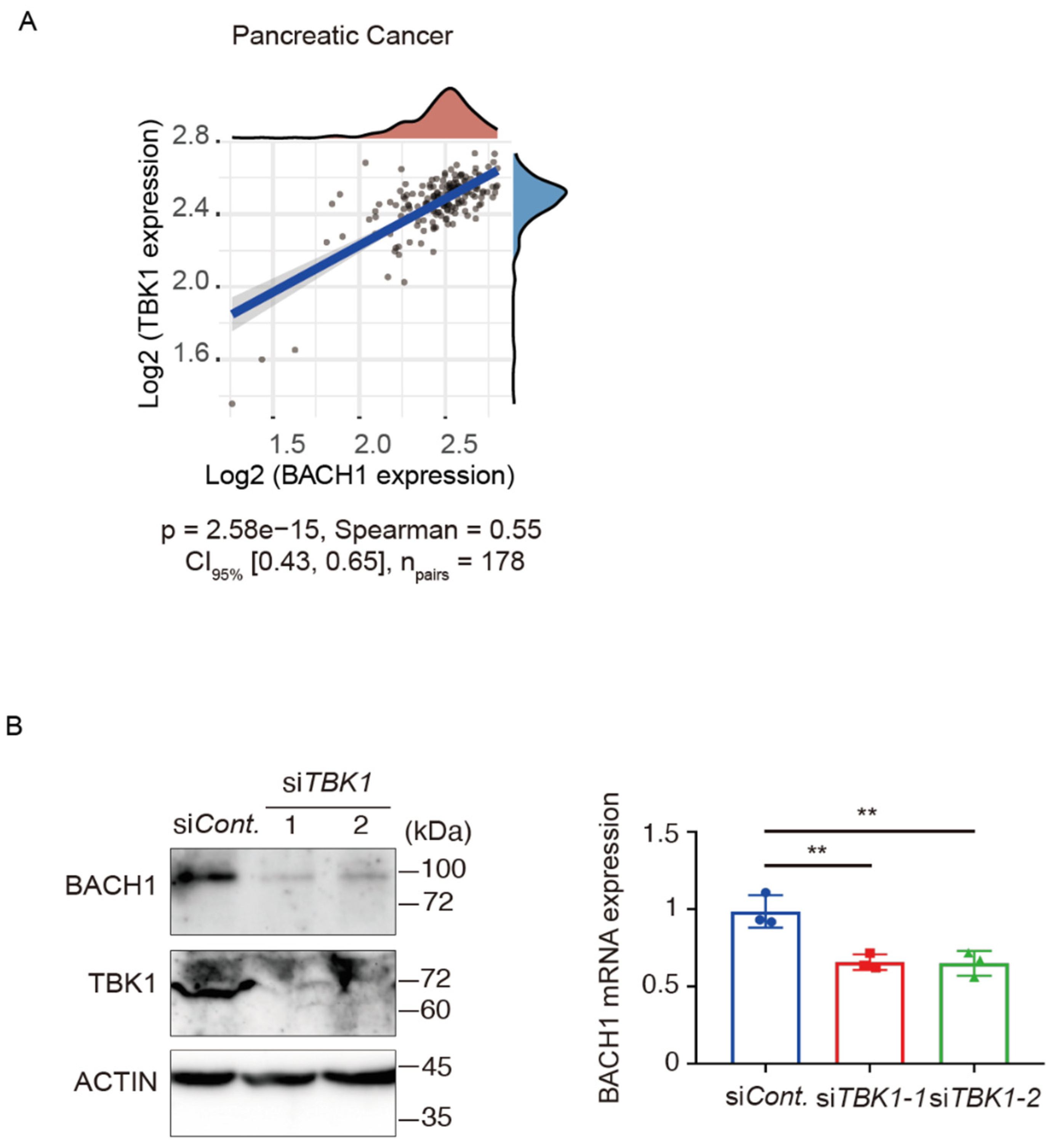
3.1. TBK1 Promotes BACH1 Expression and Accumulation

We analyzed the mRNA expression data from TCGA using the GEPIA database (Gene Expression Profiling Interactive Analysis) [72] and found that the expression of BACH1 and TBK1 are positively correlated in pancreatic cancer (Figure 1A). Since pancreatic cancer is a cancer of the digestive system, we also analyzed several other cancers of the digestive system and found similar results in bowel cancer, hepatocellular carcinoma and gastric cancer (Supplementary Figure S1A–C). To determine the roles of TBK1-mediated regulation of BACH1 in PDAC cells, we knocked down TBK1 by siRNA in human AsPC−1 PDAC cells. TBK1 knockdown led to the reduction of BACH1 protein and mRNA levels (Figure 1B). Because the change of the mRNA was smaller than that of the protein, a major regulatory mechanism of BACH1 by TBK1 may be at the protein level rather than the transcription level in AsPC−1 cells. Phosphorylation of BACH1 by TBK1 (L.L. et al., submitted) also suggested this possibility. In contrast, when TBK1 was knocked down in HEK293T cells, endogenous BACH1 protein and mRNA levels did not decrease (Supplementary Figure S2A). To test if TBK1 affects the subcellular distribution of BACH1, immunostaining was carried out. TBK1 knockdown resulted in a decrease of the endogenous BACH1 protein in AsPC−1 cells, but its subcellular distribution did not change (Supplementary Figure S2B). The cell counts did not change under the reduction of TBK1 (Supplementary Figure S2C). These results indicated that TBK1 is required for the accumulation of BACH1 protein and to promote BACH1 function in PDAC cells.

3.2. TBK1-BACH1 Pathway Regulates Iron Homeostasis and Cell Migration

To further investigate the regulatory relationship between TBK1 and BACH1, we knocked down TBK1 in AsPC−1 cells and carried out an RNA-seq analysis. Using our previously published data of RNA-seq in AsPC−1 cells with or without BACH1 knockdown [46], there were 102 common upregulated genes and 181 common downregulated genes in TBK1-silenced and BACH1-silenced AsPC−1 cells (Figure 2A,B). A GO pathway enrichment analysis of these genes showed that terms related to oxidation-reduction process, regulation of transcription from RNA polymerase II promoter in response to iron, positive regulation of sequence-specific DNA binding transcription factor activity and cellular iron ion homeostasis were enriched in genes upregulated in response to TBK1 or BACH1 knockdown (Figure 2C). Considering that TBK1 is involved in iron metabolism [66], these results suggested the involvement of TBK1 in the regulation of BACH1 to tune iron homeostasis in AsPC−1 cells. Consistent with the reduction of BACH1 protein, its known target genes were increased in their expression, including HMOX1 and SLC40A1. On the other hand, the enrichment of terms related to positive regulation of endothelial cell proliferation, wound healing and positive regulation of cell migration in the genes whose expression was decreased (Figure 2D). The expression of mesenchymal genes, such as MMP7, SNAI2, and VIM, were decreased upon TBK1 knockdown (Supplementary Figure S3A). The expression of these genes is also reduced upon BACH1 knockdown in these cells [46], suggesting that TBK1 promotes metastatic process including cell migration via enhancing the function of BACH1.

3.3. BACH1 Increases the Iron Content by Reducing the Expression of Ferritin

A reanalysis of ChIP-seq data of BACH1 in AsPC−1 cells [46] showed direct binding of BACH1 to FTL and FTH1 encoding ferritin subunits (Figure 3A). Ferritin mRNAs were increased upon knockout (Figure 3B) or knockdown (Supplementary Figure S3B) of BACH1 in AsPC−1 cells. Correspondingly, ferritin heavy and light chain proteins were increased in AsPC−1 cells with the knockout of BACH1 (Figure 3C). While BACH1 also directly binds with SLC40A1 encoding ferroportin, its mRNA was not increased (Supplementary Figure S3C,D). Two cytoplasmic iron regulatory proteins (IRP1 and IRP2) post-transcriptionally regulate cellular iron metabolism, including translational inhibition of ferritin mRNAs when the iron is reduced [20,73,74]. IRP2, but not IRP1, was also decreased under the situation of BACH1 knockdown and knockout (Supplementary Figure S3B,E), suggesting a direct or indirect regulation of IRP2 expression by BACH1.
To examine whether these alterations led to changes in iron within cells, we measured labile iron, which is not bound tightly to proteins, in mitochondria by using Mito-FerroGreen and flow cytometry (FACS) with a gating strategy shown in Supplementary Figure S4. FACS analysis showed that AsPC−1 cells with BACH1 knockout and knockdown were lower in the mean intensity of the Mito-FerroGreen signal and less in the mean numbers of Mito-FerroGreen positive cells than respective control cells (Figure 3D; Supplementary Figure S5). These results suggested that BACH1 increases mitochondrial labile iron in AsPC−1 cells by repressing the expression of ferritin genes.

3.4. Iron Availability Regulates BACH1 and E-Cadherin Expression

Since iron and heme play key roles in the regulation of BACH1 [43,75,76,77], the changes in BACH1 protein and mRNA in response to fluctuations of iron in AsPC−1 and SW1990 derived from PDAC patients was examined with ferrous sulfate (FeSO4) or iron chelator deferasirox (DFX). BACH1 protein was increased with DFX and decreased with FeSO4 (Figure 4A,B; Supplementary Figure S6A). Consistent with the protein amount, BACH1 mRNA increased in response to DFX after 12 or 48 h. Among downstream target genes of BACH1, HMOX1 mRNA decreased with DFX (Figure 4C; Supplementary Figure S6B). Taken together with the enhanced BACH1 protein degradation by heme [77], these results demonstrated that iron deficiency, which leads to a decrease in heme, induces the expression of BACH1 at both transcriptional and post-translational steps.
BACH1 is polyubiquitinated by the E3 ligase adaptor proteins F-box protein 22 (FBXO22) [47]. The mRNA of FBXO22 decreased with DFX (Figure 4C; Supplementary Figure S6B), which suggested that FBXO22-dependent BACH1 degradation was decreased under this condition, contributing to the accumulation of BACH1 protein. The next question is how TBK1 responds to a reduction of iron. TBK1 mRNA and protein were decreased when AsPC−1 cells were treated with DFX (Figure 4C,D; Supplementary Figure S6B). This observation led to the question of whether TBK1 is involved in the accumulation of BACH1 protein upon iron chelation. DFX still increased BACH1 protein even when TBK1 was reduced by siRNA (Figure 4D). However, the accumulation of BACH1 was reduced compared with DFX alone. FBXO22 protein was also reduced by DFX, which was not affected by TBK1 knockdown (Figure 4D). When taken together, these observations suggested that TBK1 and iron constitute two independent, parallel pathways to control BACH1 expression in PDAC cells. TBK1 promotes the accumulation of BACH1 protein whereas iron inhibits its accumulation.
While BACH1 reduces the expression of CDH1 encoding E-cadherin in PDAC cells to promote metastasis [46], the specific mechanism remains unclear. A recent report pointed to the role of iron in promoting EMT in HepG2 cells [78]. When taken with this report, the above observations raise the possibility that the regulation of CDH1 expression involves iron. Indeed, E-cadherin mRNA and protein were both increased by the DFX treatment (Figure 5A). The increased expression of E-cadherin upon BACH1 knockdown may involve iron reduction caused by increased expression of ferritin (Figure 5B).

4. Discussion

This study has shown that TBK1 is required to maintain the expression of BACH1 in PDAC cells. Since TBK1 phosphorylates BACH1 (LL et al., unpublished observation), this modification may increase the stability thereof. In addition, TBK1 was found to increase BACH1 mRNA. Since TBK1 is known to activate transcription factors for immune responses such as IRF3 and NF-κB [51,52,53,54,55,56], these factors may transactivate BACH1 gene expression. As iron metabolism is dynamically altered during infection [79,80,81], TBK1 and BACH1 may contribute to immune responses as well as EMT by altering iron metabolism. The links between iron and E-cadherin expression remain unclear. It has been reported that in colon cancer cells, iron chelators antagonize the reduction of E-cadherin expression in response to transforming growth factor-β, and that their effect is dependent on the induction of N-myc downstream-regulated gene 1 (NDRG1) upon iron chelation [82]. Therefore, it will be important to examine whether NDRG1 expression is altered in response to TBK1 or BACH1. TBK1 is known to promote EMT, including the reduction of E-cadherin expression, by altering the activities of downstream protein kinases such as AKT, ERK and GSK3β [83,84]. However, it was also reported that TBK1 inhibits EMT in breast cancer cells via increasing the expression of estrogen receptors [85]. Taken together with these reports, our findings suggested that TBK1 may promote EMT and metastasis of cancer cells via both BACH1-independent and BACH1-dependent mechanisms. Consistent with this hypothesis, it has been reported that TBK1 enhances the invasive and metastatic capacity of pancreatic cancer cells [86].
The expression of BACH1 in PDAC cells is strictly regulated in response to the amount of intracellular iron. As a continuously high level of BACH1 is expected to increase cellular iron levels, which may cause ferroptosis [87], an alternative mechanism to suppress overshooting of BACH1 appears to be necessary. Our results suggest that this may be achieved by the decrease of BACH1 by iron, leading to the re-expression of ferritin genes. When iron is limited, cells increase the amount of BACH1 by two distinct mechanisms. One is the increased transcription of BACH1 mRNA in response to a reduction in iron. Since BACH1 is known to be induced by the hypoxic response [88], hypoxia-inducible factors may be involved in the increased transcription of BACH1 gene in response to iron deficiency. The mechanism of the increased expression of BACH1 mRNA awaits further clarification. The other mechanism involves a decrease in the expression of FBXO22, which leads to a cessation of BACH1 degradation. As heme is a complex of iron and protoporphyrin IX, the supply of iron via the transferrin-receptor pathway limits, and thus controls, the heme synthesis rate [89]. Hence, it is also possible that DFX enhances BACH1 accumulation by reducing heme-regulated degradation of BACH1 [77]. The presence of multiple pathways to increase BACH1 in response to iron reduction probably leads to a rapid increase in BACH1 protein level, resulting in prompt changes in target gene expression.
The present observations suggest that BACH1 and iron form a negative feedback loop (Figure 5B). When it is active, BACH1 increases intracellular labile iron by repressing the expression of ferritin genes, and TBK1 is required to maintain the amount of BACH1. Ensuring an increase in mobile iron and heme then decreases the amount of BACH1 protein in part by increasing the expression of FBXO22 and promoting heme-dependent degradation of BACH1. Therefore, iron and BACH1 regulate each other by a network of multiple regulatory interactions including TBK1 and FBXO22. It will be important to investigate the universality and cell type-specific modulation of this network in various cell types including cancer cells.

5. Conclusions

We determined that BACH1 protein and mRNA levels were reduced by TBK1 knockdown in PDAC cells. A GO pathway enrichment analysis of PDAC cells with knockdown of TBK1 or BACH1 suggested that TBK1 and BACH1 both regulate cellular iron ion homeostasis and cell migration. Consistent with this, BACH1 represses the transcription of ferritin and thus, brings about an increase in cellular iron levels in these cells. In addition, iron reduces the expression of E-cadherin. These observations suggest that TBK1 and BACH1 promote metastasis of PDAC cells, in part by controlling iron metabolism.

Supplementary Materials

The following supporting information can be downloaded at: https://www.mdpi.com/article/10.3390/antiox11081460/s1, Figure S1: Spearman correlation analysis of BACH1 gene expression and TBK1 gene expression in cancer tissues; Figure S2: BACH1 subcellular distribution did not change upon TBK1 knockdown; Figure S3: BACH1 inhibits the expression of ferritin genes; Figure S4: Additional data demonstrating flow cytometry gating of dead cells; Figure S5: BACH1 increases the iron content; Figure S6: The increases of BACH1 mRNA and protein under iron deficiency.

Author Contributions

Conception and design: L.L., M.M., K.I.; Acquisition of data: L.L., M.M., B.K.K.C., Y.K., M.T.; Analysis and interpretation of data (e.g., statistical analysis, biostatistics, computational analysis): L.L., M.M., H.N., K.I.; Writing, review, and/or revision of the manuscript: L.L., M.M., K.I.; Administrative, technical, or material support (i.e., reporting or organizing data, constructing databases): L.L., M.M., M.M.-W., K.O., H.N., L.C.N., K.I.; Study supervision: K.I. All authors have read and agreed to the published version of the manuscript.

Funding

This study has been supported by Grants-in-Aid for Scientific Research from the Japan Society for the Promotion of Science (20KK0176 and 18H04021 to K.I., 20K16296 and 19K23738 to H.N., and 19K07680 and 16K07108 to M.M.), Grant-in-Aid for Joint Research by Young Researchers from Tohoku University Graduate School of Medicine (to H.N.), Gonryo Medical Foundation (to H.N.), Takeda Science Foundation (to H.N.), and Research Grant in the Natural Sciences from the Mitsubishi Foundation (to K.I.). It was also funded by the China Scholarship Council.

Institutional Review Board Statement

Not applicable.

Informed Consent Statement

Not applicable.

Data Availability Statement

RNA-seq data of siTBK1 from this study have been deposited in GEO under SuperSeries accession number GSE201307.

Acknowledgments

We thank members of the Departments of Biochemistry, Tohoku University Graduate School of Medicine for discussions, support and technical support.

Conflicts of Interest

No potential conflict of interest were disclosed.

References

  1. Robbins, E.; Pedrson, H. Iron: Its intracellular localization and possible role in cell division. Proc. Natl. Acad. Sci. USA 1970, 66, 1244–1251. [Google Scholar] [CrossRef] [PubMed] [Green Version]
  2. Rudland, P.S.; Durbin, H.; Clingan, D.; de Asua, L.J. Iron salts and transferrin are specifically required for cell division of cultured 3T6 cells. Biochem. Biophys. Res. Commun. 1977, 75, 556–562. [Google Scholar] [CrossRef]
  3. White, M.F.; Dillingham, M.S. Iron-sulphur clusters in nucleic acid processing enzymes. Curr. Opin. Struct. Biol. 2012, 22, 94–100. [Google Scholar] [CrossRef] [PubMed]
  4. Zhang, C. Essential functions of iron-requiring proteins in DNA replication, repair and cell cycle control. Protein. Cell 2014, 5, 750–760. [Google Scholar] [CrossRef] [PubMed] [Green Version]
  5. Fuss, J.O.; Tsai, C.L.; Ishida, J.P.; Tainer, J.A. Emerging critical roles of Fe-S clusters in DNA replication and repair. Biochim. Biophys. Acta 2015, 1853, 1253–1271. [Google Scholar] [CrossRef] [PubMed] [Green Version]
  6. Puig, S.; Ramos-Alonso, L.; Romero, A.M.; Martinez-Pastor, M.T. The elemental role of iron in DNA synthesis and repair. Metallomics 2017, 9, 1483–1500. [Google Scholar] [CrossRef] [Green Version]
  7. Read, A.D.; Bentley, R.E.; Archer, S.L.; Dunham-Snary, K.J. Mitochondrial iron-sulfur clusters: Structure, function, and an emerging role in vascular biology. Redox. Biol. 2021, 47, 102164. [Google Scholar] [CrossRef]
  8. Kiley, P.J.; Beinert, H. Oxygen sensing by the global regulator, FNR: The role of the iron-sulfur cluster. FEMS Microbiol. Rev. 1998, 22, 341–352. [Google Scholar] [CrossRef]
  9. Kobayashi, K.; Fujikawa, M.; Kozawa, T. Oxidative stress sensing by the iron-sulfur cluster in the transcription factor, SoxR. J. Inorg. Biochem. 2014, 133, 87–91. [Google Scholar] [CrossRef]
  10. Barton, J.K.; Silva, R.M.B.; O’Brien, E. Redox Chemistry in the Genome: Emergence of the [4Fe4S] Cofactor in Repair and Replication. Annu. Rev. Biochem. 2019, 88, 163–190. [Google Scholar] [CrossRef] [Green Version]
  11. Jiang, Y.; Li, C.; Wu, Q.; An, P.; Huang, L.; Wang, J.; Chen, C.; Chen, X.; Zhang, F.; Ma, L.; et al. Iron-dependent histone 3 lysine 9 demethylation controls B cell proliferation and humoral immune responses. Nat. Commun. 2019, 10, 2935. [Google Scholar] [CrossRef] [PubMed] [Green Version]
  12. Yi, C.; Yang, C.G.; He, C. A non-heme iron-mediated chemical demethylation in DNA and RNA. Acc. Chem. Res. 2009, 42, 519–529. [Google Scholar] [CrossRef] [PubMed] [Green Version]
  13. Lal, S.; Comer, J.M.; Konduri, P.C.; Shah, A.; Wang, T.; Lewis, A.; Shoffner, G.; Guo, F.; Zhang, L. Heme promotes transcriptional and demethylase activities of Gis1, a member of the histone demethylase JMJD2/KDM4 family. Nucleic Acids Res. 2018, 46, 215–228. [Google Scholar] [CrossRef] [PubMed] [Green Version]
  14. Shen, J.; Sheng, X.; Chang, Z.; Wu, Q.; Wang, S.; Xuan, Z.; Li, D.; Wu, Y.; Shang, Y.; Kong, X.; et al. Iron metabolism regulates p53 signaling through direct heme-p53 interaction and modulation of p53 localization, stability, and function. Cell Rep. 2014, 7, 180–193. [Google Scholar] [CrossRef] [PubMed] [Green Version]
  15. Zhang, J.; Chen, X. P53 tumor suppressor and iron homeostasis. FEBS J. 2019, 286, 620–629. [Google Scholar] [CrossRef] [Green Version]
  16. Fenton, H.J.H. LXXIII.—Oxidation of tartaric acid in presence of iron. J. Chem. Soc. Trans. 1894, 65, 899–911. [Google Scholar] [CrossRef] [Green Version]
  17. Winterbourn, C.C. Toxicity of iron and hydrogen peroxide: The Fenton reaction. Toxicol Lett. 1995, 82–83, 969–974. [Google Scholar] [CrossRef]
  18. Dixon, S.J.; Lemberg, K.M.; Lamprecht, M.R.; Skouta, R.; Zaitsev, E.M.; Gleason, C.E.; Patel, D.N.; Bauer, A.J.; Cantley, A.M.; Yang, W.S.; et al. Ferroptosis: An iron-dependent form of nonapoptotic cell death. Cell 2012, 149, 1060–1072. [Google Scholar] [CrossRef] [Green Version]
  19. Conrad, M.; Pratt, D.A. The chemical basis of ferroptosis. Nat. Chem. Biol. 2019, 15, 1137–1147. [Google Scholar] [CrossRef]
  20. Rouault, T.A. The role of iron regulatory proteins in mammalian iron homeostasis and disease. Nat. Chem. Biol. 2006, 2, 406–414. [Google Scholar] [CrossRef]
  21. Theil, E.C. Ferritin protein nanocages use ion channels, catalytic sites, and nucleation channels to manage iron/oxygen chemistry. Curr. Opin. Chem. Biol. 2011, 15, 304–311. [Google Scholar] [CrossRef] [PubMed] [Green Version]
  22. Torti, S.V.; Torti, F.M. Iron and cancer: More ore to be mined. Nat. Rev. Cancer 2013, 13, 342–355. [Google Scholar] [CrossRef] [PubMed] [Green Version]
  23. Wang, Y.F.; Zhang, J.; Su, Y.; Shen, Y.Y.; Jiang, D.X.; Hou, Y.Y.; Geng, M.Y.; Ding, J.; Chen, Y. G9a regulates breast cancer growth by modulating iron homeostasis through the repression of ferroxidase hephaestin. Nat. Commun. 2017, 8, 274. [Google Scholar] [CrossRef] [PubMed] [Green Version]
  24. Wang, Y.; Yu, L.; Ding, J.; Chen, Y. Iron Metabolism in Cancer. Int. J. Mol. Sci. 2018, 20, 95. [Google Scholar] [CrossRef] [Green Version]
  25. El Hout, M.; Dos Santos, L.; Hamai, A.; Mehrpour, M. A promising new approach to cancer therapy: Targeting iron metabolism in cancer stem cells. Semin. Cancer. Biol. 2018, 53, 125–138. [Google Scholar] [CrossRef]
  26. Prior, R.; Reifenberger, G.; Wechsler, W. Transferrin receptor expression in tumours of the human nervous system: Relation to tumour type, grading and tumour growth fraction. Virchows Arch. A Pathol. Anat. Histopathol. 1990, 416, 491–496. [Google Scholar] [CrossRef] [PubMed]
  27. Das Gupta, A.; Shah, V.I. Correlation of transferrin receptor expression with histologic grade and immunophenotype in chronic lymphocytic leukemia and non-Hodgkin’s lymphoma. Hematol. Pathol. 1990, 4, 37–41. [Google Scholar] [PubMed]
  28. Habeshaw, J.A.; Lister, T.A.; Stansfeld, A.G.; Greaves, M.F. Correlation of transferrin receptor expression with histological class and outcome in non-Hodgkin lymphoma. Lancet 1983, 1, 498–501. [Google Scholar] [CrossRef]
  29. Seymour, G.J.; Walsh, M.D.; Lavin, M.F.; Strutton, G.; Gardiner, R.A. Transferrin receptor expression by human bladder transitional cell carcinomas. Urol. Res. 1987, 15, 341–344. [Google Scholar] [CrossRef] [PubMed]
  30. Jeong, S.M.; Lee, J.; Finley, L.W.; Schmidt, P.J.; Fleming, M.D.; Haigis, M.C. SIRT3 regulates cellular iron metabolism and cancer growth by repressing iron regulatory protein 1. Oncogene 2015, 34, 2115–2124. [Google Scholar] [CrossRef] [Green Version]
  31. Leung, L.; Radulovich, N.; Zhu, C.Q.; Organ, S.; Bandarchi, B.; Pintilie, M.; To, C.; Panchal, D.; Tsao, M.S. Lipocalin2 promotes invasion, tumorigenicity and gemcitabine resistance in pancreatic ductal adenocarcinoma. PLoS ONE 2012, 7, e46677. [Google Scholar] [CrossRef] [PubMed]
  32. Kakhlon, O.; Gruenbaum, Y.; Cabantchik, Z.I. Repression of ferritin expression modulates cell responsiveness to H-ras-induced growth. Biochem. Soc. Trans. 2002, 30, 777–780. [Google Scholar] [CrossRef]
  33. Kakhlon, O.; Gruenbaum, Y.; Cabantchik, Z.I. Ferritin expression modulates cell cycle dynamics and cell responsiveness to H-ras-induced growth via expansion of the labile iron pool. Biochem. J. 2002, 363, 431–436. [Google Scholar] [CrossRef] [PubMed]
  34. Jiang, X.P.; Elliott, R.L.; Head, J.F. Manipulation of iron transporter genes results in the suppression of human and mouse mammary adenocarcinomas. Anticancer Res. 2010, 30, 759–765. [Google Scholar] [PubMed]
  35. Xue, D.; Zhou, C.X.; Shi, Y.B.; Lu, H.; He, X.Z. Decreased expression of ferroportin in prostate cancer. Oncol. Lett. 2015, 10, 913–916. [Google Scholar] [CrossRef] [PubMed] [Green Version]
  36. Oyake, T.; Itoh, K.; Motohashi, H.; Hayashi, N.; Hoshino, H.; Nishizawa, M.; Yamamoto, M.; Igarashi, K. Bach proteins belong to a novel family of BTB-basic leucine zipper transcription factors that interact with MafK and regulate transcription through the NF-E2 site. Mol. Cell Biol. 1996, 16, 6083–6095. [Google Scholar] [CrossRef] [PubMed] [Green Version]
  37. Hintze, K.J.; Katoh, Y.; Igarashi, K.; Theil, E.C. Bach1 repression of ferritin and thioredoxin reductase1 is heme-sensitive in cells and in vitro and coordinates expression with heme oxygenase1, beta-globin, and NADP(H) quinone (oxido) reductase1. J. Biol. Chem. 2007, 282, 34365–34371. [Google Scholar] [CrossRef] [PubMed] [Green Version]
  38. Nishizawa, H.; Matsumoto, M.; Shindo, T.; Saigusa, D.; Kato, H.; Suzuki, K.; Sato, M.; Ishii, Y.; Shimokawa, H.; Igarashi, K. Ferroptosis is controlled by the coordinated transcriptional regulation of glutathione and labile iron metabolism by the transcription factor BACH1. J. Biol. Chem. 2020, 295, 69–82. [Google Scholar] [CrossRef] [PubMed]
  39. Marro, S.; Chiabrando, D.; Messana, E.; Stolte, J.; Turco, E.; Tolosano, E.; Muckenthaler, M.U. Heme controls ferroportin1 (FPN1) transcription involving Bach1, Nrf2 and a MARE/ARE sequence motif at position -7007 of the FPN1 promoter. Haematologica 2010, 95, 1261–1268. [Google Scholar] [CrossRef] [Green Version]
  40. Ogawa, K.; Sun, J.; Taketani, S.; Nakajima, O.; Nishitani, C.; Sassa, S.; Hayashi, N.; Yamamoto, M.; Shibahara, S.; Fujita, H.; et al. Heme mediates derepression of Maf recognition element through direct binding to transcription repressor Bach1. EMBO J. 2001, 20, 2835–2843. [Google Scholar] [CrossRef] [Green Version]
  41. Sun, J.; Hoshino, H.; Takaku, K.; Nakajima, O.; Muto, A.; Suzuki, H.; Tashiro, S.; Takahashi, S.; Shibahara, S.; Alam, J. Hemoprotein Bach1 regulates enhancer availability of heme oxygenase-1 gene. EMBO J. 2002, 21, 5216–5224. [Google Scholar] [CrossRef] [PubMed]
  42. Hou, W.; Tian, Q.; Steuerwald, N.M.; Schrum, L.W.; Bonkovsky, H.L. The let-7 microRNA enhances heme oxygenase-1 by suppressing Bach1 and attenuates oxidant injury in human hepatocytes. Biochim. Biophys. Acta 2012, 1819, 1113–1122. [Google Scholar] [CrossRef] [PubMed] [Green Version]
  43. Igarashi, K.; Nishizawa, H.; Saiki, Y.; Matsumoto, M. The transcription factor BACH1 at the crossroads of cancer biology: From epithelial-mesenchymal transition to ferroptosis. J. Biol. Chem. 2021, 297, 101032. [Google Scholar] [CrossRef] [PubMed]
  44. Liang, Y.; Wu, H.; Lei, R.; Chong, R.A.; Wei, Y.; Lu, X.; Tagkopoulos, I.; Kung, S.Y.; Yang, Q.; Hu, G.; et al. Transcriptional network analysis identifies BACH1 as a master regulator of breast cancer bone metastasis. J. Biol. Chem. 2012, 287, 33533–33544. [Google Scholar] [CrossRef] [PubMed] [Green Version]
  45. Lee, J.; Yesilkanal, A.E.; Wynne, J.P.; Frankenberger, C.; Liu, J.; Yan, J.; Elbaz, M.; Rabe, D.C.; Rustandy, F.D.; Tiwari, P.; et al. Effective breast cancer combination therapy targeting BACH1 and mitochondrial metabolism. Nature 2019, 568, 254–258. [Google Scholar] [CrossRef] [PubMed]
  46. Sato, M.; Matsumoto, M.; Saiki, Y.; Alam, M.; Nishizawa, H.; Rokugo, M.; Brydun, A.; Yamada, S.; Kaneko, M.K.; Funayama, R.; et al. BACH1 Promotes Pancreatic Cancer Metastasis by Repressing Epithelial Genes and Enhancing Epithelial-Mesenchymal Transition. Cancer Res. 2020, 80, 1279–1292. [Google Scholar] [CrossRef] [Green Version]
  47. Lignitto, L.; LeBoeuf, S.E.; Homer, H.; Jiang, S.; Askenazi, M.; Karakousi, T.R.; Pass, H.I.; Bhutkar, A.J.; Tsirigos, A.; Ueberheide, B.; et al. Nrf2 Activation Promotes Lung Cancer Metastasis by Inhibiting the Degradation of Bach1. Cell 2019, 178, 316–329.e318. [Google Scholar] [CrossRef] [PubMed]
  48. Wiel, C.; Le Gal, K.; Ibrahim, M.X.; Jahangir, C.A.; Kashif, M.; Yao, H.; Ziegler, D.V.; Xu, X.; Ghosh, T.; Mondal, T.; et al. BACH1 Stabilization by Antioxidants Stimulates Lung Cancer Metastasis. Cell 2019, 178, 330–345.e322. [Google Scholar] [CrossRef] [PubMed]
  49. Clement, J.F.; Meloche, S.; Servant, M.J. The IKK-related kinases: From innate immunity to oncogenesis. Cell Res. 2008, 18, 889–899. [Google Scholar] [CrossRef] [Green Version]
  50. Li, S.; Wang, L.; Berman, M.; Kong, Y.Y.; Dorf, M.E. Mapping a dynamic innate immunity protein interaction network regulating type I interferon production. Immunity 2011, 35, 426–440. [Google Scholar] [CrossRef] [Green Version]
  51. Sonia, S.; Benjamin, R.; Nathalie, G.; Guo, P.Z.; Rongtuan, L.; John, H. Triggering the interferon antiviral response through an IKK-related pathway. Science 2003, 300, 1148–1151. [Google Scholar]
  52. Zhao, P.; Wong, K.I.; Sun, X.; Reilly, S.M.; Uhm, M.; Liao, Z.; Skorobogatko, Y.; Saltiel, A.R. TBK1 at the Crossroads of Inflammation and Energy Homeostasis in Adipose Tissue. Cell 2018, 172, 731–743.e712. [Google Scholar] [CrossRef] [PubMed] [Green Version]
  53. Yum, S.; Li, M.; Fang, Y.; Chen, Z.J. TBK1 recruitment to STING activates both IRF3 and NF-kappaB that mediate immune defense against tumors and viral infections. Proc. Natl. Acad. Sci. USA 2021, 118, e2100225118. [Google Scholar] [CrossRef]
  54. Balka, K.R.; Louis, C.; Saunders, T.L.; Smith, A.M.; Calleja, D.J.; D’Silva, D.B.; Moghaddas, F.; Tailler, M.; Lawlor, K.E.; Zhan, Y.; et al. TBK1 and IKKε Act Redundantly to Mediate STING-Induced NF-κB Responses in Myeloid Cells. Cell Rep. 2020, 31, 107492. [Google Scholar] [CrossRef] [PubMed]
  55. Basit, A.; Cho, M.G.; Kim, E.Y.; Kwon, D.; Kang, S.J.; Lee, J.H. The cGAS/STING/TBK1/IRF3 innate immunity pathway maintains chromosomal stability through regulation of p21 levels. Exp. Mol. Med. 2020, 52, 643–657. [Google Scholar] [CrossRef] [PubMed] [Green Version]
  56. Fitzgerald, K.A.; McWhirter, S.M.; Faia, K.L.; Rowe, D.C.; Latz, E.; Golenbock, D.T.; Coyle, A.J.; Liao, S.M.; Maniatis, T. IKKε and TBK1 are essential components of the IRF3 signaling pathway. Nat. Immunol. 2003, 4, 491–496. [Google Scholar] [CrossRef]
  57. Chien, Y.; Kim, S.; Bumeister, R.; Loo, Y.M.; Kwon, S.W.; Johnson, C.L.; Balakireva, M.G.; Romeo, Y.; Kopelovich, L.; Gale, M., Jr.; et al. RalB GTPase-mediated activation of the IkappaB family kinase TBK1 couples innate immune signaling to tumor cell survival. Cell 2006, 127, 157–170. [Google Scholar] [CrossRef] [PubMed] [Green Version]
  58. Barbie, D.A.; Tamayo, P.; Boehm, J.S.; Kim, S.Y.; Moody, S.E.; Dunn, I.F.; Schinzel, A.C.; Sandy, P.; Meylan, E.; Scholl, C.; et al. Systematic RNA interference reveals that oncogenic KRAS-driven cancers require TBK1. Nature 2009, 462, 108–112. [Google Scholar] [CrossRef] [PubMed]
  59. Ou, Y.H.; Torres, M.; Ram, R.; Formstecher, E.; Roland, C.; Cheng, T.; Brekken, R.; Wurz, R.; Tasker, A.; Polverino, T.; et al. TBK1 directly engages Akt/PKB survival signaling to support oncogenic transformation. Mol. Cell 2011, 41, 458–470. [Google Scholar] [CrossRef] [Green Version]
  60. Wei, C.; Cao, Y.; Yang, X.; Zheng, Z.; Guan, K.; Wang, Q.; Tai, Y.; Zhang, Y.; Ma, S.; Cao, Y.; et al. Elevated expression of TANK-binding kinase 1 enhances tamoxifen resistance in breast cancer. Proc. Natl. Acad. Sci. USA 2014, 111, E601–E610. [Google Scholar] [CrossRef] [PubMed] [Green Version]
  61. Cooper, J.M.; Ou, Y.H.; McMillan, E.A.; Vaden, R.M.; Zaman, A.; Bodemann, B.O.; Makkar, G.; Posner, B.A.; White, M.A. TBK1 provides context-selective support of the activated AKT/mTOR pathway in lung cancer. Cancer Res. 2017, 77, 5077–5094. [Google Scholar] [CrossRef] [PubMed] [Green Version]
  62. Sparrer, K.M.J.; Gableske, S.; Zurenski, M.A.; Parker, Z.M.; Full, F.; Baumgart, G.J.; Kato, J.; Pacheco-Rodriguez, G.; Liang, C.; Pornillos, O.; et al. TRIM23 mediates virus-induced autophagy via activation of TBK1. Nat. Microbiol. 2017, 2, 1543–1557. [Google Scholar] [CrossRef] [PubMed] [Green Version]
  63. Vargas, J.N.S.; Wang, C.; Bunker, E.; Hao, L.; Maric, D.; Schiavo, G.; Randow, F.; Youle, R.J. Spatiotemporal control of ULK1 activation by NDP52 and TBK1 during selective autophagy. Mol. Cell 2019, 74, 347–362.e346. [Google Scholar] [CrossRef] [PubMed] [Green Version]
  64. Nozawa, T.; Sano, S.; Minowa-Nozawa, A.; Toh, H.; Nakajima, S.; Murase, K.; Aikawa, C.; Nakagawa, I. TBC1D9 regulates TBK1 activation through Ca(2+) signaling in selective autophagy. Nat. Commun. 2020, 11, 770. [Google Scholar] [CrossRef] [PubMed]
  65. Hu, L.; Xie, H.; Liu, X.; Potjewyd, F.; James, L.I.; Wilkerson, E.M.; Herring, L.E.; Xie, L.; Chen, X.; Cabrera, J.C.; et al. TBK1 is a synthetic lethal target in cancer with VHL loss. Cancer Discov. 2020, 10, 460–475. [Google Scholar] [CrossRef] [PubMed]
  66. Goodwin, J.M.; Dowdle, W.E.; DeJesus, R.; Wang, Z.; Bergman, P.; Kobylarz, M.; Lindeman, A.; Xavier, R.J.; McAllister, G.; Nyfeler, B.; et al. Autophagy-independent lysosomal targeting regulated by ULK1/2-FIP200 and ATG9. Cell Rep. 2017, 20, 2341–2356. [Google Scholar] [CrossRef] [Green Version]
  67. Matsumoto, M.; Kondo, K.; Shiraki, T.; Brydun, A.; Funayama, R.; Nakayama, K.; Yaegashi, N.; Katagiri, H.; Igarashi, K. Genomewide approaches for BACH1 target genes in mouse embryonic fibroblasts showed BACH1-Pparg pathway in adipogenesis. Genes Cells 2016, 21, 553–567. [Google Scholar] [CrossRef] [Green Version]
  68. Taheem, D.K.; Foyt, D.A.; Loaiza, S.; Ferreira, S.A.; Ilic, D.; Auner, H.W.; Grigoriadis, A.E.; Jell, G.; Gentleman, E. Differential Regulation of Human Bone Marrow Mesenchymal Stromal Cell Chondrogenesis by Hypoxia Inducible Factor-1alpha Hydroxylase Inhibitors. Stem. Cells 2018, 36, 1380–1392. [Google Scholar] [CrossRef] [Green Version]
  69. Leermakers, P.A.; Remels, A.H.V.; Zonneveld, M.I.; Rouschop, K.M.A.; Schols, A.; Gosker, H.R. Iron deficiency-induced loss of skeletal muscle mitochondrial proteins and respiratory capacity; the role of mitophagy and secretion of mitochondria-containing vesicles. FASEB J. 2020, 34, 6703–6717. [Google Scholar] [CrossRef] [PubMed] [Green Version]
  70. Robinson, J.T.; Thorvaldsdóttir, H.; Winckler, W.; Guttman, M.; Lander, E.S.; Getz, G.; Mesirov, J.P. Integrative genomics viewer. Nat. Biotechnol. 2011, 29, 24–26. [Google Scholar] [CrossRef] [PubMed] [Green Version]
  71. Thorvaldsdottir, H.; Robinson, J.T.; Mesirov, J.P. Integrative Genomics Viewer (IGV): High-performance genomics data visualization and exploration. Brief Bioinform. 2013, 14, 178–192. [Google Scholar] [CrossRef] [PubMed] [Green Version]
  72. Tang, Z.; Li, C.; Kang, B.; Gao, G.; Li, C.; Zhang, Z. GEPIA: A web server for cancer and normal gene expression profiling and interactive analyses. Nucleic Acids Res. 2017, 45, W98–W102. [Google Scholar] [CrossRef] [PubMed] [Green Version]
  73. Wallander, M.L.; Leibold, E.A.; Eisenstein, R.S. Molecular control of vertebrate iron homeostasis by iron regulatory proteins. Biochim. Biophys. Acta 2006, 1763, 668–689. [Google Scholar] [CrossRef] [PubMed] [Green Version]
  74. Muckenthaler, M.U.; Galy, B.; Hentze, M.W. Systemic iron homeostasis and the iron-responsive element/iron-regulatory protein (IRE/IRP) regulatory network. Annu. Rev. Nutr. 2008, 28, 197–213. [Google Scholar] [CrossRef] [PubMed]
  75. Igarashi, K.; Watanabe-Matsui, M. Wearing red for signaling: The heme-bach axis in heme metabolism, oxidative stress response and iron immunology. Tohoku J. Exp. Med. 2014, 232, 229–253. [Google Scholar] [CrossRef] [Green Version]
  76. Suzuki, H.; Tashiro, S.; Hira, S.; Sun, J.; Yamazaki, C.; Zenke, Y.; Ikeda-Saito, M.; Yoshida, M.; Igarashi, K. Heme regulates gene expression by triggering Crm1-dependent nuclear export of Bach1. EMBO J. 2004, 23, 2544–2553. [Google Scholar] [CrossRef] [Green Version]
  77. Zenke-Kawasaki, Y.; Dohi, Y.; Katoh, Y.; Ikura, T.; Ikura, M.; Asahara, T.; Tokunaga, F.; Iwai, K.; Igarashi, K. Heme induces ubiquitination and degradation of the transcription factor Bach1. Mol. Cell Biol. 2007, 27, 6962–6971. [Google Scholar] [CrossRef] [PubMed] [Green Version]
  78. Mehta, K.J.; Sharp, P.A. Iron elevates mesenchymal and metastatic biomarkers in HepG2 cells. Sci. Rep. 2020, 10, 21926. [Google Scholar] [CrossRef]
  79. Gomes, A.C.; Moreira, A.C.; Mesquita, G.; Gomes, M.S. Modulation of Iron Metabolism in Response to Infection: Twists for All Tastes. Pharmaceuticals 2018, 11, 84. [Google Scholar] [CrossRef] [Green Version]
  80. Ratledge, C. Iron metabolism and infection. Food Nutr. Bull. 2007, 28, S515–S523. [Google Scholar] [CrossRef]
  81. Drakesmith, H.; Prentice, A. Viral infection and iron metabolism. Nat. Rev. Microbiol. 2008, 6, 541–552. [Google Scholar] [CrossRef] [PubMed]
  82. Chen, Z.; Zhang, D.; Yue, F.; Zheng, M.; Kovacevic, Z.; Richardson, D.R. The iron chelators Dp44mT and DFO inhibit TGF-beta-induced epithelial-mesenchymal transition via up-regulation of N-Myc downstream-regulated gene 1 (NDRG1). J. Biol. Chem. 2012, 287, 17016–17028. [Google Scholar] [CrossRef] [PubMed] [Green Version]
  83. Qu, H.; Liu, L.; Liu, Z.; Qin, H.; Liao, Z.; Xia, P.; Yang, Y.; Li, B.; Gao, F.; Cai, J. Blocking TBK1 alleviated radiation-induced pulmonary fibrosis and epithelial-mesenchymal transition through Akt-Erk inactivation. Exp. Mol. Med. 2019, 51, 1–17. [Google Scholar] [CrossRef]
  84. Liu, W.; Huang, Y.J.; Liu, C.; Yang, Y.Y.; Liu, H.; Cui, J.G.; Cheng, Y.; Gao, F.; Cai, J.M.; Li, B.L. Inhibition of TBK1 attenuates radiation-induced epithelial-mesenchymal transition of A549 human lung cancer cells via activation of GSK-3beta and repression of ZEB1. Lab. Investig. 2014, 94, 362–370. [Google Scholar] [CrossRef] [PubMed] [Green Version]
  85. Yang, K.M.; Jung, Y.; Lee, J.M.; Kim, W.; Cho, J.K.; Jeong, J.; Kim, S.J. Loss of TBK1 induces epithelial-mesenchymal transition in the breast cancer cells by ERalpha downregulation. Cancer Res. 2013, 73, 6679–6689. [Google Scholar] [CrossRef] [Green Version]
  86. Cruz, V.H.; Arner, E.N.; Du, W.; Bremauntz, A.E.; Brekken, R.A. Axl-mediated activation of TBK1 drives epithelial plasticity in pancreatic cancer. JCI Insight 2019, 5, e126117. [Google Scholar] [CrossRef]
  87. Jiang, L.; Kon, N.; Li, T.; Wang, S.J.; Su, T.; Hibshoosh, H.; Baer, R.; Gu, W. Ferroptosis as a p53-mediated activity during tumour suppression. Nature 2015, 520, 57–62. [Google Scholar] [CrossRef] [Green Version]
  88. Kitamuro, T.; Takahashi, K.; Ogawa, K.; Udono-Fujimori, R.; Takeda, K.; Furuyama, K.; Nakayama, M.; Sun, J.; Fujita, H.; Hida, W.; et al. Bach1 functions as a hypoxia-inducible repressor for the heme oxygenase-1 gene in human cells. J. Biol. Chem. 2003, 278, 9125–9133. [Google Scholar] [CrossRef] [Green Version]
  89. Ponka, P.; Schulman, H.M. Acquisition of iron from transferrin regulates reticulocyte heme synthesis. J. Biol. Chem. 1985, 260, 14717–14721. [Google Scholar] [CrossRef]
Figure 1. TBK1 promotes BACH1 expression and accumulation. (A) Spearman correlation analysis of BACH1 and TBK1 gene expression in 178 pairs pancreatic cancer samples. The horizontal and ordinate axes in the figure represent the expression distribution of BACH1 and TBK1, respectively, with density curves representing their distributions. p represents correlation p values, Spearman means Spearman’s rank correlation coefficient, CI95% shows 95% confidence limits. (B) Effects of TBK1 knockdown on BACH1. At 48 h after TBK1 knockdown in AsPC−1 cells, cell lysates were used for anti-BACH1, anti-TBK1 and anti-actin Western blotting (left) or measurement of BACH1 mRNA levels (right). Scrambled siRNA was used as a negative control. One-way ANOVA, n = 3 biologic replicates for each experiment. **, p < 0.01.
Antioxidants 11 01460 g001
Figure 2. TBK1 and BACH1 regulate genes for iron homeostasis and cell migration. (A,B), AsPC−1 cells with TBK1 siRNA or control siRNA were analyzed with RNA-sequence, resulting in upregulated (A) or downregulated (B) genes with TBK1 knockdown. These genes were compared with those affected with BACH1 knockdown, pointing to 102 common upregulated genes and 181 common downregulated genes in TBK1-silenced and BACH1-silenced AsPC−1 cells. (C,D), Top 10 Gene Ontology (GO) biological process terms enriched in the upregulated genes (C) or downregulated genes (D) using DAVID for each considered group.
Antioxidants 11 01460 g002aAntioxidants 11 01460 g002b
Figure 3. BACH1 increases the iron content by repressing the expression of ferritin. (A), ChIP-seq analysis of the binding of BACH1 to FTL and FTH1 genes in AsPC−1 cells and SW1990 cells. (B), Relative mRNA levels of FTL and FTH1 in BACH1 knockout and control AsPC−1 cells. All data are presented as mean ± SD, with p values from the Student’s t-test, n = 3 biologic replicates for each experiment. **, p < 0.01. (C), Western blotting of BACH1 and ferritin proteins as in (B). Cell lysates were used for anti-BACH1, anti-ferritin heavy, anti-ferritin light and anti-actin Western blotting. (D), Flow cytometry analysis for detecting mitochondrial Fe2+ with Mito-FerroGreen in BACH1 knockout and control AsPC−1 cells. Distribution (left), mean fluorescence intensity (middle) and the fraction of positive cells (right) are shown. GMFI, geometric mean fluorescent intensity. All data are presented as mean ± SD, with P values from the Student’s t-test. n = 3 biologic replicates for each experiment. **, p < 0.01.
Antioxidants 11 01460 g003
Figure 4. The increases of BACH1 mRNA and protein under iron deficiency. (A), Western blotting of BACH1 protein. AsPC−1 cells were treated with DFX (100 μM) or FeSO4 (200 μM) for 24 h or 48 h and cell lysates were used for Western blotting with anti-BACH1 and anti-actin antibodies. DMSO served as a negative control. (B), Western blotting of BACH1 protein. AsPC−1 cells were incubated with indicated concentrations of DFX for 6, 12, 24 or 48 h. Proteins were detected as in (A). (C), Relative mRNA levels of indicated genes. AsPC−1 cells were incubated with DFX (50 μM) or DMSO for 12 h. mRNA amounts were normalized using actin mRNA and presented as mean ± SD, with p values from the Student’s t-test. n = 3 biologic replicates for each experiment, *, p < 0.05; **, p < 0.01. (D), Western blotting of BACH1, TBK1 and FBXO22 proteins. At 24 h after TBK1 knockdown, cells were incubated with DFX (50 μM) or DMSO for 48 h and were used for Western blotting. DMSO served as a negative control.
Antioxidants 11 01460 g004aAntioxidants 11 01460 g004b
Figure 5. The increases of E-cadherin mRNA and protein under iron deficiency. (A), CDH1 mRNA (left) and protein (right) levels. AsPC−1 cells were incubated with DFX (50 μM) or DMSO for 48 h. All data are presented as mean ± SD, with p values from the Student’s t-test. n = 3 biologic replicates for each experiment. **, p < 0.01. (B), Proposed model for the regulation of iron and BACH1 and their connection to the regulation of E-cadherin in pancreatic cancer cells.
Antioxidants 11 01460 g005
Table 1. Sequences of siRNAs.
siRNA NameAccession NameSequence (5′-3′)
siBACH1-1NCBI Gene ID 571GGUCAAAGGACUUUCACAACAUUAA
siBACH1-2NCBI Gene ID 571GGGCACCAGGGAAGAUAGUAGUGUU
siTBK1-1NCBI Gene ID 29110GGACUACCAGAAUCUGAAUUCUUAA
siTBK1-2NCBI Gene ID 29110GCGAGAUGUGGUGGGUGGAAUGAAU
siTBK1-3NCBI Gene ID 29110GGGAACCUCUGAAUACCAUAGGAUU
Table 2. Sequences of qPCR primers.
Gene NameAccession NameForward Primer (5′-3′)Reverse Primer (5′-3′)
BACH1NCBI Gene ID 571GTTACTTCCACTCAAGAATCGTACATTTGCACACTTCATCCA
CDH1NCBI Gene ID 999TCCTGGCCTCAGAAGACAGACCTTGGCCAGTGATGCTGTA
FBXO22NCBI Gene ID 26263ATTGCTTGGTTCGCGTGGTAGCTCTCTTATGGCCACGACA
FTH1NCBI Gene ID 2495TGAAGCTGCAGAACCAACGAGGGCACACTCCATTGCATTCAGCC
FTLNCBI Gene ID 2512TACGAGCGTCTCCTGAAGATGCGGTTCAGCTTTTTCTCCAGGGC
HMOX1NCBI Gene ID 3162TTTCAGAAGGGCCAGGTGACAGTAGACAGGGGCGAAGACT
IRP1NCBI Gene ID 48TGCTTCCTCAGGTGATTGGCTACATAGCTCGGTCAGCAATGGACAACT
IRP2NCBI Gene ID 3658ACCAGAGGTGGTTGGATGTGAGTTACTCCTACTTGCCTGAGGTGCTTT
MMP7NCBI Gene ID 4316ATCATGATTGGCTTTGCGCGCCAGCGTTCATCCTCATCGA
RPL13ANCBI Gene ID 23521TCGTACGCTGTGAAGGCATCGTGGGGCAGCATACCTCG
SLC40A1NCBI Gene ID 30061GATCCTTGGCCGACTACCTGCACATCCGATCTCCCCAAGT
SNAI2NCBI Gene ID 6591CAACGCCTCCAAAAAGCCAAACAGTGATGGGGCTGTATGC
TBK1NCBI Gene ID 29110AGCGGCAGAGTTAGGTGAAACCAGTGATCCACCTGGAGAT
VIMNCBI Gene ID 7431GGACCAGCTAACCAACGACAGGGTGTTTTCGGCTTCCTCT
Table 3. DNA sequences of genes analyzed.
GeneNCBI Reference Sequence
BACH1NG_029658.2
CDH1NG_008021.1
FTH1NG_008346.1
FTLNG_008152.1
SLC40A1NG_009027.1
TBK1NG_046906.1
Publisher’s Note: MDPI stays neutral with regard to jurisdictional claims in published maps and institutional affiliations.

