Article

The Transcriptional Repressor PerR Senses Sulfane Sulfur by Cysteine Persulfidation at the Structural Zn2+ Site in Synechococcus sp. PCC7002

1
Institute of Marine Science and Technology, Shandong University, Qingdao 266237, China
2
School of Pharmaceutical Sciences, Shandong University, Jinan 250012, China
3
Joint Lab for Ocean Research and Education at Dalhousie University, Shandong University and Xiamen University, Qingdao 266237, China
4
Third Institute of Oceanography, Ministry of Natural Resources, Xiamen 361000, China
*
Author to whom correspondence should be addressed.
Antioxidants 2023, 12(2), 423; https://doi.org/10.3390/antiox12020423
Submission received: 16 January 2023 / Revised: 3 February 2023 / Accepted: 7 February 2023 / Published: 9 February 2023
(This article belongs to the Special Issue Reactive Sulfur Species in Microorganisms)

Abstract

:
Cyanobacteria can perform both anoxygenic and oxygenic photosynthesis, a characteristic which ensured that these organisms were crucial in the evolution of the early Earth and the biosphere. Reactive oxygen species (ROS) produced in oxygenic photosynthesis and reactive sulfur species (RSS) produced in anoxygenic photosynthesis are closely related to intracellular redox equilibrium. ROS comprise superoxide anion (O2●−), hydrogen peroxide (H2O2), and hydroxyl radicals (OH). RSS comprise H2S and sulfane sulfur (persulfide, polysulfide, and S8). Although the sensing mechanism for ROS in cyanobacteria has been explored, that of RSS has not been elucidated. Here, we studied the function of the transcriptional repressor PerR in RSS sensing in Synechococcus sp. PCC7002 (PCC7002). PerR was previously reported to sense ROS; however, our results revealed that it also participated in RSS sensing. PerR repressed the expression of prxI and downregulated the tolerance of PCC7002 to polysulfide (H2Sn). The reporter system indicated that PerR sensed H2Sn. Cys121 of the Cys4:Zn2+ site, which contains four cysteines (Cys121, Cys124, Cys160, and Cys163) bound to one zinc atom, could be modified by H2Sn to Cys121-SSH, as a result of which the zinc atom was released from the site. Moreover, Cys19 could also be modified by polysulfide to Cys19-SSH. Thus, our results reveal that PerR, a representative of the Cys4 zinc finger proteins, senses H2Sn. Our findings provide a new perspective to explore the adaptation strategy of cyanobacteria in Proterozoic and contemporary sulfurization oceans.

1. Introduction

The environment on Earth transformed from anaerobic to aerobic during the evolution of life [1]. Cyanobacteria, some of the oldest microorganisms on Earth that can perform both anoxygenic and oxygenic photosynthesis, were a key driving force during evolution [2]. Life is created, regulated, and sustained by reduction–oxidation (redox) reactions, and ROS and RSS are two critical kinds of signal intracellular molecules associated with the redox balance [3]. In Proterozoic oceans, cyanobacteria perform anoxygenic photosynthesis, using alternative reduced electron donors, such as hydrogen sulfide (H2S) [4]. Therefore, RSS should be the major participant in the regulation of the intracellular redox balance in cyanobacteria. Oxygenic photosynthesis, which uses solar energy to pry electrons from water, became a major part of the Earth’s ecosystems as it succeeded in oxygenating the atmosphere and the biosphere more than 3 billon years (Ga) ago [5]. The main player in redox regulation became ROS. Although the modern ocean is aerobic, there are still some areas that lack oxygen, such as oxygen-minimum zones [6] and microbial mats [7]. The versatility of cyanobacteria to perform both anoxygenic photosynthesis and oxygenic photosynthesis has therefore been conserved [8], with cyanobacteria coping with both ROS and RSS in living environments. The metabolic regulation mechanism of ROS in cyanobacteria has widely been reported [9,10,11]; however, the sensing mechanisms of RSS have remained incompletely understood.
RSS, similar to ROS, are important cellular signaling molecules [3]. ROS comprise O2●−, H2O2, and hydroxyl radicals (OH), which are products of molecular oxygen accepting electrons from cellular redox components [12]. H2S and sulfane sulfur are representatives of RSS, which are produced during the process of sulfur-containing compound metabolism [13,14,15]. Even though H2S was originally thought to be a gasotransmitter [16], emerging evidence suggests that sulfane sulfur plays a more important role in signal transduction [17,18,19,20]. Sulfane sulfur consists of various forms of zero-valent sulfur, including persulfide forms (RSSH and HSSH), polysulfide forms (RSSnH, RSSnR, and H2Sn, n ≥ 2), and elemental sulfur (S8). Sulfane sulfur operates via a similar mechanism to ROS while acting as a signaling molecule, namely, by modifying protein at a cysteine residue. In addition, the second and equally important mechanism would be performed by reacting with metal centers in proteins [21]. S-sulfhydration could protect cysteine residues from ROS-mediated damaging oxidation [22]. However, excess sulfane sulfur can initiate complex antioxidant reactions, even affecting cellular processes, which is catastrophic for cellular function [23]. Furthermore, sulfane sulfur participates in the regulation of gene expression in photosynthesis [24]. Therefore, it is important to maintain intracellular sulfane sulfur homeostasis.
Microorganisms have evolved a series of protective enzymes to maintain intracellular sulfane sulfur concentration within safe limits. Sulfide:quinone oxidoreductase (SQR) [15], persulfide dioxygenase (PDO) [25], and flavocytochrome c sulfide dehydrogenase (FCSD) [26] are involved in the process. Under normal conditions, SQR oxidizes H2S and produces sulfane sulfur, which is further oxidized by PDO. FCSD is another type of enzyme that also oxidizes H2S. However, some ROS coping strategies were also reported to be suitable for sulfane sulfur, such as superoxide dismutases (SOD), catalase, thioredoxin (Trx), glutaredoxin (Grx), and peroxiredoxin (Prx). SOD was reported to metabolize H2S and produce RSS, and catalase could also act as a H2S oxidase [27,28,29]. Trx, Grx and Prx systems also participate in the process of sulfane sulfur reduction [30,31]. Thus, there is a close relationship between RSS and ROS metabolism.
The response to sulfane sulfur in bacteria is often coordinated by transcription factors [32]. CstR [33], BigR [34], SqrR [35], and FisR [36] are major transcription factors. CstR, BigR, and SqrR are transcription repressors, negatively regulating the transcription of H2S oxidation-related genes, while FisR is a σ54-dependent transcription activator. These regulatory factors display a similar mechanism of RSS sensing; that is, RSS modifies the two cysteines on the protein to form a Cys-S-S-Cys structure. OxyR was also reported to participate in sulfane sulfur sensing [30]. Escherichia coli OxyR was the earliest transcription factor discovered; it regulates the expression of katG (encoding catalase), trxC (encoding Trx), and grxA (encoding Grx). OxyR is a transcriptional activator that acts via the formation of a disulfide bond between the C199 and C208 residues, while sensing H2O2 [37]. Sulfane sulfur modifies OxyR at Cys199 and forms a persulfide OxyR Cys199-SSH, thus activating the expression of the trx and grx genes. PerR is another type of peroxide-sensing regulator, which is complementary to OxyR; therefore, these two regulators do not usually exist in the same bacterium. PerR, which belongs to the Fur family, is a metal-dependent regulator that represses the expression of oxidative stress genes (prx and ahpc) [38,39]. PerR contains a DNA-binding region, a Zn2+-binding site consisting of cysteine residues, and a Fe2+/Mn2+-binding site consisting of histidine and aspartic acid residues. Based on the above, we deduced that PerR may also be involved in sulfane sulfur sensing, but the mechanism may be different from that of OxyR. This hypothesis remains to be explored.
Many studies have focused on the role of PerR in H2O2 sensing, but the mechanism of sulfane sulfur sensing is still unclear. Li et al. found that PerR in Synechocystis sp. PCC6803 binds to the promoter region of prx to regulate its expression in response to peroxide stress [9]. Ludwig et al. found that prx expression in PCC7002 was also regulated by PerR [40]. However, these studies did not resolve the specific regulation mechanism of PerR. The mechanism by which PerR senses H2O2 in Bacillus subtilis has been reported in detail. B. subtilis PerR contains a structural metal ion (Zn2+) binding site and a regulatory metal ion (Fe2+ or Mn2+) binding site. In the presence of excess H2O2 or O2, the two histidines that constitute the binding site of regulatory metal ions are oxidized, and the inhibitory effect of PerR is released. This is the main mechanism by which PerR senses H2O2 [38]. In addition, four conserved cysteines combine with Zn2+ to form a Cys4:Zn2+ structure, which also plays a key role in the process of redox regulation [39]. Based on this mechanism of H2O2 sensing and considering that the site of action of sulfane sulfur is cysteine [41], we speculated that the active site of sulfane sulfur on PerR may be the Cys4:Zn2+ structure. It has not been reported how, or indeed whether, the Cys4:Zn2+ structure is affected by sulfane sulfur, and thus, the mechanism needs to be explored in greater depth.
Here, we report that Synechococcus PerR senses sulfane sulfur and regulates the expression of prxI. PerR effectively decreases the tolerance of PCC7002 to sulfane sulfur by altering the expression of prxI. Sulfane sulfur modifies Cys19 and Cys121 to form Cys19-SSH and Cys121-SSH; as a result, the zinc atom is released from the Cys4:Zn2+ site, destroying the function of PerR. The discovery that sulfane sulfur acts on the Cys4:Zn2+ site of regulators is significant. Our findings reveal a new sulfane sulfur sensing mechanism, and provide a new perspective for exploring the adaptive mechanism of cyanobacteria in the evolution from an anaerobic environment to an aerobic one on Earth and the contemporary anoxic environment.

2. Materials and Methods

2.1. Strains and Culture Conditions

PCC7002 and its mutants (PCC7002ΔperR and PCC7002ΔprxIΔperR) were grown in conical flasks containing medium A+ [42] under continuous illumination of 50 μmol photons m−2·s−1, at 30 °C. To sustain normal growth, 30 µg/mL chloramphenicol was added to the medium of PCC7002ΔperR, and 50 µg/mL kanamycin and 30 µg/mL chloramphenicol were added to the medium of PCC7002ΔprxIΔperR. Escherichia coli strains were cultured in LB medium, at 37 °C. All strains and plasmids are listed in Table S1.

2.2. Construction of PCC7002 Mutants

The PCC7002ΔprxI mutant was constructed in our previous study [31]. PCC7002ΔperR and PCC7002ΔprxIΔperR were constructed by natural transformation and homologous recombination according to a previously reported method [24]. The plasmid used in perR deletion was constructed as follows. First, two segments, ~1000-bp long, immediately upstream and downstream of the perR gene, were acquired using the primer sets perR-del-1/perR-del-2 and perR-del-5/perR-del-6 (Table S2) by PCR from genomic DNA of PCC7002. The chloramphenicol resistance cartridge was amplified with the primers perR-del-3/perR-del-4. Second, the above three segments were fused by PCR, and they were connected with the pJET1.2 blunt vector by the TEDA method [43]. Then, the product was transformed into E. coli DH5α by electroporation, and correct transformants were verified by PCR and sequencing. For PCC7002ΔperR, the correct plasmid was transformed into PCC7002 by natural transformation. For PCC7002ΔprxIΔperR, the correct plasmid was transformed into PCC7002ΔprxI. Here, 30 µg/mL chloramphenicol was used to select for correct transformants. Finally, the mutants PCC7002ΔperR and PCC7002ΔprxIΔperR were verified by PCR and sequencing.

2.3. The Toxicity of H2Sn against PCC7002, PCC7002ΔperR, and PCC7002ΔprxIΔperR

H2Sn, at concentrations of 1, 3, and 5 mM, was added to the sealed centrifugation tubes containing PCC7002, PCC7002ΔperR, and PCC7002ΔprxIΔperR cells at log phase with an OD730nm of 0.6–0.7. H2Sn was prepared according to a previously reported method with minor modification [15]. Briefly, sulfur powder, NaOH, and NaHS were mixed in a 1:1:1 molar ratio and dissolved in distilled water under argon gas in sealed bottle. Then, the bottle was incubated, at 37 °C, till sulfur was completely dissolved. After 6 h incubation, at 30 °C, under continuous illumination of 50 μmol photons·m−2·s−1, cells were washed and resuspended in fresh A+ medium. Then, 10 µL of cells was placed on the A+ agar plate after diluting with A+ medium to 100, 10−1, and 10−2. The plates were cultivated at 30 °C under continuous illumination of 50 μmol photons·m−2·s−1 for 7 days.

2.4. Induction, RNA Extraction, and qRT-PCR Analysis

PCC7002 and PCC7002ΔperR cells at log phase with an OD730 nm of 0.6–0.7 were incubated with or without H2S and H2Sn (at concentrations of 250 and 500 µM) in sealed centrifuge tubes for 3 h, at 30 °C, and 50 μmol photons·m−2·s−1 illumination. H2S was prepared according to the previous report [44] and experimental requirements, and the preparation method was as follows: 56.06 mg NaHS was dissolved into 1 mL of buffer (containing 50 mMTris.HCL and 100 μM DTPA), which had been degassed with N2 prior to NaHS powder solubilization, and diluted according to the desired concentration. Then, the induced cells were harvested by centrifugation at 10,000× g, and 4 °C for 10 min. Total RNA was isolated using the TaKaRa MiniBEST Universal RNA Extraction Kit, and the concentration and quality of RNA were verified by Qubit 4 (Invitrogen, Carlsbad, CA, USA). The cDNA was acquired using the Prime Script™ RT reagent kit with gDNA Eraser (TaKaRa, Dalian, China). qRT-PCR was performed using the CFX96 Touch Real-Time PCR Detection System (Bio-Rad, Hercules, CA, USA) with the SYBR® Premix Ex Taq™ II kit (TaKaRa, Dalian, China). The primers used for the target genes are shown in Table S1. The reference gene rnpA (SYNPCC7002_A0989) was also included [45]. The results were analyzed according to the 2−ΔΔCT method [46].

2.5. Construction of the perR-Repressed Reporter System

A perR-repressed reporter system in E. coli BL21 was constructed to assess the regulatory role of PerR on prxI expression. The plasmid pBBR-perR-PprxI-egfp was constructed as follows: The perR gene was expressed under the control of the lacI promoter, and the egfp gene was expressed under the control of the prxI promoter. PerR could act on the promoter region of prxI, thus influencing the fluorescence of GFP. The effect of H2Sn and S8 on PerR were evaluated by the changes in fluorescence intensities. The plasmid was transformed to E. coli BL21 for further study. E. coli BL21 (pBBR-perR-PprxI-egfp) was cultured in LB media, at 37 °C, to logarithmic phase (OD600nm = 0.6) and 0.5 mM of isopropyl β-D-thiogalactoside (IPTG) was added to induce PerR expression. Then, H2S, H2Sn, and S8 (at concentrations of 0, 150, and 300 µM) were added to the medium and the cells were cultured for another 2 h. Finally, the cells were collected and washed twice with 50 mM PBS (pH 7.4) to detect the fluorescence of GFP at excitation and emission wavelengths of 482 nm and 515 nm.
The six cysteines of PerR in the pBBR-perR-PprxI-egfp plasmid were all mutated to serines using the primer pairs PerR-C19S-F/R, PerR-C121S-F/R, PerR-C124S-F/R, PerR-C137S-F/R, PerR-C160S-F/R, and PerR-C163S-F/R by a modified QuickChange Site-Directed Mutagenesis Method [47]. E. coli BL21 (pBBR-perRC-S-PprxI-egfp) cells at logarithmic phase were induced with 0.5 mM IPTG and incubated with 300 µM H2Sn for 2 h to investigate the role of cysteines in PerR.

2.6. Construction, Overexpression, and Purification of PerR

PerR was fused to the C-terminus of maltose binding protein (MBP) and overexpressed in the vector pMal-C2X [48]. To achieve this, the perR fragment was amplified from the PCC7002 genome using the primer pair pMal-perR-F/R, ligated to pMal-C2X by the TEDA method, and transformed into E. coli DH5α. Verified plasmid was then transformed into E. coli BL21(DE3), and the resulting pMal-perR cells were cultured in LB medium, at 37 °C, to an OD600nm of 0.6. Then, 0.5 mM IPTG was added for an additional 6 h incubation, at 30 °C. The cells were disrupted by a pressure cell homogenizer (SPCH-18; Stansted Fluid Power Ltd., London, UK). The cell debris was removed by centrifugation at 13,000× g and 4 °C for 20 min. PerR protein with the MBP (MBP-PerR) was separated by Amylose Resin Column (Invitrogen, Carlsbad, CA, USA) according to the supplier’s recommendations. PerR was released from the fusion with MBP using Factor Xa, at room temperature, for 24 h.

2.7. Zn2+ Release Assay

PAR could bind to Zn2+ and the Zn2+-PAR complex had maximum absorption at 494 nm; thus, absorption was used to indicate the amount of Zn2+. Here, 5 µM of purified PerR was treated with 10 mM H2Sn or 10 mM H2O2 in the presence of 100 µM PAR at 25 °C, and released Zn2+ ions were measured by monitoring the Zn2+-PAR complex at 494 nm every 1 s for 10 min. PerR without treatment was used as a control.

2.8. LC-MS/MS Analysis of PerR

Purified PerR at 5 mg/mL was reacted with 1 mM H2Sn sulfur or DTT for 30 min, at 25 °C. The reacted protein was treated with denaturing buffer (0.5 M Tris-HCl, 2.75 mM EDTA, 6 M guanadine-HCl, pH 8.0), then incubated with 1 M iodoacetamide (IAM) for 1 h in the dark. The sample was subsequently digested with trypsin (1:25, w/w), at 37 °C, for 20 h and subjected to C18 Zip-Tip (Millipore, Burlington, MA, USA) purification for desalting before analysis by HPLC-tandem mass spectrometry (LC-MS). A gradient of solvent A (0.1% formic acid in 2% acetonitrile) and solvent B (0.1% formic acid in 98% acetonitrile) from 0% to 100% in 100 min was used for elution in the Prominence nano-LC system (Shimadzu, Kyoto, Japan). LTQ-OrbitrapVelos Pro CID mass spectrometer (Thermo Scientific, Waltham, MA, USA) was used to ionize and electrospray the eluent, which was run in data-dependent acquisition mode with Xcalibur 2.2.0 software (Thermo Scientific, Waltham, MA, USA). Fullscan MS spectra (from 400 to 1800 m/z) were detected in the Orbitrap with a resolution of 60,000 at 400 m/z [17,36,49,50,51,52].

2.9. Phylogenetic Analysis

Cyanobacterial genomes were downloaded from the NCBI database. The sequences in Table S3 were used as queries to obtain PerR candidates. The candidates were obtained by searching the database with the standalone BLASTP algorithm, using conventional criteria (E value of ≥1 × 10⁻⁵ coverage of ≥45%, and identity of ≥30%) [53]. PerR candidates were aligned using MAFFT version 7.490 [54] with the option “-auto-maxiterate 1000”, and ambiguously aligned regions were removed using trimAl version 1.4 [55] with the “gappyout” option. Phylogenetic analysis was performed based on maximum likelihood methods using IQ-TREE [56] with automatic detection of the best-fit model with the “-MFP” option using ModelFinder [57] under the Bayesian information criterion (BIC). The topological robustness of the tree was evaluated by 1000 ultrafast bootstrap replicates. PerR proteins from Staphylococcus epidermidis, Staphylococcus haemolyticus, and Staphylococcus aureus, detailed in Table S3 were used as an outgroup.

3. Results

3.1. Phylogenetic Analysis of PerR in Cyanobacteria

To investigate the distribution of PerR in cyanobacteria, we performed a BLASTsearch of the 198 cyanobacteria genomes (downloaded from the NCBI database on 17 December 2021) with the queries (Table S3) to find PerR candidates (Figure 1). PerR genes were identified using phylogenetic tree analysis (Figure 1A). In total, 68 PerR-encoding genes were distributed among 64 cyanobacteria genomes (Table S4). The cyanobacteria PerRs were distributed among five orders, including 25 Synechococcales, 27 Nostocales, 3 Gloeobacteria, 9 Oscillatoriales, and 4 Pseudanabaenales (Figure 1B). In Gloeobacteria, which was believed to be the early diverging lineage of cyanobacteria, all three of the published genomes within this order were encoded as perR. Furthermore, the proportions of species that contained perR in Oscillatoriales and Nostocales were 81.8% and 56.3%, respectively. For Synechococcales, the proportion was 27.5%, even though the total number of PerR genes was 25. For Pseudanabaenales, the proportion was only 23.5%.

3.2. PerR Deletion Increases the Tolerance of PCC7002 to High H2Sn

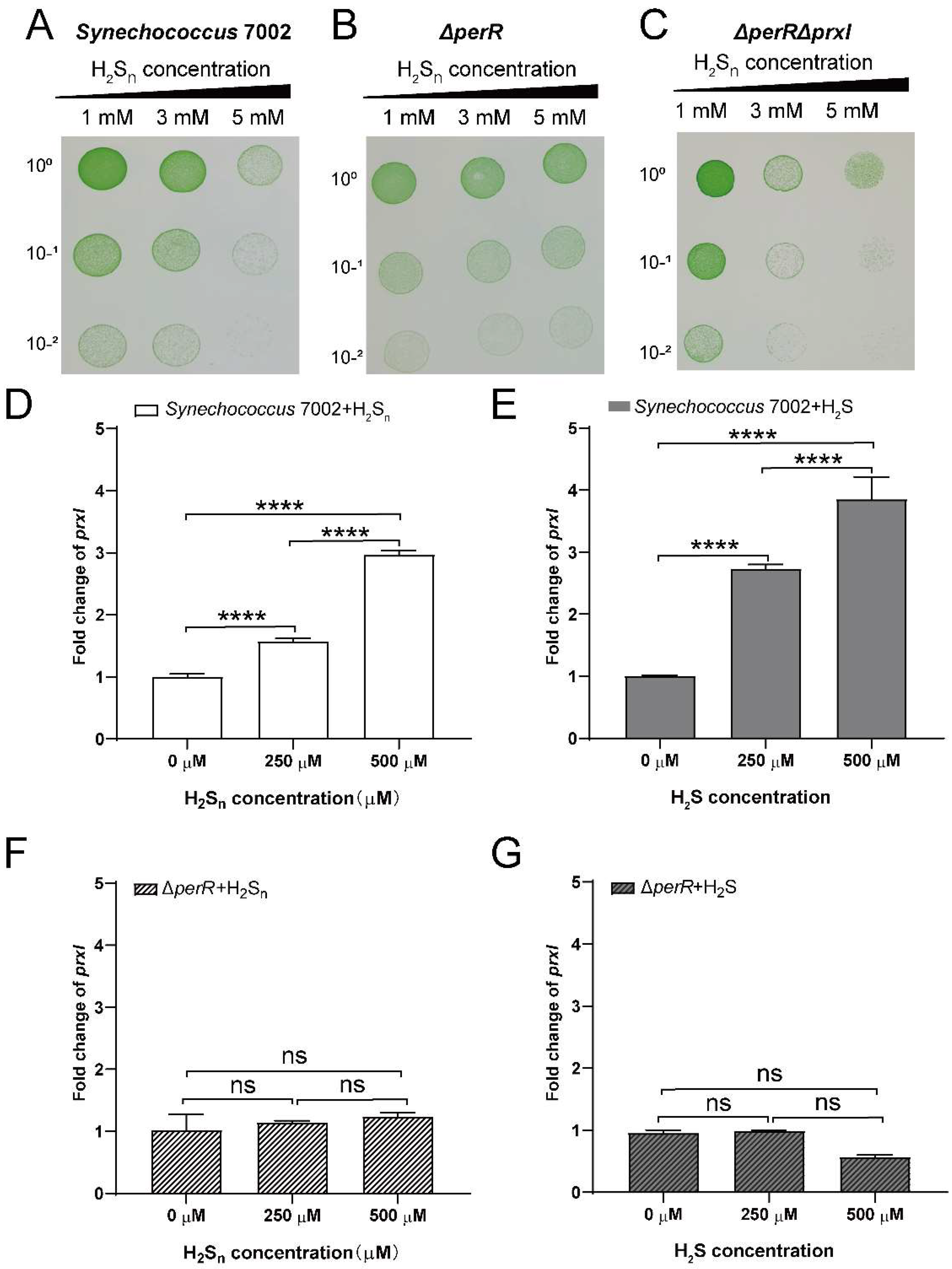
To investigate the effect of PerR on the tolerance of PCC7002 to sulfane sulfur, we constructed a single-deletion strain PCC7002ΔperR, and double-deletion strain PCC7002ΔprxIΔperR by homologous recombination (Figure S1). The mutation was verified by PCR (Figure S1A). Then, the tolerance of PCC7002, PCC7002ΔperR, and PCC7002ΔprxIΔperR to sulfane sulfur was tested (Figure 2). PCC7002ΔperR grew better than the wild-type after induction with 5 mM H2Sn, indicating that the deletion of perR increased tolerance (Figure 2A,B). However, the double-deletion mutant (PCC7002ΔprxIΔperR) showed decreased tolerance to H2Sn, and growth inhibition was apparent after induction with 3 mM H2Sn (Figure 2C). These results indicated that PerR and PrxI were all involved in H2Sn tolerance of PCC7002.

3.3. PerR Senses H2Sn and Regulates the Expression of prxI

PerR acts as a transcriptional repressor in the regulation of prxI expression, as qPCR analysis showed that the transcript level of prxI was upregulated ~100-fold in PCC7002ΔperR compared with PCC7002 (Figure S1B). Furthermore, the expression levels of prxI were analyzed in PCC7002 and PCC7002ΔperR after induction with H2Sn and H2S to verify whether PerR is involved in the regulation of H2Sn metabolism. The expression of prxI increased 1.5-fold following 250 µM H2Sn induction and 3-fold following 500 µM H2Sn induction (Figure 2D); this effect was concentration dependent. H2S induction could also increase prxI expression by 2.5-fold at concentrations of 250 µM and 4-fold at concentrations of 500 µM (Figure 2E). However, neither H2S nor H2Sn could induce the expression of prxI in PCC7002ΔperR (Figure 2F,G), which was different from the wild-type. Based on the above results, we deduced that PerR played a critical role in H2Sn sensing, thus regulating the expression of prxI.
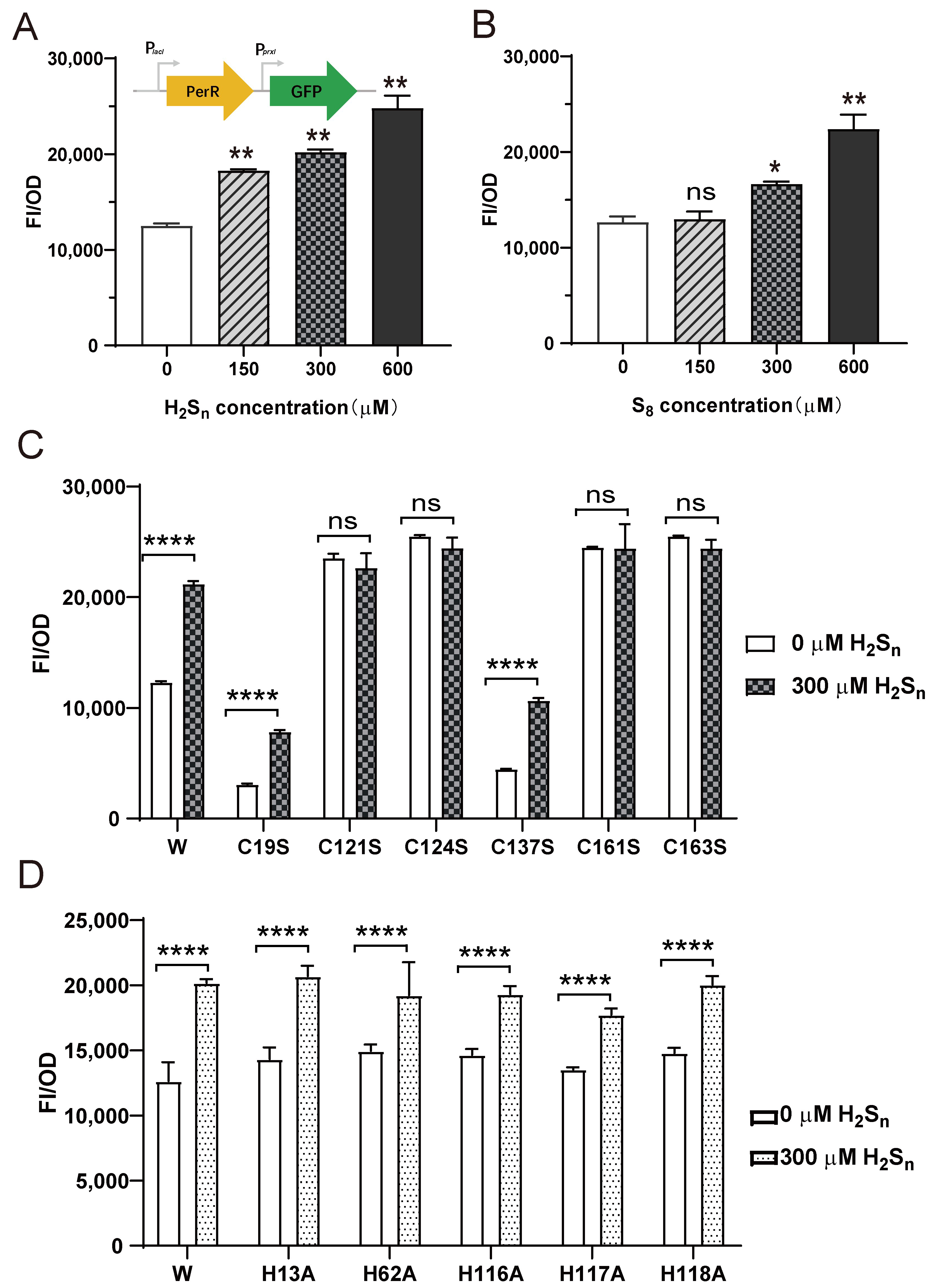
Meanwhile, a PerR-repressed reporter system in E. coli BL21 was constructed to further assess the effect of H2Sn on prxI expression regulated by PerR (Figure 3). In the reporter system, the perR gene is controlled by the lacI promoter (PlacI), and the egfp gene is controlled by the prxI promoter (PprxI) (Figure 3A). When the expression of PerR was induced by IPTG, GFP fluorescence decreased significantly, indicating that the expressed PerR could act on the prxI promoter and inhibit the expression of GFP (Figure S2). Having verified that PerR acts on the promoter of prxI to inhibit its expression, the effects of H2Sn and S8 were tested. H2Sn induction caused an increase in fluorescence intensity (Figure 2A), and S8, another form of sulfane sulfur, had a similar effect (Figure 2B).
To test the critical role of Cys residues in PerR, all six Cys residues (Cys19, Cys121, Cys123, Cys137, Cys160, and Cys163) were individually mutated to Ser. The mutation of Cys19 and Cys137 (C19S and C137S) resulted in decreased fluorescence intensity but did not affect their response to H2Sn. Cys121, Cys124, Cys160, and Cys163 were important components of the Cys4:Zn2+ site, and their mutation to Ser (C121S, C124S, C160S, and C163S) resulted in increased fluorescence intensities compared with the wild-type, indicating the inactivation of PerR (Figure 3C). As a result, PerR no longer acted on the prxI promoter to inhibit the expression of egfp, and it no longer responded to H2Sn induction. The mutation of His had no effect on PerR (Figure 3D), indicating H2Sn did not act on the Fe2+/Mn2+ site. We concluded that the expression of prxI was regulated by PerR, and the induction of S8 and H2Sn enhanced prxI expression by acting on PerR. Meanwhile, the Cys121, Cys124, Cys160, and Cys163 residues of PerR played crucial roles in H2Sn sensing.

3.4. Sulfane Sulfur Acts on the Cys4:Zn2+ Site of PerR

Furthermore, we measured the rate at which Zn2+ ions were released from PerR:Zn in the presence of H2Sn (Figure 4). An amount of 1 µM PerR contains about 0.125 µM Zn, 0.033 µM Fe and 0.006 µM Mn, as detected by ICP-MS. As the previous result (Figure 3D) confirmed that H2Sn did not act on the Fe2+/Mn2+ site, the effect on Cys4:Zn2+ site was monitored here. The formation of the colored Zn2+-PAR complex, whose absorption maximum was observed at 494 nm, was used to monitor the release of Zn2+. Thus, the Zn2+ release result showed that all selected concentrations of H2Sn induced the release of Zn2+. Based on the above results, we deduced the mechanism by which H2Sn acts on PerR, which involves H2Sn acting on the Cys4:Zn2+ site to dissociate Zn2+ from the active site, thus destroying the normal function of PerR.
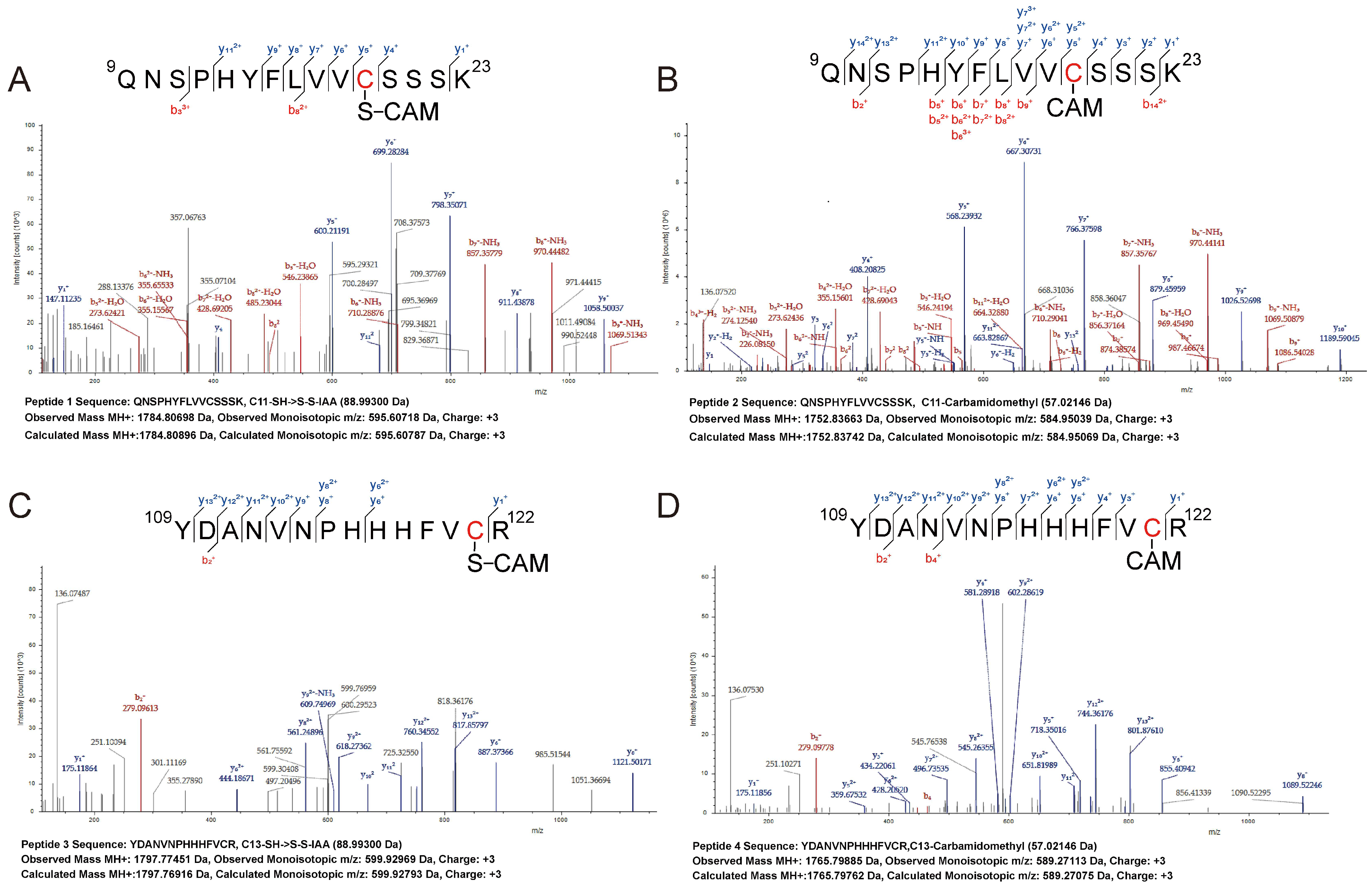
Finally, we explored the mechanism by which H2Sn acts on the Cys4:Zn2+ site by LTQ-Orbitrap tandem mass spectrometry (Figure 5). Cys19-SH of Peptide 1 in H2Sn-treated PerR was modified to Cys19-SSH (Figure 5A), while Cys19-SH of Peptide 2 in DTT-treated PerR was directly modified by acetamide (CAM) (Figure 5B). Similarly, Cys121-SH of Peptide 3 in H2Sn-treated PerR was modified to Cys121-SSH (Figure 5C), while Cys121-SH of Peptide 4 in DTT-treated PerR was also directly modified by acetamide (CAM) (Figure 5D). Among these, Cys121 was an important constituent of the Cys4:Zn2+ site, thus indicating that H2Sn acts on Cys121 to inhibit the activity of PerR. Notably, Cys19 was also modified by H2Sn, although it was not a component of the Cys4:Zn2+ site. In summary, the persulfide modification of Cys121 in the Cys4:Zn2+ site by H2Sn was the mechanism that affected PerR activity.

4. Discussion

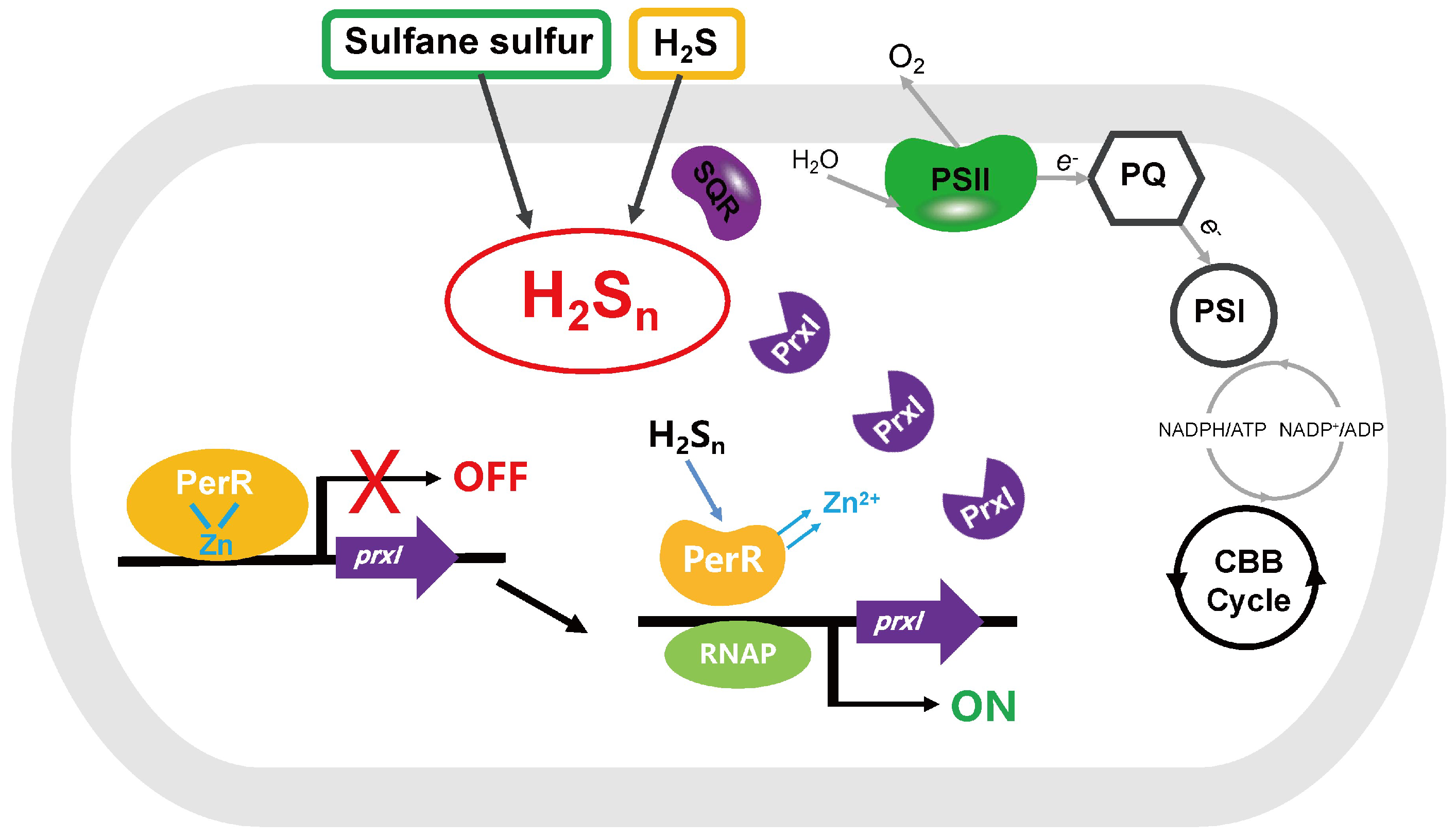
The data from our study revealed that PerR senses H2Sn and regulates the expression of prxI (Figure 6). The deletion of perR increased the tolerance for H2Sn in PCC7002 (Figure 2B), although the enhanced tolerance was not observed in the dual mutant PCC7002ΔperRΔprxI (Figure 2C), indicating that PerR functioned by acting on PrxI. Meanwhile, the induction effect of H2Sn on prxI transcription levels was only observed in the presence of PerR (Figure 2), a result which was further confirmed by the PerR-repressed reporter system (Figure 3), indicating that PerR acted on the promoter region to inhibit prxI expression. H2S had similar effect with H2Sn on prxI transcription levels (Figure 2B). This effect may be caused by H2Sn, which derive from H2S solution prepared from NaHS, as considerable levels of H2Sn may present [44]. Meanwhile, H2S may be converted to H2Sn by SQR [24]. Certainly, H2S may also act on PerR with a new mechanism directly, which needs to be further verified. Furthermore, H2Sn acted on the Cys4:Zn2+ site to release Zn2+, thus removing the inhibition (Figure 4) and allowing prxI to be expressed in large quantities to clear the excess sulfane sulfur. H2Sn modified Cys121 to form Cys121–SSH (Figure 5), destroying the structure of the Cys4:Zn2+ site and causing the release of Zn2+. Cys19 could also be modified by H2Sn. Thus, H2Sn acted on the zinc figure structure of PerR, which represents a new type of mechanism for sulfane sulfur sensing.
Zinc-binding proteins are among the most abundant transcriptional regulators in eukaryotes, harboring at least one common motif, the zinc finger, which contributes to proper protein structure and function [58,59]. Zinc finger proteins are also found in prokaryotic genomes, such as Bacillus PerR [39] and Synechococcus PerR [9]. Zinc-binding proteins display variable secondary structures and vast functional diversity, and can be classified into three classes based on their distinct structural properties: Cys2His2 (C2H2) zinc finger proteins (Class I), Cys4 (C4) zinc finger proteins (Class II), and Cys6 (C6) zinc finger proteins (Class III). Class I proteins are often referred to as the classical zinc finger [60]. Class II proteins contain four cysteine residues bound to one zinc atom [61], whereas Class III proteins contain six cysteine residues bound to two zinc atoms [62]. Thus, Synechococcus PerR belongs to Class II, and this is the first report of a zinc-binding protein being involved in sulfane sulfur sensing.
OxyR and PerR are two representative regulators that can sense both H2O2 and H2Sn. In total, 68 PerR proteins were identified among 198 sequenced cyanobacteria genomes (Figure 5), whereas only 9 OxyR proteins were identified (Table S5) [30], indicating that PerR may be the key player in cyanobacteria. Furthermore, PerR is a transcriptional inhibitor, whereas OxyR is a transcriptional activator, and their sensing mechanisms for H2O2 and H2Sn are quite different. The exact mechanism for the OxyR sensing of H2O2 is still under debate. The formation of a disulfide bond between Cys199 and Cys208 or the oxidization of Cys199 to C199-SOH in E. coli are two of the proposed mechanisms [63,64,65]. For H2Sn sensing, the Cys199 of E. coli OxyR is modified to Cys199-SSH [30]. Bacillus PerR senses H2O2 by metal-catalyzed oxidation [38], where one oxygen atom is incorporated into histidine 37 or histidine 91, which coordinates the bound Fe2+. Cysteines in the Cys4:Zn2+ site may also be oxidized by H2O2 [39]. Our results revealed that Cys121 in the Cys4:Zn2+ site of Synechococcus PerR could be modified by H2Sn to form Cys121-SSH (Figure 4), releasing one zinc atom and destabilizing the structure. Normally, PerR and OxyR do not exist in the same microbial strain, but there are some exceptions [66,67]. In cyanobacteria, PerR and OxyR did not coexist (Tables S4 and S5). Thus, although PerR and OxyR are considered functionally complementary, the mechanisms by which they function are different.
In addition to OxyR and PerR, two-component systems play an important role in H2O2 signal transduction in cyanobacteria [68]. A microarray-based study in Synechocystis PCC6803 revealed that His kinases (Hiks), namely, Hik33, Hik34, Hik16, and Hik42, are involved in the expression of a large number of H2O2-inducible genes [10]. Among the four Hiks, Hik33 was the main contributor and was responsible for the regulation of more H2O2-inducible genes than PerR. Furthermore, the response of Synechocystis to H2O2 treatment also relied on Group 2 sigma factors, namely, SigB and SigD [69]. The lack of Group 2 sigma factors meant that the strain was unable to sustain its growth under oxidative stress. Taken together, the signaling of H2O2-induced oxidative stress is based on the coordinated action of several regulators and dedicated alternative sigma factors. Whether these regulators participate in sulfane sulfur sensing requires further investigation.
The distribution of PerR proteins in cyanobacteria was also investigated. Gloeobacterales are early-branching photosynthetic cyanobacteria that are used as model species to study the physiology of early oxygenic phototrophs [70]. Gloeobacterales contain reduced photosystems that lack thylakoids and a circadian clock. However, our results revealed that all three species with published genomes within this order encoded perR, which may offer insight into the important role of PerR in primitive cyanobacteria and the evolution of oxygenic photosynthesis. Meanwhile, 81.8% of the species in Oscillatoriales and 56.3% of those in Nostocales contained PerR. Oscillatoriales and Nostocales are bloom-forming cyanobacteria that dominate among the cyanobacterial biomass of shallow polymictic eutrophic lakes [71]. The high proportion of PerR proteins among the two orders may provide insight into the survival strategies of cyanobacteria in hypoxic and sulfidic environments.
The finding that PerR senses H2Sn in cyanobacteria is significant. First, cyanobacteria have to tolerate the accumulation of sulfane sulfur in living environments. In Proterozoic oceans [2] and modern oxygen minimum zones [72], the environments in which cyanobacteria thrive are anoxic and sulfidic, and as a result, sulfane sulfur might accumulate. In cyanobacteria mats, a typical habitat for these microorganisms, the cyanobacteria are intermittently exposed to sulfane sulfur [73]. Although cyanobacteria can perform sulfur respiration and provide ATP for growth under dark and anoxic conditions by reducing sulfane sulfur [74], excess sulfane sulfur is fatal to cells [23]. Therefore, PerR sensing of H2Sn provides the opportunity for cyanobacteria to activate the expression of metabolic genes in time to scavenge excess sulfane sulfur, thus ensuring survival in such environments. Second, cyanobacteria perform anoxygenic photosynthesis under low-O2 and sulfidic conditions, using H2S as the electron donor [8,75]. In addition, cyanobacteria could produce some sulfur-containing histidine such as ergothioneine and ovothiols, which can also be used as electron donors [76,77]. As a result, sulfane sulfur was generated during the process of H2S oxidation by SQR during anoxygenic photosynthesis. Sulfane sulfur is a signal that participates in the regulation of physiology and critical gene expression in photosynthesis [24]. The PerR sensing of H2Sn may help cyanobacteria to maintain normal signal transduction and photosynthesis. Third, a previous study reported that the composition and stability of the photosynthetic machinery and the cell division process were affected by the overexpression of PerR [78], indicating that the effect of sulfane sulfur on the function of PerR may also affect the above process. Thus, the association between PerR, sulfane sulfur, photosynthesis, and cell division provides a new perspective on the significance of the PerR sensing of sulfane sulfur. In brief, the ability of PerR to sense H2Sn may ensure that cyanobacteria respond to intracellular and extracellular sulfane sulfur in a timely manner, allowing them to maintain normal photosynthesis and cell division, and adapt to environmental conditions.

5. Conclusions

In summary, we showed that PerR in PCC7002 senses H2Sn and regulates the expression of prxI. PCC7002 was able to respond in a timely manner to excess H2Sn in the environment with the help of PerR, enhancing its tolerance. H2Sn modified Cys121 of PerR to form Cys121-SSH, thus releasing Zn2+ from the Cys4:Zn2+ site, revealing a new mechanism of sulfane sulfur sensing in cyanobacteria. This is also the first report of a zinc-binding protein that participates in sulfane sulfur sensing. Our findings offer new insight into the mechanism of sulfane sulfur sensing and provide a new perspective for understanding the adaptation mechanism of cyanobacteria in anaerobic and sulfidic environments.

Supplementary Materials

The following supporting information can be downloaded at: https://www.mdpi.com/article/10.3390/antiox12020423/s1; Figure S1. The deletion of perR was verified by PCR and its effect on the transcriptional level of prxI; Figure S2. The expressed PerR could act on the prxI promoter and inhibit the expression of GFP; Table S1. Strains and plasmids used in this study; Table S2. Primers used in this study; Table S3. The queries used in the phylogenetic analysis of PerR; Table S4. The information of PerRs in cyanobacteria; Table S5. The OxyRs in cyanobacteria.

Author Contributions

Conceptualization, D.L. and H.S.; methodology, D.L.; software, H.L.; validation, D.L.; investigation, Y.L. and R.H.; data curation, D.L.; writing—original draft preparation, D.L.; writing—review and editing, H.S. and K.T.; supervision, N.J.; D.L. and J.L. All authors have read and agreed to the published version of the manuscript.

Funding

This research was funded by the Key Research and Development Program of Shandong Province, grant number 2020ZLYS04 and the Natural Science Foundation of Shandong Province, grant number ZR2022QC044.

Institutional Review Board Statement

Not applicable.

Informed Consent Statement

Not applicable.

Data Availability Statement

Data are contained within the article and supplementary material.

Acknowledgments

We thank Luying Xun for constructive discussions.

Conflicts of Interest

The authors declare no conflict of interest.

References

  1. Schopf, J.W. Geological evidence of oxygenic photosynthesis and the biotic response to the 2400–2200 ma “great oxidation event”. Biochem. Biokhimiia 2014, 79, 165–177. [Google Scholar] [CrossRef]
  2. Soo, R.M.; Hemp, J.; Parks, D.H.; Fischer, W.W.; Hugenholtz, P. On the origins of oxygenic photosynthesis and aerobic respiration in Cyanobacteria. Science 2017, 355, 1436–1440. [Google Scholar] [CrossRef]
  3. Olson, K.R. Reactive oxygen species or reactive sulfur species: Why we should consider the latter. J. Exp. Biol. 2020, 223, 196352. [Google Scholar] [CrossRef]
  4. Johnston, D.T.; Wolfe-Simon, F.; Pearson, A.; Knoll, A.H. Anoxygenic photosynthesis modulated Proterozoic oxygen and sustained Earth’s middle age. Proc. Natl. Acad. Sci. USA 2009, 106, 16925–16929. [Google Scholar] [CrossRef]
  5. Buick, R. When did oxygenic photosynthesis evolve? Philos. Trans. R. Soc. Lond. Ser. B Biol. Sci. 2008, 363, 2731–2743. [Google Scholar] [CrossRef]
  6. Oschlies, A. A committed fourfold increase in ocean oxygen loss. Nat. Commun. 2021, 12, 2307. [Google Scholar] [CrossRef]
  7. Haas, S.; de Beer, D.; Klatt, J.M.; Fink, A.; Rench, R.M.; Hamilton, T.L.; Meyer, V.; Kakuk, B.; Macalady, J.L. Low-light anoxygenic photosynthesis and Fe-S-Biogeochemistry in a microbial mat. Front. Microbiol. 2018, 9, 858. [Google Scholar] [CrossRef]
  8. Klatt, J.M.; Al-Najjar, M.A.; Yilmaz, P.; Lavik, G.; de Beer, D.; Polerecky, L. Anoxygenic photosynthesis controls oxygenic photosynthesis in a cyanobacterium from a sulfidic spring. Appl. Environ. Microbiol. 2015, 81, 2025–2031. [Google Scholar] [CrossRef]
  9. Li, H.; Singh, A.K.; McIntyre, L.M.; Sherman, L.A. Differential gene expression in response to hydrogen peroxide and the putative PerR regulon of Synechocystis sp. strain PCC 6803. J. Bacteriol. 2004, 186, 3331–3345. [Google Scholar] [CrossRef]
  10. Kanesaki, Y.; Yamamoto, H.; Paithoonrangsarid, K.; Shoumskaya, M.; Suzuki, I.; Hayashi, H.; Murata, N. Histidine kinases play important roles in the perception and signal transduction of hydrogen peroxide in the cyanobacterium, Synechocystis sp. PCC 6803. Plant J. Cell Mol. Biol. 2007, 49, 313–324. [Google Scholar] [CrossRef]
  11. Kobayashi, M.; Ishizuka, T.; Katayama, M.; Kanehisa, M.; Bhattacharyya-Pakrasi, M.; Pakrasi, H.B.; Ikeuchi, M. Response to Oxidative stress involves a novel peroxiredoxin gene in the unicellular cyanobacterium Synechocystis sp. PCC 6803. Plant Cell Physiol. 2004, 45, 290–299. [Google Scholar] [CrossRef]
  12. Latifi, A.; Ruiz, M.; Zhang, C.C. Oxidative stress in cyanobacteria. FEMS Microbiol. Rev. 2009, 33, 258–278. [Google Scholar] [CrossRef]
  13. Xia, Y.; Lü, C.; Hou, N.; Xin, Y.; Liu, J.; Liu, H.; Xun, L. Sulfide production and oxidation by heterotrophic bacteria under aerobic conditions. ISME J. 2017, 11, 2754–2766. [Google Scholar] [CrossRef]
  14. Li, K.; Xin, Y.; Xuan, G.; Zhao, R.; Liu, H.; Xia, Y.; Xun, L. Escherichia coli uses separate enzymes to produce H2S and Reactive Sulfane Sulfur from L-cysteine. Front. Microbiol. 2019, 10, 298. [Google Scholar] [CrossRef]
  15. Xin, Y.; Liu, H.; Cui, F.; Liu, H.; Xun, L. Recombinant Escherichia coli with sulfide:quinone oxidoreductase and persulfide dioxygenase rapidly oxidises sulfide to sulfite and thiosulfate via a new pathway. Environ. Microbiol. 2016, 18, 5123–5136. [Google Scholar] [CrossRef]
  16. Wang, R. Physiological implications of hydrogen sulfide: A whiff exploration that blossomed. Physiol. Rev. 2012, 92, 791–896. [Google Scholar] [CrossRef]
  17. Xuan, G.; Lü, C.; Xu, H.; Chen, Z.; Li, K.; Liu, H.; Liu, H.; Xia, Y.; Xun, L. Sulfane Sulfur is an intrinsic signal activating MexR-regulated antibiotic resistance in Pseudomonas aeruginosa. Mol. Microbiol. 2020, 114, 1038–1048. [Google Scholar] [CrossRef]
  18. Xuan, G.; Lv, C.; Xu, H.; Li, K.; Liu, H.; Xia, Y.; Xun, L. Sulfane sulfur regulates LasR-mediated quorum sensing and virulence in Pseudomonas aeruginosa PAO1. Antioxidants 2021, 10, 1498. [Google Scholar] [CrossRef]
  19. Xun, H.; Xuan, G.; Liu, H.; Xia, Y.; Xun, L. Sulfane sulfur is a strong inducer of the multiple antibiotic resistance regulator MarR in Escherichia coli. Antioxidants 2021, 10, 1778. [Google Scholar] [CrossRef]
  20. Iciek, M.; Kowalczyk-Pachel, D.; Bilska-Wilkosz, A.; Kwiecien, I.; Gorny, M.; Wlodek, L. S-sulfhydration as a cellular redox regulation. Biosci. Rep. 2015, 36, e00304. [Google Scholar] [CrossRef]
  21. Yan, F.; Fojtikova, V.; Man, P.; Stranava, M.; Martínková, M.; Du, Y.; Huang, D.; Shimizu, T. Catalytic enhancement of the heme-based oxygen-sensing phosphodiesterase EcDOS by hydrogen sulfide is caused by changes in heme coordination structure. Biomet. Int. J. Role Met. Ions Biol. Biochem. Med. 2015, 28, 637–652. [Google Scholar] [CrossRef]
  22. Zivanovic, J.; Kouroussis, E.; Kohl, J.B.; Adhikari, B.; Bursac, B.; Schott-Roux, S.; Petrovic, D.; Miljkovic, J.L.; Thomas-Lopez, D.; Jung, Y.; et al. Selective persulfide detection reveals evolutionarily conserved antiaging effects of s-sulfhydration. Cell Metab. 2019, 30, 1152–1170.e13. [Google Scholar] [CrossRef]
  23. Bhatwalkar, S.B.; Mondal, R.; Krishna, S.B.N.; Adam, J.K.; Govender, P.; Anupam, R. Antibacterial properties of organosulfur compounds of Garlic (Allium sativum). Front. Microbiol. 2021, 12, 613077. [Google Scholar] [CrossRef]
  24. Liu, D.; Zhang, J.; Lü, C.; Xia, Y.; Liu, H.; Jiao, N.; Xun, L.; Liu, J. Synechococcus sp. strain PCC7002 uses Sulfide:Quinone Oxidoreductase to detoxify exogenous sulfide and to convert endogenous sulfide to cellular sulfane sulfur. mBio 2020, 11, e03420–e03519. [Google Scholar] [CrossRef]
  25. Liu, H.; Xin, Y.; Xun, L. Distribution, diversity, and activities of sulfur dioxygenases in heterotrophic bacteria. Appl. Environ. Microbiol. 2014, 80, 1799–1806. [Google Scholar] [CrossRef]
  26. Lü, C.; Xia, Y.; Liu, D.; Zhao, R.; Gao, R.; Liu, H.; Xun, L. Cupriavidus necator H16 uses flavocytochrome c sulfide dehydrogenase to oxidize self-produced and added sulfide. Appl. Environ. Microbiol. 2017, 83, e01610–e01617. [Google Scholar] [CrossRef]
  27. Harada, M.; Akiyama, A.; Furukawa, R.; Yokobori, S.I.; Tajika, E.; Yamagishi, A. Evolution of superoxide dismutases and catalases in cyanobacteria: Occurrence of the antioxidant enzyme genes before the rise of atmospheric oxygen. J. Mol. Evol. 2021, 89, 527–543. [Google Scholar] [CrossRef]
  28. Olson, K.R.; Gao, Y.; DeLeon, E.R.; Arif, M.; Arif, F.; Arora, N.; Straub, K.D. Catalase as a sulfide-sulfur oxido-reductase: An ancient (and modern?) regulator of reactive sulfur species (RSS). Redox Biol. 2017, 12, 325–339. [Google Scholar] [CrossRef]
  29. Olson, K.R.; Gao, Y.; Arif, F.; Arora, K.; Patel, S.; DeLeon, E.R.; Sutton, T.R.; Feelisch, M.; Cortese-Krott, M.M.; Straub, K.D. Metabolism of hydrogen sulfide (H2S) and Production of Reactive Sulfur Species (RSS) by superoxide dismutase. Redox Biol. 2018, 15, 74–85. [Google Scholar] [CrossRef]
  30. Hou, N.; Yan, Z.; Fan, K.; Li, H.; Zhao, R.; Xia, Y.; Xun, L.; Liu, H. OxyR senses sulfane sulfur and activates the genes for its removal in Escherichia coli. Redox Biol. 2019, 26, 101293. [Google Scholar] [CrossRef]
  31. Liu, D.; Chen, J.; Wang, Y.; Meng, Y.; Li, Y.; Huang, R.; Xia, Y.; Liu, H.; Jiao, N.; Xun, L.; et al. Synechococcus sp. PCC7002 uses peroxiredoxin to cope with reactive sulfur species stress. mBio 2022, 13, e0103922. [Google Scholar] [CrossRef] [PubMed]
  32. Marinho, H.S.; Real, C.; Cyrne, L.; Soares, H.; Antunes, F. Hydrogen peroxide sensing, signaling and regulation of transcription factors. Redox Biol. 2014, 2, 535–562. [Google Scholar] [CrossRef]
  33. Luebke, J.L.; Shen, J.; Bruce, K.E.; Kehl-Fie, T.E.; Peng, H.; Skaar, E.P.; Giedroc, D.P. The CsoR-like sulfurtransferase repressor (CstR) is a persulfide sensor in Staphylococcus aureus. Mol. Microbiol. 2014, 94, 1343–1360. [Google Scholar] [CrossRef] [PubMed]
  34. Guimarães, B.G.; Barbosa, R.L.; Soprano, A.S.; Campos, B.M.; de Souza, T.A.; Tonoli, C.C.C.; Leme, A.F.P.; Murakami, M.T.; Benedetti, C.E. Plant pathogenic bacteria utilize biofilm growth-associated repressor (BigR), a novel winged-helix redox switch, to control hydrogen sulfide detoxification under hypoxia. J. Biol. Chem. 2011, 286, 26148–26157. [Google Scholar] [CrossRef]
  35. Shimizu, T.; Shen, J.; Fang, M.; Zhang, Y.; Hori, K.; Trinidad, J.C.; Bauer, C.E.; Giedroc, D.P.; Masuda, S. Sulfide-responsive transcriptional repressor SqrR functions as a master regulator of sulfide-dependent photosynthesis. Proc. Natl. Acad. Sci. USA 2017, 114, 2355–2360. [Google Scholar] [CrossRef]
  36. Li, H.; Li, J.; Lu, C.; Xia, Y.; Xin, Y.; Liu, H.; Xun, L.; Liu, H. FisR activates sigma(54)-dependent transcription of sulfide-oxidizing genes in Cupriavidus pinatubonensis JMP134. Mol. Microbiol. 2017, 105, 373–384. [Google Scholar] [CrossRef]
  37. Jo, I.; Chung, I.Y.; Bae, H.W.; Kim, J.S.; Song, S.; Cho, Y.H.; Ha, N.C. Structural details of the OxyR peroxide-sensing mechanism. Proc. Natl. Acad. Sci. USA 2015, 112, 6443–6448. [Google Scholar] [CrossRef]
  38. Lee, J.W.; Helmann, J.D. The PerR transcription factor senses H2O2 by metal-catalysed histidine oxidation. Nature 2006, 440, 363–367. [Google Scholar] [CrossRef]
  39. Lee, J.W.; Helmann, J.D. Biochemical characterization of the structural Zn2+ site in the Bacillus subtilis peroxide sensor PerR. J. Biol. Chem. 2006, 281, 23567–23578. [Google Scholar] [CrossRef]
  40. Ludwig, M.; Chua, T.T.; Chew, C.Y.; Bryant, D.A. Fur-type transcriptional repressors and metal homeostasis in the cyanobacterium Synechococcus sp. PCC 7002. Front. Microbiol. 2015, 6, 1217. [Google Scholar] [CrossRef] [Green Version]
  41. Ida, T.; Sawa, T.; Ihara, H.; Tsuchiya, Y.; Watanabe, Y.; Kumagai, Y.; Suematsu, M.; Motohashi, H.; Fujii, S.; Matsunaga, T.; et al. Reactive cysteine persulfides and S-polythiolation regulate oxidative stress and redox signaling. Proc. Natl. Acad. Sci. USA 2014, 111, 7606–7611. [Google Scholar] [CrossRef]
  42. Stevens, S.E.; Porter, R.D. Transformation in Agmenellum quadruplicatum. Proc. Natl. Acad. Sci. USA 1980, 77, 6052–6056. [Google Scholar] [CrossRef]
  43. Xia, Y.; Li, K.; Li, J.; Wang, T.; Gu, L.; Xun, L. T5 exonuclease-dependent assembly offers a low-cost method for efficient cloning and site-directed mutagenesis. Nucleic Acids Res. 2019, 47, e15. [Google Scholar] [CrossRef]
  44. Nagy, P.; Pálinkás, Z.; Nagy, A.; Budai, B.; Tóth, I.; Vasas, A. Chemical aspects of hydrogen sulfide measurements in physiological samples. Biochim. Biophys. Acta 2014, 1840, 876–891. [Google Scholar] [CrossRef]
  45. Szekeres, E.; Sicora, C.; Dragos, N.; Druga, B. Selection of proper reference genes for the cyanobacterium Synechococcus PCC 7002 using real-time quantitative PCR. FEMS Microbiol. Lett. 2014, 359, 102–109. [Google Scholar] [CrossRef]
  46. Livak, K.J.; Schmittgen, T.D. Analysis of relative gene expression data using real-time quantitative PCR and the 2(-Delta Delta C(T)) Method. Methods 2001, 25, 402–408. [Google Scholar] [CrossRef]
  47. Xia, Y.; Xun, L. Revised mechanism and improved efficiency of the quikchange site-directed mutagenesis method. Methods Mol. Biol. 2017, 1498, 367–374. [Google Scholar] [CrossRef]
  48. Waugh, D.S. The remarkable solubility-enhancing power of Escherichia coli maltose-binding protein. Postep. Biochem. 2016, 62, 377–382. [Google Scholar] [CrossRef]
  49. Lu, T.; Cao, Q.; Pang, X.; Xia, Y.; Xun, L.; Liu, H. Sulfane sulfur-activated actinorhodin production and sporulation is maintained by a natural gene circuit in Streptomyces coelicolor. Microb. Biotechnol. 2020, 13, 1917–1932. [Google Scholar] [CrossRef]
  50. Doka, E.; Pader, I.; Biro, A.; Johansson, K.; Cheng, Q.; Ballago, K.; Prigge, J.R.; Pastor-Flores, D.; Dick, T.P.; Schmidt, E.E.; et al. A novel persulfide detection method reveals protein persulfide- and polysulfide-reducing functions of thioredoxin and glutathione systems. Sci. Adv. 2016, 2, e1500968. [Google Scholar] [CrossRef] [Green Version]
  51. Fan, K.; Chen, Z.; Liu, H. Evidence that the ProPerDP method is inadequate for protein persulfidation detection due to lack of specificity. Sci. Adv. 2020, 6, eabb6477. [Google Scholar] [CrossRef]
  52. Dóka, É.; Arnér, E.S.J.; Schmidt, E.E.; Dick, T.P.; van der Vliet, A.; Yang, J.; Szatmári, R.; Ditrói, T.; Wallace, J.L.; Cirino, G.; et al. Comment on “Evidence that the ProPerDP method is inadequate for protein persulfidation detection due to lack of specificity”. Sci. Adv. 2021, 7, abe7006. [Google Scholar] [CrossRef] [PubMed]
  53. Gerdol, M.; Sollitto, M.; Pallavicini, A.; Castellano, I. The complex evolutionary history of sulfoxide synthase in ovothiol biosynthesis. Proc. Biol. Sci. 2019, 286, 20191812. [Google Scholar] [CrossRef] [PubMed]
  54. Katoh, K.; Standley, D.M. MAFFT multiple sequence alignment software version 7: Improvements in performance and usability. Mol. Biol. Evol. 2013, 30, 772–780. [Google Scholar] [CrossRef] [PubMed]
  55. Capella-Gutiérrez, S.; Silla-Martínez, J.M.; Gabaldón, T. trimAl: A tool for automated alignment trimming in large-scale phylogenetic analyses. Bioinformatics 2009, 25, 1972–1973. [Google Scholar] [CrossRef] [PubMed]
  56. Naser-Khdour, S.; Minh, B.Q.; Lanfear, R. Assessing confidence in root placement on phylogenies: An empirical study using non-reversible models for mammals. Syst. Biol. 2021, 71, 959–972. [Google Scholar] [CrossRef]
  57. Kalyaanamoorthy, S.; Minh, B.Q.; Wong, T.K.F.; von Haeseler, A.; Jermiin, L.S. ModelFinder: Fast model selection for accurate phylogenetic estimates. Nat. Methods 2017, 14, 587–589. [Google Scholar] [CrossRef] [PubMed]
  58. MacPherson, S.; Larochelle, M.; Turcotte, B. A fungal family of transcriptional regulators: The zinc cluster proteins. Microbiol. Mol. Biol. Rev. 2006, 70, 583–604. [Google Scholar] [CrossRef]
  59. Laity, J.H.; Lee, B.M.; Wright, P.E. Zinc finger proteins: New insights into structural and functional diversity. Curr. Opin. Struct. Biol. 2001, 11, 39–46. [Google Scholar] [CrossRef]
  60. Näär, A.M.; Ryu, S.; Tjian, R. Cofactor requirements for transcriptional activation by Sp1. Cold Spring Harb. Symp. Quant. Biol. 1998, 63, 189–199. [Google Scholar] [CrossRef]
  61. Urnov, F.D. A feel for the template: Zinc finger protein transcription factors and chromatin. Biochem. Cell Biol. 2002, 80, 321–333. [Google Scholar] [CrossRef] [PubMed]
  62. Todd, R.B.; Andrianopoulos, A. Evolution of a fungal regulatory gene family: The Zn(II)2Cys6 binuclear cluster DNA binding motif. Fungal Genet. Biol. 1997, 21, 388–405. [Google Scholar] [CrossRef] [PubMed]
  63. Kim, S.O.; Merchant, K.; Nudelman, R.; Beyer, W.F.; Keng, T.; DeAngelo, J.; Hausladen, A.; Stamler, J.S. OxyR: A molecular code for redox-related signaling. Cell 2002, 109, 383–396. [Google Scholar] [CrossRef] [PubMed]
  64. Choi, H.-J.; Kim, S.-J.; Mukhopadhyay, P.; Cho, S.; Woo, J.-R.; Storz, G.; Ryu, S.-E. Structural basis of the redox switch in the OxyR transcription factor. Cell 2001, 105, 103–113. [Google Scholar] [CrossRef]
  65. Storz, G.; Tartaglia, L.A.; Ames, B.N. Transcriptional regulator of oxidative stress-inducible genes: Direct activation by oxidation. Science 1990, 248, 189–194. [Google Scholar] [CrossRef]
  66. Tseng, H.J.; McEwan, A.G.; Apicella, M.A.; Jennings, M.P. OxyR acts as a repressor of catalase expression in Neisseria gonorrhoeae. Infect. Immun. 2003, 71, 550–556. [Google Scholar] [CrossRef]
  67. Wu, H.J.; Seib, K.L.; Srikhanta, Y.N.; Kidd, S.P.; Edwards, J.L.; Maguire, T.L.; Grimmond, S.M.; Apicella, M.A.; McEwan, A.G.; Jennings, M.P. PerR controls Mn-dependent resistance to oxidative stress in Neisseria gonorrhoeae. Mol. Microbiol. 2006, 60, 401–416. [Google Scholar] [CrossRef]
  68. Ashby, M.K.; Houmard, J. Cyanobacterial two-component proteins: Structure, diversity, distribution, and evolution. Microbiol. Mol. Biol. Rev. 2006, 70, 472–509. [Google Scholar] [CrossRef]
  69. Koskinen, S.; Hakkila, K.; Kurkela, J.; Tyystjärvi, E.; Tyystjärvi, T. Inactivation of group 2 σ factors upregulates production of transcription and translation machineries in the cyanobacterium Synechocystis sp. PCC 6803. Sci. Rep. 2018, 8, 10305. [Google Scholar] [CrossRef]
  70. Grettenberger, C.L. Novel Gloeobacterales spp. from diverse environments across the globe. mSphere 2021, 6, e0006121. [Google Scholar] [CrossRef]
  71. Briand, J.-F.; Robillot, C.; Quiblier, C.; Bernard, C. A perennial bloom of planktothrix agardhii (cyanobacteria) in a shallow eutrophic french lake: Limnological and microcystin production studies. Arch. Fur Hydrobiol. 2002, 153, 605–622. [Google Scholar] [CrossRef]
  72. Schunck, H.; Lavik, G.; Desai, D.K.; Grosskopf, T.; Kalvelage, T.; Loscher, C.R.; Paulmier, A.; Contreras, S.; Siegel, H.; Holtappels, M.; et al. Giant hydrogen sulfide plume in the oxygen minimum zone off Peru supports chemolithoautotrophy. PLoS ONE 2013, 8, e68661. [Google Scholar] [CrossRef]
  73. Klatt, J.M.; Gomez-Saez, G.V.; Meyer, S.; Ristova, P.P.; Yilmaz, P.; Granitsiotis, M.S.; Macalady, J.L.; Lavik, G.; Polerecky, L.; Bühring, S.I. Versatile cyanobacteria control the timing and extent of sulfide production in a Proterozoic analog microbial mat. ISME J. 2020, 14, 3024–3037. [Google Scholar] [CrossRef] [PubMed]
  74. Stal, L.J.; Moezelaar, R. Fermentation in cyanobacteria. FEMS Microbiol. Rev. 1997, 21, 179–211. [Google Scholar] [CrossRef]
  75. Klatt, J.M.; de Beer, D.; Hausler, S.; Polerecky, L. Cyanobacteria in sulfidic spring microbial mats can perform oxygenic and anoxygenic photosynthesis simultaneously during an entire diurnal period. Front. Microbiol. 2016, 7, 1973. [Google Scholar] [CrossRef]
  76. Liao, C.; Seebeck, F.P. Convergent evolution of ergothioneine biosynthesis in cyanobacteria. ChemBioChem 2017, 18, 2115–2118. [Google Scholar] [CrossRef]
  77. Brancaccio, M.; Tangherlini, M.; Danovaro, R.; Castellano, I. Metabolic adaptations to marine environments: Molecular diversity and evolution of ovothiol biosynthesis in bacteria. Genome Biol. Evol. 2021, 13, evab169. [Google Scholar] [CrossRef]
  78. Sevilla, E.; Sarasa-Buisan, C.; Gonzalez, A.; Cases, R.; Kufryk, G.; Peleato, M.L.; Fillat, M.F. Regulation by FurC in Anabaena links the oxidative stress response to photosynthetic metabolism. Plant Cell Physiol. 2019, 60, 1778–1789. [Google Scholar] [CrossRef]
Figure 1. Phylogenetic analysis of PerR-encoding genes in the sequenced cyanobacteria genomes. (A) Phylogenetic tree of PerRs in cyanobacteria. A total of 68 probable PerRs were found in 198 cyanobacteria genomes. The representative proteins were labeled with name of species. The PerR queries were listed in Table S3. PerRs from Staphylococcus epidermidis, Staphylococcus haemolyticus and Staphylococcus aureus in Table S3 were used as the outgroup. (B) The distribution of PerR-encoding genes in cyanobacteria genomes. In total, 68 predicted PerR-encoding genes were detected among 64 cyanobacteria genomes, including 25 Synechococcales, 27 Nostocales, 3 Gloeobacteria, 9 Oscillatoriales, and 4 Pseudanabaenalles.
Antioxidants 12 00423 g001
Figure 2. PerR decreases the tolerance of PCC7002 to H2Sn. The deletion of perR (PCC7002ΔperR) (B) increased H2Sn tolerance, while the double deletion of perR and prxI (PCC7002ΔprxIΔperR) (C) decreased H2Sn tolerance compared with the wild–type (A). PCC7002, PCC7002ΔperR, and PCC7002ΔprxIΔperR cells at log phase with an OD730 nm of 1 were treated with 1, 3, and 5 mM H2Sn, at 30 °C, and 50 μmol photons m−2·s−1 illumination for 6 h. Then, cells were diluted with A+ medium to 100, 10−1, and 10−2, and plated onto the A+ solid medium and cultured for 7 days, at 30 °C, and 50 μmol photons·m−2·s−1 illumination. The tolerance of PCC7002ΔperR and PCC7002ΔprxIΔperR to H2Sn was opposite to that of the wild-type. The expression of prxI was largely upregulated by H2Sn (D) and H2S (E) in PCC7002, while H2Sn (F) and H2S (G) showed little influence on its expression in PCC7002ΔperR. PCC7002 and PCC7002ΔperR cells at log phase were induced by H2Sn and H2S with concentrations of 250 µM and 500 µM for 3 h, and the expression of prxI was measured. The Y-axis is the fold change in prxI calculated by relative quantitative qPCR, based on the 2−ΔΔCT method, with rnp as the reference gene. All data are averages from three samples with standard deviations (error bars). The experiment was repeated at least three times. ****, p < 0.0001; ns, not significant (paired t test).
Antioxidants 12 00423 g002
Figure 3. The effect of H2Sn and S8 on the PerR-repressed reporter. (A) Schematic representation of the test plasmid (pBBR-perR-PprxI-egfp). The expression of egfp was initiated by the prxI promoter (PprxI), and the interaction between PerR and PprxI, which was associated with the inducers that affected GFP fluorescence. H2Sn (A) and S8 (B) induction increased the intensity of GFP fluorescence in E. coli BL21 (pBBR-perR-PprxI-egfp). (C) The mutation of Cys affected the function of PerR in E. coli BL21 (pBBR-perR-PprxI-egfp). C19S, C121S, C124S, C137S, C161S, and C163S represented single mutations of Cys to Ser in PerR. (D) The mutation of His did not affect the function of PerR in E. coli BL21 (pBBR-perR-PprxI-egfp). H13A, H62A, H116A, H117A and H118A represented single mutations of His to Ala in PerR. FI/OD represents the fluorescence intensity of per OD cells. All data are averages from three samples with standard deviations (error bars). The experiment was repeated at least three times. *, p < 0.1; **, p < 0.01; ****, p < 0.0001; ns, not significant (paired t test).
Antioxidants 12 00423 g003
Figure 4. H2Sn caused dissociation of zinc ions from PerR. Oxidation of the Cys4:Zn2+ site by H2Sn led to Zn2+ release. H2Sn was incubated with 100 µM PerR, and formation of the Zn2+-PAR complex was continuously monitored by measuring the absorbance at 494 nm for 10 min. All data are averages from three samples with standard deviations (error bars). The experiment was repeated at least three times.
Antioxidants 12 00423 g004
Figure 5. H2Sn acted on the cysteines of PerR. (A) The Cys19–SSH group was blocked by IAM in the peptide 1 from H2Sn–treated PerR. (B) The Cys19–SH group was blocked by IAM in the peptide 2 from DTT-treated PerR. (C) The Cys121–SSH group was blocked by IAM in the peptide 3 from H2Sn-treated PerR. (D) The Cys121–SH group was blocked by IAM in the peptide 4 from DTT–treated PerR. The purified PerR (5 mg/mL) was treated with 1 mM H2Sn and 1 mM DTT for 30 min, at 25 °C. After denaturing and incubating with IAM, the reacted protein was digested by trypsin. The generated peptides were detected by LTQ-Orbitrap tandem mass spectrometry.
Antioxidants 12 00423 g005
Figure 6. PerR senses H2Sn and regulates the expression of prxI in PCC7002. H2Sn induces the expression of prxI via PerR. PerR binds to Zn2+ and acts on the promoter of prxI to inhibit its expression, a process that can be disinhibited by H2Sn. H2Sn acts on the Cys4:Zn2+ site of PerR to relieve Zn2+, destroying the zinc finger structure. C121-SH in the Cys4:Zn2+ site is modified by H2Sn and forms C121-SSH.
Antioxidants 12 00423 g006
Disclaimer/Publisher’s Note: The statements, opinions and data contained in all publications are solely those of the individual author(s) and contributor(s) and not of MDPI and/or the editor(s). MDPI and/or the editor(s) disclaim responsibility for any injury to people or property resulting from any ideas, methods, instructions or products referred to in the content.