Article

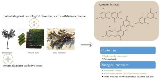
Edible Seaweeds Extracts: Characterization and Functional Properties for Health Conditions

by
Mariana Coelho
1,
Ana Patrícia Duarte
1,
Sofia Pinto
1,
Hugo M. Botelho
2,
Catarina Pinto Reis
3,4,*,
Maria Luísa Serralheiro
2,5 and
Rita Pacheco
1,6,*
1
Departamento de Engenharia Química, Instituto Superior de Engenharia de Lisboa (ISEL), Av. Conselheiro Emídio Navarro 1, 1959-007 Lisboa, Portugal
2
BioISI—Biosystems & Integrative Sciences Institute, Faculty of Sciences, Universidade de Lisboa, 1749-016 Lisboa, Portugal
3
Research Institute for Medicines (iMed.ULisboa), Faculty of Pharmacy, Universidade de Lisboa, Av. Professor Gama Pinto, 1649-003 Lisboa, Portugal
4
Instituto de Biofísica e Engenharia Biomédica (IBEB), Faculdade de Ciências, Universidade de Lisboa, Campo Grande, 1749-016 Lisboa, Portugal
5
Departamento de Química e Bioquímica, Faculty of Sciences, Universidade de Lisboa, 1749-016 Lisboa, Portugal
6
Centro de Química Estrutural—Institute of Molecular Sciences, Universidade de Lisboa, 1749-016 Lisboa, Portugal
*
Authors to whom correspondence should be addressed.
Antioxidants 2023, 12(3), 684; https://doi.org/10.3390/antiox12030684
Submission received: 14 January 2023 / Revised: 1 March 2023 / Accepted: 8 March 2023 / Published: 10 March 2023
(This article belongs to the Special Issue Antioxidant Compounds from Marine Organisms)

Abstract

:
Seaweeds are popular foods due to claimed beneficial health effects, but for many there is a lack of scientific evidence. In this study, extracts of the edible seaweeds Aramé, Nori, and Fucus are compared. Our approach intends to clarify similarities and differences in the health properties of these seaweeds, thus contributing to target potential applications for each. Additionally, although Aramé and Fucus seaweeds are highly explored, information on Nori composition and bioactivities is scarce. The aqueous extracts of the seaweeds were obtained by decoction, then fractionated and characterized according to their composition and biological activity. It was recognized that fractioning the extracts led to bioactivity reduction, suggesting a loss of bioactive compounds synergies. The Aramé extract showed the highest antioxidant activity and Nori exhibited the highest potential for acetylcholinesterase inhibition. The identification of the bioactive compounds in the extracts allowed to see that these contained a mixture of phloroglucinol polymers, and it was suggested that Nori’s effect on acetylcholinesterase inhibition may be associated with a smaller sized phlorotannins capable of entering the enzyme active site. Overall, these results suggest a promising potential for the use of these seaweed extracts, mainly Aramé and Nori, in health improvement and management of diseases, namely those associated to oxidative stress and neurodegeneration.

Graphical Abstract

1. Introduction

Seaweeds have been an important dietary constituent in Asian countries such as China, Japan, and Korea [1]. In some regions of the world, the population is growing to reach a level where food production may not be sufficient to feed the population [2], therefore the demand for alternatives, whether for human consumption or industrial processing, has increased in recent decades [3]. Seaweeds are considered an abundant, rich and sustainable marine source of macro and micronutrients, and an alternative to animal or even synthetic products [3]. Furthermore, several seaweed compounds have been reported to be bioactive, thus providing beneficial health effects [2], with their bioactive compounds showing the potential to be used as ingredients, both in functional foods and in health and food supplements.
Regarding macronutrients, seaweeds are a good source of sulfated polysaccharides (15–76% dry mass), proteins (5–47% dry mass), including all the essential amino acids, and lipids (1–5% dry mass), such as polyunsaturated fatty acids [3,4]. Concerning micronutrients, seaweeds are a good source of lipid and water-soluble vitamins, namely A, B1, B12, C, D and E and minerals (7–36% dry mass), such as calcium, iron, magnesium, copper, and iodine [3,4,5]. Seaweeds are also a source of secondary metabolites such as polyphenols [1,3,4]. Although some of these compounds, especially the latter, have reported beneficial health effects, like antioxidant, antimicrobial, anti-inflammatory, anti-cancer, anti-diabetic, anti-hypertensive, anti-hyperlipidemic, and anti-obesity effects [6], there is an immense variety of seaweeds. Numerous reports concern individual species, and there are several reviews about these organisms. The widespread belief that seaweed products have the potential to treat or prevent most human conditions has been rising. However, often there is lack of the proof of concept for the claimed effects and no association of a particular species to the bioactivities, which are then assigned to the whole group.
Seaweeds are taxonomically classified into three main groups, brown seaweeds (phylum Ochrophyta), red seaweeds (phylum Rhodophyta), and green seaweeds (phylum Chlorophyta) [7].
Seaweed colors are associated with its pigment, such as chlorophyll for green, phycobilin for red, and fucoxanthin for brown seaweeds [3]. The compound content of each seaweed depends on the species, but also on other geographical and environmental factors, such as substrate firmness, exposure to ice and waves, salinity, wave force, light and competition between seaweeds [8,9]. For this work, three seaweeds commonly used in foods or supplements were chosen to be studied, and their composition and biological activities were compared to report on their given potential for health improvement and management of diseases, especially those associated with oxidative stress.
Eisenia bicyclis, traditionally known as Aramé, is a perennial brown seaweed [10] distributed along the mid-pacific coastlines of Korea and Japan [10]. This seaweed is used industrially to extract sodium alginate, and for consumption by the population of East Asia in soups and salads [11,12]. The main bioactive compounds reported for Aramé are phenolic compounds, and polysaccharides [13,14]. The most abundant phenolic compounds are phlorotannins (eckol and other phloroglucinol derivatives), which were reported to have various biological activities, namely anti-diabetic and antioxidant activities [15]. The most abundant polysaccharides seen to be present in Aramé were fucoidan and laminarin [14]. On Ecklonia cava [16] and Sargassum vulgare [17], other brown seaweeds, these polysaccharides were reported to have antioxidant activity and anti-inflammatory activity [16,17]. Nevertheless, to the best of our knowledge, there is no research about these type of activities for Aramé fucoidan.
Fucus vesiculosus, known as bladderwrack [18], is an edible brown seaweed of the rocky bottoms of the northern temperate coastal areas [19], used to make beverage infusions, in cooked dishes and soups, or sprinkled in salads [20]. In traditional medicine Fucus is used due to its high iodine content to treat gout and aid weight loss [21]. This seaweed has bioactive compounds such as phenolic compounds [22], and sulfated polysaccharides [23]. It also contains proteins, minerals, vitamins, fatty acids, sterols, dietary fiber, and iodine [20,24]. The phenolic compounds mostly present in this seaweed are phlorotannins [25], which have reported activities such as antioxidant [18], anti-diabetic, anti-inflammatory, anti-cancer, anti-obesity, anti-lipidemic, and anti-hypertensive [20,26,27,28]. Also, Fucus polysaccharides are fucoidan and laminarin [20]. The latter was reported to have anti-inflammatory, anticoagulant, antioxidant, anti-cancer, and hypolipidemic activity [29,30,31], but again for fucoidan from Fucus there are no reports.
Porphyra tenera, known as Nori, is a red seaweed considered the most valuable maricultured seaweed in the world, as it is one of the most popular edible seaweeds used in sushi and soups [32,33]. It is known to have major bioactive compounds phenolic compounds, sulfated polysaccharides, and peptides [34,35]. It is also a source of dietary fiber, essential fatty acids, vitamins, and minerals [36]. In Nori, phlorotannins were identified and described to exhibit antioxidant activity and protection against UV light [37,38]. Other bioactive compounds present in this seaweed are sulfated polysaccharides, called porphyrans, which are reported to have hypolipidemic, anti-cancer, and anti-inflammatory activities [39]. Other polysaccharides reported in red seaweeds, such as cellulose, xylans, and manans, are not water-soluble [40].
In this work, a novel approach was used. Three of the most consumed edible seaweeds, Aramé, Fucus, and Nori, were matched for the composition and biological activities associated to the aqueous extracts of the seaweeds. Though there are various reports about Aramé [13,14], Fucus [22,23,24], and Nori [34,35], individually there are no studies comparing their composition and beneficial health effects.
With this in mind, we herein report the characterization of aqueous extracts from the three seaweeds, obtained under the same conditions, and fractions of these extracts, enriched in different bioactive compounds classes from the three seaweeds. Their biological activities, namely the antioxidant activity and enzyme acetylcholinesterase (AChE) inhibition were investigated and compared in order to evaluate their potential against oxidative stress. In this work, reported for the first time is the capacity of Nori extracts to inhibit acetylcholinesterase, an enzyme associated to gastrointestinal motility and neurodegenerative diseases, as Alzheimer’s disease (AD). The comparison of the seaweeds extracts guided the association between the exhibited activities and their composition in bioactive compounds, supporting future exploration of targets for application. Additionally, the results herein reported may encourage the development of novel and natural products with the incorporation of these seaweeds into the diet, supplements, or functional foods, particularly to prevent oxidative stress. Oxidative stress is often associated to several diseases, such as cardiovascular diseases, metabolic conditions, and neurodegenerative disorders.

2. Materials and Methods

2.1. Seaweeds

Porphyra tenera was purchased from Flavers-International Flavours Shop® (Blue Dragon line, B#JS2039J01). While Fucus vesiculosus was collected from Tagus River (38.7822 N, 9.0913 W). The dry Eisenia bicyclis seaweed was purchased in a commercial surface from the Seara brand (B# T20220405, expiration date April 2022), as previously described in [13].

2.2. Chemical

All reagents and solvents were of analytical grade unless otherwise specified and used without further purification. Roswell Park Memorial Institute (RPMI-1640), Dulbecco’s Modified Eagle Medium (DMEM), trypsin and glutamine from Biowhittaker® Lonza. Fetal Bovine Serum (FBS) from Biowest, phosphate-buffered saline (PBS) were obtained from Corning (Corning, NY, USA). Antibiotic Antimycotic Solution 100 × (10,000 U/mL penicillin, 10 mg/mL streptomycin, and 25 μg amphotericin B/mL), reagent Folin & Ciocalteu, sodium acetate, 2,2-diphenyl-1-picyl-hydroxyl (DPPH), acetylcholinesterase (AChE) (149 U/mg solid, 241 U/mg protein), and acetycholine iodide (AChI) were obtained from Sigma®Aldrich (St. Louis, MO, USA). Calcium Carbonate, Concentrated Sulfuric Acid from Merck. Phloroglucinol from Aldrich® chemistry. Polygalacturonic acid and 5-5′-Dithiobis (2-nitrobenzoic acid) (DTNB) were purchased to Alfa Aesar (Ward Hill, MA, USA). Phenol and (4,5-dimetylthiazol-1-yl)-2,5-diphenyltetrazolium (MTT) were obtained from VWR (Radnor, PA, USA). Citric acid and Magnesium Chloride-6-hydrate were obtained from Riedel-de Haën (Seelze, Germany). Sodium Chloride was obtained from Panreac (Glenview, IL, USA).

2.3. Preparation of Seaweed Extracts

The dried and milled biomass of the seaweeds was used to prepare aqueous extracts. As the biomass of Fucus vesiculosus was collected in nature, it was subjected to an extensive washing procedure and further dried in an Heto® PowerDry LL3000 freeze dryer.
For the preparation of a dry mass of Nori aqueous extract, the biomass of Porphyra tenera was mixed with distilled water (20 g/L) and the suspension was autoclaved at 121 °C for 15 min. The suspension was filtered, frozen at −20 °C and freeze dried, to obtain the dry extract (50% g/g yield). The same procedure was performed to obtain a dry mass of Fucus aqueous extract, using 10 g/L biomass of Fucus vesiculosus (38% g/g yield). The procedure for obtaining a dry mass of Aramé aqueous extract using 33 g/L biomass of Eisenia bicylis (66% g/g yield) was already reported in a previous publication from our group [13].

2.4. Extract Fractioning by Solid Phase Extraction (SPE)

Solutions of the Aramé, Nori, and Fucus extracts were prepared by resuspending the extract dried mass in water to a concentration of 30 mg/mL, 15 mg/mL and 11.8 mg/mL, respectively, loaded into Sep-Pak C18 Plus Short Cartridge (360 mg sorbent per cartridge, 55–105 µm particle size, 50/pk), which had been pre-conditioned with methanol followed by water. One mL of the extract solution was loaded to the cartridge and then water 83 mL) and methanol (5 mL) were added, the procedure was repeated at least 3 times per cartridge, a total volume of 10 mL was used per extract solution. The fractions collected in water were frozen at −20 °C, freeze dried and named Aramé H2O, Nori H2O, and Fucus H2O. The fractions collected in methanol were evaporated and named Aramé MeOH, Nori MeOH, and Fucus MeOH.

2.5. Extracts and Fractions Characterization

The extract and fraction dried mass, obtained in Section 2.3 and Section 2.4, was dissolved in water to prepare solutions that were used for further quantifications and the biological activities assays, except for the cytotoxicity assays where the dried mass was dissolved in cells growth medium.

2.5.1. Quantification of the Total Phenolic Content (TPC)

The total phenolic content was determined according to the Folin-Ciocalteu method [41], and the results were expressed as mg of phloroglucinol equivalents (PGE) per mg of dry mass (phloroglucinol 0–0.06 mg·mL−1; R2 = 0.95), as the mean of triplicates. Briefly, 1350 μL water, 30 μL Folin-Ciocalteau reagent, 30 μL sample (extract, fraction, or standard phloroglucinol) solution, and 90 μL Na2CO3 (2% w/v) were kept in an orbital shaker for 1 h at 4 °C and, afterwards, the absorbance was measured at 760 nm in an UV-Vis Shimadzu spectrophotometer against a blank containing water instead of sample.

2.5.2. Quantification of the Total Proteins

For the quantification of total proteins, the 2-D Quant Kit from GE Healthcare® was used and the procedure was followed according to manufacturer instruction [42]. Bovine serum albumin (BSA) was used as standard to obtain a calibration curve (BSA 0–40 µg; R2 = 0.98) and the results were expressed in mg total proteins/mg dry mass, as the mean of triplicates.

2.5.3. Quantification of the Total Polysaccharides

The concentration of polysaccharides in the extracts and fractions was determined according to the phenol-sulfuric acid method, as described in [43]. The results were expressed as mg of polygalacturonic acid equivalents (PE) per mg of dry mass (polygalacturonic acid 0–0.2 mg·mL−1; R2 = 0.99), as the mean of triplicates. Briefly, 50 μL sample (extract, fraction, or standard polygalacturonic acid) solution, 150 μL concentrated sulfuric acid water, and 30 μL phenol solution (5% w/v) were incubated for 5 min at 90 °C. After cooling the absorbance was measured at 490 nm in a microplate reader TECAN Sunrise.

2.6. Biological Activities

2.6.1. In Vitro Safety in Caco-2 and Hep-G2 Cells

Hepatocellular carcinoma cell line Hep-G2 (ECACC#85011430) and colorectal adenocarcinoma cell line Caco-2 (ECACC#86010202) were cultured in DMEM and RPMI-1640 medium, respectively, and supplemented with inactivated FBS 10% (DMEM) or 20% (RPMI-1640), antibiotic-antimycotic (100 U/mL penicillin-streptomycin and 0.25 μg amphotericin B), and 2 mM L-glutamine at 37 °C in an atmosphere with 5% CO2. The medium was changed every 48–72 h, and cells were harvested before reaching confluence using PBS and 1x trypsin, and grown in the supplemented medium in 96-well microplates in an incubator with 5% CO2 at 37 °C, until reaching 100% confluence.
These cells lines were used because the seaweed extracts are a food product or food supplement. This type of food goes to intestine and liver, so the aim was to evaluate the cytotoxicity of the methanol fractions.
For the in vitro evaluation of the cytotoxicity of the extracts and fractions, the 3-(4.5-dimethylthiazol-2-yl)-2.5-diphenyltetrazolium bromide (MTT) method, described in [44] was used. The cell viability was evaluated after 24 h incubation, with 100 µL solutions of the extracts and fractions at the concentration of 0.5 and 1 mg dry mass/mL in growth media. After incubation, the solutions were replaced by 100 µL of 0.5 mg/mL MTT solution in culture medium and incubated at 37 °C, 5% CO2 for 2 to 4 h. The formed formazan crystals were dissolved in 200 µL of methanol and the absorbance at 595 nm was registered against 630 nm (reference wavelength). For each solution, the percentage of growth inhibition/cytotoxicity was evaluated considering 100% of viability for the absorbance of the control (cells incubated in the same conditions solely in growth media).

2.6.2. Antioxidant Activity

The antioxidant activity of the solutions of extract and fraction dry mass was measured using an adaptation of the DPPH method described in [44]. To 1 mL of 0.002% w/v DPPH solution in methanol, 25 μL of sample solution was added and incubated for 30 min at room temperature. Afterwards, the absorbance of the mixture was measured at 517 nm and the percentage of antioxidant activity (%) was determined using Equation (1), where Abs 517 nm control is the absorbance at 517 nm of the blank DPPH solution with water instead of sample solution and Abs 517 nm Sample is the absorbance at 517 nm of the sample solution. The assays were carried out in triplicate.
% = 100 × A b s 517   n m   c o n t r o l A b s 517   n m   s a m p l e A b s 517   n m   c o n t r o l
For the Aramé extract, which showed the highest antioxidant activity, the EC50 value was also calculated. EC50 is the concentration of the extract showing 50% of DPPH-free radical scavenging activity, calculated by plotting the antioxidant activity for different concentration of the Aramé extract solutions.

2.6.3. AChE Inhibitory Activity

The inhibition of acetylcholinesterase (AChE) enzymatic activity was measured using the Ellman’s colorimetric method with some alterations [44]. Briefly, 325 μL of 50 mM Tris–HCl buffer (pH 8), 100 μL of the extract solution, and 25 μL of AChE (0.1 U/mL) in 50 mM Tris–HCl buffer pH 8 were incubated for 15 min. Subsequently, 75 μL of acetylthiocholine iodide (AChI) (0.023 mg/mL) and 475 μL of 3 mM 5,5′-dithiobis(2-nitrobenzoic acid) (DTNB) in Tris–HCl buffer (pH 8) containing 0.05 M NaCl and 0.021 M MgCl2 were added to initiate the reaction. The initial rate of the enzymatic reaction was quantified by measuring the absorbance at 405 nm for 5 min (V[compound]). A control reaction was carried out using water instead of the extract solution, and this initial rate was considered 100% of the enzymatic activity, Vcontrol. The percentage of AChE inhibition (I) for the extracts was determined as the ratio of V[compound] and Vcontrol. All the assays were carried out in triplicate. The concentration of the extracts used was 1 mg/mL.

2.7. LC-HRMS/MS Extract Analysis

For the LC/HRMS analysis, a LiChrospher® 100 RP-18 (5 μm) LiChroCART® 250-4 mm column and a mobile phase constituted by a binary system of formic acid 0.1% MeOH at a rate of 1 mL/min. The method used was as follows: 0 min 80% formic acid 20% MeOH; 20 min 20% formic acid 80% MeOH; 25 min 20% formic acid 80% MeOH; 30 min 80% formic acid 20% MeOH. High resolution mass spectra were acquired using negative ESI mode because the goal was to identify phenolic compounds that are majority found in this mode. The results were analyzed by the mass higher than 100 (m/z) and intensities around 100%. All the extracts were injected with a concentration of 1 mg/mL. Mass spectra were acquired, in the range of 120–1000 m/z and the mass spectrometer parameters were adjusted to optimize the signal-to-noise (S/N) ratio for the ions of interest. Briefly, the flow rates of nebulization and auxiliary gas (nitrogen) were 40 and 20 arbitrary units, the capillary temperature was set at 250 °C and the collision energies at 40 and 45 eV. For the analysis of the mass spectrometry results, DataAnalysis software developed by Bruker® (Darmstadt, Germany) was used.

2.8. Data Analysis

The software used for treatment was Microsoft® Excel (Microsoft Office 365) and the results were expressed as average ± standard deviation. Additional analysis of variance was carried out using one-way ANOVA for values comparison, difference between mean values were considered significant when p < 0.05.

3. Results

3.1. Extracts and Fractions Characterization

Extracts were prepared by first performing a water extraction from Arame, Nori, and Fucus biomass. The extracts were fractionated using SPE in water and methanol to obtain MeOH and H2O fractions for each extract. Methanol, according to ICH Q3, is limited to 3000 ppm per day (Class 2) due to its inherent toxicity, and therefore it was completely evaporated to obtain a dried mass of the fractions [45]. For comparison, the same procedure was performed both for the extract and water fraction.

3.1.1. Quantification of the TPC

The results of the phenolic quantification of the Arame, Nori, and Fucus extracts, as well as its MeOH and H2O fractions, are shown in Figure 1. The extract showing higher TPC was Aramé, with 0.062 ± 0.005 mg PGE/mg dry mass [13]. However, the methanol fraction of Aramé showed the highest TPC per mg dry mass amongst all the analyzed samples. In the case of the water fraction of Nori, it was seen to have a low TPC below the detection limit. This behavior suggests a different composition of phenolic compounds for each seaweed extract, appearing to be mostly hydrophobic in the Nori extracts, and with the Aramé and Fucus extracts also having water-soluble phenolic compounds.

3.1.2. Quantification of Total Proteins

Proteins were found at low concentration in the Fucus extract (0.0074 ± 0.0004 mg/mg dry mass), being below the detection limit in all other samples.
Although seaweeds are considered a rich source of proteins, their aqueous extracts are not, as already reported for the Aramé extract [13]. This may be caused by the complex cell walls hampering protein extraction from the crude biomass [46] but also by the temperature used for biomass extraction, which causes protein denaturation.

3.1.3. Quantification of Total Polysaccharides

The results for polysaccharides quantification are shown in Figure 2, reported as mg of polygalacturonic acid equivalents (PE)/dry mass. In the case of the extracts, Nori showed the highest quantity and Aramé the lowest. Regarding Nori and Aramé extract fractions, these showed highest content of polysaccharides per mg dry mass. In the case of Nori, the fractions of methanol were enriched with these compounds, as were both fractions of Aramé, containing higher content in polysaccharides than the extract per mg dry mass. This was not the case with Fucus samples.
As mentioned, sulfated polysaccharides such as fucoidan have already been found in brown seaweeds and others, such as laminarin [47]. In the case of red seaweeds, porphyran is present in the cell walls of the red macroalgae Porphyra [48].

3.2. Biological Activities

3.2.1. In Vitro Safety of Seaweed Extracts in Caco-2 and Hep-G2 Cells

As the seaweed extracts and their fractions may contain some compounds at very high concentration, the safety of these was addressed to eliminate concerns regarding hepatic toxicity or intestinal damage associated either to the dose or to the type of compounds present. Liver (Hep-G2) [49] and intestinal epithelial cells (Caco-2) [50] were used to evaluate the safety of the extracts and fractions for consumption. Hep-G2 cell line is widely accepted by regulatory agencies for medicines and food supplements to assess liver toxicity. The same applies for the Caco-2 cell system, a well characterized intestinal in vitro model with morphologic resemblance to intestinal epithelia.
According to the literature and ISO 10993, extracts or mixtures of compounds are considered not to be toxic to humans if its IC50, the concentration of extract to reduce 50% cell viability, is above a concentration of 0.1 mg/mL [51].
As can be seen in Figure 3 and Figure 4, both liver (Hep-G2) and intestine (Caco-2) cell viability against extract and fraction concentrations of 0.5 and 1 mg/mL, during 24 h, was always above this threshold. Thus, it can be concluded that the seaweed extracts and fractions obtained are not cytotoxic, which was relevant to evaluate before proceeding to other stages of the work. For Fucus only the extract was tested due to the consistent low compounds content in the previous trials.

3.2.2. Antioxidant Activity

The antioxidant activity of the seaweed extracts and fractions are shown in Table 1. For the Aramé extract, the antioxidant activity for 0.25 mg/mL was 65 ± 3% [13], and an EC50, the concentration of the extract for achieving 50% antioxidant activity, of 0.174 mg/mL was obtained. Nori samples showed the lowest antioxidant activities.
For Aramé and Fucus, the antioxidant activity of the seaweed extracts was significantly higher than for the corresponding fractions, although, as seen previously, some of these fractions had higher TPC or polysaccharides per mg of dried mass relative to the extract. Evidently, the synergy of compounds in the extract mixture is an important feature of the exhibited antioxidant activity, as already seen in other cases [52]. As a result, only the extracts were analyzed for their capacity to inhibit acetylcholinesterase (AChE) enzyme.

3.2.3. AChE Inhibitory Activity

AChE enzyme inhibition by the extracts was evaluated and the results are shown in Figure 5 for 1 mg dry mass/mL solutions of the extract. Though all the extracts showed a mild capacity to inhibit this enzyme, the highest value of inhibition was obtained for the Nori extract (28 ± 2%). Therefore, this suggests that the compounds in the Nori extract may be seen as a promising natural option for increasing gastrointestinal motility, when included in diet, or to ameliorate neurodegenerative disorders, as AD, as AChE inhibition is the target for pharmacological treatment of this disease.

3.3. LC-HRMS/MS Extracts Characterization

The characterization of the compounds present in the extracts was performed using liquid chromatography-high resolution tandem mass spectrometry (LC-HRMS/MS).
The chromatographic profiles of the samples were compared, and between the extracts at the same concentration some of the m/z peaks differed in intensity, and other peaks were only present in some of the extracts. The chromatograms were obtained in positive and negative modes, but only the negative mode is presented here (Supporting Information-Figure S1). The compounds were tentatively identified based on the MS fragmentation patterns, literature sources [8,53,54], molecular masses, and predicted molecular formula (Table 2). Assuming that in phlorotannins with ether bonds, such as phloroethols or eckols, fragmentation can occur on either side of the ether bond of the attached phloroglucinol units, the expected MS/MS spectra of these compounds often present [M-H] at m/z 125, 141, and 110, corresponding to phloroglucinol, tetrahydroxy benzene, or resorcinol, as well as their combination with additional phloroglucinol, for example m/z 233, corresponding to two phloroglucinol units [53,54]. In the case of eckols, due to the presence of dibenzodioxin structures, the MS/MS spectra of compounds may present an [M-H] at m/z 261 corresponding to fragmentation of the ether bonds linking these structures to phloroglucinol units. All these [M-H] 125, 139, 111, 233, and 261 were seen in the MS fragmentation pattern for compounds m/z 575 and 596, and therefore these two compounds were tentatively assigned as eckol derivatives.
The m/z 265 was tentatively identified as a fuhalol derivative, corresponding to a tetrahydroxy benzene and its combination with phloroglucinol [53,54].
Finally, considering the MS fragmentation patterns of m/z 325, 311, and 339, and that in fucols, having only C-C bonds between its phloroglucinol units, is more likely to occur cross-ring cleavages [53,54], as well as their combinations with additional phloroglucinol, (e.g., [M-H] at m/z 165), and/or with water moieties (e.g., [M-H] at m/z 183), it is suggested that these phloroglucinol derivatives could be fucol type phlorotannins.
A heatmap representing the variation of the intensity of the tentatively assigned compounds between the extracts was also obtained to better understand the differences between the extracts (Table 2). In the extracts, several phlorotannins derivatives, such as eckol, fuhalol, and other phloroglucinol derivatives, were tentatively identified. Additionally, in the Fucus extracts, citric acid and glycidil compound were identified by comparing with those previously identified in our previous work [8,55].
Regarding phlorotannins, amongst the extracts, the Aramé extracts showed the highest intensity peak of m/z 575, and this was tentatively identified as an eckol derivative. This and other eckol derivatives were not detected in the other extracts. The Nori extract showed the highest intensity peaks for lower molecular weight phlorotannins, tentatively assigned as fucol type phloroglucinol derivatives, m/z 325, 311, and 339. These had lower intensity in the Aramé extract and were not detected in the Fucus extract. In the case of the Fucus compounds, the highest intensity was detected with m/z 265, proposed as a fuhalol derivative. This compound was also seen to be present in other Fucus extracts [8,53].
As previously seen, the highest activities were dependent on the extract; antioxidant activity was better for the Aramé extract and the Nori extract showed the highest capacity to inhibit AChE. This seems to indicate that higher molecular weight compounds, suggested as eckol derivatives in the Aramé rich extract, may be important for antioxidant activity. These, synergistically with lower molecular weight fucol type phloroglucinol derivatives, may explain the highest antioxidant activity achieved in this extract. On the other hand, these lower weight phlorotannins may well be also associated to the AChE inhibitory activity of the Nori extract. These smaller structures will be more prone to fit inside the enzyme’s active site. It is well known that AChE is inhibited by compounds having phenolic moieties in their structure due to the establishment of π–π interactions at the active site [56] and this may be the case for phlorotannins. However, the unit of phlorotannins, phloroglucinol, was used as standard and assayed for AChE inhibition and it was seen that 0.1 mg/mL solution showed 15 ± 2% inhibition, which is considered very small.

4. Discussion

As mentioned, seaweeds have been an important part of folk medicine and a source for dietary intake in Asian countries, which have been rapidly expanding globally in previous years. The demand for seaweeds has increased in proportion to the renewed interest in sustainable and alternative ways to provide sufficient healthy food for the growing global population. Seaweeds thus appear to contribute to the possibility of reaching the goal set by the United Nations for sustainable development, as several cultivation plants are addressing the sustainable cultivation of seaweed biomass across Europe [7,57,58]. Seaweed growth is not very demanding. They do not require fertilizers and their biomass captures carbon dioxide, having a negative carbon footprint. Their growth rate is higher than plants and they are less likely to be infected with pests and other diseases [59].
The increased interest in seaweeds was also significantly influenced by their claimed health benefits, which are often associated with the presence of several bioactive compounds. However, due to differences amongst several species and the variety of compounds present in these organisms, most of their bioactive compounds are yet to be identified and associated to their potential effects, hindering the proof–of-concept necessary for their application, either in therapeutic areas or as functional foods.
This work aims to increase knowledge about the future trends and perspectives for the application of three of the most common edible seaweeds: Aramé, Nori, and Fucus. A novel approach to address this issue was used. The seaweed extracts and their compounds, separated in enriched fractions, were characterized and compared in terms of composition and exhibited biological activities.
The total phenolic content, proteins, and polysaccharides content of the extracts and corresponding fractions were evaluated, and it was seen that fractionating the compounds present in the extracts had a major impact on the extract’s composition and in the exhibited biological properties.
Regarding the DPPH antioxidant activity, both the Aramé and Fucus extracts exhibited good antioxidant activity. The Aramé extract demonstrated the strongest antioxidant activity, and the opposite was seen for the Nori extract. Additionally, a clear reduction was seen in the antioxidant activity of the fractions, although some of the fractions, relative to the extract, were richer in compounds, such as phenolic compounds, often associated to high antioxidant activity [60], and polysaccharides [31]. This type of behavior indicates that bioactivity depends on the combinations of compounds present in the extract, and the synergistic effect of the mixture of compounds is evidently missing in the fractions causing poorest performance for the latter.
Mass spectrometry tentative identification of compounds present in the extracts showed that the Aramé extract, exhibiting the highest antioxidant activity, contained a combination of eckols, fuhalol, and other phloroglucionol derivatives of lower molecular weight. As shown by Brand-Williams and colleagues [61], there is a correlation between the interaction with DPPH radical antioxidant capacity and the structural conformation of the compounds tested. The capacity for DPPH scavenging, associated to phlorotannins, may be correlated to the extent of electron donating groups, such as the hydroxyl group, especially at the ortho or para position, occurring in the phloroglucinol unit. Additionally, phlorotannins with a lower polymerization degree, such as eckol and phloroeckol, show an increased antioxidant activity relative to higher molecular weight, such dieckol and 8,8’-bieckol, as higher polymerization condenses electron donating groups [62]. As for the analysed extracts, but mainly for the Aramé extract, a combination of lower molecular weight phlorotannins was identified, and therefore in accordance with the previous observations. The Nori extract was seen to contain lower TPC and a less significant array of phlorotannins, and both of these circumstances affected the antioxidant capacity.
Regarding the AChE activity, overall, the extracts showed milder inhibitory capacity, with this being the first report for a Nori extract. The Nori extract showed the best results for AChE inhibition, therefore its major bioactive compounds, tentatively identified as smaller sized fucol type phloroglucinol derivatives, could be suggested to be further explored for its potential to improve gastrointestinal motility and in the treatment/prevention of Alzheimer’s disease (AD). AChE is an enzyme located in the neuromuscular junctions and in the neurosynaptic gaps [63]. AChE inhibition may improve digestion [64], and there is also clinical evidence that the inhibition of AChE activity is an effective therapeutic target for the management of AD [65,66]. The inhibition of AChE increases the levels of ACh in synaptic cleft, which attenuates the cholinergic deficit associated to AD and improves cognition and memory function [67]. Also, it is reported that dementia is associated with poor nutrition, so it is suggested that the use of small size phlorotannins from seaweed extracts, as Nori extract phloroglucinol derivatives, could be effective both to inhibit the enzyme, improving symptoms in AD management, and a safe diet complement [68]. Oxidative stress has also been implicated in AD, because brain cells are predisposed to free radical attack due to their content and inability to synthesize antioxidant enzymes [69,70], which can lead to free radical attack at cell biomolecules [71]. Oxidative stress is associated to other chronic and degenerative diseases, such as cancer [72], cardiovascular associated diseases [73], multiple sclerosis [74], and rheumatoid arthritis [75]; as a debilitated antioxidant system within the organism may affect the elimination of reactive species formed during cell metabolism.
Thus, considering the results presented in this work, as the bioactive compounds from seaweed extracts demonstrated strong antioxidant activity, especially the mixture of eckols, fuhalol, and other phloroglucinol derivatives from brown seaweed Aramé, it is projected that the inclusion of these extracts or bioactive compounds in either a healthy diet, supplements, or functional foods is prone to improve health conditions.
The extracts did not show cytotoxicity in intestinal Caco-2 and liver Hep-G2 cell lines, which was expected as these seaweeds have been used this way in the diet for many years. The bioactive compounds incorporation into upcoming supplements or functional foods requires supplementary validation steps according to international standards. Among these, safety tests should be performed in normal cell lines, as the cell lines, Caco-2 [76] and Hep-G2 [49] used in this work have the metabolic profile characteristic of immortalized cells.

5. Conclusions

Seaweeds are a promising source of bioactive compounds, but their composition and associated biological activities may vary depending on the species and other attributes. Considering that most of these issues continue to be underexplored, the present study aimed to elaborate a scientific comparison between extracts of the three of the most consumed seaweeds Aramé, Nori, and Fucus, regarding the commonly claimed qualities and biological activities associated to seaweeds.
It was seen that these seaweed extracts might have therapeutic potential against oxidative stress and neurological disorders, such as Alzheimer’s disease. It was additionally seen that, for all seaweeds, the mixture of bioactive compounds, obtained by extraction in hot water—as often used for consumption—had the most promising antioxidant activity, relative to extract fractions containing the separated compounds. The Aramé extract, seen to be a mixture of phlorotannins, tentatively assigned as eckols, fulahol, and other phloroglucinol derivatives, showed the highest antioxidant potential, therefore promising against oxidative stress associated conditions. Results also showed that the red seaweed Nori extract, containing smaller sized phlorotannins, showed the lowest antioxidant activity. On the other hand, the Nori extract demonstrated the highest AChE inhibitory capacity when compared with the other seaweeds; therefore, Nori bioactive compounds emerge as promising to improve digestion and complement AD management.
In conclusion, given the interesting outcomes with the extract’s bioactive compounds association to health improvement and management of diseases, mainly those associated with oxidative stress and neurodegeneration, future trends and perspectives are to persist towards the validation of the extract’s capacity to be used as dietary supplements or functional food, pursuing the development of seaweed-based food or novel and natural products with the incorporation of these seaweeds.

Supplementary Materials

The following supporting information can be downloaded at: https://www.mdpi.com/article/10.3390/antiox12030684/s1, Figure S1: Chromatographic profiles of the compounds in the three extracts, Nori (N), Aramé (A), and Fucus (F) obtained by using LC/HRMS-MS in ESI negative mode. Extracts at 1 mg dry mass/mL.

Author Contributions

Conceptualization and methodology, R.P., C.P.R., M.L.S. and H.M.B.; formal analysis and investigation, M.C., A.P.D. and S.P.; resources, R.P., C.P.R., M.L.S. and H.M.B.; data curation, M.C.; writing—original draft preparation, M.C.; writing—review and editing, H.M.B., C.P.R., R.P., M.L.S. and A.P.D.; supervision and project administration, R.P.; funding acquisition, R.P., M.L.S., C.P.R. and H.M.B. All authors have read and agreed to the published version of the manuscript.

Funding

This research was funded by Centro de Química Estrutural is a Research Unit (RU) funded by Fundação para a Ciência e Tecnologia, projects UIDB/00100/2020 and UIDP/00100/2020. Institute of Molecular Sciences is an Associate Laboratory funded by FCT, project LA/P/0056/2020. Research Institute for Medicines is a RU funded by FCT, project UIDB/04138/2020 and UIDP/04138/2020. IBEB, is a RU funded by FCT, project UIDB/00645/2020 and UIDP/00645/2020. BioISI is a RU funded by FCT, projects UIDB/04046/2020 and UIDP/04046/2020. Authors also acknowledge financial support by project PTDC/BIA-BQM/28355/2017.

Institutional Review Board Statement

Not applicable.

Informed Consent Statement

Not applicable.

Data Availability Statement

Not applicable.

Acknowledgments

The authors would like to acknowledge the support of the BioISI-MassSpec Facility, Faculdade de Ciências, Universidade de Lisboa.

Conflicts of Interest

The authors declare no conflict of interest.

References

  1. Gómez-Zorita, S.; González-Arceo, M.; Trepiana, J.; Eseberri, I.; Fernández-Quintela, A.; Milton-Laskibar, I.; Aguirre, L.; González, M.; Portillo, M.P. Anti-Obesity Effects of Macroalgae. Nutrients 2020, 12, 2378. [Google Scholar] [CrossRef] [PubMed]
  2. Leandro, A.; Pacheco, D.; Cotas, J.; Marques, J.C.; Pereira, L.; Gonçalves, A.M.M. Seaweed’s Bioactive Candidate Compounds to Food Industry and Global Food Security. Life 2020, 10, 140. [Google Scholar] [CrossRef] [PubMed]
  3. Peñalver, R.; Lorenzo, J.M.; Ros, G.; Amarowicz, R.; Pateiro, M.; Nieto, G. Seaweeds as a Functional Ingredient for a Healthy Diet. Mar. Drugs 2020, 18, 301. [Google Scholar] [CrossRef] [PubMed]
  4. Cotas, J.; Leandro, A.; Monteiro, P.; Pacheco, D.; Figueirinha, A.; Gonçalves, A.M.M.; da Silva, G.J.; Pereira, L. Seaweed Phenolics: From Extraction to Applications. Mar. Drugs 2020, 18, 384. [Google Scholar] [CrossRef] [PubMed]
  5. Cherry, P.; O’Hara, C.; Magee, P.J.; McSorley, E.M.; Allsopp, P.J. Risks and benefits of consuming edible seaweeds. Nutr. Rev. 2019, 77, 307–329. [Google Scholar] [CrossRef] [Green Version]
  6. Lomartire, S.; Gonçalves, A.M.M. An Overview of Potential Seaweed-Derived Bioactive Compounds for Pharmaceutical Applications. Mar. Drugs 2022, 20, 141. [Google Scholar] [CrossRef]
  7. Bizzaro, G.; Vatland, A.K.; Pampanin, D.M. The One-Health approach in seaweed food production. Environ. Int. 2022, 158, 106948. [Google Scholar] [CrossRef]
  8. André, R.; Guedes, L.; Melo, R.; Ascensão, L.; Pacheco, R.; Vaz, P.D.; Serralheiro, M.L. Effect of Food Preparations on In Vitro Bioactivities and Chemical Components of Fucus vesiculosus. Foods 2020, 9, 955. [Google Scholar] [CrossRef]
  9. Kim, S.; Choi, S.K.; Van, S.; Kim, S.T.; Kang, Y.H.; Park, S.R. Geographic Differentiation of Morphological Characteristics in the Brown Seaweed Sargassum thunbergii along the Korean Coast: A Response to Local Environmental Conditions. J. Mar. Sci. Eng. 2022, 10, 549. [Google Scholar] [CrossRef]
  10. Jung, H.A.; Roy, A.; Jung, J.H.; Choi, J.S. Evaluation of the inhibitory effects of eckol and dieckol isolated from edible brown alga Eisenia bicyclis on human monoamine oxidases A and B. Arch. Pharmacal Res. 2017, 40, 480–491. [Google Scholar] [CrossRef]
  11. Kim, H.J.; Dasagrandhi, C.; Kim, S.H.; Kim, B.G.; Eom, S.H.; Kim, Y.M. In Vitro Antibacterial Activity of Phlorotannins from Edible Brown Algae, Eisenia bicyclis Against Streptomycin-Resistant Listeria monocytogenes. Indian J. Microbiol. 2018, 58, 105–108. [Google Scholar] [CrossRef] [PubMed]
  12. Kim, K.A.; Kim, S.M.; Kang, S.W.; Jeon, S.I.; Um, B.H.; Jung, S.H. Edible seaweed, Eisenia bicyclis, protects retinal ganglion cells death caused by oxidative stress. Mar. Biotechnol. 2012, 14, 383–395. [Google Scholar] [CrossRef] [PubMed]
  13. Pinto, S.; Gaspar, M.M.; Ascensão, L.; Faísca, P.; Reis, C.P.; Pacheco, R. Nanoformulation of Seaweed Eisenia bicyclis in Albumin Nanoparticles Targeting Cardiovascular Diseases: In Vitro and In Vivo Evaluation. Mar. Drugs 2022, 20, 608. [Google Scholar] [CrossRef] [PubMed]
  14. Hentati, F.; Tounsi, L.; Djomdi, D.; Pierre, G.; Delattre, C.; Ursu, A.V.; Fendri, I.; Abdelkafi, S.; Michaud, P. Bioactive Polysaccharides from Seaweeds. Molecules 2020, 25, 3152. [Google Scholar] [CrossRef]
  15. Maheswari, V.; Babu, P.A.S. Phlorotannin and its Derivatives, a Potential Antiviral Molecule from Brown Seaweeds, an Overview. Russ. J. Mar. Biol. 2022, 48, 309–324. [Google Scholar] [CrossRef]
  16. Jesumani, V.; Du, H.; Aslam, M.; Pei, P.; Huang, N. Potential Use of Seaweed Bioactive Compounds in Skincare—A Review. Mar. Drugs 2019, 17, 688. [Google Scholar] [CrossRef] [Green Version]
  17. Saraswati; Giriwono, P.E.; Iskandriati, D.; Tan, C.P.; Andarwulan, N. Sargassum Seaweed as a Source of Anti-Inflammatory Substances and the Potential Insight of the Tropical Species: A Review. Mar. Drugs 2019, 17, 590. [Google Scholar] [CrossRef] [Green Version]
  18. Catarino, M.D.; Silva, A.M.S.; Mateus, N.; Cardoso, S.M. Optimization of Phlorotannins Extraction from Fucus vesiculosus and Evaluation of Their Potential to Prevent Metabolic Disorders. Mar. Drugs 2019, 17, 162. [Google Scholar] [CrossRef] [Green Version]
  19. Balina, K.; Romagnoli, F.; Blumberga, D. Chemical Composition and Potential Use of Fucus Vesiculosus from Gulf of Riga. Energy Procedia 2016, 95, 43–49. [Google Scholar] [CrossRef] [Green Version]
  20. André, R.; Pacheco, R.; Bourbon, M.; Serralheiro, M.L. Brown Algae Potential as a Functional Food against Hypercholesterolemia: Review. Foods 2021, 10, 234. [Google Scholar] [CrossRef]
  21. Lomartire, S.; Marques, J.C.; Gonçalves, A.M.M. An Overview to the Health Benefits of Seaweeds Consumption. Mar. Drugs 2021, 19, 341. [Google Scholar] [CrossRef] [PubMed]
  22. Kumar, Y.; Tarafdar, A.; Badgujar, P.C. Seaweed as a Source of Natural Antioxidants: Therapeutic Activity and Food Applications. J. Food Qual. 2021, 2021, 5753391. [Google Scholar] [CrossRef]
  23. Usov, A.I.; Bilan, M.I.; Ustyuzhanina, N.E.; Nifantiev, N.E. Fucoidans of Brown Algae: Comparison of Sulfated Polysaccharides from Fucus vesiculosus and Ascophyllum nodosum. Mar. Drugs 2022, 20, 638. [Google Scholar] [CrossRef] [PubMed]
  24. Catarino, M.D.; Silva, A.; Cruz, M.T.; Mateus, N.; Silva, A.M.S.; Cardoso, S.M. Phlorotannins from Fucus vesiculosus: Modulation of Inflammatory Response by Blocking NF-κB Signaling Pathway. Int. J. Mol. Sci. 2020, 21, 6897. [Google Scholar] [CrossRef]
  25. Imbs, T.I.; Zvyagintseva, T.N. Phlorotannins are Polyphenolic Metabolites of Brown Algae. Russ. J. Mar. Biol. 2018, 44, 263–273. [Google Scholar] [CrossRef]
  26. Beaulieu, L. Insights into the Regulation of Algal Proteins and Bioactive Peptides Using Proteomic and Transcriptomic Approaches. Molecules 2019, 24, 1708. [Google Scholar] [CrossRef] [Green Version]
  27. Negara, B.F.; Sohn, J.H.; Kim, J.-S.; Choi, J.-S. Effects of Phlorotannins on Organisms: Focus on the Safety, Toxicity, and Availability of Phlorotannins. Foods 2021, 10, 452. [Google Scholar] [CrossRef]
  28. Kumar, L.R.G.; Paul, P.T.; Anas, K.K.; Tejpal, C.S.; Chatterjee, N.S.; Anupama, T.K.; Mathew, S.; Ravishankar, C.N. Phlorotannins–bioactivity and extraction perspectives. J. Appl. Phycol. 2022, 34, 2173–2185. [Google Scholar] [CrossRef]
  29. Patil, N.P.; Le, V.; Sligar, A.D.; Mei, L.; Chavarria, D.; Yang, E.Y.; Baker, A.B. Algal Polysaccharides as Therapeutic Agents for Atherosclerosis. Front. Cardiovasc. Med. 2018, 5, 153. [Google Scholar] [CrossRef] [Green Version]
  30. Kimura, Y.; Watanabe, K.; Okuda, H. Effects of soluble sodium alginate on cholesterol excretion and glucose tolerance in rats. J. Ethnopharmacol. 1996, 54, 47–54. [Google Scholar] [CrossRef]
  31. Park, J.; Yeom, M.; Hahm, D.H. Fucoidan improves serum lipid levels and atherosclerosis through hepatic SREBP-2-mediated regulation. J. Pharm. Sci. 2016, 131, 84–92. [Google Scholar] [CrossRef] [PubMed] [Green Version]
  32. Ichihara, T.; Wanibuchi, H.; Taniyama, T.; Okai, Y.; Yano, Y.; Otani, S.; Imaoka, S.; Funae, Y.; Fukushima, S. Inhibition of liver glutathione S-transferase placental form-positive foci development in the rat hepatocarcinogenesis by Porphyra tenera (Asakusa-nori). Cancer Lett. 1999, 141, 211–218. [Google Scholar] [CrossRef] [PubMed]
  33. Cian, R.E.; Drago, S.R.; de Medina, F.S.; Martínez-Augustin, O. Proteins and Carbohydrates from Red Seaweeds: Evidence for Beneficial Effects on Gut Function and Microbiota. Mar. Drugs 2015, 13, 5358–5383. [Google Scholar] [CrossRef] [PubMed] [Green Version]
  34. Carpena, M.; Garcia-Perez, P.; Garcia-Oliveira, P.; Chamorro, F.; Otero, P.; Lourenço-Lopes, C.; Cao, H.; Simal-Gandara, J.; Prieto, M.A. Biological properties and potential of compounds extracted from red seaweeds. Phytochem. Rev. 2022, 1, 1–32. [Google Scholar] [CrossRef] [PubMed]
  35. Carpena, M.; Caleja, C.; Pereira, E.; Pereira, C.; Ćirić, A.; Soković, M.; Soria-Lopez, A.; Fraga-Corral, M.; Simal-Gandara, J.; Ferreira, I.; et al. Red Seaweeds as a Source of Nutrients and Bioactive Compounds: Optimization of the Extraction. Chemosensors 2021, 9, 132. [Google Scholar] [CrossRef]
  36. Ismail, M.M.; Alotaibi, B.S.; El-Sheekh, M.M. Therapeutic Uses of Red Macroalgae. Molecules 2020, 25, 4411. [Google Scholar] [CrossRef]
  37. Kalasariya, H.S.; Pereira, L.; Patel, N.B. Pioneering Role of Marine Macroalgae in Cosmeceuticals. Phycology 2022, 2, 172–203. [Google Scholar] [CrossRef]
  38. Yang, K.; Kim, S.-Y.; Park, J.-H.; Ahn, W.-G.; Jung, S.H.; Oh, D.; Park, H.C.; Choi, C. Topical Application of Phlorotannins from Brown Seaweed Mitigates Radiation Dermatitis in a Mouse Model. Mar. Drugs 2020, 18, 377. [Google Scholar] [CrossRef]
  39. Liu, Z.; Gao, T.; Yang, Y.; Meng, F.; Zhan, F.; Jiang, Q.; Sun, X. Anti-Cancer Activity of Porphyran and Carrageenan from Red Seaweeds. Molecules 2019, 24, 4286. [Google Scholar] [CrossRef] [Green Version]
  40. Gotteland, M.; Riveros, K.; Gasaly, N.; Carcamo, C.; Magne, F.; Liabeuf, G.; Beattie, A.; Rosenfeld, S. The Pros and Cons of Using Algal Polysaccharides as Prebiotics. Front. Nutr. 2020, 7, 163. [Google Scholar] [CrossRef]
  41. Arantes, A.A.; Falé, P.L.; Costa, L.C.; Pacheco, R.; Ascensão, L.; Serralheiro, M.L. Inhibition of Hmg-Coa Redutase Activity and Cholesterol Permeation Through Caco-2 Cells by Caffeoylquinic Acids from Vernonia Condensata Leaves. Rev. Bras. Farmacogn. 2016, 26, 738–743. [Google Scholar] [CrossRef] [Green Version]
  42. Merck. Protein Determination Using 2-D Quant Kit. Available online: http://www.sigmaaldrich.com/PT/en/technical-documents/protocol/protein-biology/protein-quantitation/protein-determination-using-2-d-quant-kit (accessed on 20 December 2022).
  43. Masuko, T.; Minami, A.; Iwasaki, N.; Majima, T.; Nishimura, S.; Lee, Y.C. Carbohydrate analysis by a phenol-sulfuric acid method in microplate format. Anal. Biochem. 2005, 339, 69–72. [Google Scholar] [CrossRef] [PubMed]
  44. Falé, P.L. Antioxidant and Anti-Acetylcholinesterase Activity of Commercially Available Medicinal Infusions after In Vitro Gastrointestinal Digestion. J. Med. Plants Res. 2013, 7, 1370–1378. [Google Scholar]
  45. European Medicines Agency. Ich Guideline Q3 on Impurites: Guideline for Residual Solvents; European Medicines Agency: Amsterdam, The Netherlands, 2022; p. 11.
  46. O’ Connor, J.; Meaney, S.; Williams, G.A.; Hayes, M. Extraction of Protein from Four Different Seaweeds Using Three Different Physical Pre-Treatment Strategies. Molecules 2020, 25, 2005. [Google Scholar] [CrossRef]
  47. Ermakova, S.; Men’shova, R.; Vishchuk, O.; Kim, S.-M.; Um, B.-H.; Isakov, V.; Zvyagintseva, T. Water-soluble polysaccharides from the brown alga Eisenia bicyclis: Structural characteristics and antitumor activity. Algal Res. 2013, 2, 51–58. [Google Scholar] [CrossRef]
  48. Bhatia, S.; Rathee, P.; Sharma, K.; Chaugule, B.B.; Kar, N.; Bera, T. Immuno-modulation effect of sulphated polysaccharide (porphyran) from Porphyra vietnamensis. Int. J. Biol. Macromol. 2013, 57, 50–56. [Google Scholar] [CrossRef]
  49. Arzumanian, V.A.; Kiseleva, O.I.; Poverennaya, E.V. The Curious Case of the HepG2 Cell Line: 40 Years of Expertise. Int. J. Mol. Sci. 2021, 22, 13135. [Google Scholar] [CrossRef] [PubMed]
  50. Angelis, I.D.; Turco, L. Caco-2 cells as a model for intestinal absorption. Curr. Protoc. Toxicol. 2011, 47, 20.6.1–20.6.15. [Google Scholar] [CrossRef]
  51. Oonsivilai, R.; Mario, G. Antioxidant activity and Cytotoxicity of Rang Chuet (Thunbergia laurifolia Lindl.) Extracts. J. Food Agro-Ind. 2008, 1, 116–128. [Google Scholar]
  52. Junio, H.A.; Sy-Cordero, A.A.; Ettefagh, K.A.; Burns, J.T.; Micko, K.T.; Graf, T.N.; Richter, S.J.; Cannon, R.E.; Oberlies, N.H.; Cech, N.B. Synergy-directed fractionation of botanical medicines: A case study with goldenseal (Hydrastis canadensis). J. Nat. Prod. 2011, 74, 1621–1629. [Google Scholar] [CrossRef] [Green Version]
  53. Catarino, M.D.; Pires, S.M.G.; Silva, S.; Costa, F.; Braga, S.S.; Pinto, D.C.G.A.; Silva, A.M.S.; Cardoso, S.M. Overview of Phlorotannins’ Constituents in Fucales. Mar. Drugs 2022, 20, 754. [Google Scholar] [CrossRef] [PubMed]
  54. Chouh, A.; Nouadri, T.; Catarino, M.D.; Silva, A.M.S.; Cardoso, S.M. Phlorotannins of the Brown Algae Sargassum vulgare from the Mediterranean Sea Coast. Antioxidants 2022, 11, 1055. [Google Scholar] [CrossRef] [PubMed]
  55. Nunes, D.; André, R.; Ressaissi, A.; Duarte, B.; Melo, R.; Serralheiro, M.L. Influence of Gender and Age of Brown Seaweed (Fucus vesiculosus) on Biochemical Activities of Its Aqueous Extracts. Foods 2022, 11, 39. [Google Scholar] [CrossRef] [PubMed]
  56. Jang, C.; Yadav, D.K.; Subedi, L.; Venkatesan, R.; Venkanna, A.; Afzal, S.; Lee, E.; Yoo, J.; Ji, E.; Kim, S.Y.; et al. Identification of novel acetylcholinesterase inhibitors designed by pharmacophore-based virtual screening, molecular docking and bioassay. Sci. Rep. 2018, 8, 14921. [Google Scholar] [CrossRef] [PubMed] [Green Version]
  57. Araújo, R.; Vázquez Calderón, F.; Sánchez López, J.; Azevedo, I.C.; Bruhn, A.; Fluch, S.; Garcia Tasende, M.; Ghaderiardakani, F.; Ilmjärv, T.; Laurans, M.; et al. Current Status of the Algae Production Industry in Europe: An Emerging Sector of the Blue Bioeconomy. Front. Mar. Sci. 2021, 7, 1247. [Google Scholar] [CrossRef]
  58. Lopes, D.; Rey, F.; Leal, M.C.; Lillebø, A.I.; Calado, R.; Domingues, M.R. Bioactivities of Lipid Extracts and Complex Lipids from Seaweeds: Current Knowledge and Future Prospects. Mar. Drugs 2021, 19, 686. [Google Scholar] [CrossRef]
  59. Choudhary, B.; Chauhan, O.P.; Mishra, A. Edible Seaweeds: A Potential Novel Source of Bioactive Metabolites and Nutraceuticals With Human Health Benefits. Front. Mar. Sci. 2021, 8, 1–17. [Google Scholar] [CrossRef]
  60. Shibata, T.; Ishimaru, K.; Kawaguchi, S.; Yoshikawa, H.; Hama, Y. Antioxidant activities of phlorotannins isolated from Japanese Laminariaceae. J. Appl. Phycol. 2007, 20, 705–711. [Google Scholar] [CrossRef]
  61. Brand-Williams, W.; Cuvelier, M.E.; Berset, C. Use of a free radical method to evaluate antioxidant activity. LWT—Food Sci. Technol. 1995, 28, 25–30. [Google Scholar] [CrossRef]
  62. Hermund, D.B.; Plaza, M.; Turner, C.; Jónsdóttir, R.; Kristinsson, H.G.; Jacobsen, C.; Nielsen, K.F. Structure dependent antioxidant capacity of phlorotannins from Icelandic Fucus vesiculosus by UHPLC-DAD-ECD-QTOFMS. Food Chem. 2018, 240, 904–909. [Google Scholar] [CrossRef] [Green Version]
  63. Müller, T.C.; Rocha, J.B.; Morsch, V.M.; Neis, R.T.; Schetinger, M.R. Antidepressants inhibit human acetylcholinesterase and butyrylcholinesterase activity. Biochim. Biophys. Acta 2002, 1587, 92–98. [Google Scholar] [CrossRef] [PubMed] [Green Version]
  64. Bharucha, A.E.; Low, P.; Camilleri, M.; Veil, E.; Burton, D.; Kudva, Y.; Shah, P.; Gehrking, T.; Zinsmeister, A.R. A randomised controlled study of the effect of cholinesterase inhibition on colon function in patients with diabetes mellitus and constipation. Gut 2013, 62, 708–715. [Google Scholar] [CrossRef] [PubMed] [Green Version]
  65. Grossberg, G.T. Cholinesterase inhibitors for the treatment of Alzheimer’s disease: Getting on and staying on. Curr. Res. Clin. Exp. 2003, 64, 216–235. [Google Scholar] [CrossRef] [Green Version]
  66. Darreh-Shori, T.; Soininen, H. Effects of cholinesterase inhibitors on the activities and protein levels of cholinesterases in the cerebrospinal fluid of patients with Alzheimer’s disease: A review of recent clinical studies. Curr. Alzheimer. Res. 2010, 7, 67–73. [Google Scholar] [CrossRef] [PubMed]
  67. Jivad, N.; Rabiei, Z. A review study on medicinal plants used in the treatment of learning and memory impairments. Asian Pac. J. Trop. Biomed. 2014, 4, 780–789. [Google Scholar] [CrossRef] [Green Version]
  68. Polidori, M.C. Preventive benefits of natural nutrition and lifestyle counseling against Alzheimer’s disease onset. J. Alzheimers Dis. 2014, 42 (Suppl. 4), S475–S482. [Google Scholar] [CrossRef] [PubMed]
  69. Oboh, G.; Olasehinde, T.A.; Ademosun, A.O. Essential oil from lemon peels inhibit key enzymes linked to neurodegenerative conditions and pro-oxidant induced lipid peroxidation. J. Oleo Sci. 2014, 63, 373–381. [Google Scholar] [CrossRef] [Green Version]
  70. Uttara, B.; Singh, A.V.; Zamboni, P.; Mahajan, R.T. Oxidative stress and neurodegenerative diseases: A review of upstream and downstream antioxidant therapeutic options. Curr. Neuropharmacol. 2009, 7, 65–74. [Google Scholar] [CrossRef] [PubMed] [Green Version]
  71. Rahman, K. Studies on free radicals, antioxidants, and co-factors. Clin. Interv. Aging 2007, 2, 219–236. [Google Scholar] [PubMed]
  72. Valko, M.; Izakovic, M.; Mazur, M.; Rhodes, C.J.; Telser, J. Role of oxygen radicals in DNA damage and cancer incidence. Mol. Cell. Biochem. 2004, 266, 37–56. [Google Scholar] [CrossRef]
  73. Dhalla, N.S.; Temsah, R.M.; Netticadan, T. Role of oxidative stress in cardiovascular diseases. J. Hypertens. 2000, 18, 655–673. [Google Scholar] [CrossRef] [PubMed]
  74. Witherick, J.; Wilkins, A.; Scolding, N.; Kemp, K. Mechanisms of oxidative damage in multiple sclerosis and a cell therapy approach to treatment. Autoimmune Dis. 2010, 2011, 164608. [Google Scholar] [CrossRef] [PubMed]
  75. Vasanthi, P.; Nalini, G.; Rajasekhar, G. Status of oxidative stress in rheumatoid arthritis. Int. J. Rheum. Dis. 2009, 12, 29–33. [Google Scholar] [CrossRef] [PubMed]
  76. Larregieu, C.A.; Benet, L.Z. Drug discovery and regulatory considerations for improving in silico and in vitro predictions that use Caco-2 as a surrogate for human intestinal permeability measurements. AAPS J. 2013, 15, 483–497. [Google Scholar] [CrossRef] [Green Version]
Figure 1. Total phenolic content per amount of dry mass for extracts and fractions purified water (H2O) and methanol (MeOH). Letters a–g correspond to values that are statistically different between the samples under study (p < 0.05). * results reported in [13].
Antioxidants 12 00684 g001
Figure 2. Polysaccharides content per amount of dry mass for extracts and fractions. Letters a–g correspond to values that are statistically different between the samples under study (p < 0.05). * results reported in [13].
Antioxidants 12 00684 g002
Figure 3. Caco-2 cell cytotoxicity for the Aramé, Nori, and Fucus extracts and purified fractions at 0.5 and 1 mg/mL. Letters a–e correspond to values that are statistically different between the samples under study (p < 0.05). * results reported in [13].
Antioxidants 12 00684 g003
Figure 4. Hep-G2 cell cytotoxicity for the Aramé, Nori, and Fucus extracts and purified fractions at 0.5 and 1 mg/mL. Letters a–g correspond to values that are statistically different between the samples under study (p < 0.05). * results reported in [13].
Antioxidants 12 00684 g004
Figure 5. AChE Inhibitory activity for the seaweed extracts 1 mg dry mass/mL. Letters a–c correspond to values that are statistically different between the samples under study (p <0.05). * results reported in [13].
Antioxidants 12 00684 g005
Table 1. Antioxidant activity of the extracts and fractions at 0.25 mg/mL. Letters a–d correspond to values that are statistically different between the samples under study (p < 0.05). * results reported in [13].
AraméNoriFucusAramé
MeOH
Nori
MeOH
Fucus
MeOH
Aramé
H2O
Nori
H2O
Fucus
H2O
65.1 ± 2.7 a *2.0 ± 0.4 b20.8 ± 0.1 c17.4 ± 1.0 c,d2.0 ± 1.6 b7.8 ± 1.0 d9.8 ± 1.4 d4.0 ± 0.9 b7.6 ± 0.04 d
Table 2. Tentative identification of several compounds presents in the extracts of Aramé, Nori, and Fucus MS fragmentation pattern, Molecular Formula, and corresponding heatmap representing the intensity of the compounds in different extracts. The color gradient of the heatmap illustrates different intensities between 19812 and 908170, from lighter to darker blue.
Rt (min)[M-H] m/zMS FragmentationMolecular FormulaExtractsTentative Identification
AraméNoriFucus
2191.019657.03 (100%); 87.00 (86.0%); 111.00 (53%)C6H8O7 Citric acid [8]
551.1821505.18 (100%); 59.01 (59.5%);179.06 (34.8%); 71.01 (15.6%); 101.02 (10.4%)C19H36O18 Glycidil compound [55]
5.9575.0126495.06 (81.6%); 233.05 (59.9%); 139.00 (13.9%); 261.00 (13.5%); 125.02 (10.2%); 111 (10.1%)C30H24O12 Eckol derivative
7.3596.0338261.00(100%); 369.02 (36%); 139.00 (33.2%); 125.02 (27.4%); 111.00 (18.9%); 556.06 (13.3%)C30H28O13 Eckol derivative
13.2265.148296.96 (100%); 79.96 (32.8%)C12H10O7 Fuhalol derivative
14311.1691183.01 (47.6%); 79.96 (28.6%); 119.05 (27.8%)C19H20O4 Phloroglucinol derivative (Fucol type)
15.1325.1841183.01 (42.6%); 119.05 (32.6%);79.96 (29%)C19H18O5 Phloroglucinol derivative (Fucol type)
16.3339.2002183.01 (34%); 119.05 (23.5%); 79.96 (18.9%)C19H16O6 Phloroglucinol derivative (Fucol type)
Disclaimer/Publisher’s Note: The statements, opinions and data contained in all publications are solely those of the individual author(s) and contributor(s) and not of MDPI and/or the editor(s). MDPI and/or the editor(s) disclaim responsibility for any injury to people or property resulting from any ideas, methods, instructions or products referred to in the content.

