Article

Influence of Redox and Dopamine Regulation in Cocaine-Induced Phenotypes Using Drosophila

by
Ana Filošević Vujnović
*,
Marko Rubinić
,
Ivona Starčević
and
Rozi Andretić Waldowski
Department of Biotechnology, University of Rijeka, 51000 Rijeka, Croatia
*
Author to whom correspondence should be addressed.
Antioxidants 2023, 12(4), 933; https://doi.org/10.3390/antiox12040933
Submission received: 8 March 2023 / Revised: 8 April 2023 / Accepted: 10 April 2023 / Published: 14 April 2023

Abstract

:
Reactive Oxidative Species (ROS) are produced during cellular metabolism and their amount is finely regulated because of negative consequences that ROS accumulation has on cellular functioning and survival. However, ROS play an important role in maintaining a healthy brain by participating in cellular signaling and regulating neuronal plasticity, which led to a shift in our understanding of ROS from being solely detrimental to having a more complex role in the brain. Here we use Drosophila melanogaster to investigate the influence of ROS on behavioral phenotypes induced by single or double exposure to volatilized cocaine (vCOC), sensitivity and locomotor sensitization (LS). Sensitivity and LS depend on glutathione antioxidant defense. Catalase activity and hydrogen peroxide (H2O2) accumulation play a minor role, but their presence is necessary in dopaminergic and serotonergic neurons for LS. Feeding flies the antioxidant quercetin completely abolishes LS confirming the permissive role of H2O2 in the development of LS. This can only partially be rescued by co-feeding H2O2 or the dopamine precursor 3,4-dihydroxy-L-phenylalanine (L-DA) showing coordinate and similar contribution of dopamine and H2O2. Genetic versatility of Drosophila can be used as a tool for more precise dissection of temporal, spatial and transcriptional events that regulate behaviors induced by vCOC.

1. Introduction

The importance of cellular metabolism that involves a finely regulated balance of oxidation and reduction for optimal cell functioning has been studied extensively for many decades. Increase in reactive oxidative species (ROS), such as hydrogen peroxide (H2O2), the superoxide anion (O2•−), and the hydroxyl radical (OH) occurs normally during ageing. Similarly, prolonged and tissue specific increase in ROS can be a cause, or a consequence in the development of neuropsychiatric and neurodegenerative diseases [1,2]. Similarly, substance abuse that leads to addiction is devastating for the brain due to an increase in oxidative molecules that damage proteins, lipids and DNA, causing neurotoxicity [3,4,5]. However, the emphasis on negative effects of ROS has recently shifted to ROS molecules as important cellular signals involved in many physiological processes, such as neurodevelopment and neuronal plasticity [6].
Cocaine (COC) is a powerful stimulant drug [7] that blocks the reuptake of neurotransmitters, primarily dopamine (DA) leading to the extended activation of postsynaptic neurons [8]. Neuronal activity is metabolically demanding and leads to production of ROS. Additional sources of ROS include cocaine oxidative metabolites [9], and auto-oxidation of released synaptic dopamine which can generate ROS through redox cycling [10]. The activation of the cellular endogenous antioxidant defense, glutathione and catalase (CAT)/superoxide dismutase (SOD) pathway is not sufficient to remove excessive accumulation of redox intermediates induced by drug taking and commonly leads to gradual neuronal degeneration [5,11]. One indicator of extended oxidative stress is the formation of advanced glycation end products (AGEs), a group of complex molecules that are formed when proteins, lipids, and nucleic acids react with sugars in a process known as glycation [12]. Increased levels of AGEs are recorded in the blood of cocaine-dependent individuals [13], while chronic cocaine use increases levels of AGEs in the brain, leading to cognitive deficits and neuroinflammation [14]. A contributing factor for the elevation of AGEs in COC users is increased metabolism of dopamine [15].
Exogenous antioxidants have been used to combat the harmful effects of ROS accumulation in substance abuse. Extensive laboratory and clinical studies with N-acetylcysteine (NAC) have shown effectiveness in treating the symptoms of addiction to different drugs, but also other neurological diseases, including COVID-19. The positive effects of NAC are attributed to the restoration of glutamatergic tone and its antioxidative activity [16]. Other experimental studies that indicated a connection between ROS accumulation and substance abuse in rodents include non-specific ROS scavenger N-tert-butyl-α-phenylnitrone (PBN), and superoxide specific scavenger 4-hydroxy-2,2,6,6-tetramethylpiperidine-1-oxyl (TEMPOL). These molecules resulted in reduced oxidative stress and changes in dopamine signaling [17,18,19]. Most importantly, these studies show that substances with antioxidant properties change not only cellular, but also behavioral phenotypes and can thus be useful in treating symptoms of substance abuse. An obstacle that remains for the wide applications of antioxidants in treating substance abuse is the lack of knowledge about the precise cellular role of ROS signaling in the development of neuronal plasticity and addiction.
A potential explanation for the observed effects that ROS plays in the development of addiction comes from the electrophysiological and genetic studies of neuronal plasticity. It was shown that ROS affects calcium signaling mediated by N-methyl-D-aspartate (NMDA) receptors, voltage gated calcium channels and calcium release from the endoplasmic reticulum [20,21], while H2O2 mediated oxidative stress reduces long term potentiation (LTP) at the single cell level [22]. LTP can be disrupted either by an increase or decrease in ROS [23], disposing of simplistic explanations about the detrimental role of ROS in cellular functioning [24], and showing that synaptic plasticity is regulated in a complex manner dependent on a pre-existing redox state and transient change in H2O2 levels [25]. In D. melanogaster, ROS act as an obligate signal to regulate activity-mediated neuronal plasticity at the larval neuromuscular junction [26,27], and mediate plastic adjustments (dendritic plasticity) downstream of neuronal activity [28]. Furthermore, ROS regulate the activity of the K+ channel Kvβ subunit Hyperkinetic which regulates sleep and circadian light sensing in Drosophila [29,30].
To address the role of ROS in neuroplastic changes induced by cocaine we utilized D. melanogaster that has over the last decades significantly contributed to our understanding of the genetic basis of substance abuse [31]. We measured several indicators of oxidative status in the wild type (wt) Drosophila after treatment with brief, volatilized exposure to cocaine. Our aim was to show if cocaine changes redox levels and how genetic or pharmacological intervention reflects on behavioral phenotypes. We have measured sensitivity to a single, brief exposure to volatilized cocaine (vCOC), and locomotor sensitization after two doses of vCOC. Behavioral sensitization to psychostimulants or other abused substances is a relatively long-lasting phenotype that engages mechanisms of neuronal plasticity. It is a consequence of changes in signaling within and among neurons that persist due to changes in gene expression. Due to genetic versatility of D. melanogaster our results provide a basis for further studies of the spatial and temporal changes in ROS molecules, in particular H2O2, as mediators of neuronal plasticity that is involved in the development of addiction.

2. Materials and Methods

2.1. Drosophila Strains and Culture

Wild type (wt) Canton S strain was generously donated by C. Förster and ddc-Gal4 line by S. Birman. Flies carrying transgenic constructs UAS fused with specific RNA interference sequence (RNAi) were: catalase (CAT) (VDRC/6283) and Glutamate-Cysteine Ligase Catalytic Subunit (GCLC) (VDRC/33512) from the Vienna Drosophila Resource Center. Elav-Gal4 flies were obtained from the Bloomington Drosophila Stock Center (BDSC_458). All experiments were done on male flies grown at 25 °C, 70% humidity, and 12 h dark-light cycle (lights on at 08:00, lights off at 20:00) on a standard cornmeal media with 10% anti-fungal agent methyl parahydroxybenzoate (≥98 %, Carl Roth, Karlsruhe, Germany) dissolved in 96 % ethanol (GramMol, Zagreb, Croatia) and propionic acid (≥99.5%, Sigma Aldrich, Gewerbegebiet, Germany).

2.2. Oral Pretreatment of Flies

4.80 mM quercetin dihydrate (QUE, ≥95%, Sigma Aldrich, Bangalore, Karnataka, India), 130 mM hydrogen peroxide (H2O2, 30%, Sigma Aldrich, Gewerbegebiet, Germany), 5 mM 3,4-dihydroxy-L-phenylalanine (L-DA, ≥98%, Sigma Aldrich, Shanghai, China) and 1.50 mM cocaine hydrochloride (COC, ≥97.5%, Sigma Aldrich, Oakville, ON, Canada) were incorporated into molasses media and the flies were fed 24 h before behavioral and biochemical testing and for the duration of the experiment. The flies were fed 1.50 mM COC for 48 h before biochemical testing. Molasses-based media consisted of: molasses (3.00%), table sugar (1.50%), yeast extract powder (3.50%) and agar type 1 (Genesee Scientific, San Diego, CA, USA) (1.00%), with 10% anti-fungal agent methyl parahydroxybenzoate (≥98%, Carl Roth, Karlsruhe, Geramany) dissolved in 96% ethanol (GramMol, Zagreb, Croatia) and propionic acid (≥99.5%, Sigma Aldrich, Gewerbegebiet, Germany).

2.3. vCOC Administration

Flies were exposed to vCOC using a high-throughput and objective system for quantifying locomotor activity of individual flies before and after exposure to volatilized chemical substances, that we developed in the past and named the FlyBong [32]. FlyBong consists of two main components: a drug delivery and a locomotor activity monitoring part. The drug delivery part consists of volatilization chamber (a three-neck flask, 250 mL, VWR, Darmstadt, Germany), connected on one side to an air pump (Crawfish 1800 air pump), and on the other to a Gas Distribution Manifold (TriKinetics, Walthman, MA, USA). The locomotor monitoring part consists of a manifold that delivers air to polycarbonate tubes that house individual flies, while tubes are in a vertical Drosophila Activity Monitor (all from TriKinetics, Walthman, MA, USA). Every tube has food on one end to prevent starvation or dehydration of flies. Locomotor activity counts are collected every minute using a PSIU9 Power Supply Interface Unit (TriKinetics, Walthman, MA, USA) connected to the computer. The connection between the volatilization chamber and the monitor with flies is clamped, except during the exposure of flies to a mild stream of air (control) or vCOC. Cocaine dissolved in ethanol is added to the flask that sits in a heat cap (SAF, LabHEAT, KM-G) 4–6 h before drug administration to allow the ethanol to vaporize. The flask is then closed and heated for 8 min to 185 °C, heat is turned off, the clamp removed, and the aerosol is pushed into the tubes with flies for one minute using an air pump with a flow rate of 2.5 L/min. Three to five days old male flies were exposed to 75 μg volatilized cocaine (vCOC) hydrochloride (≥97.5%, Sigma Aldrich, Oakvile, ON, Canada) for one minute at 09:00 and 15:00 in the FlyBong for behavioral tests. All experiments were performed on 5 × 32 flies and percent of flies that satisfied the criteria for the sensitivity and for the locomotor sensitization were presented as average ± standard error mean (SEM). For biochemical analysis, vCOC was administered at 07:00 and 13:00 to allow sample collection during the light phase, at 13:00 and 19:00 h.
Locomotor activity (measured as the number of times that a fly crosses the midline of the tube) were collected in one-minute resolution 30 min before, and 30 min after each administration. Individual flies satisfy the criteria for sensitivity if they increase locomotor activity in counts/minute during the 5-min interval after vCOC exposure relative to the 5-min interval before the exposure. Individual flies satisfy the criteria for locomotor sensitization if they increase locomotor activity in counts/min after the first exposure to vCOC relative to the baseline, and then show a further increase after the second vCOC exposure relative to the first. Data is presented as percent of flies that satisfy the criteria relative to all exposed flies. Control values, or percentage of flies that are sensitive versus those that develop locomotor sensitization always differs because a higher percentage of flies is sensitive to the first exposure and among those only a subset develops locomotor sensitization to the second dose.

2.4. Sample Collection and Preparation

Samples of whole-body homogenates were collected at 13:00 and 19:00 in triplicate for control samples. Experimental samples were collected at the same time as the control in triplicate, 6 h after one 75 μg vCOC or two 75 μg vCOC exposures (Figure 1). Heads of flies fed cocaine and controls were collected at 09:00, 24 or 48 h after feeding on the media containing the supplement. Flies were frozen and then decapitated using manual dissection. For whole body homogenates we used 5 adult flies, and for head homogenate approximately 32 heads. Samples were prepared using lysis buffer consisting of 1 × phosphate-buffered saline (1 × PBS) at pH 7.4 in the presence of 0.1% (v/v) Triton X-100 (Sigma Aldrich, Zwijndrecht, The Netherlands). The volume of extraction buffer was determined as the proportion of 300 µL of buffer and 5mg tissue sample. NaCl, KCl, Na2HPO4, and KH2PO4, were of analytical grade. After mechanical homogenization, samples were incubated in an ice bath for 20 min and centrifuged at 14,000 rpm for 30 min at 4 °C. Supernatants were taken for biochemical assays (Figure 1). We measured the specific enzyme activity of superoxide dismutase (SOD) and catalase (CAT), and total ROS and H2O2 amount with fluorescent redox sensitive dyes, 2′,7′-dichlorofluorescein (DCF) and dihydroethidium. (DHE) in the whole body homogenates. Fluorescent advanced end glycation products (fAGEs) were measured in the whole body and head homogenates. From the head homogenates we have measured only H2O2 amount using DHE.

2.4.1. Protein Quantification

Protein quantification was carried out using the Bradford method [33]. The absorbance was measured using a 48-well plate on a Infinite 200PRO microplate reader (Tecan, Switzerland) at 595 nm, and the concentration of protein was determined using bovine serum albumin (BSA, Sigma Aldrich, St. Louis, MO, USA) as the standard.

2.4.2. Specific Enzyme Activity

Catalase activity was measured following decomposition of hydrogen peroxide in the presence of tissue homogenate using Cary 60 UV-Vis spectrophotometer (Agilent, Wilmington, Delaware, USA) [34]. Three mL of reaction mixture contained 1 × PBS (pH 7.4), 10 μL of tissue homogenate and 15 mM of substrate, 30% hydrogen peroxide (H2O2) (Sigma Aldrich, Gewerbegebiet, Germany). 15 mM H2O2 was used because the absorbance for this concentration is in the range from 0.5 to 0.75, which is optimal for spectrophotometric monitoring of the kinetics of the decomposition of hydrogen peroxide into water and oxygen. The decrease in H2O2 concentration was monitored for 5 min at 240 nm and expressed as mmol of H2O2 decomposed/min/mg protein. The experimentally determined molar extinction coefficient of H2O2 used in the calculation was 0.03528 mM−1cm−1.
Superoxide dismutase (SOD) activity was assayed by monitoring the inhibition of quercetin (QUE) auto-oxidation in the presence of N,N,N′,N′-tetramethylethylenediamine (TEMED) (≥99.5%, SigmaAldrich, Shanghai, China) [35]. QUE oxidation was carried out in the reaction buffer (1 × PBS; pH 10; 0.80 mM TEMED; 0.08 mM ethylenediaminetetraacetic acid (EDTA)) mixed with 13.20 µM QUE in a 48-well plate. Samples were prepared by mixing the reaction buffer, 13.20 µM QUE and 10 μL of tissue homogenate to a final volume of 1 mL. The reactions were monitored for 10 min at 406 nm on Infinite 200PRO microplate reader (Tecan, Switzerland) and specific enzyme activity was expressed as amount of protein required to inhibit 50% of quercetin auto-oxidation.

2.4.3. Reactive Oxygen Species

Reactive oxygen species (ROS) in the whole body homogenates were measured using fluorescent dye 2′,7′-dichlorofluorescein diacetate (H2DCF-DA, ≥97%, Sigma Aldrich, Gewerbegebiet, Germany) [36]. The reaction mixture contained 1 × PBS (pH 7.4), 50 μM H2DCF-DA and 10 μL of body homogenate with a final volume of 200 µL. The reaction mixture was incubated for 30 min in the dark at 37 °C. The breakdown of H2DCF-DA to the fluorescent product DCF was measured in a microplate reader Infinite 200PRO with an excitation wavelength of 488 nm and emission at 520 nm. Using auto-oxidation of H2DCF-DA as control, we normalized all measured values as % of relative DCF fluorescence units.

2.4.4. Hydrogen Peroxide Concentration Measurement

H2O2 concentration in the tissue homogenates was determined using the calibration curve for dihydroethidium (DHE, ≥95%, Sigma Aldrich, Buchs, Switzerland) with known H2O2 concentration [36]. To eliminate DHE oxidation induced by the environment, each of the measured sample relative fluorescence units (RFU) were corrected for dilution, and RFU of DHE was subtracted from the RFU of the sample. The reaction mixture contained 1 × PBS (pH 7.4), 10 μM DHE and 5 μL of body homogenate with a final volume of 200 µL. The microplate with samples was incubated for 30 min at 37 °C in the dark. The amount of H2O2 in the head homogenates was measured using the microplate reader Infinite 200PRO (Tecan, Switzerland) with an excitation wavelength of 480 nm and emission at 625 nm.

2.4.5. fAGEs Measurement

fAGEs quantification was done using fAGEs-BSA calibration curve on the Infinite 200PRO microplate reader [15]. 4μL of sample was added to 196μL of Na2HPO4 (0.2 M; pH 7.4) in a 96-well black plate, in triplicate. The fluorescence was recorded using 360 nm excitation and 440 nm emission.

2.5. Data Analysis and Statistics

All biochemical data were calculated using MS Excel. Raw behavioral data were collected using DAMSystem3 Data Collection Software and extracted into txt files using the FileScan program (www.trikinetics.com accessed on 15 June 2022). Population and individual behavior data analysis for activity, sleep and vCOC behavior were performed using software developed in our laboratory [37]. All statistical analyses and visualizations were performed using Prism 9.5.0 (GraphPad, La Jolla, CA, USA). Differences between genotype or treatments were analyzed using unpaired t-test, one-way ANOVA or two-way ANOVA, followed by Tukey’s multiple comparisons test, depending on the data set. All data were tested for normality using Bartlett’s test or Brown–Forsythe’s test. Differences were considered significant if p < 0.05.

3. Results

3.1. vCOC Disrupts Redox Regulation

A single dose of volatilized cocaine (vCOC) in wild type (wt) flies leads to a dose-dependent increase in locomotion, governed by changes in dopamine signaling, metabolism and synthesis [32,38]. These events are accompanied by a change in the intracellular and extracellular redox status, depending on the concentration and frequency of administration that can contribute to neurotoxic outcome. Based on the evidence that ROS participates in cellular events involved in neuronal plasticity we were interested if a brief, one-minute exposure to vCOC triggers accumulation of oxygen radicals and activates an endogenous antioxidative response.
We measured the specific enzyme activity of two antioxidant enzymes, superoxide dismutase (SOD) and catalase (CAT), and with fluorescent redox sensitive dyes, 2′,7′-dichlorofluorescein (DCF) and dihydroethidium (DHE) we measured the total ROS and H2O2 amount in the whole body homogenates. The flies were exposed either to one, or two doses of 75 μg vCOC given six hours apart as this is the time interval that leads to the development of locomotor sensitization. Whole-body homogenates were prepared six hours after each vCOC exposure to provide sufficient delay for the modulation of the enzymatic activity. This meant that for the single exposure vCOC was administered at 07:00 h and samples were prepared at 13:00, while for two exposures, the same group of flies received vCOC at 07:00 and 13:00 h and samples were prepared at 19:00 h.
A single dose of vCOC did not affect the specific enzymatic activity of SOD (Figure 2.B), but it significantly increased the activity of CAT (Figure 2D) compared to the untreated control. ROS measured as a change in the relative fluoresce units (RFU) increased in vCOC exposed flies (Figure 2A), while there was no change in the H2O2 concentration (Figure 2C).
Exposing flies to two doses of vCOC caused a significant change in all four measures (Figure 3). SOD specific enzyme activity increased relative to the control (Figure 3B) and led to the accumulation of H2O2 (Figure 3C). CAT specific enzyme activity (Figure 3D) and ROS production (Figure 3A) were similarly significantly increased. Interestingly, all four measures were lower in the controls at the end of the day (19:00 h), than at mid-day (13:00 h), suggesting the influence of the time of the day on redox regulation. The difference in the percentage of ROS between control groups at 13:00 and 19:00 (Figure 2A and Figure 3A) is based on different proportion of oxidized versus reduced H2DCF-DA. This indirectly determines the amount of oxidative species and gives the semiquantitative information of the oxidative status.
These results show that although a single, brief one-minute exposure to vCOC is sufficient to disturb the redox balance and activate antioxidant defense, exposure to two doses leads to significant accumulation of ROS and activation of the antioxidative response. This change in the oxidative events coincides with the development of locomotor sensitization and provides the basis for the investigation of the mechanism that oxidative events play in the drug induced neuronal plasticity.
To show that the disruption of redox regulation represents a general cellular response to cocaine and is not specific for the volatilized exposure, we varied the administration route, length, and concentration of cocaine. We exposed flies orally to 1.50 mM cocaine for 24 and 48 h and measured CAT specific enzyme activity in the whole-body homogenates. The level of specific CAT activity increased to similar levels after 24 h and 48 h of feeding on the substrate containing COC (Figure 4A). We also measured fluorescent advanced end glycation products (fAGEs), an indicator of the post-translational modification of proteins caused by oxidative stress. The concentration of fAGEs after 24 h did not change relative to the control, however, a significant increase occurred after 48 h (Figure 4B). This confirms that increase in fAGEs is not an immediate consequence of increased oxidative stress [15].

3.2. CAT Activity in Dopaminergic and Serotonergic Neurons Is Required for the Development of Locomotor Sensitization to vCOC

Given that a single vCOC exposure increases CAT activity and ROS amount in the whole body, we wondered if there is a specific brain area where the antioxidant defense against H2O2 is important for the regulation of sensitivity and locomotor sensitization to vCOC. Furthermore, to gage the relative contribution of CAT/SOD activity versus the glutathione pathway, we used the Glutamate-Cysteine Ligase Catalytic Subunit (GCLC), a key enzyme in the glutathione synthesis and H2O2 removal. We tested sensitivity to a single exposure (an increase in locomotion measured as counts/min after the first vCOC administration relative to the baseline levels of locomotion before the administration) and locomotor sensitization to the second exposure of vCOC given six hours later (incremental increase in locomotion measured as counts/min between the baseline, first exposure and second exposure to vCOC in the same fly). The phenotypes were tested in transgenic flies with inactivated CAT and GCLC enzymes using RNAi specific constructs either in all neurons, using the pan neuronal GLA4 driver Elav, or selectively in the dopaminergic and serotonergic neurons, using ddc driver (controls for transgenic constructs in Appendix D, Figure A4A,B).
Abolishing GCLC function significantly affected sensitivity to a single vCOC exposure. A stronger decrease in the sensitivity was present when GCLC expression was inactivated in the entire brain than in the ddc neurons (Figure 5A) suggesting that the role of glutathione in the regulation of sensitivity to vCOC is not specific for dopaminergic and serotonergic neurons. Inactivation of the CAT enzyme either in all or only in ddc neurons did not change the sensitivity, arguing for a minor role of the CAT enzyme in the regulation of sensitivity to vCOC (Figure 5A).
In contrast, development of the locomotor sensitization to the second vCOC exposure requires activity of both the CAT and Glutathione (GSH) pathways. The effects of inactivation of the GCLC were again more pronounced than the inactivation of the CAT enzyme. Transgenic flies with a non-functional GCLC subunit in either all, or in the ddc neurons alone, completely abolished the development of locomotor sensitization (Figure 5B). The inactivation of the CAT enzyme in the entire brain led to a significant decrease in locomotor sensitization, but restricting CAT inactivation to ddc neurons had an even stronger effect.
These results show that: first, two exposures lead to stronger redox imbalance than a single exposure, second, the glutathione pathway is a dominant antioxidative pathway in the fly brain in the response to vCOC, and third, CAT activity, and consequently the presence of H2O2 in ddc neurons might have a unique role in regulating locomotor sensitization to vCOC.

3.3. H2O2 Is Neccesary for The Development of Locomotor Sensitization to vCOC

Our results with inactivating the CAT and GCLC subunits suggest that H2O2 accumulation has a permissive role in the long-term brain changes that occur after vCOC administration. To correlate the neuronal concentration of H2O2 with the vCOC behavioral phenotype we used a pharmacological approach and modulated the amount of H2O2.
To decrease H2O2 we fed the flies the natural phenolic compound quercetin (QUE) before exposing them to vCOC. QUE is a plant flavonoid with demonstrated antioxidant properties, both in vivo and in vitro [39]. The flies were fed 4.80 mM QUE based on experiments showing that 4.80 mM was non-toxic for flies in a long-term survival test (unpublished results). QUE lowered the amount of H2O2 in head homogenates (Figure 6A) and had a dramatic effect on vCOC-induced behaviors. Sensitivity to a single dose of vCOC was significantly reduced in QUE fed flies (Figure 6B), while locomotor sensitization was completely abolished (Figure 6C). This result shows that the removal of oxygen radicals prevents the sequence of molecular events that lead to the expression of locomotor sensitization.
Because the antioxidant feeding attenuated vCOC-induced behaviors, we postulated that increasing the amount of H2O2 will have the opposite effect. Thus, we fed flies 130 mM H2O2 for 24 h before vCOC exposure. As expected, this significantly increased the amount of H2O2 in in the head homogenates (Figure 6A), but the behavioral effect was similar to the QUE feed flies. Both, the sensitivity and locomotor sensitization were significantly reduced (Figure 6B,C), although the change was less pronounced than in QUE alone (Figure 6C). This shows that removal, as well as excess of H2O2 affects the development of locomotor sensitization.
To explain the dramatic effects that QUE feeding had on vCOC-induced behaviors we quantified locomotor activity and sleep (Appendix A), and monoamine concentration (Appendix B) in flies fed 4.80 mM QUE for 24 h. QUE increased sleep (Figure A1A) and decreased activity (Figure A1B). Additionally, QUE decreased the amount of dopamine in the brain homogenates and increased the oxidized form 3,4-dihydroxyphenylacetic acid (DOPAC) (Figure A2A). We also measured the fAGEs concentration but observed no difference (Figure A2B). These results suggested that at least part of QUE’s effect on sensitivity and locomotor sensitization could be explained by decreased dopamine in QUE treated flies.
Dopamine is an essential neurotransmitter that mediates many psychostimulant induced behaviors [40], so we investigated the effects that increasing or decreasing the amount of dopamine has on vCOC-induced behaviors (Appendix C). We used the precursor of dopamine synthesis L-3,4-dihydroxyphenylalanine (L-DA), known to increase DA levels, 3-iodo-tyrosine (3IY) which reduces synthesis of dopamine, and reserpine, which blocks the Vesicular Monoamine Transporter 2 (VMAT 2). Surprisingly, pharmacological increase of dopamine using L-DA did not affect sensitivity or locomotor sensitization (Figure 6B,C), but significantly increased the concentration of H2O2 in their brains (Figure 6A). Decreasing dopamine synthesis with 3IY or decreasing storage and release of the monoaminergic vesicles using reserpine significantly decreased sensitivity and locomotor sensitization to vCOC exposure (Figure A3A,B). This shows that dopamine synthesis, storage and release are important mediators of vCOC-induced locomotor sensitization.
To define the contribution that H2O2 versus dopamine have in QUE treated flies we tried to rescue QUE phenotypes by feeding flies a cocktail of QUE and H2O2 or QUE and L-DA. QUE + H2O2 fully reversed both the QUE effect on the amount of H2O2 in the head and sensitivity to a single dose of vCOC. This showed that H2O2 feeding was effective in replenishing H2O2 in the brain and that the physiological levels of H2O2 are permissive for the regulation of sensitivity. QUE + H2O2 did not rescue locomotor sensitization deficit of QUE feeding. Similarly, the cocktail of QUE + L-DA fully rescued QUE sensitivity, but only partially rescued locomotor sensitization. Although locomotor sensitization in QUE + L-DA fed flies did not return to the control levels, it was significantly higher than in the QUE or QUE + H2O2 fed flies, suggesting the higher relevance of DA than H2O2 in the regulation of locomotor sensitization.

4. Discussion

Increased evidence about the importance of redox regulation, and the role that redox intermediates play in the regulation of neuronal plasticity prompted us to examine several redox parameters in Drosophila, a laboratory organism that over the last decades has significantly advanced our knowledge about the genetic mechanisms that underlie addiction related phenotypes [41]. We show that brief exposures to volatilized cocaine are sufficient to significantly change the amount of oxygen radicals and H2O2 in D. melanogaster with accompanying activation of endogenous antioxidant enzymes CAT and SOD in the whole body homogenates. As expected, two exposures to vCOC led to stronger activation of the antioxidant defense and accompanying accumulation of oxygen radicals. Since the timing of vCOC-induced redox regulation coincides with the development of locomotor sensitization, it suggests a role for redox modulation in the development of neuronal plasticity that underlies vCOC locomotor sensitization. Using a genetic approach, we show that H2O2 has a permissive role in the development of locomotor sensitization to vCOC, particularly in the ddc neurons. Disturbances in the redox modulation that result in either excessive increase or decrease of H2O2 measured in the head homogenates, interfere with the development of locomotor sensitization. Although dopamine has a dominant role in the regulation of sensitivity and locomotor sensitization to vCOC, delicate and balanced regulation of oxidative and antioxidative processes is important for the neuronal functioning that underlies development of locomotor sensitization.
Normal sensitivity to a single, brief exposure of vCOC, measured as an increase in the locomotor activity, requires physiological levels of H2O2. First, this is evident from the QUE and H2O2 feeding experiment, where either an increase or decrease in the level of H2O2 in the head led to decreased sensitivity. Second, sensitivity was rescued in QUE+ H2O2 fed flies when the brain concentration of H2O2 was similar to the one in control flies. Additionally, a single exposure to vCOC did not increase the H2O2 level in the whole body, nor did it activated CAT specific enzyme activity, likely due to efficiency and the dominant role that the glutathione pathway has in removing oxidative radicals. This is supported by the result showing that the number of neurons in which GCLC was inactivated correlated with the size of the effect on decreasing sensitization; inactivation in the entire brain had a stronger effect than inactivation in ddc neurons. Further support comes from the observation that CAT inactivation in the ddc neurons, or the entire brain did not affect sensitization. Thus, CAT activity is not necessary for the regulation of sensitivity to vCOC because the glutathione pathway ensures physiological levels of H2O2.
H2O2 has a permissive role for the development of locomotor sensitization to vCOC and we have several results to support this conclusion. The most specific comes from the inactivation of CAT in the brain that led to a significant decrease in the development of locomotor sensitization (Figure 5). Inactivation of CAT in ddc neurons had a stronger negative effect on locomotor sensitization than the inactivation in the entire brain. This result indicates that the presence of elevated levels of H2O2 in the ddc neurons is permissive for the development of locomotor sensitization, while H2O2 in other regions of the brain might have a restrictive role, potentially through inhibitory interaction due to elevated H2O2 content. Despite the CAT effect on the regulation of locomotor sensitization, the dominant role in regulating H2O2 levels is achieved through the activity of the glutathione pathway. GCLC inactivation in either ddc neurons or the entire brain completely abolished locomotor sensitization.
The systemic effect on increasing or decreasing the H2O2 level by feeding either QUE or H2O2 abolished locomotor sensitization, similar to the effect on sensitivity. The biochemical measurements showed that QUE and H2O2 had expected effects on levels of H2O2 in the brain, but unexpectedly, increasing H2O2 had a similar effect on behavior to decreasing H2O2 using QUE. A similar observation was reported for another mechanism that regulates neuronal plasticity, long-term potentiation (LTP), where H2O2 shows opposite effects depending on the interaction between the pre-existing redox status and the transient change in H2O2 level [25]. This provides one potential explanation of our results where pre-treatment with QUE or H2O2 led to adaptation in redox status that was then perturbed by vCOC administration. Alternatively, considering the permissive role for H2O2 in the ddc neurons, the systemic effect of H2O2 due to feeding might have triggered molecular events in the rest of the brain that prevented development of locomotor sensitization. While temporal and spatial specificity of redox changes needs to be further explored, our results indicate that locomotor sensitization is sensitive to disruption of the redox balance, primarily the level of H2O2.
Another important consideration in our experiments is that QUE lowered both H2O2 and dopamine in the brain homogenate. In agreement with decreased dopamine levels, we show expected behavioral consequences, namely decreased activity and increased sleep [42,43]. However, observed behavioral consequence can also be due to a decrease in H2O2 [44]. It was reported that H2O2 feeding, or injections lead to increased locomotor activity and disrupted daily rhythm of activity. Since both DA and H2O2 show the same effect on activity and sleep, and QUE feeding decreased levels of both, we tried to parse out effects of dopamine from those of H2O2 in the context of locomotor sensitization after QUE feeding.
We decided to combine QUE feeding with either L-DA or H2O2 to differentiate the behavioral and biochemical outcome. QUE + L-DA and QUE + H2O2 had similar effects on behavior: they did not change sensitivity, but both cocktails partially restored the locomotor sensitization that was abolished by QUE. However, QUE + L-DA had a better restorative effect on locomotor sensitization than QUE + H2O2. While the simplest answer is that this effect is a consequence of the concentrations that we used, this does not preclude an alternative explanation. QUE + L-DA was not only better in restoring locomotor sensitization, but it also increased H2O2 levels above those measured in wt flies head homogenates. Indeed, L-DA feeding raised H2O2 levels in head homogenates higher than H2O2 feeding, confirming that dopaminergic signaling leads to significant production of ROS. Thus, when flies are fed QUE + L-DA they might benefit from two factors that are required for the development of locomotor sensitization, dopamine and H2O2 as a byproduct of dopaminergic metabolism. Another speculative, but complementary explanation is that L-DA feeding has an effect in a specific area where it is required for locomotor sensitization, in ddc neurons. Only those neurons contain the machinery to store and release dopamine and excessive dopaminergic concentration following vCOC exposure which would restore or elevate H2O2 levels in the same neurons for which we showed that inactivation of CAT activity has the greatest effect on behavior.
Increase in oxygen radicals and H2O2 is a universal response to cocaine administration and is not specific just for vCOC. Oral administration of cocaine for 24 and 48 h leads to activation of the endogenous antioxidative defense, which is not sufficient to prevent accumulation of fAGEs. Accumulation of fAGEs has been linked to neurodegenerative diseases and ageing and is an indicator of the detrimental effects for protein function [45]. Here we show that fAGEs accumulation due to cocaine administration can potentially accelerate the progression of other diseases by a general negative effect on protein function.
Our work has several implications for the understanding of the neural mechanism underlying substance abuse and potential therapeutic interventions. First, changes in cellular redox metabolism have been linked to gene activation and long term changes involving epigenetic mechanisms that affect neuronal plasticity [46,47]. Second, cellular metabolism is easily modulated by our lifestyle and food intake. However, first we need to precisely define the spatial and temporal roles of different ROS species and their metabolism in the context of brain functioning as it has been confirmed by many studies that ROS signaling is an integral element involved in healthy function and disease progression.

5. Conclusions

Using the model organism Drosophila melanogaster, we show that vCOC induced phenotypes are dependent on the joint roles of dopamine and H2O2.Long-term changes in the brain functioning, and consequent changes in behavior following cocaine administrations involve complex change in the signaling and metabolism of catecholamines, but also glutamate that has been studied extensively in the context of neuronal plasticity. In our work we have focused on dopamine, and we have shown that dopamine signaling is required for the expression of behaviors induced by vCOC administration in Drosophila. This leads to production of ROS and we show that H2O2 has a permissive role, specifically in the serotonergic and dopaminergic neurons, for the development of locomotor sensitization, an expression of neuronal plasticity in the brain. The dramatic effect of the oral administration of antioxidant QUE on cocaine induced behaviors suggests that our lifestyle and diet can significantly affect neuronal plasticity and consequently behavior. However, we need to better understand the specific roles of different types of ROS in the brain, as ROS signaling is important for healthy brain functioning and the progression of diseases such as addiction.
The main highlights of our work are: (I) vCOC disrupts redox homeostasis and activates endogenous antioxidative defense in Drosophila melanogaster; (II) locomotor sensitization to the repeated administrations of vCOC represents a behavioral expression of neuronal plasticity that can be studied in Drosophila melanogaster; (III) feeding quercetin abolishes development of locomotor sensitization through coordinate action on dopamine metabolism and H2O2 levels; (IV) activity of antioxidant enzyme catalase in the dopaminergic and serotonergic neurons has a permissive role for the development of locomotor sensitization and (V) modulation of redox metabolism suggests that nutritional interventions can be an aid in the prevention and treatment of addiction.

Author Contributions

Conceptualization, A.F.V. and R.A.W.; methodology, A.F.V.; validation, A.F.V. and M.R.; formal analysis, A.F.V., M.R. and I.S.; investigation, A.F.V., M.R. and I.S.; resources, A.F.V. and M.R.; data curation, A.F.V.; writing—original draft preparation, A.F.V.; writing—review and editing, R.A.W.; visualization, A.F.V.; supervision, R.A.W.; project administration, A.F.V. and R.A.W.; funding acquisition, R.A.W. All authors have read and agreed to the published version of the manuscript.

Funding

This research was funded by the Croatian Science Foundation, grant number #2794.

Institutional Review Board Statement

Not applicable.

Informed Consent Statement

Not applicable.

Data Availability Statement

The data present in this study are available on request from the corresponding author.

Acknowledgments

We acknowledge Filip Vranješević for his help in setting up the HPLC method and Dean Marković for enabling us to work on the HPLC instrument in his laboratory.

Conflicts of Interest

The authors declare no conflict of interest.

Appendix A

Quantification of the Locomotor Activity and Sleep

Locomotor activity was tracked using Drosophila activity monitors connected to the DAM system (TriKinetics, USA). The DAM monitor has 32 glass tubes, each containing one fly [48]. Activity is defined as the interruption of an infrared beam that passes through the midline of each glass tube, and data is collected with one-minute resolution. Locomotor activity counts are collected on a computer using a PSIU9 Power Supply Interface Unit (TriKinetics, USA) which are extracted by DAMFileScan software. Raw data, in the form of txt files, collected by the DAM system were analyzed either as sleep or activity with software developed in our lab [37]. Sleep was defined as a lack of movement during a five minute time interval and calculated as total minutes of sleep during 24 h. Locomotor activity was expressed as a sum of minutes during 24 h when a fly crossed the middle of the tube at least once during a one minute time interval.
Figure A1. Diet supplemented with QUE increases sleep and reduces the activity in wt males. Wild type (wt) CantonS flies without treatment (CTRL) and orally fed for 24 h with 4.80 mM quercetin (QUE) were monitored for the amount of sleep and activity using DAM system at one minute resolution, at 25 °C, 70% humidity, and 12 h dark-light cycle. Results were presented as average ± standard error mean (SEM). (A) Total average sleep amount during 24 h (n = 32). (B) Total average activity amount during 24 h (n = 32). **: p < 0.01, unpaired Student’s t-test.
Antioxidants 12 00933 g0a1

Appendix B

Measurement of Monoamines

We optimized the protocol according to published methods for dopamine quantification in Drosophila head homogenates and for quantification of oxidized dopamine forms [49,50]. 15 heads per sample were collected and mechanically homogenized on ice in 300 μL of 0.1 M perchloric acid (95%, Sigma Aldrich, Buchs, Switzerland) for 15–20 s. Immediately after homogenization, samples were centrifuged for 45 min at 14,000 rpm and 4 °C. Samples were then filtered through a cellulose filter with 0.20 μm pores (Macherey-Nagel, Chromafil Xtra RC-20/13, 0, 20 μm, 13 mm) into a 1 mL transparent vial and closed with a stopper containing a septum. Monoamine concentration was measured using the 1290 Infinity II LC system (Agilent, Wilmington, Delaware, USA), with a quaternary pump including thermostat and degasser, auto sampler, a sample injector, a diode array UV detector and the C18 reverse-phase column (Agilent ZORBAX RRHD SB-C18 1.8 μM, 2.1 mm id × 50 mm Wilmington, Delaware, USA). Aqueous mobile phase was 0.1% formic acid (98–100%, Sigma Aldrich, Gewerbegebiet, Germany) in ultrapure water (HPLC grade, Fisher Scientific, Branchburg, NY, USA) and organic mobile phase 0.1% formic acid in acetonitrile (HPLC grade, Fisher Scientific, Branchburg, NY, USA). The method was isocratic elution with 70% of aqueous and 30% of organic mobile phase using a flow rate of 0.3 mL/min at 25 °C and an injection volume of 5 μL. The diode array detector at 280 nm detected separated compounds from samples simultaneously. Signals from the UV detector were recorded and integrated by a PC (OpenLab Agilent ChemStation, Wilmington, Delaware, USA). The concentration of analytes: dopamine hydrochloride (DA, 98%, Sigma Aldrich, Gewerbegebiet, Germany), and oxidative metabolites of dopamine 3,4-dihydroxyphenylacetic acid (DOPAC, 97.0%, Sigma Aldrich, Buchs, Switzerland) and 4-hydroxy-3-methoxybenzeneacetic acid or homovanillic acid (HVA, 97.0%,Sigma Aldrich, Shanghai, China) were calculated by comparing the peak area between the sample and standard. The identities of the DA, DOPAC and HVA peaks in head homogenates were confirmed based on comparison of their retention times with those of the standard mixture and concentrations of DA, DOPAC and HVA were expressed in nanomoles. Samples were prepared in triplicate and each replica was measured three time.
Figure A2. Diet supplemented with QUE decreases dopamine concentration and increases the oxidized metabolite DOPAC. Brain extracts without (CTRL) and after 4.80 mM QUE feeding for 24 h were collected at 09:00 and measured in triplicate (n = 9). (A) Using high performance liquid chromatography active form of dopamine (DA), 3,4-dihydroxyphenylacetic acid (DOPAC) and homovanillic acid (HVA). *: p < 0.05, two-way ANOVA with Tukey’s multiple comparison post-hoc test. (B) Fluorescent advanced end glycation products (fAGEs) concentration determined using calibration curve for standard calibrator fAGEs-BSA (bovine serum albumin). Statistical analysis was done using unpaired t-test.
Antioxidants 12 00933 g0a2

Appendix C

Oral Pretreatment of Flies

16,28 mM of 3-iodo-L-tyrosine (3IY, ≥95%, Sigma Aldrich, Buchs, Switzerland) or 10 μM of reserpine (≥99%, Sigma Aldrich, Shanghai, China) was incorporated into molasses media and the flies were fed 48 h before behavioral testing [38,43]. Molasses-based media consisted of: molasses (3%), table sugar (1.50%), yeast extract powder (3.50%) and agar type 1 (Genesee Scientific, San Diego, CA, USA) (1%), with 10% anti-fungal agent methyl parahydroxybenzoate (≥98 %, Carl Roth, Karlsruhe, Germany) dissolved in 96 % ethanol (GramMol, Zagreb, Croatia) and propionic acid (≥99.5%, Sigma Aldrich, Gewerbegebiet, Germany).
Figure A3. Dopamine modulates sensitivity and locomotor sensitization to vCOC. wt flies without treatment (CTRL) and with oral supplementation with 5 mg/mL of 3-iodo-L-tyrosine (3IY) or 10 μM of reserpine for 48 h were exposed to two doses vCOC. (A) Percent of individual flies that increased locomotor activity relative to baseline activity after single exposure to 75 μg vCOC at 09:00 (sensitivity). (B) Percent of individual flies that increased locomotor activity relative to baseline activity after single exposure at 09:00 and further increased activity after second exposure at 15:00 to 75 μg vCOC (locomotor sensitization). Data show five replicates (n = 160) as average ± standard error mean (SEM). *: p < 0.05 and **: p < 0.01. One-way ANOVA with Tukey’s multiple post hoc test.
Antioxidants 12 00933 g0a3

Appendix D

FlyBong Control Experiments

The phenotypes of sensitivity to one dose of vCOC and locomotor sensitization to two doses of vCOC were tested on parental UAS-CAT RNAi and UAS-GCLC RNAi lines, and cell type specific driver linens for either all neurons, using the pan neuronal Elav-GLA4, or selectively in the dopaminergic and serotonergic neurons, using ddc-Gal4 driver. Experimental setup, data collection and analysis were the same as it is described in methods 2.3 vCOC administration.
Figure A4. Main driver lines Elav-Gal4 and ddc-Gal4 and UAS-lines carrying RNAi constructs for CAT and GCLC show sensitivity and locomotor sensitization to vCOC similar to wt. (A) Percent of individual flies that increased locomotor activity relative to baseline activity after single exposure to 75 μg vCOC at 09:00 (sensitivity). (B) Percent of individual flies that increased locomotor activity relative to baseline activity after single exposure at 09:00 and further increased activity after second exposure at 15:00 to 75 μg vCOC (locomotor sensitization). Data show one replica (n = 32).
Antioxidants 12 00933 g0a4

References

  1. Gandhi, S.; Abramov, A.Y. Mechanism of oxidative stress in neurodegeneration. Oxidative Med. Cell. Longev. 2012, 2012, 428010. [Google Scholar] [CrossRef] [PubMed] [Green Version]
  2. Kim, G.H.; Kim, J.E.; Rhie, S.J.; Yoon, S. The Role of Oxidative Stress in Neurodegenerative Diseases. Exp. Neurobiol. 2015, 24, 325–340. [Google Scholar] [CrossRef] [Green Version]
  3. Dietrich, J.-B.; Mangeol, A.; Revel, M.-O.; Burgun, C.; Aunis, D.; Zwiller, J. Acute or repeated cocaine administration generates reactive oxygen species and induces antioxidant enzyme activity in dopaminergic rat brain structures. Neuropharmacology 2005, 48, 965–974. [Google Scholar] [CrossRef] [PubMed]
  4. Uys, J.D.; Knackstedt, L.; Hurt, P.; Tew, K.D.; Manevich, Y.; Hutchens, S.; Townsend, D.M.; Kalivas, P.W. Cocaine-Induced Adaptations in Cellular Redox Balance Contributes to Enduring Behavioral Plasticity. Neuropsychopharmacology 2011, 36, 2551–2560. [Google Scholar] [CrossRef] [Green Version]
  5. Wen, S.; Aki, T.; Funakoshi, T.; Unuma, K.; Uemura, K. Role of Mitochondrial Dynamics in Cocaine’s Neurotoxicity. Int. J. Mol. Sci. 2022, 23, 5418. [Google Scholar] [CrossRef] [PubMed]
  6. Massaad, C.A.; Klann, E. Reactive Oxygen Species in the Regulation of Synaptic Plasticity and Memory. Antioxid. Redox Signal. 2011, 14, 2013–2054. [Google Scholar] [CrossRef] [Green Version]
  7. Nestler, E. The Neurobiology of Cocaine Addiction. Sci. Pract. Perspect. 2005, 3, 4–10. [Google Scholar] [CrossRef] [Green Version]
  8. Bravo, R.R.; Faria, A.C.; Brito-Da-Costa, A.M.; Carmo, H.; Mladěnka, P.; da Silva, D.D.; Remião, F.; Researchers, O.B.O.T.O. Cocaine: An Updated Overview on Chemistry, Detection, Biokinetics, and Pharmacotoxicological Aspects including Abuse Pattern. Toxins 2022, 14, 278. [Google Scholar] [CrossRef]
  9. Boess, F.; Ndikum-Moffor, F.M.; A Boelsterli, U.; Roberts, S.M. Effects of cocaine and its oxidative metabolites on mitochondrial respiration and generation of reactive oxygen species. Biochem. Pharmacol. 2000, 60, 615–623. [Google Scholar] [CrossRef] [PubMed]
  10. Jîtcă, G.; Ősz, B.E.; Tero-Vescan, A.; Vari, C.E. Psychoactive Drugs—From Chemical Structure to Oxidative Stress Related to Dopaminergic Neurotransmission. A Review. Antioxidants 2021, 10, 381. [Google Scholar] [CrossRef]
  11. Womersley, J.S.; Uys, J.D. S-Glutathionylation and Redox Protein Signaling in Drug Addiction. Prog. Mol. Biol. Transl. Sci. 2015, 137, 87–121. [Google Scholar] [CrossRef] [Green Version]
  12. Gkogkolou, P.; Böhm, M. Advanced glycation end products. Dermato-Endocrinology 2012, 4, 259–270. [Google Scholar] [CrossRef] [PubMed] [Green Version]
  13. Treweek, J.B.; Dickerson, T.J.; Janda, K.D. Drugs of Abuse That Mediate Advanced Glycation End Product Formation: A Chemical Link to Disease Pathology. Acc. Chem. Res. 2009, 42, 659–669. [Google Scholar] [CrossRef] [PubMed] [Green Version]
  14. Münch, G.; Gerlach, M.; Sian, J.; Wong, A.; Riederer, P. Advanced glycation end products in neurodegeneration: More than early markers of oxidative stress? Ann. Neurol. 1998, 44, S85–S88. [Google Scholar] [CrossRef]
  15. Vujnović, A.F.; Jović, K.; Pištan, E.; Waldowski, R.A. Influence of Dopamine on Fluorescent Advanced Glycation End Products Formation Using Drosophila melanogaster. Biomolecules 2021, 11, 453. [Google Scholar] [CrossRef] [PubMed]
  16. Womersley, J.S.; Townsend, D.M.; Kalivas, P.W.; Uys, J.D. Targeting redox regulation to treat substance use disorder using N-acetylcysteine. Eur. J. Neurosci. 2018, 50, 2538–2551. [Google Scholar] [CrossRef] [PubMed]
  17. Zegers-Delgado, J.; Blanlot, C.; Calderon, F.; Yarur, H.E.; Novoa, J.; Vega-Quiroga, I.; Bastias, C.P.; Gysling, K. Reactive oxygen species modulate locomotor activity and dopamine extracellular levels induced by amphetamine in rats. Behav. Brain Res. 2022, 427, 113857. [Google Scholar] [CrossRef]
  18. Jang, E.Y.; Yang, C.H.; Hedges, D.M.; Kim, S.P.; Lee, J.Y.; Ekins, T.G.; Garcia, B.T.; Kim, H.Y.; Nelson, A.C.; Kim, N.J.; et al. The role of reactive oxygen species in methamphetamine self-administration and dopamine release in the nucleus accumbens. Addict. Biol. 2016, 22, 1304–1315. [Google Scholar] [CrossRef]
  19. Involvement of Reactive Oxygen Species in Cocaine-Taking Behaviors in Rats-Jang-2015-Addiction Biology—Wiley Online Library. Available online: https://onlinelibrary.wiley.com/doi/abs/10.1111/adb.12159 (accessed on 22 February 2023).
  20. Baev, A.Y.; Vinokurov, A.Y.; Novikova, I.N.; Dremin, V.V.; Potapova, E.V.; Abramov, A.Y. Interaction of Mitochondrial Calcium and ROS in Neurodegeneration. Cells 2022, 11, 706. [Google Scholar] [CrossRef]
  21. Beckhauser, T.F.; Francis-Oliveira, J.; De Pasquale, R. Reactive Oxygen Species: Physiological and Physiopathological Effects on Synaptic Plasticity. J. Exp. Neurosci. 2016, 10 (Suppl. 1), 23–48. [Google Scholar] [CrossRef] [Green Version]
  22. Kalinichenko, A.L.; Jappy, D.; Solius, G.M.; Maltsev, D.I.; Bogdanova, Y.A.; Mukhametshina, L.F.; Sokolov, R.A.; Moshchenko, A.A.; Shaydurov, V.A.; Rozov, A.V.; et al. Chemogenetic emulation of intraneuronal oxidative stress affects synaptic plasticity. Redox Biol. 2023, 60, 102604. [Google Scholar] [CrossRef]
  23. Kishida, K.T.; Klann, E. Sources and Targets of Reactive Oxygen Species in Synaptic Plasticity and Memory. Antioxid. Redox Signal. 2007, 9, 233–244. [Google Scholar] [CrossRef] [PubMed] [Green Version]
  24. Kamlser, A.; Segal, M. Hydrogen Peroxide Modulation of Synaptic Plasticity. J. Neurosci. 2003, 23, 269–276. [Google Scholar] [CrossRef]
  25. Kamsler, A.; Segal, M. Paradoxical Actions of Hydrogen Peroxide on Long-Term Potentiation in Transgenic Superoxide Dismutase-1 Mice. J. Neurosci. 2003, 23, 10359–10367. [Google Scholar] [CrossRef] [PubMed] [Green Version]
  26. Sobrido-Cameán, D.; Oswald, M.C.W.; Bailey, D.M.D.; Mukherjee, A.; Landgraf, M. Activity-regulated growth of motoneurons at the neuromuscular junction is mediated by NADPH oxidases. Front. Cell. Neurosci. 2023, 16, 1106593. [Google Scholar] [CrossRef]
  27. Oswald, M.C.; Brooks, P.S.; Zwart, M.F.; Mukherjee, A.; West, R.J.; Giachello, C.N.; Morarach, K.; A Baines, R.; Sweeney, S.T.; Landgraf, M. Reactive oxygen species regulate activity-dependent neuronal plasticity in Drosophila. eLife 2018, 7, 39393. [Google Scholar] [CrossRef]
  28. Dhawan, S.; Myers, P.; Bailey, D.M.D.; Ostrovsky, A.D.; Evers, J.F.; Landgraf, M. Reactive Oxygen Species Mediate Activity-Regulated Dendritic Plasticity Through NADPH Oxidase and Aquaporin Regulation. Front. Cell. Neurosci. 2021, 15, 641802. [Google Scholar] [CrossRef]
  29. Hong, G.; Pachter, R.; Ritz, T. Coupling Drosophila melanogaster Cryptochrome Light Activation and Oxidation of the Kvβ Subunit Hyperkinetic NADPH Cofactor. J. Phys. Chem. B 2018, 122, 6503–6510. [Google Scholar] [CrossRef]
  30. Kempf, A.; Song, S.M.; Talbot, C.B.; Miesenböck, G. A potassium channel β-subunit couples mitochondrial electron transport to sleep. Nature 2019, 568, 230–234. [Google Scholar] [CrossRef]
  31. Lathen, D.R.; Merrill, C.B.; Rothenfluh, A. Flying Together: Drosophila as a Tool to Understand the Genetics of Human Alcoholism. Int. J. Mol. Sci. 2020, 21, 6649. [Google Scholar] [CrossRef]
  32. Filošević, A.; Al-Samarai, S.; Waldowski, R.A. High Throughput Measurement of Locomotor Sensitization to Volatilized Cocaine in Drosophila melanogaster. Front. Mol. Neurosci. 2018, 11, 25. [Google Scholar] [CrossRef] [Green Version]
  33. Bradford, M.M. A rapid and sensitive method for the quantitation of microgram quantities of protein utilizing the principle of protein-dye binding. Anal. Biochem. 1976, 72, 248–254. [Google Scholar] [CrossRef] [PubMed]
  34. Trawczyńska, I. New Method of Determining Kinetic Parameters for Decomposition of Hydrogen Peroxide by Catalase. Catalysts 2020, 10, 323. [Google Scholar] [CrossRef] [Green Version]
  35. Kostyuk, V.A.; I Potapovich, A. Superoxide—Driven oxidation of quercetin and a simple sensitive assay for determination of superoxide dismutase. Biochem. Int. 1989, 19, 1117–1124. [Google Scholar] [PubMed]
  36. Wang, Y.-C.; Lee, C.-M.; Lee, L.-C.; Tung, L.-C.; Hsieh-Li, H.-M.; Lee-Chen, G.-J.; Su, M.-T. Mitochondrial Dysfunction and Oxidative Stress Contribute to the Pathogenesis of Spinocerebellar Ataxia Type 12 (SCA12). J. Biol. Chem. 2011, 286, 21742–21754. [Google Scholar] [CrossRef] [Green Version]
  37. Rigo, F.; Filošević, A.; Petrović, M.; Jović, K.; Waldowski, R.A. Locomotor sensitization modulates voluntary self-administration of methamphetamine in Drosophila melanogaster. Addict. Biol. 2020, 26, 12963. [Google Scholar] [CrossRef] [PubMed]
  38. Bainton, R.J.; Tsai, L.T.-Y.; Singh, C.M.; Moore, M.S.; Neckameyer, W.S.; Heberlein, U. Dopamine modulates acute responses to cocaine, nicotine and ethanol in Drosophila. Curr. Biol. 2000, 10, 187–194. [Google Scholar] [CrossRef] [Green Version]
  39. Gupta, A.; Birhman, K.; Raheja, I.; Sharma, S.K.; Kar, H.K. Quercetin: A wonder bioflavonoid with therapeutic potential in disease management. Asian Pac. J. Trop. Dis. 2016, 6, 248–252. [Google Scholar] [CrossRef]
  40. Scofield, M.D.; Heinsbroek, J.A.; Gipson, C.D.; Kupchik, Y.M.; Spencer, S.; Smith, A.C.W.; Roberts-Wolfe, D.; Kalivas, P.W. The Nucleus Accumbens: Mechanisms of Addiction across Drug Classes Reflect the Importance of Glutamate Homeostasis. Pharmacol. Rev. 2016, 68, 816–871. [Google Scholar] [CrossRef] [Green Version]
  41. Philyaw, T.J.; Rothenfluh, A.; Titos, I. The Use of Drosophila to Understand Psychostimulant Responses. Biomedicines 2022, 10, 119. [Google Scholar] [CrossRef]
  42. Fernandez-Chiappe, F.; Hermann-Luibl, C.; Peteranderl, A.; Reinhard, N.; Senthilan, P.R.; Hieke, M.; Selcho, M.; Yoshii, T.; Shafer, O.T.; Muraro, N.I.; et al. Dopamine Signaling in Wake-Promoting Clock Neurons Is Not Required for the Normal Regulation of Sleep in Drosophila. J. Neurosci. 2020, 40, 9617–9633. [Google Scholar] [CrossRef]
  43. Andretic, R.; van Swinderen, B.; Greenspan, R.J. Dopaminergic Modulation of Arousal in Drosophila. Curr. Biol. 2005, 15, 1165–1175. [Google Scholar] [CrossRef] [Green Version]
  44. Grover, D.; Ford, D.; Brown, C.; Hoe, N.; Erdem, A.; Tavaré, S.; Tower, J. Hydrogen Peroxide Stimulates Activity and Alters Behavior in Drosophila melanogaster. PLoS ONE 2009, 4, e7580. [Google Scholar] [CrossRef] [PubMed]
  45. Perrone, A.; Giovino, A.; Benny, J.; Martinelli, F. Advanced Glycation End Products (AGEs): Biochemistry, Signaling, Analytical Methods, and Epigenetic Effects. Oxidative Med. Cell. Longev. 2020, 2020, 3818196. [Google Scholar] [CrossRef] [Green Version]
  46. Niu, Y.; DesMarais, T.L.; Tong, Z.; Yao, Y.; Costa, M. Oxidative stress alters global histone modification and DNA methylation. Free. Radic. Biol. Med. 2015, 82, 22–28. [Google Scholar] [CrossRef] [Green Version]
  47. Nayak, M.; Das, D.; Pradhan, J.; Ahmed, R.; Laureano-Melo, R.; Dandapat, J. Epigenetic signature in neural plasticity: The journey so far and journey ahead. Heliyon 2022, 8, e12292. [Google Scholar] [CrossRef]
  48. Chiu, J.C.; Low, K.H.; Pike, D.H.; Yildirim, E.; Edery, I. Assaying Locomotor Activity to Study Circadian Rhythms and Sleep Parameters in Drosophila. J. Vis. Exp. 2010, 43, e2157. [Google Scholar] [CrossRef] [Green Version]
  49. Bogomolova, E.V.; Rauschenbach, I.; Adonyeva, N.V.; Alekseev, A.A.; Faddeeva, N.V.; Gruntenko, N.E. Dopamine down-regulates activity of alkaline phosphatase in Drosophila: The role of D2-like receptors. J. Insect Physiol. 2010, 56, 1155–1159. [Google Scholar] [CrossRef]
  50. Faust, K.; Gehrke, S.; Yang, Y.; Yang, L.; Beal, M.F.; Lu, B. Neuroprotective effects of compounds with antioxidant and anti-inflammatory properties in a Drosophila model of Parkinson’s disease. BMC Neurosci. 2009, 10, 109. [Google Scholar] [CrossRef] [PubMed] [Green Version]
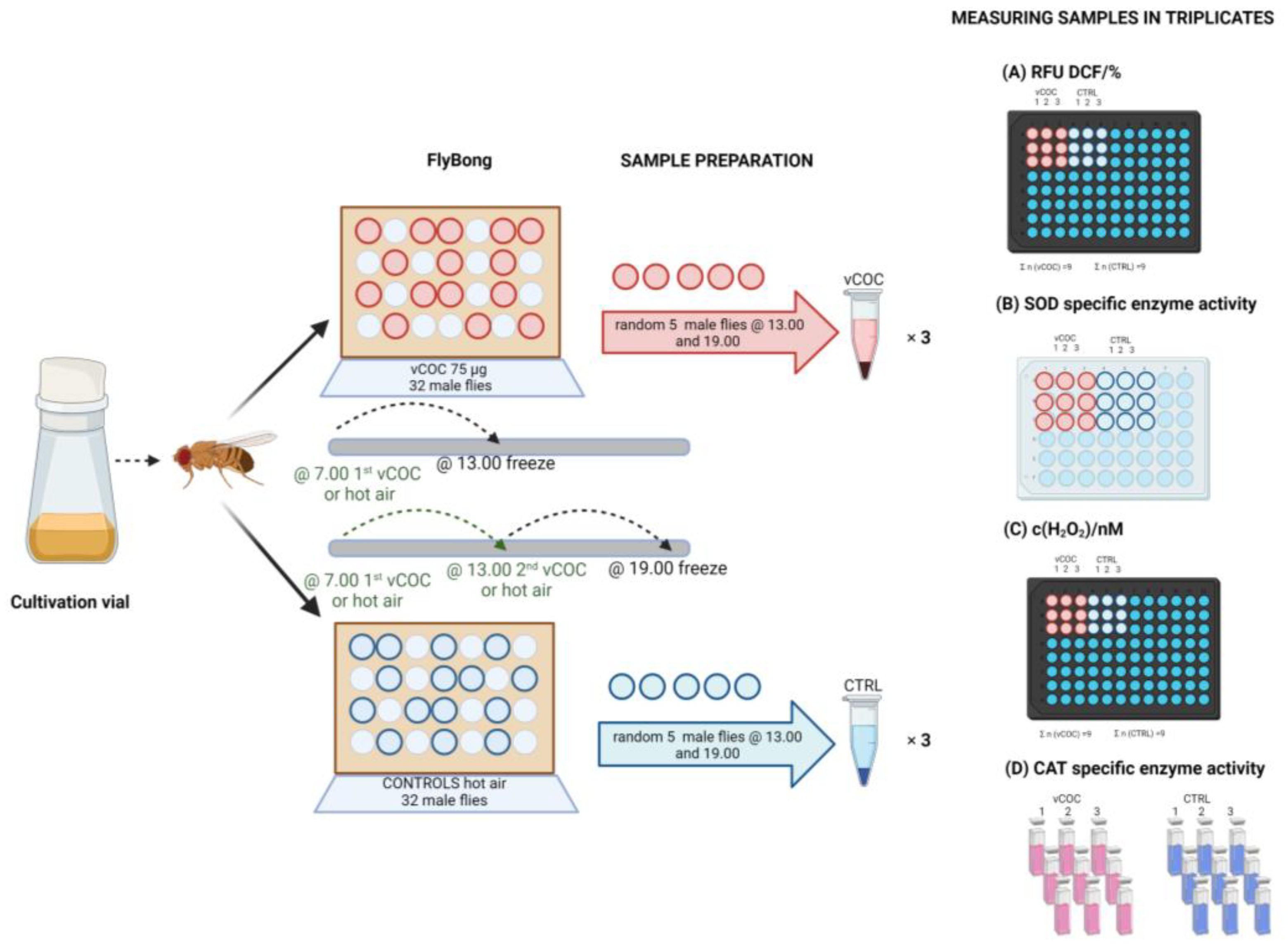
Figure 1. Whole body sample preparation and biochemical measurement flowchart. wt males were exposed to hot air (control) or vCOC using the FlyBong (described in the Section 2.3). Here only a front cross-section of the vertical DAMS with single IR beam is presented. After single or double vCOC administration control and experimental samples were collected and prepared for the measurement of: (A) total ROS using fluorescent redox sensitive dye 2′,7′-dichlorofluorescein (DCF) expressed as percent of 2′,7′-dichlorofluorescein (DCF) relative fluorescence units (RFU), (B) superoxide dismutase (SOD) specific enzyme activity as % of quercetin oxidation inhibition in the presence of N,N,N′,N′-tetramethylethylenediamine (TEMED), (C) hydrogen peroxide (H2O2) concentration using calibration curve for dihydroethidium (DHE) and (D) catalase (CAT) specific enzyme activity as time depended decline in H2O2 concentration.
Antioxidants 12 00933 g001
Figure 2. Single vCOC exposure increases ROS levels and CAT enzyme activity in the whole body of wt flies. Male flies were exposed to 75 μg of volatilized cocaine (vCOC), or hot air control (CTRL) at 07:00. Six hours later we measured: (A) reactive oxygen species (ROS) as percent of 2′,7′-dichlorofluorescein (DCF) relative fluorescence units (RFU), (B) superoxide dismutase (SOD) specific enzyme activity as % of quercetin oxidation inhibition, (C) hydrogen peroxide (H2O2) concentration using calibration curve for dihydroethidium (DHE) and (D) catalase (CAT) specific enzyme activity as decline in H2O2 concentration. All measurements were performed in triplicate (n = 9). *: p < 0.05 using unpaired Student’s t-test.
Antioxidants 12 00933 g002
Figure 3. Two exposures to vCOC lead to stronger perturbation of redox balance relative to a single exposure. Male flies were exposed to 75 μg of volatilized cocaine (vCOC) at 07:00 and 13:00 or hot air, control (CTRL). The following parameters were measured in whole body homogenates six hours after the second exposure: (A) reactive oxygen species (ROS) production, (B) superoxide dismutase (SOD) specific enzyme activity, (C) hydrogen peroxide (H2O2) concentration and (D) catalase (CAT) specific enzyme activity. All measurements were performed in triplicate (n = 9). *: p < 0.05, **: p < 0.01 using unpaired Student’s t-test.
Antioxidants 12 00933 g003
Figure 4. Oral feeding of cocaine leads to an increase in CAT activity and accumulation of fAGE. Male flies were fed either 1.50 mM cocaine (COC) for 24 or 48 h or left untreated, control (CTRL). Whole body homogenates were prepared at 09:00. (A) Catalase (CAT) specific enzyme activity. (B) Fluorescent advanced end glycation products (fAGEs) concentration determined using calibration curve for standard calibrator fAGEs-BSA (bovine serum albumin). All measurements were performed in triplicate (n = 9). **: p < 0.01 using one-way ANOVA with Tukey’s multiple comparison post-hoc test.
Antioxidants 12 00933 g004
Figure 5. Oxygen and glutathione based redox modulation in dopaminergic and serotonergic (ddc) neurons influences the development of locomotor sensitization to vCOC. CAT and GCLC expression were prevented using RNAi (UAS-CAT RNAi or UAS-GCLC RNAi) constructs in all neurons or dopaminergic and serotoninergic neurons (Elav-Gal4 or ddc-Gal4). (A) Percent of individual flies that increased locomotor activity relative to baseline activity after single exposure to 75 μg vCOC at 09:00 (sensitivity). (B) Percent of individual flies that increased locomotor activity relative to baseline activity after single exposure at 09:00 and further increased activity after the second exposure at 15:00 to 75 μg vCOC (locomotor sensitization). Data show five replicates (n = 160) as average ± standard error mean (SEM). **: p < 0.01 One-way ANOVA with Tukey’s multiple comparison post-hoc test.
Antioxidants 12 00933 g005
Figure 6. QUE feeding prevents development of locomotor sensitization to vCOC. wt flies without treatment (CTRL), orally fed for 24 h with 4.80 mM quercetin (QUE), 130 mM hydrogen peroxide (H2O2), 5 mM L-3,4-dihydroxyphenylalanine (L-DA), or a combination of QUE (4.80 mM) + H2O2 (130 mM) and QUE (4.80 mM) + L-DA (5 mM). (A) Hydrogen peroxide (H2O2) concentration was measured in head homogenates at 09:00 in triplicate (n = 9). (B) Percent of individual flies that increase locomotor activity relative to baseline activity after single exposure to 75 μg vCOC at 09:00 (sensitivity). (C) Percent of individual flies that increase locomotor activity relative to baseline activity after single exposure at 09:00 and further increase activity after the second exposure at 15:00 to 75 μg vCOC (locomotor sensitization). Data show five replicates (n = 160) as average ± standard error mean (SEM). *: p < 0.05 and **: p < 0.01. One-way ANOVA with Tukey’s multiple comparison post-hoc test.
Antioxidants 12 00933 g006
Disclaimer/Publisher’s Note: The statements, opinions and data contained in all publications are solely those of the individual author(s) and contributor(s) and not of MDPI and/or the editor(s). MDPI and/or the editor(s) disclaim responsibility for any injury to people or property resulting from any ideas, methods, instructions or products referred to in the content.