Review

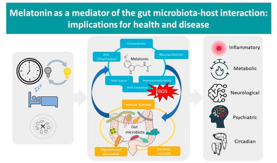
Melatonin as a Mediator of the Gut Microbiota–Host Interaction: Implications for Health and Disease

by
María-Ángeles Bonmatí-Carrión
1,2,* and
Maria-Angeles Rol
1,2
1
Chronobiology Laboratory, Department of Physiology, College of Biology, Mare Nostrum Campus, University of Murcia, Instituto Universitario de Investigación en Envejecimiento, Instituto Murciano de Investigación Biosanitaria-Arrixaca, 30100 Murcia, Spain
2
Ciber Fragilidad y Envejecimiento Saludable (CIBERFES), Instituto de Salud Carlos III, 28029 Madrid, Spain
*
Author to whom correspondence should be addressed.
Antioxidants 2024, 13(1), 34; https://doi.org/10.3390/antiox13010034
Submission received: 30 November 2023 / Revised: 20 December 2023 / Accepted: 21 December 2023 / Published: 23 December 2023
(This article belongs to the Section Health Outcomes of Antioxidants and Oxidative Stress)

Abstract

:
In recent years, the role played by melatonin on the gut microbiota has gained increasingly greater attention. Additionally, the gut microbiota has been proposed as an alternative source of melatonin, suggesting that this antioxidant indoleamine could act as a sort of messenger between the gut microbiota and the host. This review analyses the available scientific literature about possible mechanisms involved in this mediating role, highlighting its antioxidant effects and influence on this interaction. In addition, we also review the available knowledge on the effects of melatonin on gut microbiota composition, as well as its ability to alleviate dysbiosis related to sleep deprivation or chronodisruptive conditions. The melatonin–gut microbiota relationship has also been discussed in terms of its role in the development of different disorders, from inflammatory or metabolic disorders to psychiatric and neurological conditions, also considering oxidative stress and the reactive oxygen species-scavenging properties of melatonin as the main factors mediating this relationship.

Graphical Abstract

1. Introduction

Melatonin (N-acetyl-5-methoxytryptamine) is a pleiotropic molecule chemically classified as an indoleamine. It exerts multiple actions, both directly and through different receptors (MT1 and MT2, G-protein-coupled membrane receptors; MT3, cytosolic enzyme quinone reductase 2, which has potent antioxidant effects when bound to melatonin). In mammals, this hormone is produced by the pineal gland, located in the brain, as well as by a great variety of extrapineal sources, including the retina and the immune and gastrointestinal systems (reviewed in [1]).
Among the functional properties of melatonin, we can find potent antioxidant (to be reviewed in the next section), immunomodulatory and anti-inflammatory actions, as well as specific antiproliferative, antiangiogenic and proapoptotic effects in tumour cells (reviewed in [1]). Apart from these local effects, melatonin is also considered to be the most important chronobiotic hormone when released by the pineal gland into the blood circulation, sending time-of-day information and acting as a chemical signal of darkness (reviewed in [2]). This hormone presents an endogenous circadian variation, with a peak during the night and undetectable levels during the day. In addition, pineal melatonin can be acutely suppressed by light at night. Both processes are essential for this molecule to inform about the arrival of darkness at night. Although melatonin locally produced in other tissues has also shown different circadian patterns, the interplay between the pineal and extrapineal melatonin rhythms is not yet clear (reviewed in [1]) (Figure 1).
The most important extrapineal source of melatonin is precisely the gastrointestinal tract, where the concentrations can reach levels as much as 400 times greater than plasma melatonin levels [3]. Also, a healthy gut contains an enormous population of microorganisms working in close relationship with our body and physiology. Traditionally, the gut microbiota has been known to be involved in metabolic activities that result in energy and nutrient management and protection of the host against invasion by pathogenic microorganisms. However, in recent years, the gut microbiota has been shown to be involved in a myriad of functions. Indeed, dysbiosis (abnormal gut bacterial composition) has been linked to the pathogenesis of several inflammatory disorders, as well as to the emergence of certain neurological conditions and even cancer. It is now well known that changes in our physiology can affect the gut microbiota and, conversely, that changes in the gut microbiota will affect our physiology. So, knowing the particular processes involved in this interplay will be essential in order to treat and prevent a number of health issues.
In this sense, maintaining a healthy microbiome composition becomes crucial to preserving health and wellbeing. Although nutrition is thought to be the primary factor affecting the gut microbiota, other aspects may also compromise the balance between commensal and pathogenic gut bacteria. It has been shown that a lack of sleep alters melatonin levels in the gut [4] and plasma [5], as well as the gut microbiota composition [4,5,6,7,8,9,10]. It is precisely in the relationship between the gut microbiota, sleep and circadian rhythms where melatonin may play an important role in diurnal species, such as humans. Indeed, sleep deprivation and artificial light at night [11] have been demonstrated to suppress melatonin and to alter the microbiota composition, an effect that can be counteracted via the administration of exogenous melatonin [12].
Considering that melatonin has been identified in different and distant taxa of unicellular organisms, including bacteria [13], it is not surprising to find that the gut microbiota is also a source of melatonin [14] and contains melatonin receptors. This means that melatonin might also act as a signal to transfer both external and internal information between the gut microbiota and the host.
Therefore, melatonin may play a significant role in the relationship between sleep deprivation or artificial light at night, dysbiosis and those health problems that may result from alterations of the microbiota composition. It would follow that among other multiple functions of melatonin, maintenance of the microbiota composition could be another crucial role of this pleiotropic molecule.
All of this suggests that the gut microbiota might be acting through melatonin as a link between two essential aspects of health: nutrition and sleep, both framed within the circadian system. Here, we will review the current scientific evidence on the relationship between melatonin and the gut microbiota, as well as the effects of that interplay on the development of different health issues, excluding cancer, which has already been extensively reviewed in [1].

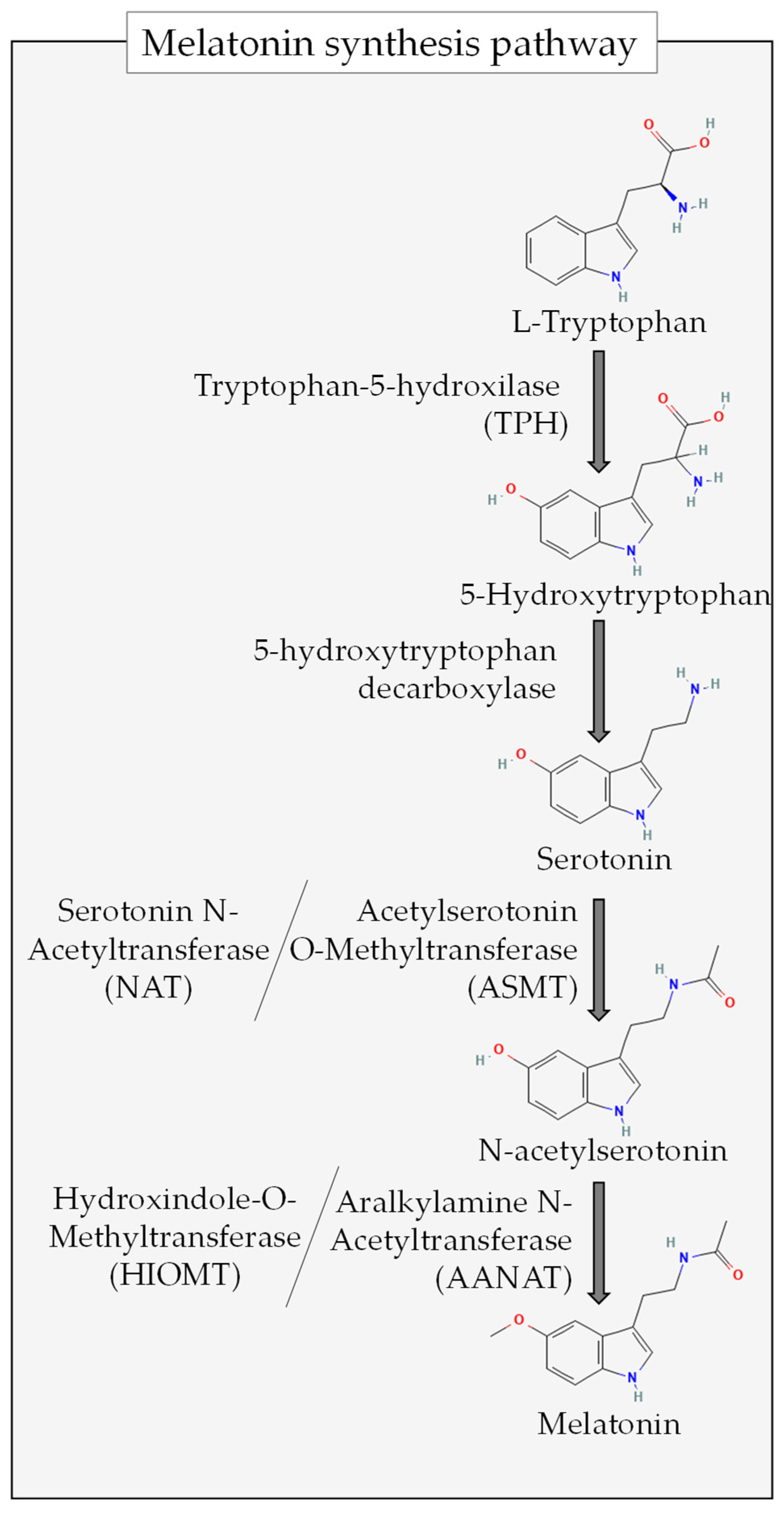
2. Antioxidant Properties of Melatonin
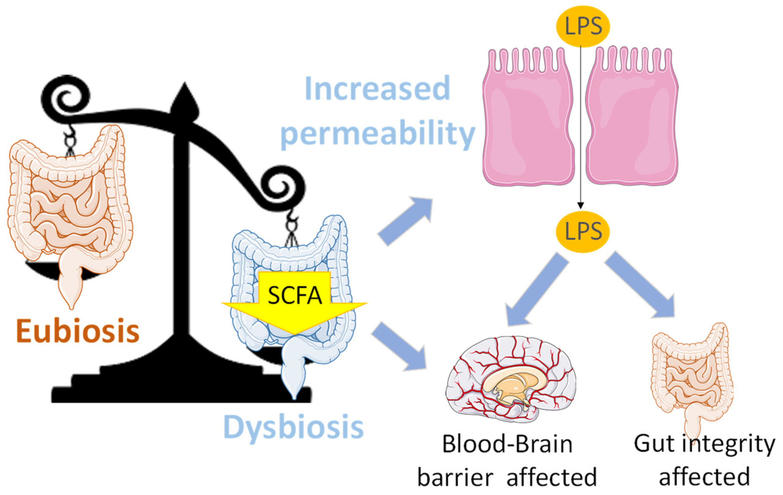
Maintaining a balance between the production and removal of free radicals is crucial for overall health, as an imbalance leads to oxidative stress. This condition results in macromolecular damage, potentially affecting the integrity of the gut barrier. Melatonin, the original function of which in unicellular organisms is speculated to be as an antioxidant, plays a protective role against oxidative stress at molecular, cellular, tissue, organ and organ system levels [15] through direct free radical scavenging and indirect modulation of antioxidant enzyme expression. Additionally, melatonin has the ability to repair oxidised biomolecules [16].
Antioxidants can be categorised based on their chemical mechanisms of action against oxidative stress. Melatonin has been proposed to be a Type IV or multifunctional antioxidant. Also, as a Type I antioxidant, melatonin can detoxify various reactive oxygen species (ROS) and reactive nitrogen species (RNS) by directly reacting with them or through the generation of less reactive species. The indole moiety of melatonin plays a crucial role in its antioxidant capacity, as it interacts with free radicals due to its resonance stability. Melatonin can scavenge multiple reactive species through a “free radical scavenging cascade”, making it a highly effective antioxidant [17]. This cascade starts once melatonin interacts with reactive species, generating intermediaries that are, in turn, free radical scavengers with different efficiencies and specificities (reviewed in [18]). As a result of this mechanism, melatonin becomes a very effective antioxidant [19,20]. These metabolites include [18] N-acetylserotonin, 5-methoxytryptamine, cyclic 3-hydroxymelatonin, N1-acetyl-N2-formyl-5-methoxykynuramine, N1-acetyl-5-methoxykynuramine, 6-hydroxymelatonin, 4-hydroxymelatonin and 2-hydroxy melatonin.
Melatonin also acts as a Type II antioxidant by quenching singlet oxygen (1O2) [21,22], chelating metal ions [23] and inhibiting lipid peroxidation and the protein damage induced by metal interactions [24,25,26]. Additionally, as a Type III antioxidant, melatonin regenerates other antioxidants, such as glutathione, ascorbic acid and Trolox, through electron transfer processes [27,28]. Melatonin can repair oxidised DNA via electron transfer [29,30] and also by enhancing DNA repair mechanisms [31,32,33,34], activating antioxidant enzymes [35,36,37,38,39,40,41] and inhibiting pro-oxidative enzymes in non-tumour cells [42,43].
These unique antioxidant properties of melatonin, including the free radical-scavenging cascade, place it as a central molecule in terms of antioxidant activity. Considering the concentrations required for these purposes, it is important to note that it is extrapineal melatonin, synthesised outside the pineal gland, which plays a significant role in antioxidant and anti-inflammatory processes. However, we cannot ignore the potential relevance of the interplay between pineal and extrapineal melatonin, which remains an important area of research to better understand the regulation of the antioxidant functions of melatonin.

3. ROS in the Gut

Reactive oxygen species (ROS) can be beneficial in low and moderate concentrations. However, when ROS levels increase, they can have detrimental effects, and their impact varies depending on the concentration and the specific tissues involved. ROS play a role in the health of the intestine and the diversity of the gut microbiota (reviewed in [44]). Maintaining the health of this organ relies to an important extent on the intestine’s ability to effectively regulate the excessive production of ROS. Indeed, the balance of gut microbiota composition is closely and bidirectionally associated with the redox equilibrium. In this sense, oxidative stress has been demonstrated to alter the gut microbiota composition in mice, increasing Bacteroidetes and decreasing Firmicutes, Clostridiales, Ruminococcaceae and Oscillospira [45]. On the other hand, the presence of gut injury and dysbiosis can lead to the suppression of antioxidant activity in the gastrointestinal tract [46]. Under such circumstances, melatonin can exert its antioxidant activity, restoring the redox balance and thereby enhancing the composition of the gut microbiota, as will be detailed in the following sections.
The regulation of the gut microbiome and systemic oxidative stress is interconnected and complex. Oxidative stress occurring in the gut can result in damage to the structural integrity of the intestinal barrier. Specifically, damage to mitochondrial DNA in intestinal epithelial cells can lead to excessive local production of ROS, contributing to mitochondrial dysfunction. This further exacerbates the increase in ROS levels and oxidative damage and, in addition, leads to a decrease in the expression of tight junction proteins (Occludin, Claudin-1 and ZO-1) and the death of intestinal epithelial cells [47].
Indeed, an important factor in the pathogenesis of gut microbes is bacterial translocation due to the compromised integrity of the intestinal barrier. The lipopolysaccharide (LPS) response serves as a significant marker signal in this process, which can activate abnormal immune signals by binding to the TLR-4 complex, subsequently increasing the production of ROS/RNS and proinflammatory cytokines [48,49]. Also, LPS can stimulate the expression of oxidative stress-related enzymes, such as inducible nitric oxide synthase [50] or NADPH oxidase, resulting in excessive RNS/ROS production and activation of the downstream NF-κB pathway [51,52]. Furthermore, specific gut microbes can induce the production of ROS in intestinal epithelial cells, and it has been shown that Lactobacillus can induce the production of ROS by intestinal phagocytes depending on the presence of NADPH oxidase 1 [53].
ROS levels in the gut are counteracted by the antioxidant activity of gut microbes, facilitated by short-chain fatty acids (SCFAs), among other metabolites. SCFAs inhibit peroxisomes and activate the Nrf2 pathway, contributing to the maintenance of redox balance in the gut [54]. Thus, gut microbiota play a crucial role in regulating this balance, which is essential for gut health and overall wellbeing.

Gut Melatonin and ROS

Melatonin acts in the gut not only by scavenging highly toxic ROS but also by upregulating different antioxidant enzymes, including glutathione peroxidase, catalase and superoxide dismutase. Additionally, melatonin downregulates pro-oxidative enzymes, further contributing to the maintenance of a favourable redox environment in the gut. Melatonin can exert its effects through direct interactions with ROS, as well as via membrane and nuclear receptors that act as mediators for its indirect antioxidant actions. Through this mechanism, melatonin activates various stress-responsive genes, such as Sirt, HIFa and AMPK, triggering an increase in the expression of multiple antioxidant enzymes [55]. Consequently, the significant impact of melatonin on the enteric microenvironment stems largely from the reduction in oxidative stress exerted on the gut microbiota through the various pathways mediated by melatonin.
As examples that confirm this evidence, exposure to imidacloprid (a neonicotinoid insecticide) has been shown to induce gut toxicity in zebrafish via oxidative stress, concomitantly with a decrease in melatonin and serotonin levels. In this study, the prolonged darkness that increased melatonin levels also attenuated said toxicity [56]. Melatonin administration can also ameliorate skin oxidative stress indirectly through the regulation of propionic acid as observed in a model of sleep-restricted mice [57]. Also in sleep-restricted mice, melatonin has been shown to prevent intestinal dysbiosis by palliating oxidative stress [5,6,8,9]. In mice with DSS-induced colitis, melatonin has also been demonstrated to improve oxidative stress resistance while regulating the gut microbiota and improving overall intestinal health [58].

4. Melatonin Effects on Gut Microbiota Composition

The gastrointestinal tract has an intimate relation with gut microbiota, and it is well known that keeping this dynamic population of microorganisms in good shape contributes to maintaining immune status and metabolic homeostasis. The gut microbiota also helps to defend the host against pathogens, reducing the probability of infections through the intestinal barrier. In this sense, and as reviewed below, both host and exogenous melatonin have been shown to have an impact on the gut microbiota, producing compositional, metabolic and circadian changes.
Two characteristics of the microbial gut microbiota are (i) its abundance (more than 100 trillion microorganisms, 1011–1012 per millilitre) and (ii) its diversity. To date, the Human Microbiome Project and MetaHit data have presented the most complete picture of the human-associated microbial repertoire [59,60]. These studies made it possible to identify 2172 human-isolated species that belong to 12 phyla, with 93.5% corresponding to Proteobacteria, Firmicutes, Actinobacteria and Bacteroidetes. One of these publications [59] also included a comprehensive inventory of the functional potential of the human gut microbiome, finding 9,879,896 genes that present a high level of redundancy.
Despite the high degree of redundancy found in gut microbial gene expression, an imbalance in this community can lead to health impairment. This alteration in microbiota composition is known as “dysbiosis” and has been linked to a variety of diseases, such as inflammatory bowel disease (IBD), metabolic disorders (e.g., obesity or diabetes), allergies, cancer and neurologic conditions, such as autism spectrum disorders (reviewed in [61]). Given the importance of maintaining the gut microbiota composition and preventing/reverting dysbiosis, the factors involved in these processes have recently been gaining attention.
Effects of exogenous melatonin
Although the exact mechanism has not yet been unravelled, exogenous melatonin administration causes different changes in gut microbiota composition (see Table 1). Indeed, melatonin has been shown to reduce the Firmicutes/Bacteroidetes ratio (which is considered an indicator of intestinal homeostasis; decreased Firmicutes/Bacteroidetes ratios are associated with dysbiosis) and also to increase the abundance of Akkermansia, a mucin-degrading bacteria related to a healthy mucosa. Exogenous melatonin has been demonstrated to revert 14 of the 69 operational taxonomic units (OTUs) altered in mice fed a high-fat diet, including Desulfovibrionaceae, Bacteroides, Ruminococcaceae, Helicobacteraceae, Porphyromonadaceae and Christensenellaceae [62]. Other authors found that melatonin had the effect of increasing Clostridiales, Bacteroidales and Enterobacteriales [63], Alistipes [64] and Bacteroidetes [65] and decreasing Firmicutes in rats [63] and mice [64,65] fed a high-fat diet.
Ren and colleagues (2018) [12] explored the effects of melatonin supplementation in mice suffering from weanling stress, finding that 0.2 mg/mL for 2 weeks was enough to influence the gut microbiota composition and to increase its richness indices, with an increase in the abundance of Lactobacillus, while reducing the load of enterotoxigenic Escherichia coli [12,66]. Similar effects were reported by Jing and colleagues in 2019, who demonstrated an increase in the relative abundance of Lactobacillales and Lactobacillus after injecting melatonin in a mouse model of spinal cord injury suffering from the consequent dysbiosis. In this case, the relative abundance of Clostridiales was reduced by the melatonin treatment [67]. Other studies that found a protective effect of melatonin in colitis symptoms discovered that although its administration reduced the variety and richness of the gut microbiota, it also increased the amount of the probiotic Bifidobacterium and at the same time decreased the relative abundance of pathogenic bacterial taxa, such as Desulfovibrio, Peptococcaceae and Lachnospiraceae [68]. In suckling piglets, melatonin has been shown to increase the relative abundance of Actinobacteria while reducing Selenomonadales [69].
Table 1. Effects of melatonin on gut microbiota composition.
StudyAnimal ModelTreatment to Alter Gut Microbiota CompositionMelatonin Treatment/Intervention (Route, Dosage, Time Period)/
Measurement
Effects of Melatonin Treatment
Xu et al., 2017 [62]MouseHigh-fat dietBy gavage
50 mg/kg body
weight
Once daily, 10 weeks
Revert 14/69 OTUs altered
Firmicutes/Bacteroidetes
Akkermansia
Yildirim et al., 2019 [63]RatHigh-fat dietIn drinking water
4 mg/kg per day
2 weeks
Clostridiales
Bacteroidales
Enterobacteriales
Yin et al., 2018 [64]MouseHigh-fat dietIn drinking water
0.4 mL/mL
2 weeks
Bacteroides
Alistipes
Ren et al., 2018 [12]MouseWeanling mouse modelIn drinking water
0.2 mg/mL
2 weeks
↑ Richness
Lactobacillus
Escherichia coli
Jing et al., 2019 [67]MouseMouse model of spinal cord injuryIntraperitoneal injection
10 mg/kg
Twice a day
Lactobacillales
Lactobacillus
Clostridiales
Li et al., 2021 [70]SheepBrucella infectionOverexpression of ASMT in transgenic sheep↓ Abundance of microbes related to infectious diseases
Zhang et al., 2022 [71]Mouse-Endogenous melatonin reduction
(Aanat-knockout (Aanat−/−))
Microbiota dysbiosis
↑ Gut permeability
↑ Systemic inflammation
Zhao et al., 2022 [68]MouseInduced colitis (oxazolone)50 mg/kg body weight
By gavage
1 week before induction of colitis
Bifidobacterium
Desulfovibrio
Peptococcaceae
Lachnospiraceae
Xia et al., 2022 [69]Sucking pigletsHealthy10 mL oral melatonin solution (1 mg/mL)
21 days
Actinobacteria
Selenomonadales
Ouyang et al., 2021 [72]Lactating cowsHealthyRuminal melatonin
In vitro
(+) Muribaculaceae,
Succinivibrionaceae, Rikenellaceae,
unidentified Cyanobacteria, Defluviitaleaceae, Veillonellaceae, Spirochaetaceae and Prevotellaceae
Prevotellaceae
Muribaculaceae
Succinivibrionaceae
Veillonellaceae
Yin et al., 2020 [65]MouseHigh-fat diet Bacteroidetes
Firmicutes
Manipulated endogenous melatonin production
Another approach to studying the possible effects of melatonin on gut microbiota composition is through the use of transgenic animals over- or underexpressing limiting enzymes for the synthesis of melatonin (e.g., aASMT, Figure 2). The overexpression of these enzymes has been shown to reduce the abundance of microbes related to infectious diseases in transgenic sheep [70]. In contrast, the models with endogenous melatonin reduction (EMR) suffer from microbiota dysbiosis, together with systemic inflammation and increased gut permeability [71].

5. Effects of Melatonin on Rhythmic Variations of Microbiota

The gut microbiota imbalance, known as dysbiosis, should not be confused with the normal variation of gut microbiota composition, considering that this microbial population is not an isolated and static bacterial community. Instead, its composition and metabolism depend on different factors. Some of these factors are exogenous, such as diet; some are endogenous, such as host genetics; and some have an exogenous component mediated by endogenous physiological processes. An example of the latter is the light–dark cycle or photoperiod, the effects of which are mediated by melatonin secreted in a circadian manner by the pineal gland. Thus, the gut microbiota shows a daily metabolic rhythmicity, partly influenced by the host circadian system, which is also affected by the rhythmic variations of microbiota in a bidirectional way. The physiology of a healthy gut microbiota includes this rhythmicity, with daily variations in community populations as well as circadian variations in their functional and metabolite expression [73].
Although the evidence so far is limited, some results support the potential beneficial effect of melatonin supplementation in restoring the lost rhythmicity of Firmicutes under a high-fat diet [65], for example. This study includes a detailed analysis of the rhythmicity exhibited by different phyla and genera under an HFD and melatonin supplementation, with varying results.
Another interesting area of study is endogenous melatonin of extrapineal origin. Although the information is limited, the role of melatonin produced by the gastrointestinal tract (which can have a concentration of up to 400 times that of melatonin from pineal origin) has been investigated in ruminants by Ouyang et al. (2021). They found that melatonin in the rumen fluid oscillates in a diurnal manner and so does the relative abundance of 9% of total rumen bacterial OTUs. Furthermore, ruminal melatonin seems to be positively correlated with Prevotellaceae, Muribaculaceae, Veillonellaceae and Succinivibrionaceae. Apart from this in vivo experiment, these authors also confirmed the effect of melatonin on bacterial composition with an in vitro assay, finding that melatonin can increase the relative abundance of Muribaculaceae and Prevotellaceae while reducing that of Veillonellaceae and Succinivibrionaceae. According to these results, ruminant microbiota seems to maintain a circadian rhythm associated with melatonin profiles [72].

6. Effects of Melatonin on Other Aspects of Gut Microbiota

Composition or rhythmicity are not the only aspects of the gut microbiota to be affected by melatonin. In fact, in vitro swarming and motility of Enterobacter aerogenes can be influenced by melatonin (1 nM), which also produces a synchronising effect of this rhythmic behaviour. This effect might be mediated by the presence of melatonin receptors found in this prokaryote [74]. Although Klebsiella pneumoniae and Escherichia coli do not seem to be sensitive to the effect of this indoleamine [74], the latter has also shown a dose-dependent repellent response to melatonin (<0.1 mM) [66].
Short-chain fatty acids (SCFAs), the main metabolites produced by the gut microbiota, exert many different and relevant functions in our body. They are produced by anaerobic fermentation of indigestible polysaccharides (e.g., dietary fibre or resistant starch) [75], and their presence in plasma also exhibits a diurnal rhythm that is disrupted under chronodisruptive conditions, such as night shift work [76].

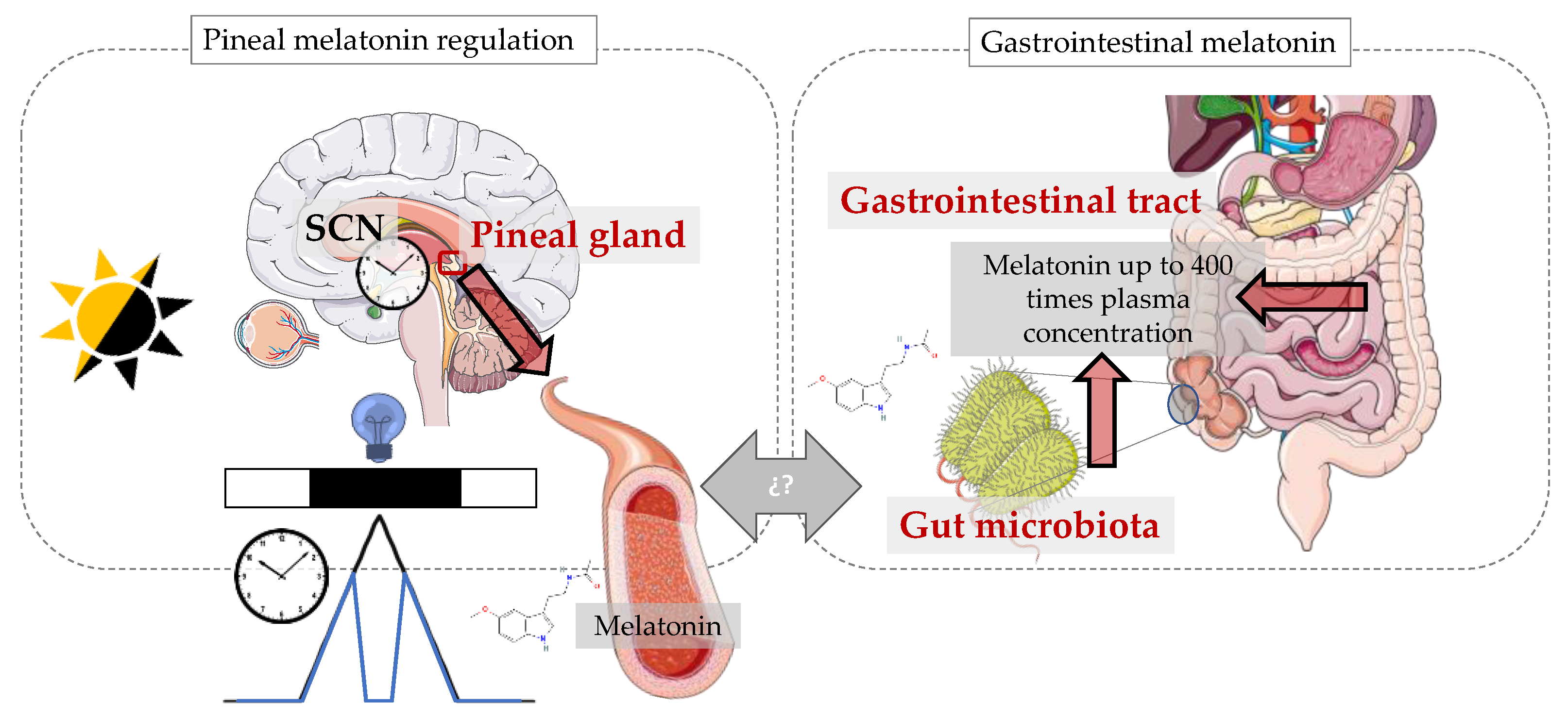
7. Gut Microbiota as an Extrapineal Source of Melatonin

Some of the demonstrated effects of melatonin on the gut microbiota have been described in previous sections. However, the relationship between melatonin and the gut microbiota has been shown to be bidirectional, so this population of microbes can also affect melatonin levels in the host. Indeed, since microorganisms can produce large amounts of melatonin [13,77], intuitively, we may infer that the intestinal microbiota could be another source of melatonin [14].
One approach to demonstrating the ability of the gut microbiota to produce melatonin has consisted of injecting a precursor of the indoleamine (5-Hydroxytryptophan, 5-HTP) into the gastrointestinal tract and evaluating whether melatonin increases at different levels (see Figure 2 for melatonin synthesis pathway). Pan et al. (2021) identified the highest concentrations of the hormone in the colon, which suggested that the microbiota present at that level of the gastrointestinal tract may participate in melatonin production. These authors also demonstrated that melatonin produced in the gut can also reach the bloodstream via the portal vein, since sheep injected with 5-HTP also exhibited higher plasma melatonin concentrations [78]. However, considering that the possible interplay between melatonin from pineal and extrapineal sources has not yet been elucidated, this statement might be considered questionable.
Another indirect method for detecting possible actions of the gut microbiota on the host’s melatonin levels is through the effects of microbial metabolites. In this sense, butyrate (an SCFA produced by gut microbiota) has been shown to induce N-acetylserotonin and melatonin synthesis in the gut through the induction of aralkylamine N-acetyltransferase (AANAT) and acetylserotonin O-methyltransferase (HIOMT) (two limiting enzymes for melatonin production; see Figure 2), suggesting that a possible effect of this SCFA might be mediated via its induction of the melatonergic pathway, not only in the microbiota itself but also in (e.g.,) immune cells [79]. Also, the induction of this indoleamine through butyrate may exert feedback on the gut microbiota through the mechanisms described in Section 3.
Another way in which microbiota might influence the effects of melatonin on the host is by inducing the expression of melatonin receptors. In this sense, Wang et al. demonstrated in 2019 that gut microbiota is able to promote melatonin receptor expression in the host’s colonic cells, and they suggested that SCFAs may be relevant actors in the mechanism involved [80].

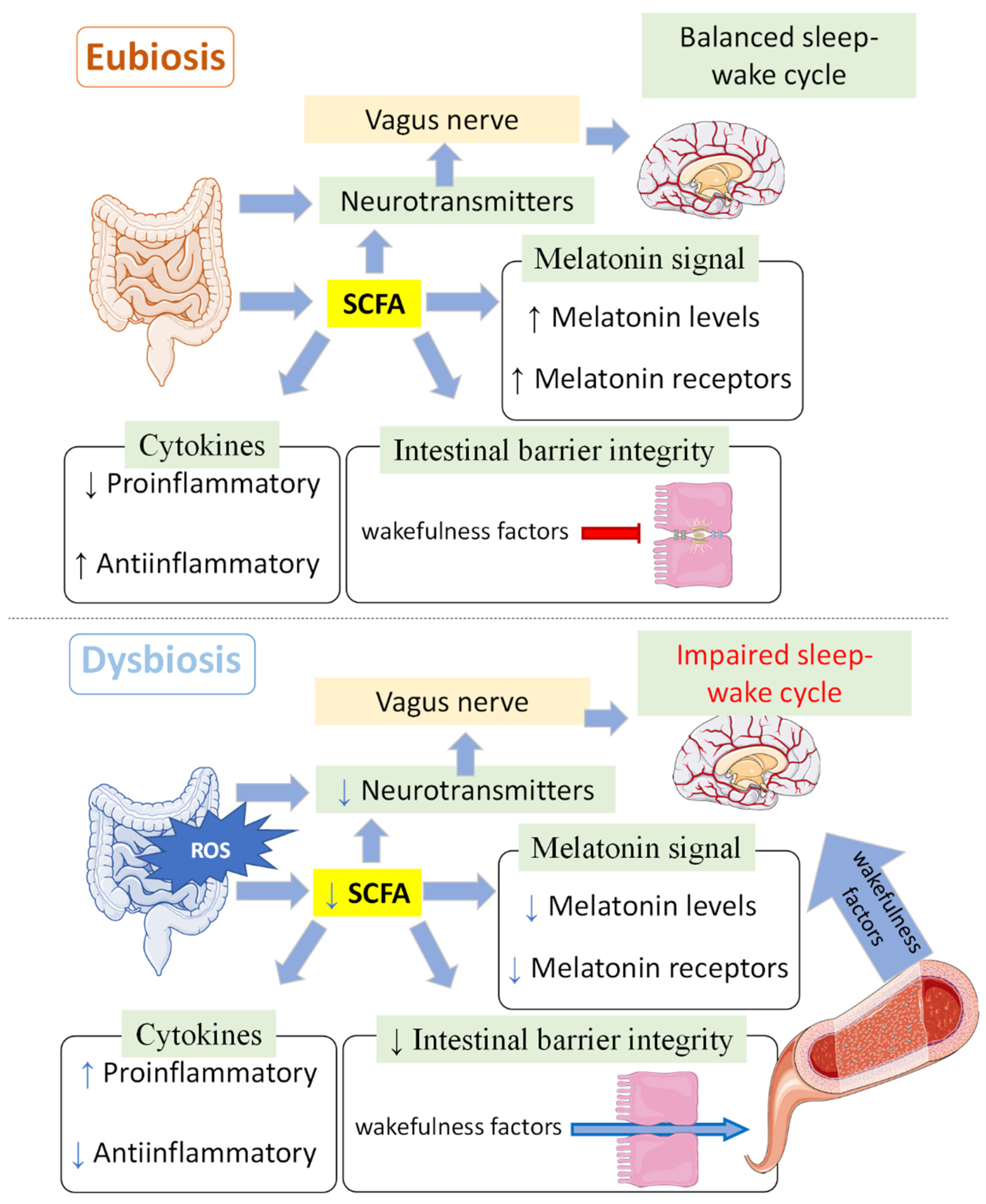
8. Sleep, Melatonin and Microbiota

There is increasing evidence that suggests that sleep deprivation produces gut microbiota disorders and that melatonin might be a possible mediator between the two processes [4,5,6,7,8,9,10] (Table 2).
In 2019, Gao et al. [6] demonstrated that 72 h sleep deprivation can produce colitis in mice, with a reduction in the diversity and richness of the gut microbiota, especially decreasing probiotics such as Akkermansia, Bacteroides and Faecalibacterium while increasing the pathogen Aeromonas. Concomitant with these changes, an increase in nor-epinephrine and a reduction in plasma melatonin were detected, with the consequent reduction in antioxidant ability and down- and upregulation of anti-inflammatory and proinflammatory cytokines, respectively, which led to colonic mucosal injury. Supplementation with 20 and 40 mg/kg of melatonin was able to reverse these changes, palliating mucosal injury and dysbiosis.
These authors [5] also reported that melatonin supplementation increased plasma melatonin concentration and OTUs, improving the diversity and richness of jejunal microbiota by increasing the presence of Bacteroidaceae, Prevotellaceae, Moraxellaceae and Aeromonadaceae and increasing the Firmicutes/Bacteroidetes ratio. Melatonin also increased anti-inflammatory cytokines (IL-22), decreasing proinflammatory cytokines (IL-17) and ROS, which resulted in a reduction in inflammation and oxidative stress. Similarly, a protocol of 20 h of sleep deprivation during 48 days in mice produced a reduction in plasma melatonin (48.91%), antioxidant enzymes (and total antioxidant capacity in intestinal tissues) and anti-inflammatory cytokines (IL10 and IFNγ). This protocol also produced an increase in glucose, norepinephrine, corticosterone and proinflammatory cytokines (IL6 and TNFα). However, it also increased α-diversity by increasing Helicobacter and Clostridium and reducing Bacteroidetes and Lactobacillus. Again, melatonin supplementation was able to restore these phenotypes [9]. These authors also described the possible role of corticosterone, which is increased in sleep-deprived mice, in mediating the colitogenic effect of sleep deprivation through microbiota. Indeed, the beneficial effects of melatonin might be related to its weakening action on glucocorticoid receptor feedback through the MT2 receptor [8].
These authors went a step further and found that sleep-deprived mice showed gut microbiota disorders and mucosa injuries, together with a decrease in plasma melatonin concentration, Card9 expression and Faecalibacterium, with the consequent reduction in butyrate levels. Melatonin supplementation, however, reversed all of these effects produced by sleep deprivation. The fact that the colitis induced in healthy (non-sleep-deprived) mice via faecal transplantation from sleep-deprived animals was reverted through the supplementation of butyrate, with no change in melatonin concentration, demonstrates that melatonin should be acting in mucosal integrity through the restoration of the microbiota [7].
The role of the interactions between Aeromonas and goblet cells has been also explored in mice, finding that Aeromonas veronii and LPS supplementation (naturally increased in sleep-deprived animals) can mimic the reduced number of goblet cells and mucin protein produced during sleep deprivation, while melatonin is capable of reversing these effects, probably through MT2 receptors [10].
Park and colleagues [4] also demonstrated the interplay between sleep deprivation, melatonin and microbiota in mice, revealing a decrease in melatonin levels—this time measured in faeces and the colon—concomitant with a shift to a colitogenic microbiota (increasing Erysipelotrichales and Enterobacteriales and decreasing Lactobacillales). This microbiota shift was reverted through melatonin supplementation, with an increased presence of Akkermansia muciniphila and Lactobacillus, while Bacteroides massiliensis and Erysipelotrichaceae were reduced.
However, the exact mechanism involved in the relationship between microbiota and sleep homeostasis and the role of melatonin has yet to be determined (Figure 3). Considering that most of the sleep deprivation protocols produce an impairment of antioxidant ability, an effect that can be reverted via melatonin administration, the antioxidant properties of this molecule cannot be excluded from this possible mechanism. Indeed, various studies have examined the beneficial impact of administering melatonin on oxidative status following the implementation of diverse sleep deprivation protocols [5,6,9].
In this sense, most of the physiological conditions and pathologies leading to poor sleep quality have been related to enhanced oxidative stress. Sleep deprivation has been demonstrated to increase oxidative stress either by increasing ROS generation or by decreasing antioxidant capacity [81,82,83,84,85]. Not only deprivation but also sleep fragmentation can be responsible for an increase in oxidative stress [86]. Pathologies like insomnia [87,88] or conditions like circadian disruption [89], a widely accepted cause for poor sleep quality, have been related to higher levels of oxidative stress. Therefore, and considering diurnal animals such as humans (where melatonin serves not only as a chronobiotic but also as a sleep inducer [90,91,92]), it becomes plausible that the potential positive impact of melatonin on sleep quality [93] could extend to improvements in microbiota status. This amelioration may be mediated by the enhancement of oxidative status, suggesting a multifaceted role for melatonin in promoting overall health.
On the other hand, healthy microbiota will maintain the production of neurotransmitters, both directly and indirectly, through SCFA production, which will activate enterochromaffin cells, which in turn secrete different neurotransmitters [94]. However, most neurotransmitters cannot pass the intestinal and the blood–brain barriers [95], so those molecules produced in the intestine will act through the vagus nerve, which contains receptors and can send the signal to the brain [96]. The melatonin signal will also be enhanced not only by direct melatonin production but also through the upregulation of its receptors mediated by SCFAs [80]. SCFAs are also immunomodulators that reduce proinflammatory cytokines and increase anti-inflammatory cytokines [75,97,98,99], which, together with the better union between epithelial cells [98,100,101], will prevent different wakefulness factors from being included in systemic circulation. When the gut microbiota composition/homeostasis is disrupted (dysbiosis), the reduction in SCFAs, together with the increase in lipopolysaccharides, will produce an increase in proinflammatory cytokines and will make both the blood–brain barrier and the intestinal barrier more permeable, favouring the passage of these molecules to systemic circulation [101], activating the hypothalamic–pituitary–adrenal axis and producing wakefulness neurotransmitters/hormones (e.g., norepinephrine, cortisol) [96,101,102].
At this point, it should be mentioned that most of the publications linking sleep deprivation, melatonin suppression and gut microbiota disorders have focused on mice, a nocturnal animal model, so it could be of interest to extrapolate these results to humans as diurnal animals.
To the best of our knowledge, there is not much research on humans that directly evaluates sleep, melatonin and microbiota composition in a polyhedral manner; however, there are some studies that attribute sleep benefits to the consumption of pro- (live microorganisms), pre- (foods that promote the growth of beneficial bacteria in the gut) and postbiotics (bioactive compounds resulting from the digestion of prebiotics by gut microbiota). Although there is some controversy, most of these studies show that the consumption of pre-, pro- and postbiotics improves sleep quality in humans (extensively reviewed in [103]).

9. Circadian System, Melatonin and Gut Microbiota

Sleep is regulated through two different types of processes, homeostatic and circadian, and it is not in vain that the sleep–wake cycle is considered to be one of the major outputs of the circadian system, also acting as a zeitgeber (zeitnemer, since it can act as a circadian input and output). Therefore, the circadian system must be highly involved in the interplay between sleep, melatonin and gut microbiota. Firstly, SCFAs show day/night fluctuations in mice, with increased levels during the active periods [104]. Furthermore, although the literature is limited, SCFAs have been demonstrated to synchronise Per2/Bmal1 (two core pieces of the molecular clock) in hepatocytes [105] and to produce phase advance in all clock genes in the kidney and in Per2 and Bmal1 in submandibular gland cells when administered in the middle of the day [104], also inhibiting histone deacetylation [75]. Therefore, SCFAs, the main metabolites of gut microbiota, could act as a signal on the circadian clock.
Unconjugated and secondary bile acids have been shown to promote circadian rhythmicity by increasing Per and Cry expression in the ileum and colon [106,107]. Interestingly, the bacteria present in healthy gut microbiota (e.g., Lactobacillus, Bifidobacterium, Clostridium and Enterococcus) produce bile salt hydrolase (BSH), which in turn will deconjugate bile acids, allowing them to exert their beneficial effects on the circadian system [106]. Sleep disruption, on the other hand, will reduce the presence of Lactobacillus and Bifidobacterium and, therefore, the production of BSH [108].
Influence of circadian cues on microbiota composition and effects on (and of) melatonin
Although it would require a more complex experimental design and the conclusions are rather speculative, some authors have tried to assess the possible effects of endogenous melatonin through the manipulation of environmental conditions (Table 3).
The light/dark cycle, produced by the alternation of day and night driven by the Earth’s rotational movement, is considered to be the main input of the circadian system. Melatonin produced by the pineal gland—mainly at night and in darkness—is closely tied to this cycle, presenting an endogenous rhythm driven by the suprachiasmatic nuclei (central pacemaker), which is also synchronised, thanks to this cycle. Apart from this endogenous rhythmicity, melatonin secretion is also acutely suppressed by light at night (Figure 1). The sleep-promoting effects of melatonin are restricted to diurnal animals, so its suppression in nocturnal animals related to sleep deprivation may cause different downstream effects.
With this in mind, different studies on nocturnal animal models have pointed out the obesogenic effect of light at night, precisely mediated by melatonin suppression and its deleterious effects on microbiota composition (Table 3). Namely, this disruptive environmental condition has been demonstrated to increase weight, insulin resistance and lipid influx and to produce gut microbiota dysbiosis in mice, specifically by inducing a reduction in the richness of Blautia, Ruminiclostridium, Lachnospiraceae, Lactobacillus, Eubacterium, Roseburia and Bacteroides (all negatively correlated with obesity). Melatonin supplementation was effective in improving circadian rhythm homeostasis and reverting these relative abundances while decreasing the abundance of Anaerotruncus, Alloprevotella and Faecalibaculum [11], which are related to obesity in mice [1,11]. Other authors have investigated the effect of different photoperiods on gut microbiota, finding a protective effect of 16L:8D (16 h of light, 8 h of darkness), probably mediated by the appropriate melatonin secretion and enhanced expression of melatonin receptors in laying ducks [109].
Similar results were found in jet-lag-induced mice, with an increase in lipid uptake and fat accumulation in the white adipose tissue as well as in the presence of Escherichia coli and LPS, concomitant with a reduction in angiopoietin-like 4. Oral melatonin supplementation reversed these phenotypes, probably through gut microbiota, since the treatment was not successful in microbiota-depleted animals [110].
Seasonal changes in photoperiod seem to be also related to seasonal changes in microbiota composition. In order to explore the factors involved in this apparent relationship, Shor and colleagues [111] divided pinealectomised and intact hamsters into long- or short-photoperiod groups. They found that pinealectomised animals placed on a short-day photoperiod had significantly more Prevotella (associated with improved glucose tolerance [112], decreased adiposity [113] and inflammatory responses [114,115]) and Clostridium and a lower abundance of Desulfovibrio as compared to animals with an intact pineal gland. The genus Hungatella (related to carbohydrate metabolism and energy harvest) [116], however, was enriched in pinealectomised animals under long-day photoperiods. Although these authors attribute these effects to melatonin from the pineal gland, considering the possible interplay between both local gut melatonin and pineal melatonin, indirect regulation of the former cannot be ruled out.
Feeding can also be a powerful circadian cue, especially for peripheral oscillators. In this sense, Wang et al. (2021) recently explored the effects on microbiota of restricting feeding in piglets (diurnal animals) to day or night. Although a causal relationship between melatonin and microbiota effects cannot be established, the authors found reduced concentrations of melatonin in the night-time-restricted feeding group, as well as an alteration in the diurnal rhythm and composition of the gut microbiota, with increased log ratios of Catenibacterium:Butyrivibrio and Streptococcus:Butyrivibrio [117], demonstrating that nocturnal fasting has a potentially protective effect on diurnal animals.
Table 3. Circadian cues and their effects on microbiota.
StudyCircadian CueAnimal ModelEffects on Gut MicrobiotaOther EffectsEffects of Melatonin Treatment
Hong et al., 2020
[11]
Constant lightMouseBlautia
Ruminiclostridium
Lachnospiraceae
Lactobacillus
Eubacterium
Roseburia
Bacteroides
↑ Weight
↑ Insulin resistance
↑ Lipid influx
Reversion of dysbiosis and
Anaerotruncus
Alloprevotella
Faecalibaculum
Cui et al., 2022 [109]Long/short photoperiodsLaying ducks
(diurnal)
20 Light:4 Darkness
↓ Microbiota α-diversity
16 Light:8 Darkness
Relative abundance of:
= Actinobacteria
= Fusobacteria
= Proteobacteria
= Fusobacterium
= Clostridium_sensu_stricto_1
= Pectobacterium
16 Light:8 Darkness
↑ Acetate
↑ Propionate,
↑ Butyrate and
↑ Total SCFA
(Ileal chyme)
↓ Melatonin with increasing photoperiods.
-
Short et al., 2020
[111]
Long/short photoperiodsHamsterPinealectomised, short-day photoperiod (compared to sham-pinealectomised)
Prevotella
Clostridium
Desulfovibrio
Pinealectomised, long-day photoperiod (compared to sham-pinealectomised)
Hungatella
(Treatments did not uniformly affect OTU abundances—see paper for details).
In the presence of the pineal gland, animals with short-day photoperiods lost more weight than those with long-day photoperiods-
Rong et al., 2021 [110]Induced jet lagMouseEscherichia coli (↑ LPS)↑ Lipid uptake
↑ Fat accumulation in white adipose tissue
↓ Angiopoietin-like 4
Reversed those phenotypes through gut microbiota (only in non-microbiota-depleted animals).
Wang et al., 2021
[117]
Feeding time
night-restricted
Piglets (diurnal)Night-restricted
↑ Log Catenibacterium/Butyrivibrio
Log Streptococcus/Butyrivibrio
Night-restricted
↓ Melatonin (day and night)
↓ Ghrelin
↓ Dopamine
↓ Serotonin
-
“=” symbol means appropriate balance; “↑” means increase; “↓” means reduction.

10. Melatonin, Gut Microbiota and Disease

10.1. Inflammatory Bowel Disease

Chronic and recurring intestinal inflammation is a hallmark of inflammatory bowel disease (IBD), which is a catch-all name for ulcerative colitis (UC) and Crohn’s disease (CD). In 2015, IBD was estimated to affect around 3 million individuals in the United States, and its incidence and prevalence have grown globally over the past few years [118]. Since it is a complex illness with an unknown pathogenesis, it is difficult to completely treat IBD. The intricate interaction of genetic, immunologic, microbiological and environmental variables that lead to IBD makes it challenging to treat this pathology.
It is well known, however, that the gut microbiota is very much implicated in this disease, since alterations in composition and metabolism can lead to histological and anatomical effects on the gastrointestinal epithelium. Indeed, recent studies have reported that disturbances in the microbiota–host relationship are linked to IBD [119,120]. In healthy conditions, the immune system eliminates pathogenic bacteria while tolerating habitual gut microbiota [121]. If this balance is disrupted, the host’s immune response will be triggered, initiating a diseased status [122,123]. The increase in harmful bacteria [124] and the secretion of enterotoxins in the gastrointestinal tract can damage the intestinal mucosal barrier and lead to the production of inflammatory factors, which eventually will cause intestinal inflammation and immune dysfunction [123]. Faecal microbiota therapy and its beneficial effect have confirmed the essential role of the gut microbiota in IBD [125,126]. In addition, the reduced diversity of beneficial bacteria and alterations in the microbiota composition typical of these patients have been linked to an increase in ROS production and a compromised defence system in the intestinal mucosa [46]. In this sense, several studies have demonstrated that in IBD, chronic intestinal inflammation is associated with the overproduction of ROS [127,128,129]. Thus, it is not surprising that melatonin administration could be beneficial with regard to its antioxidant properties, as indicated below.
Although colitis/IBD can be triggered by sleep deprivation (as discussed in Section 8), melatonin has been proven to alleviate abdominal pain in irritable bowel syndrome patients independently of its actions on sleep [130,131]. Whether these effects of melatonin are mediated by its actions on the gut microbiota composition or other aspects of metabolism is a quite recently opened field of research. Here, we include some recent studies in which melatonin administration has been proven to alleviate colitis symptoms while affecting microbiota composition (Table 4).
The effects of melatonin on colitis have also been explored in chemically induced models of this disease. Zhu et al. (2018) showed that melatonin was effective in improving antioxidant capability in mice with colitis induced by dextran sulphate sodium (DSS), while the gut microbiota diversity, abundance and coverage did not change with the treatment. Firmicutes (e.g., Coprococcus and Ruminococcaceae) presented higher levels in mice treated with melatonin, while the untreated DSS group had more Bacteroidetes [58]. Zhao et al. (2022) explored the effects of the indoleamine in the oxazolone-induced colitis model, finding that melatonin can counteract body weight loss, colon shortening and neutrophil infiltration by suppressing the type 2 immune response. Interestingly, they suggested that these effects are mediated by the gut microbiota, whose richness and diversity at the OTU level were reduced under melatonin treatment, increasing the abundance of Bifidobacterium, a well-known probiotic, and reducing the abundance of different harmful bacterial genera, such as Lachnospiraceae, Peptococcaceae and Desulfovibrio [68].
Melatonin has also been administered using a nanotechnology-based treatment composed of hyaluronic acid (HA) aggregated with this indoleamine (MT) that has been demonstrated to accumulate in the inflamed colon epithelium of colitis mice, alleviating symptoms, repairing the damaged intestinal barrier and inhibiting colon inflammation. Regarding its effects on the microbiota, this conjugate (HA-MT) can restore the Firmicutes/Bacteroidetes ratio by improving the richness and diversity of the gut microbiota in mice with colitis. When analysing gut microbiota at the species level, the abundance of Lactobacillus increased, while that of Bacteroides, Blautia and Streptococcus was reduced [132].
But what is the exact mechanism by which melatonin exerts its effects on microbiota and colitis? This question has been recently addressed by Kim et al. (2020), who found that the anticolitic effects of this indoleamine require Toll-like receptor 4 (TLR4) signalling, since it alleviated induced colitis and reverted microbial dysbiosis only in wild-type but not in TLR4 knockout mice. In this experiment, melatonin significantly suppressed Proteobacteria (Gram-negative phylum, including Escherichia coli and Salmonella) and increased Ruminococcaceae family strains, which include butyrate-producing Gram-positive bacteria that are reduced in IBD faecal microbiota. These authors also demonstrated that melatonin induces Reg3b expression, an antimicrobial peptide, as well as goblet cell differentiation, through melatonin receptors and TLR4 signalling [133]. In any case, research has also demonstrated an improvement of the antioxidant capability after treatment with melatonin, so the antioxidant properties of this molecule cannot be discarded as a potential mechanism to alleviate colitis symptoms and dysbiosis.
In line with these results in animal models, descriptive studies in humans have found that melatonin concentrations are higher in faecal samples of IBD patients than in healthy control samples [134,135], and higher levels of melatonin have been negatively correlated with visceral hypersensitivity in patients with diarrhoea. Butyrate produced by Clostridium in IBD has been suggested to be the link for this melatonin upregulation in BON-1 cells (human neuroendocrine tumour cell lines) [135].

10.2. Melatonin, Gut Microbiota and Metabolic Disorders

Appetite and food intake are also influenced by the host circadian system, including its inputs and outputs [136]. In this sense, different studies have demonstrated that changes in the light–dark cycle can affect microbiota composition, which in turn can significantly impact appetite, among other aspects of the host physiology [137,138]. In parallel, melatonin production and the expression of melatonin receptors, also modulated by photoperiod, are linked to appetite/food intake regulation mediated by leptin [139,140,141,142,143].
It is well recognised that dietary composition and feeding timing also play a significant role in regulating microbiota composition and metabolism [144,145]. These changes in the gut microbiota seem to affect host physiological processes in different ways, including the modulation of food intake and appetite [146]. Therefore, a specific dietary adjustment, such as the use of probiotics, has the potential to modify the microbiota composition and functionality, which may ultimately have different effects on the host’s metabolism and control over food consumption. In zebrafish, probiotic administration of Lactobacillus rhamnosus seems to confirm its ability to modulate not only appetite markers but also the expression of melatonin receptors, confirming the relationship between microbial metabolism, melatonin and appetite.
On the other hand, melatonin has been detected and quantified in many different foodstuffs. Good dietary sources of melatonin include cereals, seeds and nuts [147], among which we can highlight pistachio, with 226–233 µg/g [148]. A wide range of melatonin concentrations has been also detected in fermented foods, and Saccharomyces cerevisiae seems to have a relevant role in melatonin formation (reviewed in [149]).
Melatonin taken orally is absorbed from the gut and metabolised in the liver [150]. Different studies have shown that plasma melatonin concentrations and its metabolite in urine, 6-sulphatoxymelatonin, increase after the intake of foods containing indoleamine [151,152,153]. There is also evidence concerning the typical melatonin effects after the ingestion of these foods, such as the increased total antioxidative capability of serum [152]. A field to explore is thus whether melatonin contained in food can reshape the gut microbiota or whether the concentrations detected in most food are enough to produce those effects and, also, whether the effect of different food components on the microbiota could affect melatonin production in the gut.

10.2.1. Breastfeeding, Melatonin and Microbiota

Breastfeeding is a particularly interesting situation in terms of the effects of orally administered melatonin on gut microbiota composition also because the melatonin concentration in breast milk is not static but rather varies following a circadian pattern. The typical night-time increase in melatonin secreted by the pineal gland is transferred through the breast milk to the nursing infant [154], transforming breast milk into a potent chrononutrient [155]. Breast milk acts as the first circadian stimulation when the baby’s circadian machinery is not yet entirely functional, entraining their developing circadian rhythms. Furthermore, apart from other actions regulating antioxidant activity, inflammation and immunity [155], the melatonin contained in breast milk could play a role in shaping the gut microbiota composition, richness and variation over time, contributing to the modulation of the absorption of different molecules in the host. In addition, melatonin from breast milk influences weight gain in infants, and it has been suggested that it limits the occurrence of sudden infant death syndrome [156] and the development of metabolic dysregulation and comorbidities, [154] such as cardiovascular diseases, over the long term [155]. It is also interesting to note that different factors in breast milk can act to regulate melatonin production, suggesting that the varying breast milk elements may differentially modulate the levels of gut melatonin, with its potential local actions on microbiota [157].
Additional evidence of the benefits of melatonin during this stage of life might be the fact that melatonin supplementation (provided in drinking water at a dosage of 0.2 mg/mL for 2 weeks) has been demonstrated to be useful in alleviating weanling stress in mice, improving body weight gain and intestinal morphology through increased richness of intestinal microbiota and by shaping its composition [12].

10.2.2. Obesity

The role of melatonin in regulating energy metabolism (especially through glucose and lipid metabolism regulation) has been clearly demonstrated. In terms of oxidative stress, obesity is also characterised by an imbalance between the production of ROS and the antioxidant defence systems, resulting in a condition of oxidative stress. This has been related to the promotion of insulin resistance and metabolic syndrome by disrupting the regulation of adipokines and proinflammatory cytokines. Therefore, combining therapeutic antioxidant strategies with weight loss strategies could be beneficial [158]. In this sense, melatonin, among other antioxidants, occupies a unique position due to its antioxidant (see Section 2 of this article) and anti-inflammatory properties, as well as its role as a metabolic regulator. By modulating multiple processes involved in obesity and its associated metabolic abnormalities, melatonin shows potential therapeutic value in the treatment of obesity. Considering the topic of this review, we will focus on the possible role of microbiota in mediating the effects of melatonin in obesity.
In the previous sections, we have reviewed the actions of melatonin on gut microbiota composition. Some of these modifications can contribute to alleviating different signs of metabolic disorders, such as insulin resistance, liver steatosis, weight gain and low-grade inflammation [62]. The fact that the gut microbiota shows rhythmicity under a light/dark cycle may explain why this population of microbes may act as a possible link between the circadian clock and lipid metabolism [65]. Indeed, as previously mentioned, melatonin can influence the diurnal rhythms of gut microbiota, contribute to shaping its composition and promote lipid efflux from the intestine [64,159]. Melatonin also affects energy metabolism through its interaction with microbial metabolites, particularly with SCFAs, such as butyrate. In addition, enhancing the melatonergic pathway as a result of butyrate action also demonstrates the reciprocal influence of gut bacteria and melatonin. Some effects of melatonin seem to be mediated through the alpha-7 nicotinic receptor, while both melatonin and butyrate may control obesity through the opioidergic system [159]. Melatonin has also been shown to improve lipid metabolism in high-fat diet-fed mice, probably via microbiota along a pathway that includes acetic acid production, which shows a marked correlation with the relative abundances of Alistipes and Bacteroides [64].
Regarding the possible underlying molecular mechanisms, the circadian nuclear transcription factor, interleukin-3-regulated (NFIL3), which regulates lipid absorption and export in intestinal epithelial cells and can be activated by gut microbiota, may play a role. According to previous research, orally administered melatonin reduces the amount of LPS produced by E. coli, which can decrease the transcriptional inhibition of angiopoietin-like 4 (ANGPTL4) in the ileum, induced by NFIL3 through toll-like receptor 4 (TLR4)/interleukin-22 (IL-22)/STAT3 signalling. All of this, in turn, reduces ileal lipid intake and decreases fat accumulation in epididymal white adipose tissue in mice exposed to jet lag and treated with melatonin [110].
Apart from these direct effects of melatonin on lipid metabolism through microbiota, its microbiota-mediated actions may also include an indirect effect on muscle composition and metabolism in mice, limiting skeletal muscle frailty and allowing prolonged physical performance. Indeed, the concept of the gut–muscle axis has been recently proposed based on the reciprocal relationship between muscles and microbiota composition (extensively reviewed in [160]). In this sense, the reduction in physical activity in sarcopenic individuals could be related to different microbiota compositions and metabolisms [161,162], while overly intense physical exercise may dysregulate microbiota composition, leading to inflammation and increased risk of peptic ulcers [163]. Regarding the bones, melatonin has been shown to have a beneficial effect mediated by gut microbiota (and consequent butyrate production) on induced osteolysis [164].
To summarise, gut microbiota-mediated actions of melatonin in terms of obesity involve, so far, the circadian system, microbiota metabolites and skeletal muscle.

10.2.3. Other Metabolic Alterations

Different epidemiological studies have revealed that circadian disruption might increase the risk of metabolic disorders. In parallel, circadian disruption has been demonstrated to affect gut permeability and microbial ecology [137,165,166,167,168]. In this sense, oral melatonin supplementation seems to be able to reverse the changes in gut microbial ecology induced by circadian disruption.
In nocturnal rodents, melatonin has been shown to have a general beneficial role in glucose homeostasis [169], although the mechanism behind this potential relationship remains unknown. In this sense, considering that obesity and lipid dysmetabolism are known risk factors for type 2 diabetes, the previously discussed microbiota-mediated effects of melatonin might be related to a possible beneficial effect of this molecule in glucose metabolism. Apart from this indirect effect, in their matched case–control study in humans, Huang and colleagues (2022) found an altered gut microbial composition in type 2 diabetes cases, together with lower serum melatonin levels. According to their results, the underlying mechanism for this relationship may involve Bifidobacterium- and Coprococcus-mediated Trp metabolites [169]. However, the situation is controversial, since both loss-of-function and gain-of-function melatonin receptor gene MTNR1B variants seem to impair insulin secretion and increase the risk of type 2 diabetes (reviewed in [170]).

10.3. Oxidative Stress, Gut Microbiota and Neurological Disorders: Possible Role of Melatonin

Oxidative stress exerts harmful effects primarily by causing lipid peroxidation, damaging nucleic acids and oxidising proteins, which can have consequences on various signal transduction pathways in the central nervous system. The brain is particularly vulnerable to oxidative stress due to its high oxygen demand, especially during ATP generation through the electron transport chain. Also, different regions of the brain contain high levels of iron, which further contributes to the generation of hydroxyl radicals. Additionally, the brain has a higher concentration of polyunsaturated fatty acids, which are very affected by oxidative stress, thus making it more susceptible.
Interestingly, together with oxidative stress, the gut microbiota also seems to be an important factor in the pathogenesis of neurological disorders in a bidirectional manner (reviewed in [171]). The intestinal barrier’s homeostasis is influenced by the oxidative reduction potential of the gut microbiota, which refers to its ability to acquire electrons. Additionally, the brain/central nervous system (CNS) regulates the levels of oxidative stress in the gut through the vagal cholinergic anti-inflammatory pathway [172,173,174].
The complex interactions between gut microbiota and the host can influence the oxidative state of the CNS through the production of metabolites or the modulation of other neurotransmitters or molecules, such as melatonin. These substances can reach the CNS through systemic circulation or the vagus nerve, triggering microglia activation and neuroinflammation, increasing the production of ROS and also impacting the antioxidant systems, both directly and indirectly [173,175].
Notably, oxidative stress plays a role in the pathology of various chronic brain disorders, such as Alzheimer’s or Parkinson’s diseases, multiple sclerosis and depression, among others. Although speculative, these mechanisms support the hypothesis that the gut microbiota plays a role in regulating the brain’s oxidative state. In this context, based on melatonin’s antioxidant actions, this might be a molecule of interest in the study of these pathologies, considering both prevention and treatment.
On the other hand, Gerwyn et al. recently suggested a relationship between the permeability of the brain–blood barrier (BBB) and the gut [176]. First, a reduction in SCFA producers increases intestinal permeability and inflammation by changing the distribution of junction proteins and increasing the transfer of LPS, which, in turn, affects the BBB and gut integrity. It should also be noted that SCFAs show anti-inflammatory action by decreasing the activity of macrophages, dendritic cells and T-lymphocytes. SCFAs are also known to be essential for the formation and maintenance of the BBB, since they modulate different pathways involved in the gut–brain axis (Figure 4).
In this section, we will review different neurological disorders in terms of their relationship with melatonin and the gut microbiota.

10.3.1. Migraines

Migraines, microbiota and melatonin (the “3M” tripod) are intimately intricated [177]. Anderson [178] recently suggested that the butyrate reduction observed in dysbiosis could be related to a reduction in the availability of melatonin, with the consequent relative increase in the N-acetylserotonin/melatonin ratio. This could have consequences for the heightened glutamatergic excitatory transmission in migraines. The suboptimal functioning of the mitochondria and metabolism could produce alterations in satellite glial cells and astrocytes, which would reinforce the changes in vasoregulation and nociception observed in migraines.

10.3.2. Multiple Sclerosis

Gut dysbiosis has been also proposed as a possible cause of multiple sclerosis (MS), with melatonin also being involved. As previously reviewed, gut dysbiosis has been demonstrated to lower the production of butyrate, which is a significant positive regulator of mitochondrial function and also acts by suppressing the levels and effects of ceramide, considered an important driver of multiple sclerosis pathophysiology via its effects on glial mitochondrial function and melatonin and orexin production processes. Indeed, altered regulation of the local melatonergic pathway has been suggested as an important factor for the pathophysiology of MS. According to some data, the pathophysiology of MS might be supported by changes in ceramide and mitochondrial function, particularly in glial and immune cells [179].
Melatonin also increases antimicrobial peptides, especially Reg3β, which could be useful in controlling the microbiota composition. Indeed, melatonin could exert a beneficial effect on people suffering from MS, thus presenting as a promising candidate for the treatment of this disease [180]. Also, when demyelination occurs, it has been suggested that neurons are susceptible to ROS/RNS effects [181], so, in this case, the connection microbiota–oxidative stress–melatonin might also play a role.

10.3.3. Myalgic Encephalomyelitis/Chronic Fatigue Syndrome (ME/CFS)

The interplay between microbiota composition and melatonin may also play a role in the pathophysiology of myalgic encephalomyelitis/chronic fatigue syndrome, in which dysbiosis has been described [182]. As suggested and extensively reviewed by Anderson & Males [183], decreased butyrate produced by dysbiosis would attenuate the rapid epigenetic upregulation of the μ-opioid receptor, which would impact the mitochondria and immune/glial cells. This would also produce circadian effects via the β-endorphin induced by pineal melatonin, with all of this potentially being relevant to mood and cognitive deficits. Reduced levels of μ-opioid receptors and ligands may also contribute to changes in amygdala and prefrontal cortex activity that are evident in this pathology [184], as well as to emergent depression and suppressed cognition. It is precisely these effects of dysbiosis in relation to melatonin that have also been proposed to play a role in the aetiology of borderline personality disorder. In this case, the reduction in melatonin synthesis also involves the loss of the inhibition of the oestrogen receptor alpha (ERα), potentially contributing to the dysregulating effects of cyclical oestrogen in this pathology [185].

10.3.4. Autism Spectrum Disorder

The human gut microbiota is currently considered to be an important factor in the development of autism spectrum disorder (ASD) in children. Although the authors clarify that it would not necessarily involve differences in the number of enzymes, the bacterial neurometabolic signature recently studied in ASD children has shown that the gut microbiota composition in these children presents decreased contents of bacterial genes for enzymes involved in the metabolism of compounds with neuroactive properties, such as melatonin [186]. Differences in mitochondrial function have been detected in autistic children compared to controls, which could be a cause of the observed elevated oxidative stress in these children (reviewed in [187]). In this regard, the antioxidant properties of melatonin potentially position it as a molecule involved in this pathology. Also, children with ASD and sleep disorder show decreased levels of melatonin and lower excretion rates of melatonin metabolites in urine [188,189,190,191], as well as reduced proportions of Faecalibacterium and Agathobacter, both butyrate producers. Melatonin levels have been found to be positively correlated with both genera [192]. Children with ASD have also shown a reduced abundance of both Bifidobacteria and Prevotella, which could reduce folate production [193,194,195]. Alterations in the gut microbiota also contribute to altered tryptophan metabolism, yielding increased levels of indolyl 3-acetic acid and indolyl lactate and the increased transformation of tryptophan into xanthurenic acid and quinolinic acid (catabolites of the kynurenine pathway) at the expense of kynurenic acid and, especially, melatonin [196].
Aside from the possible alterations in microbiota composition, some acetylserotonin O-methyltransferase (ASMT) variations have been associated with ASD [197,198,199]. These mutations significantly decrease the activity of the enzyme ASMT [199] (for details on the melatonin synthesis pathway; see Figure 2). This indicates that a weakened melatonin metabolism could be involved in impaired sleep in ASD [189]. It is not surprising then that individuals with ASD present significantly more impaired circadian rhythms [200,201] and sleep parameters, including sleep onset latency and sleep duration and efficiency [202,203]. Consistent with this, randomised double-blind controlled trials have shown a statistically significant benefit of melatonin for sleep in subjects with ASD as compared to a placebo [204,205,206].
Good sleep hygiene and a healthy bedtime routine instilled by behavioural training from the parents have yielded good results in improving sleep in children with ASD [207,208]. Among pharmacological treatments, melatonin shows good evidence of being effective [200,201,208,209], particularly when combined with cognitive-behavioural therapy [209]. All of these findings led to the development of a recently published hypothesis [210], which states that melatonin, circadian system functioning and microbiota are relevant factors contributing to the aetiology of ASD.

10.3.5. Bipolar Disorder

Melatonin and its effect on the microbiota have also been associated with bipolar disorder due to the seasonal pattern this disorder presents [211]. Studies on this association are currently ongoing [212].

10.3.6. Alzheimer’s and Parkinson’s Diseases

The role of oxidative stress in the pathogenesis of neurodegenerative diseases such as Alzheimer’s has been gaining attention in recent years [213,214,215,216,217]. In particular, the relationship between oxidative stress and the gut microbiota has attracted the attention of scientists as a possible factor in the pathoaetiology of these neurodegenerative disorders. In this regard, individuals suffering from Alzheimer’s disease present decreased populations of commensal bacteria, such as Bifidobacterium and Firmicutes, while an increased abundance of Escherichia, Shigella and Bacteriodetes has been detected, followed by increased inflammation and protein amyloid-beta accumulation [218]. The interaction between the gut microbiota, oxidative stress and the development of these diseases has been extensively reviewed in [171].
Although in the case of Alzheimer’s disease, there is no direct evidence, Zhang et al. [71] found that a mouse model with endogenous melatonin reduction, which shows microbiota dysbiosis and increased gut permeability (among other metabolic alterations), also presented Alzheimer’s disease-like phenotypes. These authors suggest that melatonin reduction may be a pathogenic factor for Alzheimer’s disease (as well as for obesity) via gut microbiota dysbiosis. Despite different studies having suggested the potential utility of therapeutic strategies aimed at reducing oxidative stress in neurodegenerative disorders [219,220,221], it is still to be conclusively demonstrated whether melatonin’s role in the pathogenesis of Alzheimer’s disease is specifically linked to its antioxidant properties.
The scientific evidence accumulated on the effects of dysbiosis and gut permeability and melatonin on mitochondrial functioning also suggests a potential role of both aspects of physiology in the aetiology of Parkinson’s disease and may open new avenues in terms of treatment [222,223].

10.3.7. Stress

Melatonin could alleviate colonic microbiota dysbiosis in mice induced by “restraint stress”, as well as the consequent intestinal inflammation, specifically by inhibiting the activation of the NF-κB pathway [224]. Also, patients with anxiety and depression have been shown to experience milder symptoms after restoring their gut microbiota [225].

10.3.8. Chronic Pain

Recently, the possible interplay between sleep deprivation, melatonin suppression and gut dysbiosis has been reviewed in relation to chronic orofacial pain, finding several connections that might be explored [226]. Probiotics (aimed at restoring the gut microbiota composition) and melatonin supplementation have been also explored as possible therapeutic options in the management of chronic pain in fibromyalgia [227].

10.4. Reproductive System and Development

The beneficial effects of melatonin on the reproductive system have also been suggested to be mediated by the gut microbiota. Indeed, dietary fibre has been shown to protect against follicular atresia, at least in part by increasing melatonin and serotonin synthesis in serum and follicular fluid in a pig model [228]. Furthermore, melatonin can reduce prediabetes symptoms as well as defects in spermatogenesis while restoring normal microbiota composition and sphingosine levels altered by a rich diet in sheep, with the involvement of the gut microbiota (demonstrated via faecal transplantation) [229].
The interplay between melatonin and the gut microbiota has also been found in foetal development and placental functions in mice, with the beneficial effects of this hormone relieving barrier injury, endoplasmic reticulum stress and mitophagy through modulation of gut microbiota composition [230].
In addition, dietary melatonin supplementation is currently considered to be the first hormone associated with altering the composition (β diversity) of the bovine vaginal microbiota, leading to an increase in aerobic genera [231].

11. Conclusions and Future Perspectives

The multifunctional nature of melatonin makes this molecule a versatile factor in mediating different aspects of physiology and metabolism. Among these mediating functions, its multipurpose antioxidant ability should be highlighted in relation to its influence on the reciprocal interaction between the gut microbiota and other aspects of health. This influence of melatonin seems to be clear in nocturnal animal models. However, in order to confidently extrapolate results to humans, it is mandatory to also perform causal studies on diurnal animal models, especially when considering markedly circadian aspects, such as melatonin actions and sleep.
Also, human data are scarce regarding the melatonin–microbiota–health interaction, so epidemiological projects involving massive faecal and saliva/urine samplings would be desirable to correlate melatonin levels and gut microbiota composition in different populations. Human studies on probiotic effects would also be necessary to establish truly causal relationships between microbiota composition, melatonin and different aspects of health.

Author Contributions

M.-Á.B.-C. wrote the first manuscript. M.-A.R. revised the manuscript. All authors have read and agreed to the published version of the manuscript.

Funding

This research was funded by the Ministry of Economy and Competitiveness, the Instituto de Salud Carlos III, through a CIBERFES grant (CB16/10/00239) and DiabfrailLatAm (European Union Horizon 2020 research and innovation programme no. 825546) (all co-financed by FEDER). Grant PID2022-136577OB-I00 was financially supported by MCIN/AEI/10.13039/501100011033/FEDER, UE and by “ERDF A way of making Europe”, sponsored by the “European Union” or by the “European Union NextGenerationEU/PRTR”.

Conflicts of Interest

The authors declare no conflict of interest.

References

  1. Bonmati-Carrion, M.-A.; Tomas-Loba, A. Melatonin and Cancer: A Polyhedral Network Where the Source Matters. Antioxidants 2021, 10, 210. [Google Scholar] [CrossRef] [PubMed]
  2. Bonmati-Carrion, M.A.; Arguelles-Prieto, R.; Martinez-Madrid, M.J.; Reiter, R.; Hardeland, R.; Rol, M.A.; Madrid, J.A. Protecting the Melatonin Rhythm through Circadian Healthy Light Exposure. Int. J. Mol. Sci. 2014, 15, 23448–23500. [Google Scholar] [CrossRef] [PubMed]
  3. Raikhlin, N.T.; Kvetnoy, I.M.; Tolkachev, V.N. Melatonin May Be Synthesised in Enterochromaffin Cells. Nature 1975, 255, 344–345. [Google Scholar] [CrossRef] [PubMed]
  4. Park, Y.S.; Kim, S.H.; Park, J.W.; Kho, Y.; Seok, P.R.; Shin, J.H.; Choi, Y.J.; Jun, J.H.; Jung, H.C.; Kim, E.K. Melatonin in the Colon Modulates Intestinal Microbiota in Response to Stress and Sleep Deprivation. Intest. Res. 2020, 18, 325–336. [Google Scholar] [CrossRef] [PubMed]
  5. Gao, T.; Wang, Z.; Cao, J.; Dong, Y.; Chen, Y. Melatonin Attenuates Microbiota Dysbiosis of Jejunum in Short-Term Sleep Deprived Mice. J. Microbiol. 2020, 58, 588–597. [Google Scholar] [CrossRef] [PubMed]
  6. Gao, T.; Wang, Z.; Dong, Y.; Cao, J.; Lin, R.; Wang, X.; Yu, Z.; Chen, Y. Role of Melatonin in Sleep Deprivation-Induced Intestinal Barrier Dysfunction in Mice. J. Pineal Res. 2019, 67, e12574. [Google Scholar] [CrossRef]
  7. Gao, T.; Wang, Z.; Dong, Y.; Cao, J.; Chen, Y. Melatonin-Mediated Colonic Microbiota Metabolite Butyrate Prevents Acute Sleep Deprivation-Induced Colitis in Mice. Int. J. Mol. Sci. 2021, 22, 11894. [Google Scholar] [CrossRef]
  8. Gao, T.; Wang, Z.; Cao, J.; Dong, Y.; Chen, Y. Melatonin Ameliorates Corticosterone-Mediated Oxidative Stress-Induced Colitis in Sleep-Deprived Mice Involving Gut Microbiota. Oxid. Med. Cell. Longev. 2021, 2021, 9981480. [Google Scholar] [CrossRef]
  9. Wang, T.; Wang, Z.; Cao, J.; Dong, Y.; Chen, Y. Melatonin Prevents the Dysbiosis of Intestinal Microbiota in Sleep-Restricted Mice by Improving Oxidative Stress and Inhibiting Inflammation. Saudi J. Gastroenterol. 2022, 28, 209–217. [Google Scholar] [CrossRef]
  10. Gao, T.; Wang, Z.; Cao, J.; Dong, Y.; Chen, Y. The Role of Aeromonas-Goblet Cell Interactions in Melatonin-Mediated Improvements in Sleep Deprivation-Induced Colitis. Oxid. Med. Cell. Longev. 2022, 2022, e8133310. [Google Scholar] [CrossRef]
  11. Hong, F.; Pan, S.; Xu, P.; Xue, T.; Wang, J.; Guo, Y.; Jia, L.; Qiao, X.; Li, L.; Zhai, Y. Melatonin Orchestrates Lipid Homeostasis through the Hepatointestinal Circadian Clock and Microbiota during Constant Light Exposure. Cells 2020, 9, 489. [Google Scholar] [CrossRef] [PubMed]
  12. Ren, W.; Wang, P.; Yan, J.; Liu, G.; Zeng, B.; Hussain, T.; Peng, C.; Yin, J.; Tan, B.; Li, T.; et al. Melatonin Alleviates Weanling Stress in Mice: Involvement of Intestinal Microbiota. J. Pineal Res. 2018, 64, e12448. [Google Scholar] [CrossRef] [PubMed]
  13. Tan, D.-X.X.; Hardeland, R.; Manchester, L.C.; Paredes, S.D.; Korkmaz, A.; Sainz, R.M.; Mayo, J.C.; Fuentes-Broto, L.; Reiter, R.J. The Changing Biological Roles of Melatonin during Evolution: From an Antioxidant to Signals of Darkness, Sexual Selection and Fitness. Biol. Rev. Camb. Philos. Soc. 2010, 85, 607–623. [Google Scholar] [CrossRef] [PubMed]
  14. Acuña-Castroviejo, D.; Escames, G.; Venegas, C.; Díaz-Casado, M.E.; Lima-Cabello, E.; López, L.C.; Rosales-Corral, S.; Tan, D.X.; Reiter, R.J. Extrapineal Melatonin: Sources, Regulation, and Potential Functions. Cell. Mol. Life Sci. 2014, 71, 2997–3025. [Google Scholar] [CrossRef] [PubMed]
  15. Reiter, R.; Tan, D.-X.; Cabrera, J.; D’Arpa, D.; Sainz, R.; Mayo, J.; Ramos, S. The Oxidant/Antioxidant Network: Role of Melatonin. Neurosignals 1999, 8, 56–63. [Google Scholar] [CrossRef] [PubMed]
  16. Mahal, H.S.; Sharma, H.S.; Mukherjee, T. Antioxidant Properties of Melatonin: A Pulse Radiolysis Study. Free Radic. Biol. Med. 1999, 26, 557–565. [Google Scholar] [CrossRef]
  17. Reiter, R.J.; Calvo, J.R.; Karbownik, M.; Qi, W.; Tan, D.X. Melatonin and Its Relation to the Immune System and Inflammation. Ann. N. Y. Acad. Sci. 2000, 917, 376–386. [Google Scholar] [CrossRef]
  18. Galano, A.; Reiter, R.J. Melatonin and Its Metabolites vs Oxidative Stress: From Individual Actions to Collective Protection. J. Pineal Res. 2018, 65, e12514. [Google Scholar] [CrossRef]
  19. Gultekin, F.; Delibas, N.; Yasar, S.; Kilinc, I. In Vivo Changes in Antioxidant Systems and Protective Role of Melatonin and a Combination of Vitamin C and Vitamin E on Oxidative Damage in Erythrocytes Induced by Chlorpyrifos-Ethyl in Rats. Arch. Toxicol. 2001, 75, 88–96. [Google Scholar] [CrossRef]
  20. Montilla, P.; Cruz, A.; Padillo, F.J.; Túnez, I.; Gascon, F.; Muñoz, M.C.; Gómez, M.; Pera, C. Melatonin versus Vitamin E as Protective Treatment against Oxidative Stress after Extra-Hepatic Bile Duct Ligation in Rats. J. Pineal Res. 2001, 31, 138–144. [Google Scholar] [CrossRef]
  21. Matuszak, Z.; Bilska, M.A.; Reszka, K.J.; Chignell, C.F.; Bilski, P. Interaction of Singlet Molecular Oxygen with Melatonin and Related Indoles. Photochem. Photobiol. 2003, 78, 449. [Google Scholar] [CrossRef]
  22. Cagnoli, C.M.; Atabay, C.; Kharlamova, E.; Manev, H. Melatonin Protects Neurons from Singlet Oxygen-induced Apoptosis. J. Pineal Res. 1995, 18, 222–226. [Google Scholar] [CrossRef] [PubMed]
  23. Limson, J.; Nyokong, T.; Daya, S. The Interaction of Melatonin and Its Precursors with Aluminium, Cadmium, Copper, Iron, Lead, and Zinc: An Adsorptive Voltammetric Study. J. Pineal Res. 1998, 24, 15–21. [Google Scholar] [CrossRef] [PubMed]
  24. Parmar, P.; Limson, J.; Nyokong, T.; Daya, S. Melatonin Protects against Copper-Mediated Free Radical Damage. J. Pineal Res. 2002, 32, 237–242. [Google Scholar] [CrossRef] [PubMed]
  25. Mayo, J.C.; Tan, D.X.; Sainz, R.M.; Natarajan, M.; Lopez-Burillo, S.; Reiter, R.J. Protection against Oxidative Protein Damage Induced by Metal-Catalyzed Reaction or Alkylperoxyl Radicals: Comparative Effects of Melatonin and Other Antioxidants. Biochim. Et Biophys. Acta Gen. Subj. 2003, 1620, 139–150. [Google Scholar] [CrossRef] [PubMed]
  26. Romero, A.; Ramos, E.; De Los Ríos, C.; Egea, J.; Del Pino, J.; Reiter, R.J. A Review of Metal-Catalyzed Molecular Damage: Protection by Melatonin. J. Pineal Res. 2014, 56, 343–370. [Google Scholar] [CrossRef] [PubMed]
  27. Poeggeler, B.; Reiter, R.J.; Hardeland, R.; Sewerynek, E.; Melchiorri, D.; Barlow-Walden, L.R. Melatonin, a Mediator of Electron Transfer and Repair Reactions, Acts Synergistically with the Chain-Breaking Antioxidants Ascorbate, Trolox and Glutathione. Neuroendocrinol. Lett. 1995, 17, 87–92. [Google Scholar]
  28. Gitto, E.; Tan, D.-X.; Reiter, R.J.; Karbownik, M.; Manchester, L.C.; Cuzzocrea, S.; Fulia, F.; Barberi, I. Individual and Synergistic Antioxidative Actions of Melatonin: Studies with Vitamin E, Vitamin C, Glutathione and Desferrrioxamine (Desferoxamine) in Rat Liver Homogenates. J. Pharm. Pharmacol. 2001, 53, 1393–1401. [Google Scholar] [CrossRef]
  29. Sliwinski, T.; Rozej, W.; Morawiec-Bajda, A.; Morawiec, Z.; Reiter, R.; Blasiak, J. Protective Action of Melatonin against Oxidative DNA Damage-Chemical Inactivation versus Base-Excision Repair. Mutat. Res. Genet. Toxicol. Environ. Mutagen. 2007, 634, 220–227. [Google Scholar] [CrossRef]
  30. Davanipour, Z.; Poulsen, H.E.; Weimann, A.; Sobel, E. Endogenous Melatonin and Oxidatively Damaged Guanine in DNA. BMC Endocr. Disord. 2009, 9, 22. [Google Scholar] [CrossRef]
  31. Ferreira, S.G.; Peliciari-Garcia, R.A.; Takahashi-Hyodo, S.A.; Rodrigues, A.C.; Amaral, F.G.; Berra, C.M.; Bordin, S.; Curi, R.; Cipolla-Neto, J. Effects of Melatonin on DNA Damage Induced by Cyclophosphamide in Rats. Braz. J. Med. Biol. Res. 2013, 46, 278–286. [Google Scholar] [CrossRef] [PubMed]
  32. Liu, R.; Fu, A.; Hoffman, A.E.; Zheng, T.; Zhu, Y. Melatonin Enhances DNA Repair Capacity Possibly by Affecting Genes Involved in DNA Damage Responsive Pathways. BMC Cell Biol. 2013, 14, 1. [Google Scholar] [CrossRef] [PubMed]
  33. Valizadeh, M.; Shirazi, A.; Izadi, P.; Tavakkoly Bazzaz, J.; Rezaeejam, H. Expression Levels of Two Dna Repair-Related Genes under 8 Gy Ionizing Radiation and 100 Mg/Kg Melatonin Delivery in Rat Peripheral Blood. J. Biomed. Phys. Eng. 2017, 7, 27–36. [Google Scholar] [PubMed]
  34. Santoro, R.; Marani, M.; Blandino, G.; Muti, P.; Strano, S. Melatonin Triggers P53 Ser Phosphorylation and Prevents DNA Damage Accumulation. Oncogene 2012, 31, 2931–2942. [Google Scholar] [CrossRef] [PubMed]
  35. Tomás-Zapico, C.; Coto-Montes, A. A Proposed Mechanism to Explain the Stimulatory Effect of Melatonin on Antioxidative Enzymes. J. Pineal Res. 2005, 39, 99–104. [Google Scholar] [CrossRef] [PubMed]
  36. Wu, H.J.; Liu, C.; Duan, W.X.; Xu, S.C.; He, M.D.; Chen, C.H.; Wang, Y.; Zhou, Z.; Yu, Z.P.; Zhang, L.; et al. Melatonin Ameliorates Bisphenol A-Induced DNA Damage in the Germ Cells of Adult Male Rats. Mutat. Res. Genet. Toxicol. Environ. Mutagen. 2013, 752, 57–67. [Google Scholar] [CrossRef] [PubMed]
  37. Ortiz-Franco, M.; Planells, E.; Quintero, B.; Acuña-Castroviejo, D.; Rusanova, I.; Escames, G.; Molina-López, J. Effect of Melatonin Supplementation on Antioxidant Status and DNA Damage in High Intensity Trained Athletes. Int. J. Sports Med. 2017, 38, 1117–1125. [Google Scholar] [CrossRef]
  38. Reiter, R.J.; Tan, D.X.; Manchester, L.C.; Terron, M.P.; Flores, L.J.; Koppisepi, S. Medical Implications of Melatonin: Receptor-Mediated and Receptor-Independent Actions. Adv. Med. Sci. 2007, 52, 11–28. [Google Scholar]
  39. Reiter, R.J.; Tan, D.X.; Osuna, C.; Gitto, E. Actions of Melatonin in the Reduction of Oxidative Stress: A Review. J. Biomed. Sci. 2000, 7, 444–458. [Google Scholar] [CrossRef]
  40. Barlow-Walden, L.R.; Reiter, R.J.; Abe, M.; Pablos, M.; Menendez-Pelaez, A.; Chen, L.D.; Poeggeler, B. Melatonin Stimulates Brain Glutathione Peroxidase Activity. Neurochem. Int. 1995, 26, 497–502. [Google Scholar] [CrossRef]
  41. Pablos, M.I.; Agapito, M.T.; Gutierrez, R.; Recio, J.M.; Reiter, R.J.; Barlow-Walden, L.; Acuña-Castroviejo, D.; Menendez-Pelaez, A. Melatonin Stimulates the Activity of the Detoxifying Enzyme Glutathione Peroxidase in Several Tissues of Chicks. J. Pineal Res. 1995, 19, 111–115. [Google Scholar] [CrossRef] [PubMed]
  42. Laothong, U.; Pinlaor, P.; Hiraku, Y.; Boonsiri, P.; Prakobwong, S.; Khoontawad, J.; Pinlaor, S. Protective Effect of Melatonin against Opisthorchis Viverrini-Induced Oxidative and Nitrosative DNA Damage and Liver Injury in Hamsters. J. Pineal Res. 2010, 49, 271–282. [Google Scholar] [CrossRef] [PubMed]
  43. Sokolovic, D.; Djordjevic, B.; Kocic, G.; Stoimenov, T.J.; Stanojkovic, Z.; Sokolovic, D.M.; Veljkovic, A.; Ristic, G.; Despotovic, M.; Milisavljevic, D.; et al. The Effects of Melatonin on Oxidative Stress Parameters and DNA Fragmentation in Testicular Tissue of Rats Exposed to Microwave Radiation. Adv. Clin. Exp. Med. 2015, 24, 429–436. [Google Scholar] [CrossRef] [PubMed]
  44. Ballard, J.W.O.; Towarnicki, S.G. Mitochondria, the Gut Microbiome and ROS. Cell. Signal. 2020, 75, 109737. [Google Scholar] [CrossRef]
  45. Yardeni, T.; Tanes, C.E.; Bittinger, K.; Mattei, L.M.; Schaefer, P.M.; Singh, L.N.; Wu, G.D.; Murdock, D.G.; Wallace, D.C. Host Mitochondria Influence Gut Microbiome Diversity: A Role for ROS. Sci. Signal. 2019, 12, eaaw3159. [Google Scholar] [CrossRef]
  46. Ahlawat, S.; Kumar, P.; Mohan, H.; Goyal, S.; Sharma, K.K. Inflammatory Bowel Disease: Tri-Directional Relationship between Microbiota, Immune System and Intestinal Epithelium. Crit. Rev. Microbiol. 2021, 47, 254–273. [Google Scholar] [CrossRef]
  47. Hu, Q.; Ren, J.; Li, G.; Wu, J.; Wu, X.; Wang, G.; Gu, G.; Ren, H.; Hong, Z.; Li, J. The Mitochondrially Targeted Antioxidant MitoQ Protects the Intestinal Barrier by Ameliorating Mitochondrial DNA Damage via the Nrf2/ARE Signaling Pathway. Cell Death Dis. 2018, 9, 403. [Google Scholar] [CrossRef]
  48. Berg, R.D.; Garlington, A.W. Translocation of Certain Indigenous Bacteria from the Gastrointestinal Tract to the Mesenteric Lymph Nodes and Other Organs in a Gnotobiotic Mouse Model. Infect. Immun. 1979, 23, 403–411. [Google Scholar] [CrossRef]
  49. Wiest, R.; Garcia-Tsao, G. Bacterial Translocation (BT) in Cirrhosis. Hepatology 2005, 41, 422–433. [Google Scholar] [CrossRef]
  50. Iovine, N.M.; Pursnani, S.; Voldman, A.; Wasserman, G.; Blaser, M.J.; Weinrauch, Y. Reactive Nitrogen Species Contribute to Innate Host Defense against Campylobacter Jejuni. Infect. Immun. 2008, 76, 986–993. [Google Scholar] [CrossRef]
  51. Check, J.; Byrd, C.L.; Menio, J.; Rippe, R.A.; Hines, I.N.; Wheeler, M.D. Src Kinase Participates in LPS-Induced Activation of NADPH Oxidase. Mol. Immunol. 2010, 47, 756–762. [Google Scholar] [CrossRef] [PubMed]
  52. Lin, W.-N.; Lin, C.-C.; Cheng, H.-Y.; Yang, C.-M. Regulation of Cyclooxygenase-2 and Cytosolic Phospholipase A2 Gene Expression by Lipopolysaccharide through the RNA-Binding Protein HuR: Involvement of NADPH Oxidase, Reactive Oxygen Species and Mitogen-Activated Protein Kinases. Br. J. Pharmacol. 2011, 163, 1691–1706. [Google Scholar] [CrossRef] [PubMed]
  53. Jones, R.M.; Luo, L.; Ardita, C.S.; Richardson, A.N.; Kwon, Y.M.; Mercante, J.W.; Alam, A.; Gates, C.L.; Wu, H.; Swanson, P.A.; et al. Symbiotic Lactobacilli Stimulate Gut Epithelial Proliferation via Nox-Mediated Generation of Reactive Oxygen Species. EMBO J. 2013, 32, 3017–3028. [Google Scholar] [CrossRef] [PubMed]
  54. El-Deeb, O.S.; Elesawy, R.O.; Eltokhy, A.K.; Al-Shenawy, H.A.; Ghanem, H.B.; Rizk, F.H.; Barhoma, R.A.; Shalaby, R.H.; Abdelsattar, A.M.; Mashal, S.S.; et al. Moderating Gut Microbiome/Mitochondrial Axis in Oxazolone Induced Ulcerative Colitis: The Evolving Role of β-Glucan and/or, Aldose Reductase Inhibitor, Fidarestat. Int. J. Mol. Sci. 2023, 24, 2711. [Google Scholar] [CrossRef] [PubMed]
  55. Pal, P.K.; Sarkar, S.; Chattopadhyay, A.; Tan, D.X.; Bandyopadhyay, D. Enterochromaffin Cells as the Source of Melatonin: Key Findings and Functional Relevance in Mammals. Melatonin Res. 2019, 2, 61–82. [Google Scholar] [CrossRef]
  56. Huang, Y.; Hong, Y.; Wu, S.; Yang, X.; Huang, Q.; Dong, Y.; Xu, D.; Huang, Z. Prolonged Darkness Attenuates Imidacloprid Toxicity through the Brain-Gut-Microbiome Axis in Zebrafish, Danio Rerio. Sci Total Environ. 2023, 881, 163481. [Google Scholar] [CrossRef] [PubMed]
  57. Li, W.; Wang, Z.; Cao, J.; Dong, Y.; Chen, Y. Melatonin Improves Skin Barrier Damage Caused by Sleep Restriction through Gut Microbiota. J. Pineal Res. 2023, 75, e12874. [Google Scholar] [CrossRef]
  58. Zhu, D.; Ma, Y.; Ding, S.; Jiang, H.; Fang, J. Effects of Melatonin on Intestinal Microbiota and Oxidative Stress in Colitis Mice. BioMed Res. Int. 2018, 2018, 6. [Google Scholar] [CrossRef]
  59. Li, J.; Jia, H.; Cai, X.; Zhong, H.; Feng, Q.; Sunagawa, S.; Arumugam, M.; Kultima, J.R.; Prifti, E.; Nielsen, T.; et al. An Integrated Catalog of Reference Genes in the Human Gut Microbiome. Nat. Biotechnol. 2014, 32, 834–841. [Google Scholar] [CrossRef]
  60. Hugon, P.; Dufour, J.C.; Colson, P.; Fournier, P.E.; Sallah, K.; Raoult, D. A Comprehensive Repertoire of Prokaryotic Species Identified in Human Beings. Lancet Infect. Dis. 2015, 15, 1211–1219. [Google Scholar] [CrossRef]
  61. Degruttola, A.K.; Low, D.; Mizoguchi, A.; Mizoguchi, E. Current Understanding of Dysbiosis in Disease in Human and Animal Models. Inflamm. Bowel Dis. 2016, 22, 1137. [Google Scholar] [CrossRef] [PubMed]
  62. Xu, P.; Wang, J.; Hong, F.; Wang, S.; Jin, X.; Xue, T.; Jia, L.; Zhai, Y. Melatonin Prevents Obesity through Modulation of Gut Microbiota in Mice. J. Pineal Res. 2017, 62, e12399. [Google Scholar] [CrossRef] [PubMed]
  63. Yildirim, A.; Arabacl Tamer, S.; Sahin, D.; Bagriacik, F.; Kahraman, M.M.; Onur, N.D.; Cayirli, Y.B.; Cilingir Kaya, Ö.T.; Aksu, B.; Akdeniz, E.; et al. The Effects of Antibiotics and Melatonin on Hepato-Intestinal Inflammation and Gut Microbial Dysbiosis Induced by a Short-Term High-Fat Diet Consumption in Rats. Br. J. Nutr. 2019, 122, 841–855. [Google Scholar] [CrossRef] [PubMed]
  64. Yin, J.; Li, Y.; Han, H.; Chen, S.; Gao, J.; Liu, G.; Wu, X.; Deng, J.; Yu, Q.; Huang, X.; et al. Melatonin Reprogramming of Gut Microbiota Improves Lipid Dysmetabolism in High-Fat Diet-Fed Mice. J. Pineal Res. 2018, 65, e12524. [Google Scholar] [CrossRef] [PubMed]
  65. Yin, J.; Li, Y.; Han, H.; Ma, J.; Liu, G.; Wu, X.; Huang, X.; Fang, R.; Baba, K.; Bin, P.; et al. Administration of Exogenous Melatonin Improves the Diurnal Rhythms of the Gut Microbiota in Mice Fed a High-Fat Diet. mSystems 2020, 5, e00002-20. [Google Scholar] [CrossRef] [PubMed]
  66. Lopes, J.G.; Sourjik, V. Chemotaxis of Escherichia Coli to Major Hormones and Polyamines Present in Human Gut. ISME J. 2018, 12, 2736. [Google Scholar] [CrossRef] [PubMed]
  67. Jing, Y.; Yang, D.; Bai, F.; Zhang, C.; Qin, C.; Li, D.; Wang, L.; Yang, M.; Chen, Z.; Li, J. Melatonin Treatment Alleviates Spinal Cord Injury-Induced Gut Dysbiosis in Mice. J. Neurotrauma 2019, 36, 2646–2664. [Google Scholar] [CrossRef]
  68. Zhao, Z.X.; Yuan, X.; Cui, Y.Y.; Liu, J.; Shen, J.; Jin, B.Y.; Feng, B.C.; Zhai, Y.J.; Zheng, M.Q.; Kou, G.J.; et al. Melatonin Mitigates Oxazolone-Induced Colitis in Microbiota-Dependent Manner. Front. Immunol. 2022, 12, 783806. [Google Scholar] [CrossRef]
  69. Xia, S.; Gao, W.; Li, Y.; Ma, J.; Gong, S.; Gao, Z.; Tang, W.; Tian, W.; Tang, S. Effects of Melatonin on Intestinal Function and Bacterial Compositions in Sucking Piglets. J. Anim. Physiol. Anim. Nutr. 2022, 106, 1139–1148. [Google Scholar] [CrossRef]
  70. Li, G.; Lv, D.; Yao, Y.; Wu, H.; Wang, J.; Deng, S.; Song, Y.; Guan, S.; Wang, L.; Ma, W.; et al. Overexpression of ASMT Likely Enhances the Resistance of Transgenic Sheep to Brucellosis by Influencing Immune-Related Signaling Pathways and Gut Microbiota. FASEB Journal Off. Publ. Fed. Am. Soc. Exp. Biol. 2021, 35, e21783. [Google Scholar] [CrossRef]
  71. Zhang, B.; Chen, T.; Cao, M.; Yuan, C.; Reiter, R.J.; Zhao, Z.; Zhao, Y.; Chen, L.; Fan, W.; Wang, X.; et al. Gut Microbiota Dysbiosis Induced by Decreasing Endogenous Melatonin Mediates the Pathogenesis of Alzheimer’s Disease and Obesity. Front. Immunol. 2022, 13, 900132. [Google Scholar] [CrossRef] [PubMed]
  72. Ouyang, J.; Wang, M.; Bu, D.; Ma, L.; Liu, F.; Xue, C.; Du, C.; Aboragah, A.; Loor, J.J. Ruminal Microbes Exhibit a Robust Circadian Rhythm and Are Sensitive to Melatonin. Front. Nutr. 2021, 8, 760578. [Google Scholar] [CrossRef] [PubMed]
  73. Paulose, J.K.; Wright, J.M.; Patel, A.G.; Cassone, V.M. Human Gut Bacteria Are Sensitive to Melatonin and Express Endogenous Circadian Rhythmicity. PLoS ONE 2016, 11, e0146643. [Google Scholar] [CrossRef] [PubMed]
  74. Paulose, J.K.; Cassone, V.M. The Melatonin-Sensitive Circadian Clock of the Enteric Bacterium Enterobacter Aerogenes. Gut Microbes 2016, 7, 424. [Google Scholar] [CrossRef] [PubMed]
  75. Silva, Y.P.; Bernardi, A.; Frozza, R.L. The Role of Short-Chain Fatty Acids from Gut Microbiota in Gut-Brain Communication. Front. Endocrinol. 2020, 11, 25. [Google Scholar] [CrossRef] [PubMed]
  76. Swanson, G.R.; Siskin, J.; Gorenz, A.; Shaikh, M.; Raeisi, S.; Fogg, L.; Forsyth, C.; Keshavarzian, A. Disrupted Diurnal Oscillation of Gut-Derived Short Chain Fatty Acids in Shift Workers Drinking Alcohol: Possible Mechanism for Loss of Resiliency of Intestinal Barrier in Disrupted Circadian Host. Transl. Res. 2020, 221, 97–109. [Google Scholar] [CrossRef] [PubMed]
  77. Manchester, L.C.; Poeggeler, B.; Alvares, F.L.; Ogden, G.B.; Reiter, R.J. Melatonin Immunoreactivity in the Photosynthetic Prokaryote Rhodospirillum Rubrum: Implications for an Ancient Antioxidant System. Cell Mol. Biol. Res. 1995, 41, 391–395. [Google Scholar] [PubMed]
  78. Pan, J.; Li, F.; Wang, C.; Li, X.; Zhang, S.; Zhang, W.; Zhao, G.; Ma, C.; Liu, G.; Yang, K. Effects of Duodenal 5-Hydroxytryptophan Perfusion on Melatonin Synthesis in GI Tract of Sheep. Molecules 2021, 26, 5275. [Google Scholar] [CrossRef]
  79. Anderson, G. Gut Dysbiosis Dysregulates Central and Systemic Homeostasis via Decreased Melatonin and Suboptimal Mitochondria Functioning: Pathoetiological and Pathophysiological Implications. Melatonin Res. 2019, 2, 70–85. [Google Scholar] [CrossRef]
  80. Wang, B.; Zhang, L.; Zhu, S.W.; Zhang, J.D.; Duan, L.P. Short Chain Fatty Acids Contribute to Gut Microbiota-Induced Promotion of Colonic Melatonin Receptor Expression. J. Biol. Regul. Homeost. Agents 2019, 33, 767–771. [Google Scholar]
  81. Noguti, J.; Andersen, M.L.; Cirelli, C.; Ribeiro, D.A. Oxidative Stress, Cancer, and Sleep Deprivation: Is There a Logical Link in This Association? Sleep Breath 2013, 17, 905–910. [Google Scholar] [CrossRef] [PubMed]
  82. Ramanathan, L.; Gulyani, S.; Nienhuis, R.; Siegel, J.M. Sleep Deprivation Decreases Superoxide Dismutase Activity in Rat Hippocampus and Brainstem. Neuroreport 2002, 13, 1387–1390. [Google Scholar] [CrossRef] [PubMed]
  83. Neculicioiu, V.S.; Colosi, I.A.; Costache, C.; Toc, D.A.; Sevastre-Berghian, A.; Colosi, H.A.; Clichici, S. Sleep Deprivation-Induced Oxidative Stress in Rat Models: A Scoping Systematic Review. Antioxidants 2023, 12, 1600. [Google Scholar] [CrossRef] [PubMed]
  84. Atrooz, F.; Salim, S. Sleep Deprivation, Oxidative Stress and Inflammation. Adv. Protein Chem. Struct. Biol. 2020, 119, 309–336. [Google Scholar] [CrossRef] [PubMed]
  85. Rajendiran, S.; Swetha Kumari, A.; Nimesh, A.; Soundararaghavan, S.; Ananthanarayanan, P.H.; Dhiman, P. Markers of Oxidative Stress in Pregnant Women with Sleep Disturbances. Oman Med. J. 2015, 30, 264–269. [Google Scholar] [CrossRef] [PubMed]
  86. Grubač, Ž.; Šutulović, N.; Šuvakov, S.; Jerotić, D.; Puškaš, N.; Macut, D.; Rašić-Marković, A.; Simić, T.; Stanojlović, O.; Hrnčić, D. Anxiogenic Potential of Experimental Sleep Fragmentation Is Duration-Dependent and Mediated via Oxidative Stress State. Oxid. Med. Cell. Longev. 2021, 2021, 2262913. [Google Scholar] [CrossRef]
  87. Gulec, M.; Ozkol, H.; Selvi, Y.; Tuluce, Y.; Aydin, A.; Besiroglu, L.; Ozdemir, P.G. Oxidative Stress in Patients with Primary Insomnia. Prog. Neuropsychopharmacol. Biol. Psychiatry 2012, 37, 247–251. [Google Scholar] [CrossRef]
  88. Bin Heyat, M.B.; Akhtar, F.; Sultana, A.; Tumrani, S.; Teelhawod, B.N.; Abbasi, R.; Amjad Kamal, M.; Muaad, A.Y.; Lai, D.; Wu, K. Role of Oxidative Stress and Inflammation in Insomnia Sleep Disorder and Cardiovascular Diseases: Herbal Antioxidants and Anti-Inflammatory Coupled with Insomnia Detection Using Machine Learning. Curr. Pharm. Des. 2022, 28, 3618–3636. [Google Scholar] [CrossRef]
  89. Duan, W.; Ye, P.; Leng, Y.-Q.; Liu, D.-H.; Sun, J.-C.; Tan, X.; Wang, W.-Z. Oxidative Stress in the RVLM Mediates Sympathetic Hyperactivity Induced by Circadian Disruption. Neurosci. Lett. 2022, 791, 136917. [Google Scholar] [CrossRef]
  90. Zisapel, N. New Perspectives on the Role of Melatonin in Human Sleep, Circadian Rhythms and Their Regulation. Br. J. Pharmacol. 2018, 175, 3190–3199. [Google Scholar] [CrossRef]
  91. Claustrat, B.; Leston, J. Melatonin: Physiological Effects in Humans. Neurochirurgie 2015, 61, 77–84. [Google Scholar] [CrossRef] [PubMed]
  92. Cajochen, C.; Kräuchi, K.; Wirz-Justice, A. Role of Melatonin in the Regulation of Human Circadian Rhythms and Sleep. J. Neuroendocr. 2003, 15, 432–437. [Google Scholar] [CrossRef] [PubMed]
  93. Fatemeh, G.; Sajjad, M.; Niloufar, R.; Neda, S.; Leila, S.; Khadijeh, M. Effect of Melatonin Supplementation on Sleep Quality: A Systematic Review and Meta-Analysis of Randomized Controlled Trials. J. Neurol. 2022, 269, 205–216. [Google Scholar] [CrossRef] [PubMed]
  94. Fukumoto, S.; Tatewaki, M.; Yamada, T.; Fujimiya, M.; Mantyh, C.; Voss, M.; Eubanks, S.; Harris, M.; Pappas, T.N.; Takahashi, T. Short-Chain Fatty Acids Stimulate Colonic Transit via Intraluminal 5-HT Release in Rats. Am. J. Physiol. Regul. Integr. Comp. Physiol. 2003, 284, R1269–R1276. [Google Scholar] [CrossRef] [PubMed]
  95. Johnson, K.V.-A.; Foster, K.R. Why Does the Microbiome Affect Behaviour? Nat. Rev. Microbiol. 2018, 16, 647–655. [Google Scholar] [CrossRef] [PubMed]
  96. Breit, S.; Kupferberg, A.; Rogler, G.; Hasler, G. Vagus Nerve as Modulator of the Brain-Gut Axis in Psychiatric and Inflammatory Disorders. Front. Psychiatry 2018, 9, 44. [Google Scholar] [CrossRef] [PubMed]
  97. Arpaia, N.; Campbell, C.; Fan, X.; Dikiy, S.; van der Veeken, J.; deRoos, P.; Liu, H.; Cross, J.R.; Pfeffer, K.; Coffer, P.J.; et al. Metabolites Produced by Commensal Bacteria Promote Peripheral Regulatory T-Cell Generation. Nature 2013, 504, 451–455. [Google Scholar] [CrossRef]
  98. Cryan, J.F.; O’Riordan, K.J.; Cowan, C.S.M.; Sandhu, K.V.; Bastiaanssen, T.F.S.; Boehme, M.; Codagnone, M.G.; Cussotto, S.; Fulling, C.; Golubeva, A.V.; et al. The Microbiota-Gut-Brain Axis. Physiol. Rev. 2019, 99, 1877–2013. [Google Scholar] [CrossRef]
  99. Dinan, T.G.; Stanton, C.; Cryan, J.F. Psychobiotics: A Novel Class of Psychotropic. Biol. Psychiatry 2013, 74, 720–726. [Google Scholar] [CrossRef]
  100. Nicholson, J.K.; Holmes, E.; Kinross, J.; Burcelin, R.; Gibson, G.; Jia, W.; Pettersson, S. Host-Gut Microbiota Metabolic Interactions. Science 2012, 336, 1262–1267. [Google Scholar] [CrossRef]
  101. Simkin, D.R. Microbiome and Mental Health, Specifically as It Relates to Adolescents. Curr. Psychiatry Rep. 2019, 21, 93. [Google Scholar] [CrossRef] [PubMed]
  102. Imeri, L.; Opp, M.R. How (and Why) the Immune System Makes Us Sleep. Nat. Rev. Neurosci. 2009, 10, 199–210. [Google Scholar] [CrossRef] [PubMed]
  103. Haarhuis, J.E.; Kardinaal, A.; Kortman, G.A.M. Probiotics, Prebiotics and Postbiotics for Better Sleep Quality: A Narrative Review. Benef. Microbes 2022, 13, 169–182. [Google Scholar] [CrossRef] [PubMed]
  104. Tahara, Y.; Yamazaki, M.; Sukigara, H.; Motohashi, H.; Sasaki, H.; Miyakawa, H.; Haraguchi, A.; Ikeda, Y.; Fukuda, S.; Shibata, S. Gut Microbiota-Derived Short Chain Fatty Acids Induce Circadian Clock Entrainment in Mouse Peripheral Tissue. Sci. Rep. 2018, 8, 1395. [Google Scholar] [CrossRef] [PubMed]
  105. Leone, V.; Gibbons, S.M.; Martinez, K.; Hutchison, A.L.; Huang, E.Y.; Cham, C.M.; Pierre, J.F.; Heneghan, A.F.; Nadimpalli, A.; Hubert, N.; et al. Effects of Diurnal Variation of Gut Microbes and High-Fat Feeding on Host Circadian Clock Function and Metabolism. Cell Host Microbe 2015, 17, 681–689. [Google Scholar] [CrossRef] [PubMed]
  106. Foley, M.H.; O’Flaherty, S.; Barrangou, R.; Theriot, C.M. Bile Salt Hydrolases: Gatekeepers of Bile Acid Metabolism and Host-Microbiome Crosstalk in the Gastrointestinal Tract. PLoS Pathog 2019, 15, e1007581. [Google Scholar] [CrossRef] [PubMed]
  107. Govindarajan, K.; MacSharry, J.; Casey, P.G.; Shanahan, F.; Joyce, S.A.; Gahan, C.G.M. Unconjugated Bile Acids Influence Expression of Circadian Genes: A Potential Mechanism for Microbe-Host Crosstalk. PLoS ONE 2016, 11, e0167319. [Google Scholar] [CrossRef]
  108. Bowers, S.J.; Vargas, F.; González, A.; He, S.; Jiang, P.; Dorrestein, P.C.; Knight, R.; Wright, K.P.; Lowry, C.A.; Fleshner, M.; et al. Repeated Sleep Disruption in Mice Leads to Persistent Shifts in the Fecal Microbiome and Metabolome. PLoS ONE 2020, 15, e0229001. [Google Scholar] [CrossRef]
  109. Cui, Y.M.; Wang, J.; Zhang, H.J.; Qi, G.H.; Qiao, H.Z.; Gan, L.P.; Wu, S.G. Effect of Changes in Photoperiods on Melatonin Expression and Gut Health Parameters in Laying Ducks. Front. Microbiol. 2022, 13, 819427. [Google Scholar] [CrossRef]
  110. Rong, B.; Wu, Q.; Reiter, R.J.; Sun, C. The Mechanism of Oral Melatonin Smeliorates Intestinal and Adipose Lipid Dysmetabolism through Reducing Escherichia Coli-Derived Lipopolysaccharide. Cell. Mol. Gastroenterol. Hepatol. 2021, 12, 1643–1667. [Google Scholar] [CrossRef]
  111. Shor, E.K.; Brown, S.P.; Freeman, D.A. A Novel Role for the Pineal Gland: Regulating Seasonal Shifts in the Gut Microbiota of Siberian Hamsters. J. Pineal Res. 2020, 69, e12696. [Google Scholar] [CrossRef] [PubMed]
  112. Kovatcheva-Datchary, P.; Nilsson, A.; Akrami, R.; Lee, Y.S.; De Vadder, F.; Arora, T.; Hallen, A.; Martens, E.; Björck, I.; Bäckhed, F. Dietary Fiber-Induced Improvement in Glucose Metabolism Is Associated with Increased Abundance of Prevotella. Cell Metab. 2015, 22, 971–982. [Google Scholar] [CrossRef] [PubMed]
  113. Furet, J.P.; Kong, L.C.; Tap, J.; Poitou, C.; Basdevant, A.; Bouillot, J.L.; Mariat, D.; Corthier, G.; Doré, J.; Henegar, C.; et al. Differential Adaptation of Human Gut Microbiota to Bariatric Surgery–Induced Weight Loss: Links with Metabolic and Low-Grade Inflammation Markers. Diabetes 2010, 59, 3049. [Google Scholar] [CrossRef] [PubMed]
  114. Dillon, S.M.; Lee, E.J.; Kotter, C.V.; Austin, G.L.; Gianella, S.; Siewe, B.; Smith, D.M.; Landay, A.L.; McManus, M.C.; Robertson, C.E.; et al. Gut Dendritic Cell Activation Links an Altered Colonic Microbiome to Mucosal and Systemic T-Cell Activation in Untreated HIV-1 Infection. Mucosal Immunol. 2016, 9, 24–37. [Google Scholar] [CrossRef] [PubMed]
  115. Scher, J.U.; Sczesnak, A.; Longman, R.S.; Segata, N.; Ubeda, C.; Bielski, C.; Rostron, T.; Cerundolo, V.; Pamer, E.G.; Abramson, S.B.; et al. Expansion of Intestinal Prevotella Copri Correlates with Enhanced Susceptibility to Arthritis. eLife 2013, 2013, e01202. [Google Scholar] [CrossRef]
  116. Ohara, T. Identification of the Microbial Diversity after Fecal Microbiota Transplantation Therapy for Chronic Intractable Constipation Using 16s rRNA Amplicon Sequencing. PLoS ONE 2019, 14, e0214085. [Google Scholar] [CrossRef]
  117. Wang, Q.J.; Guo, Y.; Yao, C.Y.; Zhang, K.H.; Li, Q.; Shan, C.H.; Liu, P.; Wang, M.Z.; Zhu, F.; An, L.; et al. Loss of Diurnal Behavioral Rhythms and Impaired Lipid Metabolism in Growing Pigs with Mistimed Feeding. FASEB J. Off. Publ. Fed. Am. Soc. Exp. Biol. 2021, 35, e21972. [Google Scholar] [CrossRef]
  118. Walensky, R.P.; Houry, D. Centers for Disease Control and Prevention 2022. Available online: https://www.cdc.gov/ibd/data-and-statistics/prevalence.html (accessed on 1 September 2023).
  119. Ilott, N.E.; Bollrath, J.; Danne, C.; Schiering, C.; Shale, M.; Adelmann, K.; Krausgruber, T.; Heger, A.; Sims, D.; Powrie, F. Defining the Microbial Transcriptional Response to Colitis through Integrated Host and Microbiome Profiling. ISME J. 2016, 10, 2389–2404. [Google Scholar] [CrossRef]
  120. Forbes, J.D.; Van Domselaar, G.; Bernstein, C.N. The Gut Microbiota in Immune-Mediated Inflammatory Diseases. Front. Microbiol. 2016, 7, 1081. [Google Scholar] [CrossRef]
  121. Zhang, L.; Gong, J.T.; Zhang, H.Q.; Song, Q.H.; Xu, G.H.; Cai, L.; Tang, X.D.; Zhang, H.F.; Liu, F.-E.; Jia, Z.S.; et al. Melatonin Attenuates Noise Stress-Induced Gastrointestinal Motility Disorder and Gastric Stress Ulcer: Role of Gastrointestinal Hormones and Oxidative Stress in Rats. J. Neurogastroenterol. Motil. 2015, 21, 189–199. [Google Scholar] [CrossRef]
  122. Zhang, Y.-J.; Li, S.; Gan, R.-Y.; Zhou, T.; Xu, D.-P.; Li, H.-B. Impacts of Gut Bacteria on Human Health and Diseases. Int. J. Mol. Sci. 2015, 16, 7493–7519. [Google Scholar] [CrossRef] [PubMed]
  123. Shen, Z.-H.; Zhu, C.-X.; Quan, Y.-S.; Yang, Z.-Y.; Wu, S.; Luo, W.-W.; Tan, B.; Wang, X.-Y. Relationship between Intestinal Microbiota and Ulcerative Colitis: Mechanisms and Clinical Application of Probiotics and Fecal Microbiota Transplantation. World J. Gastroenterol. 2018, 24, 5–14. [Google Scholar] [CrossRef] [PubMed]
  124. Zeng, M.Y.; Inohara, N.; Nuñez, G. Mechanisms of Inflammation-Driven Bacterial Dysbiosis in the Gut. Mucosal Immunol. 2017, 10, 18–26. [Google Scholar] [CrossRef] [PubMed]
  125. de Vrieze, J. Medical Research. The Promise of Poop. Science 2013, 341, 954–957. [Google Scholar] [CrossRef] [PubMed]
  126. Pigneur, B.; Sokol, H. Fecal Microbiota Transplantation in Inflammatory Bowel Disease: The Quest for the Holy Grail. Mucosal Immunol. 2016, 9, 1360–1365. [Google Scholar] [CrossRef] [PubMed]
  127. Dudzińska, E.; Gryzinska, M.; Ognik, K.; Gil-Kulik, P.; Kocki, J. Oxidative Stress and Effect of Treatment on the Oxidation Product Decomposition Processes in IBD. Oxid. Med. Cell. Longev. 2018, 2018, 7918261. [Google Scholar] [CrossRef] [PubMed]
  128. Pereira, C.; Grácio, D.; Teixeira, J.P.; Magro, F. Oxidative Stress and DNA Damage: Implications in Inflammatory Bowel Disease. Inflamm. Bowel Dis. 2015, 21, 2403–2417. [Google Scholar] [CrossRef] [PubMed]
  129. Tian, T.; Wang, Z.; Zhang, J. Pathomechanisms of Oxidative Stress in Inflammatory Bowel Disease and Potential Antioxidant Therapies. Oxid. Med. Cell. Longev. 2017, 2017, 4535194. [Google Scholar] [CrossRef]
  130. Song, G.H.; Leng, P.H.; Gwee, K.A.; Moochhala, S.M.; Ho, K.Y. Melatonin Improves Abdominal Pain in Irritable Bowel Syndrome Patients Who Have Sleep Disturbances: A Randomised, Double Blind, Placebo Controlled Study. Gut 2005, 54, 1402. [Google Scholar] [CrossRef]
  131. Lu, W.Z.; Gwee, K.A.; Moochhalla, S.; Ho, K.Y. Melatonin Improves Bowel Symptoms in Female Patients with Irritable Bowel Syndrome: A Double-Blind Placebo-Controlled Study. Aliment. Pharmacol. Ther. 2005, 22, 927–934. [Google Scholar] [CrossRef]
  132. Jing, W.; Zhu, M.; Wang, F.; Zhao, X.; Dong, S.; Xu, Y.; Wang, S.; Yang, J.; Wang, K.; Liu, W. Hyaluronic Acid-Melatonin Nanoparticles Improve the Dysregulated Intestinal Barrier, Microbiome and Immune Response in Mice with Dextran Sodium Sulfate-Induced Colitis. J. Biomed. Nanotechnol. 2022, 18, 175–184. [Google Scholar] [CrossRef] [PubMed]
  133. Kim, S.W.; Kim, S.; Son, M.; Cheon, J.H.; Park, Y.S. Melatonin Controls Microbiota in Colitis by Goblet Cell Differentiation and Antimicrobial Peptide Production through Toll-like Receptor 4 Signalling. Sci. Rep. 2020, 10, 2232. [Google Scholar] [CrossRef] [PubMed]
  134. Tanaka, Y.; Yamashita, R.; Kawashima, J.; Mori, H. Omics Profiles of Fecal and Oral Microbiota Change in Irritable Bowel Syndrome Patients with Diarrhea and Symptom Exacerbation. J. Gastroenterol. 2022, 57, 748–760. [Google Scholar] [CrossRef] [PubMed]
  135. Wang, B.; Zhu, S.; Liu, Z.; Wei, H.; Zhang, L.; He, M.; Pei, F.; Zhang, J.; Sun, Q.; Duan, L. Increased Expression of Colonic Mucosal Melatonin in Patients with Irritable Bowel Syndrome Correlated with Gut Dysbiosis. Genom. Proteom. Bioinform. 2020, 18, 708. [Google Scholar] [CrossRef] [PubMed]
  136. Bubenik, G.A.; Pang, S.F. The Role of Serotonin and Melatonin in Gastrointestinal Physiology: Ontogeny, Regulation of Food Intake, and Mutual Serotonin-Melatonin Feedback. J. Pineal Res. 1994, 16, 91–99. [Google Scholar] [CrossRef] [PubMed]
  137. Cheng, W.Y.; Lam, K.L.; Li, X.; Kong, A.P.S.; Cheung, P.C.K. Circadian Disruption-Induced Metabolic Syndrome in Mice Is Ameliorated by Oat β-Glucan Mediated by Gut Microbiota. Carbohydr. Polym. 2021, 267, 118216. [Google Scholar] [CrossRef] [PubMed]
  138. Buonfiglio, D.; Parthimos, R.; Dantas, R.; Silva, R.C.; Gomes, G.; Andrade-Silva, J.; Ramos-Lobo, A.; Amaral, F.G.; Matos, R.; Sinésio, J.; et al. Melatonin Absence Leads to Long-Term Leptin Resistance and Overweight in Rats. Front. Endocrinol. 2018, 9, 122. [Google Scholar] [CrossRef]
  139. López-Olmeda, J.F.; Madrid, J.A.; Sánchez-Vázquez, F.J. Melatonin Effects on Food Intake and Activity Rhythms in Two Fish Species with Different Activity Patterns: Diurnal (Goldfish) and Nocturnal (Tench). Comp. Biochem. Physiol. Part A Mol. Integr. Physiol. 2006, 144, 180–187. [Google Scholar] [CrossRef]
  140. Piccinetti, C.C.; Migliarini, B.; Olivotto, I.; Simoniello, M.P.; Giorgini, E.; Carnevali, O. Melatonin and Peripheral Circuitries: Insights on Appetite and Metabolism in Danio Rerio. Zebrafish 2013, 10, 275–282. [Google Scholar] [CrossRef]
  141. Piccinetti, C.C.; Migliarini, B.; Olivotto, I.; Coletti, G.; Amici, A.; Carnevali, O. Appetite Regulation: The Central Role of Melatonin in Danio Rerio. Horm. Behav. 2010, 58, 780–785. [Google Scholar] [CrossRef]
  142. Angelakis, E.; Yasir, M.; Bachar, D.; Azhar, E.I.; Lagier, J.C.; Bibi, F.; Jiman-Fatani, A.A.; Alawi, M.; Bakarman, M.A.; Robert, C.; et al. Gut Microbiome and Dietary Patterns in Different Saudi Populations and Monkeys. Sci. Rep. 2016, 6, 32191. [Google Scholar] [CrossRef] [PubMed]
  143. Telle-Hansen, V.H.; Holven, K.B.; Ulven, S.M. Impact of a Healthy Dietary Pattern on Gut Microbiota and Systemic Inflammation in Humans. Nutrients 2018, 10, 1783. [Google Scholar] [CrossRef] [PubMed]
  144. Turnbaugh, P.J.; Ley, R.E.; Mahowald, M.A.; Magrini, V.; Mardis, E.R.; Gordon, J.I. An Obesity-Associated Gut Microbiome with Increased Capacity for Energy Harvest. Nature 2006, 444, 1027–1031. [Google Scholar] [CrossRef] [PubMed]
  145. Liu, J.; Zhong, Y.; Luo, X.M.; Ma, Y.; Liu, J.; Wang, H. Intermittent Fasting Reshapes the Gut Microbiota and Metabolome and Reduces Weight Gain More Effectively Than Melatonin in Mice. Front. Nutr. 2021, 8, 784681. [Google Scholar] [CrossRef] [PubMed]
  146. Meng, X.; Li, Y.; Li, S.; Zhou, Y.; Gan, R.Y.; Xu, D.P.; Li, H.B. Dietary Sources and Bioactivities of Melatonin. Nutrients 2017, 9, 367. [Google Scholar] [CrossRef] [PubMed]
  147. Yılmaz, C.; Gökmen, V. Neuroactive Compounds in Foods: Occurrence, Mechanism and Potential Health Effects. Food Res. Int. 2020, 128, 108744. [Google Scholar] [CrossRef] [PubMed]
  148. Johns, N.P.; Johns, J. Bioavailability of Dietary Phytomelatonin in Animals and Humans. In Serotonin and Melatonin; CRC Press: Boca Raton, FL, USA, 2016; ISBN 978-1-315-36933-4. [Google Scholar]
  149. Johns, N.P.; Johns, J.; Porasuphatana, S.; Plaimee, P.; Sae-Teaw, M. Dietary Intake of Melatonin from Tropical Fruit Altered Urinary Excretion of 6-Sulfatoxymelatonin in Healthy Volunteers. J. Agric. Food Chem. 2013, 61, 913–919. [Google Scholar] [CrossRef]
  150. Maldonado, M.D.; Moreno, H.; Calvo, J.R. Melatonin Present in Beer Contributes to Increase the Levels of Melatonin and Antioxidant Capacity of the Human Serum. Clin. Nutr. 2009, 28, 188–191. [Google Scholar] [CrossRef]
  151. Reiter, R.J.; Manchester, L.C.; Tan, D.X. Melatonin in Walnuts: Influence on Levels of Melatonin and Total Antioxidant Capacity of Blood. Nutrition 2005, 21, 920–924. [Google Scholar] [CrossRef]
  152. Wirtz, P.H.; Spillmann, M.; Bärtschi, C.; Ehlert, U.; Von Känel, R. Oral Melatonin Reduces Blood Coagulation Activity: A Placebo-Controlled Study in Healthy Young Men. J. Pineal Res. 2008, 44, 127–133. [Google Scholar] [CrossRef]
  153. Scheer, F.A.J.L.; Van Montfrans, G.A.; Van Someren, E.J.W.; Mairuhu, G.; Buijs, R.M. Daily Nighttime Melatonin Reduces Blood Pressure in Male Patients with Essential Hypertension. Hypertension 2004, 43, 192–197. [Google Scholar] [CrossRef] [PubMed]
  154. Wang, M.; Monaco, M.H.; Donovan, S.M. Impact of Early Gut Microbiota on Immune and Metabolic Development and Function. Semin. Fetal Neonatal Med. 2016, 21, 380–387. [Google Scholar] [CrossRef] [PubMed]
  155. Gombert, M.; Codoñer-Franch, P. Melatonin in Early Nutrition: Long-Term Effects on Cardiovascular System. Int. J. Mol. Sci. 2021, 22, 6809. [Google Scholar] [CrossRef] [PubMed]
  156. Anderson, G.; Vaillancourt, C.; Maes, M.; Reiter, R.J. Breast Feeding and Melatonin: Implications for Improving Perinatal Health. J. Breastfeed. Biol. 2016, 1, 8–20. [Google Scholar] [CrossRef]
  157. Anderson, G.; Vaillancourt, C.; Maes, M.; Reiter, R.J. Breastfeeding and the Gut-Brain Axis: Is There a Role for Melatonin? Biomol. Concepts 2017, 8, 185–195. [Google Scholar] [CrossRef] [PubMed]
  158. Bonnefont-Rousselot, D. Obesity and Oxidative Stress: Potential Roles of Melatonin as Antioxidant and Metabolic Regulator. Endocr. Metab. Immune Disord. Drug Targets 2014, 14, 159–168. [Google Scholar] [CrossRef]
  159. Ivanov, D.O.; Evsyukova, I.I.; Mazzoccoli, G.; Anderson, G.; Polyakova, V.O.; Kvetnoy, I.M.; Carbone, A.; Nasyrov, R.A. The Role of Prenatal Melatonin in the Regulation of Childhood Obesity. Biology 2020, 9, 72. [Google Scholar] [CrossRef]
  160. Stacchiotti, A.; Favero, G.; Rodella, L.F. Impact of Melatonin on Skeletal Muscle and Exercise. Cells 2020, 9, 288. [Google Scholar] [CrossRef]
  161. Fielding, R.A.; Reeves, A.R.; Jasuja, R.; Liu, C.; Barrett, B.B.; Lustgarten, M.S. Muscle Strength Is Increased in Mice That Are Colonized with Microbiota from High-Functioning Older Adults. Exp. Gerontol. 2019, 127, 110722. [Google Scholar] [CrossRef]
  162. Ticinesi, A.; Nouvenne, A.; Cerundolo, N.; Catania, P.; Prati, B.; Tana, C.; Meschi, T. Gut Microbiota, Muscle Mass and Function in Aging: A Focus on Physical Frailty and Sarcopenia. Nutrients 2019, 11, 1633. [Google Scholar] [CrossRef]
  163. Yuan, X.; Xu, S.; Huang, H.; Liang, J.; Wu, Y.; Li, C.; Yuan, H.; Zhao, X.; Lai, X.; Hou, S. Influence of Excessive Exercise on Immunity, Metabolism, and Gut Microbial Diversity in an Overtraining Mice Model. Scand. J. Med. Sci. Sports 2018, 28, 1541–1551. [Google Scholar] [CrossRef] [PubMed]
  164. Wu, Y.; He, F.; Zhang, C.; Zhang, Q.; Su, X.; Zhu, X.; Liu, A.; Shi, W.; Lin, W.; Jin, Z.; et al. Melatonin Alleviates Titanium Nanoparticles Induced Osteolysis via Activation of Butyrate/GPR109A Signaling Pathway. J. Nanobiotechnology 2021, 19, 170. [Google Scholar] [CrossRef] [PubMed]
  165. Marti, A.R.; Meerlo, P.; Grønli, J.; Johan Van Hasselt, S.; Mrdalj, J.; Pallesen, S.; Pedersen, T.T.; Henriksen, E.G.; Skrede, S. Shift in Food Intake and Changes in Metabolic Regulation and Gene Expression during Simulated Night-Shift Work: A Rat Model. Nutrients 2016, 8, 712. [Google Scholar] [CrossRef] [PubMed]
  166. Voigt, R.M.; Forsyth, C.B.; Green, S.J.; Mutlu, E.; Engen, P.; Vitaterna, M.H.; Turek, F.W.; Keshavarzian, A. Circadian Disorganization Alters Intestinal Microbiota. PLoS ONE 2014, 9, e97500. [Google Scholar] [CrossRef] [PubMed]
  167. Zarrinpar, A.; Chaix, A.; Yooseph, S.; Panda, S. Diet and Feeding Pattern Affect the Diurnal Dynamics of the Gut Microbiome. Cell Metab. 2014, 20, 1006–1017. [Google Scholar] [CrossRef] [PubMed]
  168. Karamitri, A.; Jockers, R. Melatonin in Type 2 Diabetes Mellitus and Obesity. Nat. Rev. Endocrinol. 2019, 15, 105–125. [Google Scholar] [CrossRef]
  169. Huang, X.; Qiu, Y.; Gao, Y.; Zhou, R.; Hu, Q.; He, Z.; Lv, Y.; Wang, X.; Chen, W.; Deng, Y.; et al. Gut Microbiota Mediate Melatonin Signalling in Association with Type 2 Diabetes. Diabetologia 2022, 65, 1627–1641. [Google Scholar] [CrossRef]
  170. Garaulet, M.; Qian, J.; Florez, J.C.; Arendt, J.; Saxena, R.; Scheer, F.A.J.L. Melatonin Effects on Glucose Metabolism: Time to Unlock the Controversy. Trends Endocrinol. Metab. 2020, 31, 192–204. [Google Scholar] [CrossRef]
  171. Dumitrescu, L.; Popescu-Olaru, I.; Cozma, L.; Tulbă, D.; Hinescu, M.E.; Ceafalan, L.C.; Gherghiceanu, M.; Popescu, B.O. Oxidative Stress and the Microbiota-Gut-Brain Axis. Oxid. Med. Cell. Longev. 2018, 2018, 2406594. [Google Scholar] [CrossRef]
  172. Ravcheev, D.A.; Thiele, I. Systematic Genomic Analysis Reveals the Complementary Aerobic and Anaerobic Respiration Capacities of the Human Gut Microbiota. Front. Microbiol. 2014, 5, 674. [Google Scholar] [CrossRef]
  173. Bonaz, B.; Bazin, T.; Pellissier, S. The Vagus Nerve at the Interface of the Microbiota-Gut-Brain Axis. Front. Neurosci. 2018, 12, 49. [Google Scholar] [CrossRef] [PubMed]
  174. Jones, R.M.; Mercante, J.W.; Neish, A.S. Reactive Oxygen Production Induced by the Gut Microbiota: Pharmacotherapeutic Implications. Curr. Med. Chem. 2012, 19, 1519–1529. [Google Scholar] [CrossRef] [PubMed]
  175. Tse, J.K.Y. Gut Microbiota, Nitric Oxide, and Microglia as Prerequisites for Neurodegenerative Disorders. ACS Chem Neurosci 2017, 8, 1438–1447. [Google Scholar] [CrossRef] [PubMed]
  176. Morris, G.; Fernandes, B.S.; Puri, B.K.; Walker, A.J.; Carvalho, A.F.; Berk, M. Leaky Brain in Neurological and Psychiatric Disorders: Drivers and Consequences. Aust. N. Z. J. Psychiatry 2018, 52, 924–948. [Google Scholar] [CrossRef] [PubMed]
  177. Boutrid, N.; Rahmoune, H. “3M”: Migraine, Microbiota and Melatonin. Med. Hypotheses 2019, 127, 90. [Google Scholar] [CrossRef]
  178. Anderson, G. Integrating Pathophysiology in Migraine: Role of the Gut Microbiome and Melatonin. Curr. Pharm. Des. 2019, 25, 3550–3562. [Google Scholar] [CrossRef]
  179. Anderson, G.; Rodriguez, M.; Reiter, R.J. Multiple Sclerosis: Melatonin, Orexin, and Ceramide Interact with Platelet Activation Coagulation Factors and Gut-Microbiome-Derived Butyrate in the Circadian Dysregulation of Mitochondria in Glia and Immune Cells. Int. J. Mol. Sci. 2019, 20, 5500. [Google Scholar] [CrossRef]
  180. Muñoz-Jurado, A.; Escribano, B.M.; Caballero-Villarraso, J.; Galván, A.; Agüera, E.; Santamaría, A.; Túnez, I. Melatonin and Multiple Sclerosis: Antioxidant, Anti-Inflammatory and Immunomodulator Mechanism of Action. Inflammopharmacology 2022, 30, 1569–1596. [Google Scholar] [CrossRef]
  181. Cobb, C.A.; Cole, M.P. Oxidative and Nitrative Stress in Neurodegeneration. Neurobiol. Dis. 2015, 84, 4–21. [Google Scholar] [CrossRef]
  182. Giloteaux, L.; Hanson, M.R.; Keller, B.A. A Pair of Identical Twins Discordant for Myalgic Encephalomyelitis/Chronic Fatigue Syndrome Differ in Physiological Parameters and Gut Microbiome Composition. Am. J. Case Rep. 2016, 17, 720–729. [Google Scholar] [CrossRef]
  183. Anderson, G.; Maes, M. Mitochondria and Immunity in Chronic Fatigue Syndrome. Prog. Neuropsychopharmacol. Biol. Psychiatry 2020, 103, 109976. [Google Scholar] [CrossRef] [PubMed]
  184. Nakatomi, Y.; Mizuno, K.; Ishii, A.; Wada, Y.; Tanaka, M.; Tazawa, S.; Onoe, K.; Fukuda, S.; Kawabe, J.; Takahashi, K.; et al. Neuroinflammation in Patients with Chronic Fatigue Syndrome/Myalgic Encephalomyelitis: An 11C-(R)-PK11195 PET Study. J. Nucl. Med. 2014, 55, 945–950. [Google Scholar] [CrossRef] [PubMed]
  185. Anderson, G. Pathoetiology and Pathophysiology of Borderline Personality: Role of Prenatal Factors, Gut Microbiome, Mu- and Kappa-Opioid Receptors in Amygdala-PFC Interactions. Prog. Neuro-Psychopharmacol. Biol. Psychiatry 2020, 98, 109782. [Google Scholar] [CrossRef] [PubMed]
  186. Averina, O.V.; Kovtun, A.S.; Polyakova, S.I.; Savilova, A.M.; Rebrikov, D.V.; Danilenko, V.N. The Bacterial Neurometabolic Signature of the Gut Microbiota of Young Children with Autism Spectrum Disorders. J. Med. Microbiol. 2020, 69, 558–571. [Google Scholar] [CrossRef] [PubMed]
  187. Hu, T.; Dong, Y.; He, C.; Zhao, M.; He, Q. The Gut Microbiota and Oxidative Stress in Autism Spectrum Disorders (ASD). Oxid. Med. Cell. Longev. 2020, 2020, 8396708. [Google Scholar] [CrossRef] [PubMed]
  188. Tordjman, S.; Anderson, G.M.; Bellissant, E.; Botbol, M.; Charbuy, H.; Camus, F.; Graignic, R.; Kermarrec, S.; Fougerou, C.; Cohen, D.; et al. Day and Nighttime Excretion of 6-Sulphatoxymelatonin in Adolescents and Young Adults with Autistic Disorder. Psychoneuroendocrinology 2012, 37, 1990–1997. [Google Scholar] [CrossRef]
  189. Wu, Z.-Y.; Huang, S.-D.; Zou, J.-J.; Wang, Q.-X.; Naveed, M.; Bao, H.-N.; Wang, W.; Fukunaga, K.; Han, F. Autism Spectrum Disorder (ASD): Disturbance of the Melatonin System and Its Implications. Biomed. Pharmacother. 2020, 130, 110496. [Google Scholar] [CrossRef]
  190. Pagan, C.; Delorme, R.; Callebert, J.; Goubran-Botros, H.; Amsellem, F.; Drouot, X.; Boudebesse, C.; Le Dudal, K.; Ngo-Nguyen, N.; Laouamri, H.; et al. The Serotonin-N-Acetylserotonin-Melatonin Pathway as a Biomarker for Autism Spectrum Disorders. Transl. Psychiatry 2014, 4, e479. [Google Scholar] [CrossRef]
  191. Han, P.P.; Zou, M.Y.; Yang, X.L.; Liu, X.C.; Liang, S.; Sun, C.H.; Xia, W.; Wu, L.J. Sleep problems and the association with the levels of 6-sulfatoxymelatonin in children with autism spectrum disorder. Zhonghua Er Ke Za Zhi 2017, 55, 911–915. [Google Scholar] [CrossRef]
  192. Hua, X.; Zhu, J.; Yang, T.; Guo, M.; Li, Q.; Chen, J.; Li, T. The Gut Microbiota and Associated Metabolites Are Altered in Sleep Disorder of Children with Autism Spectrum Disorders. Front. Psychiatry 2020, 11, 855. [Google Scholar] [CrossRef]
  193. Frye, R.E.; Slattery, J.C.; Quadros, E.V. Folate Metabolism Abnormalities in Autism: Potential Biomarkers. Biomark. Med. 2017, 11, 687–699. [Google Scholar] [CrossRef] [PubMed]
  194. Kang, D.-W.; Park, J.G.; Ilhan, Z.E.; Wallstrom, G.; LaBaer, J.; Adams, J.B.; Krajmalnik-Brown, R. Reduced Incidence of Prevotella and Other Fermenters in Intestinal Microflora of Autistic Children. PLoS ONE 2013, 8, e68322. [Google Scholar] [CrossRef] [PubMed]
  195. Coretti, L.; Paparo, L.; Riccio, M.P.; Amato, F.; Cuomo, M.; Natale, A.; Borrelli, L.; Corrado, G.; De Caro, C.; Comegna, M.; et al. Gut Microbiota Features in Young Children with Autism Spectrum Disorders. Front. Microbiol. 2018, 9, 3146. [Google Scholar] [CrossRef] [PubMed]
  196. Gevi, F.; Zolla, L.; Gabriele, S.; Persico, A.M. Urinary Metabolomics of Young Italian Autistic Children Supports Abnormal Tryptophan and Purine Metabolism. Mol. Autism 2016, 7, 47. [Google Scholar] [CrossRef] [PubMed]
  197. Toma, C.; Rossi, M.; Sousa, I.; Blasi, F.; Bacchelli, E.; Alen, R.; Vanhala, R.; Monaco, A.P.; Järvelä, I.; Maestrini, E.; et al. Is ASMT a Susceptibility Gene for Autism Spectrum Disorders? A Replication Study in European Populations. Mol. Psychiatry 2007, 12, 977–979. [Google Scholar] [CrossRef] [PubMed]
  198. Wang, L.; Li, J.; Ruan, Y.; Lu, T.; Liu, C.; Jia, M.; Yue, W.; Liu, J.; Bourgeron, T.; Zhang, D. Sequencing ASMT Identifies Rare Mutations in Chinese Han Patients with Autism. PLoS ONE 2013, 8, e53727. [Google Scholar] [CrossRef] [PubMed]
  199. Melke, J.; Goubran Botros, H.; Chaste, P.; Betancur, C.; Nygren, G.; Anckarsäter, H.; Rastam, M.; Ståhlberg, O.; Gillberg, I.C.; Delorme, R.; et al. Abnormal Melatonin Synthesis in Autism Spectrum Disorders. Mol. Psychiatry 2008, 13, 90–98. [Google Scholar] [CrossRef]
  200. Rossignol, D.A.; Frye, R.E. Melatonin in Autism Spectrum Disorders: A Systematic Review and Meta-Analysis. Dev. Med. Child Neurol. 2011, 53, 783–792. [Google Scholar] [CrossRef]
  201. Rossignol, D.A.; Frye, R.E. Melatonin in Autism Spectrum Disorders. Curr. Clin. Pharmacol. 2014, 9, 326–334. [Google Scholar] [CrossRef]
  202. Díaz-Román, A.; Zhang, J.; Delorme, R.; Beggiato, A.; Cortese, S. Sleep in Youth with Autism Spectrum Disorders: Systematic Review and Meta-Analysis of Subjective and Objective Studies. Evid. Based Ment. Health 2018, 21, 146–154. [Google Scholar] [CrossRef]
  203. Morgan, B.; Nageye, F.; Masi, G.; Cortese, S. Sleep in Adults with Autism Spectrum Disorder: A Systematic Review and Meta-Analysis of Subjective and Objective Studies. Sleep Med. 2020, 65, 113–120. [Google Scholar] [CrossRef] [PubMed]
  204. Garstang, J.; Wallis, M. Randomized Controlled Trial of Melatonin for Children with Autistic Spectrum Disorders and Sleep Problems. Child Care Health Dev. 2006, 32, 585–589. [Google Scholar] [CrossRef] [PubMed]
  205. Parker, A.; Beresford, B.; Dawson, V.; Elphick, H.; Fairhurst, C.; Hewitt, C.; Scantlebury, A.; Spiers, G.; Thomas, M.; Wright, K.; et al. Oral Melatonin for Non-Respiratory Sleep Disturbance in Children with Neurodisabilities: Systematic Review and Meta-Analyses. Dev. Med. Child Neurol. 2019, 61, 880–890. [Google Scholar] [CrossRef] [PubMed]
  206. Wright, B.; Sims, D.; Smart, S.; Alwazeer, A.; Alderson-Day, B.; Allgar, V.; Whitton, C.; Tomlinson, H.; Bennett, S.; Jardine, J.; et al. Melatonin versus Placebo in Children with Autism Spectrum Conditions and Severe Sleep Problems Not Amenable to Behaviour Management Strategies: A Randomised Controlled Crossover Trial. J. Autism Dev. Disord. 2011, 41, 175–184. [Google Scholar] [CrossRef] [PubMed]
  207. Johnson, C.R.; Turner, K.S.; Foldes, E.; Brooks, M.M.; Kronk, R.; Wiggs, L. Behavioral Parent Training to Address Sleep Disturbances in Young Children with Autism Spectrum Disorder: A Pilot Trial. Sleep Med. 2013, 14, 995–1004. [Google Scholar] [CrossRef] [PubMed]
  208. Malow, B.; Adkins, K.W.; McGrew, S.G.; Wang, L.; Goldman, S.E.; Fawkes, D.; Burnette, C. Melatonin for Sleep in Children with Autism: A Controlled Trial Examining Dose, Tolerability, and Outcomes. J. Autism Dev. Disord. 2012, 42, 1729–1737. [Google Scholar] [CrossRef] [PubMed]
  209. Cortesi, F.; Giannotti, F.; Sebastiani, T.; Panunzi, S.; Valente, D. Controlled-Release Melatonin, Singly and Combined with Cognitive Behavioural Therapy, for Persistent Insomnia in Children with Autism Spectrum Disorders: A Randomized Placebo-Controlled Trial. J. Sleep Res. 2012, 21, 700–709. [Google Scholar] [CrossRef]
  210. Gernert, C.; Falkai, P.; Falter-Wagner, C.M. The Generalized Adaptation Account of Autism. Front. Neurosci. 2020, 14, 534218. [Google Scholar] [CrossRef]
  211. Maruani, J.; Anderson, G.; Etain, B.; Lejoyeux, M.; Bellivier, F.; Geoffroy, P.A. The Neurobiology of Adaptation to Seasons: Relevance and Correlations in Bipolar Disorders. Chronobiol. Int. 2018, 35, 1335–1353. [Google Scholar] [CrossRef]
  212. Manchia, M.; Squassina, A.; Pisanu, C.; Congiu, D.; Garzilli, M.; Guiso, B.; Suprani, F.; Paribello, P.; Pulcinelli, V.; Novella Iaselli, M.; et al. Investigating the Relationship between Melatonin Levels, Melatonin System, Microbiota Composition and Bipolar Disorder Psychopathology across the Different Phases of the Disease. Int. J. Bipolar Disord. 2019, 7, 27. [Google Scholar] [CrossRef]
  213. Rummel, N.G.; Butterfield, D.A. Altered Metabolism in Alzheimer Disease Brain: Role of Oxidative Stress. Antioxid Redox Signal 2022, 36, 1289–1305. [Google Scholar] [CrossRef] [PubMed]
  214. Bai, R.; Guo, J.; Ye, X.-Y.; Xie, Y.; Xie, T. Oxidative Stress: The Core Pathogenesis and Mechanism of Alzheimer’s Disease. Ageing Res. Rev. 2022, 77, 101619. [Google Scholar] [CrossRef] [PubMed]
  215. Tönnies, E.; Trushina, E. Oxidative Stress, Synaptic Dysfunction, and Alzheimer’s Disease. J. Alzheimers Dis. 2017, 57, 1105–1121. [Google Scholar] [CrossRef] [PubMed]
  216. Chen, Z.; Zhong, C. Oxidative Stress in Alzheimer’s Disease. Neurosci. Bull. 2014, 30, 271–281. [Google Scholar] [CrossRef] [PubMed]
  217. Ionescu-Tucker, A.; Cotman, C.W. Emerging Roles of Oxidative Stress in Brain Aging and Alzheimer’s Disease. Neurobiol Aging 2021, 107, 86–95. [Google Scholar] [CrossRef] [PubMed]
  218. Vogt, N.M.; Kerby, R.L.; Dill-McFarland, K.A.; Harding, S.J.; Merluzzi, A.P.; Johnson, S.C.; Carlsson, C.M.; Asthana, S.; Zetterberg, H.; Blennow, K.; et al. Gut Microbiome Alterations in Alzheimer’s Disease. Sci. Rep. 2017, 7, 13537. [Google Scholar] [CrossRef] [PubMed]
  219. Kamat, P.K.; Kalani, A.; Rai, S.; Swarnkar, S.; Tota, S.; Nath, C.; Tyagi, N. Mechanism of Oxidative Stress and Synapse Dysfunction in the Pathogenesis of Alzheimer’s Disease: Understanding the Therapeutics Strategies. Mol. Neurobiol. 2016, 53, 648–661. [Google Scholar] [CrossRef] [PubMed]
  220. Plascencia-Villa, G.; Perry, G. Preventive and Therapeutic Strategies in Alzheimer’s Disease: Focus on Oxidative Stress, Redox Metals, and Ferroptosis. Antioxid. Redox Signal 2021, 34, 591–610. [Google Scholar] [CrossRef]
  221. Pohanka, M. Oxidative Stress in Alzheimer Disease as a Target for Therapy. Bratisl. Lek. Listy 2018, 119, 535–543. [Google Scholar] [CrossRef]
  222. Bjørklund, G.; Dadar, M.; Anderson, G.; Chirumbolo, S.; Maes, M. Preventive Treatments to Slow Substantia Nigra Damage and Parkinson’s Disease Progression: A Critical Perspective Review. Pharmacol. Res. 2020, 161, 105065. [Google Scholar] [CrossRef]
  223. Anderson, G.; Seo, M.; Berk, M.; Carvalho, A.F.; Maes, M. Gut Permeability and Microbiota in Parkinson’s Disease: Role of Depression, Tryptophan Catabolites, Oxidative and Nitrosative Stress and Melatonergic Pathways. Curr. Pharm. Des. 2016, 22, 6142–6151. [Google Scholar] [CrossRef] [PubMed]
  224. Lin, R.; Wang, Z.; Cao, J.; Gao, T.; Dong, Y.; Chen, Y. Role of Melatonin in Murine “Restraint Stress”-Induced Dysfunction of Colonic Microbiota. J. Microbiol. 2021, 59, 500–512. [Google Scholar] [CrossRef] [PubMed]
  225. Sarwar, H.; Rafiqi, S.I.; Ahmad, S.; Jinna, S.; Khan, S.A.; Karim, T.; Qureshi, O.; Zahid, Z.A.; Elhai, J.D.; Levine, J.C.; et al. Hyperinsulinemia Associated Depression. Clin. Med. Insights Endocrinol. Diabetes 2022, 15, 117955142210902. [Google Scholar] [CrossRef] [PubMed]
  226. Lassmann, Ł.; Pollis, M.; Żółtowska, A.; Manfredini, D. Gut Bless Your Pain—Roles of the Gut Microbiota, Sleep, and Melatonin in Chronic Orofacial Pain and Depression. Biomedicines 2022, 10, 1528. [Google Scholar] [CrossRef] [PubMed]
  227. Haddad, H.W.; Mallepalli, N.R.; Scheinuk, J.E.; Bhargava, P.; Cornett, E.M.; Urits, I.; Kaye, A.D. The Role of Nutrient Supplementation in the Management of Chronic Pain in Fibromyalgia: A Narrative Review. Pain Ther. 2021, 10, 827–848. [Google Scholar] [CrossRef] [PubMed]
  228. Zhuo, Y.; Cao, M.; Gong, Y.; Tang, L.; Jiang, X.; Li, Y.; Yang, M.; Xu, S.; Li, J.; Che, L.; et al. Gut Microbial Metabolism of Dietary Fibre Protects against High Energy Feeding Induced Ovarian Follicular Atresia in a Pig Model. Br. J. Nutr. 2021, 125, 38–49. [Google Scholar] [CrossRef]
  229. Sun, Y.; Sun, P.; Hu, Y.; Shan, L.; Geng, Q.; Gong, Y.; Fan, H.; Zhang, T.; Zhou, Y. Elevated Testicular Apoptosis Is Associated with Elevated Sphingosine Driven by Gut Microbiota in Prediabetic Sheep. BMC Biol. 2022, 20, 121. [Google Scholar] [CrossRef]
  230. Zhang, H.; Liu, X.; Zheng, Y.; Zha, X.; Elsabagh, M.; Zhang, Y.; Ma, Y.; Loor, J.J.; Wang, M.; Wang, H. Effects of the Maternal Gut Microbiome and Gut-Placental Axis on Melatonin Efficacy in Alleviating Cadmium-Induced Fetal Growth Restriction. Ecotoxicol. Environ. Saf. 2022, 237, 113550. [Google Scholar] [CrossRef]
  231. Messman, R.D.; Contreras-Correa, Z.E.; Paz, H.A.; Lemley, C.O. Melatonin-Induced Changes in the Bovine Vaginal Microbiota during Maternal Nutrient Restriction. J. Anim. Sci. 2021, 99, skab098. [Google Scholar] [CrossRef]
Figure 1. Pineal and gastrointestinal melatonin regulation. This figure was built with SMART resources (Servier Medical Art), licensed under a Creative Common Attribution 3.0 Generic License. See http://smart.servier.com/ (accessed on 1 September 2023).
Antioxidants 13 00034 g001
Figure 2. Melatonin synthesis pathway. Arrows mean the direction of the synthesis pathway.
Antioxidants 13 00034 g002
Figure 3. Mechanisms involved in the relationship between dysbiosis and an impaired sleep–wake cycle. SCFAs: short-chain fatty acids. This figure was created with SMART resources (Servier Medical Art), licensed under a Creative Common Attribution 3.0 Generic License. See http://smart.servier.com/ (accessed on 1 September 2023).
Antioxidants 13 00034 g003
Figure 4. Effects of gut dysbiosis (blue gut) on gut permeability and blood–brain barrier integrity. SCFAs: short-chain fatty acids; LPS: lypopolysaccharide. This figure was created using SMART resources (Servier Medical Art), licensed under a Creative Common Attribution 3.0 Generic License. See http://smart.servier.com/ (accessed on 1 September 2023).
Antioxidants 13 00034 g004
Table 2. Sleep deprivation (SD), gut microbiota and melatonin as a possible link.
StudyAnimal Model/
Duration of Sleep Deprivation (SD)
Effect of SD on the Gastrointestinal TractEffect of SD on Gut MicrobiotaOther EffectsMelatonin Treatment (Route, Dosage, Time Period)/
Measurement
Effects of Melatonin Treatment
Gao et al., 2019 [6] and Gao et al., 2020 [5]Mouse
72 h
ColitisAkkermansia
Bacteroides
Faecalibacterium
(probiotics)
Aeromonas
↑ Norepinephrine
↓ Plasma melatonin
↓ Antioxidant ability
↓ Anti-inflammatory cytokines
↑ Proinflammatory cytokines
20 and 40 mg/kg
Intraperitoneal injections once 60 min before SD, and a single dose per day at 7:00 am for a total of 3 days
Reverse changes
Improve mucosal injury and dysbiosis
↑ Plasma melatonin
↑ OTUs
↑ Diversity and richness
Bacteroidaceae
Prevotellaceae
Firmicutes/Bacteroidetes
↑ Moraxellaceae
Aeromonadaceae
↑ Anti-inflammatory cytokines
↓ Proinflammatory cytokines
↓ ROS
Wang et al., 2022
[9]
Mouse
20 h/day for 28 days
-↑ α-diversity
↑ OTUs
Helicobacter
Clostridium
Bacteroidetes
Lactobacillus
↓ Plasma melatonin (48.91%)
↓ Antioxidant enzymes
↓ Total antioxidant capacity in intestinal tissues
↓ Anti-inflammatory cytokines (IL10, IFNγ),
↑ Glucose
↑ Norepinephrine
↑ Corticosterone
↑ Proinflammatory cytokines (IL6 and TNFα)
10−5 mol/L, drinking waterReverse changes
↓ Oxidative stress
↓ Inflammatory response
↓ Dysbiosis
Gao et al., 2021 [7]Mouse, 72 hMucosa injuryFaecalibacterium↓ Plasma melatonin
↓ Card9 expression
↓ Butyrate
20 mg/kg.
Intraperitoneal injections once 60 min before SD, and a single dose per day at 7:00 am for a total of 3 days.
Reverse effects
Gao et al., 2022 [10]Mouse
72-h
No SD—effects mimicked by:
Aeromonas veronii
LPS supplementation
Mucosa injury-↓ Goblet cells
↓ Mucin protein
↓ Villin
↓ Tff3 mRNA
↑ TLR4
↑ MyD
88↓ p-GSK-3β
↓ β-catenin
20 and 40 mg/kg
Intraperitoneal injections once 60 min before SD, and a single dose per day at 7:00 am for a total of 3 days
Reverse effects (through MT2) of SD
Table 4. Effects of melatonin on microbiota composition and colitis symptoms.
StudyAnimal ModelChemical to Induce Colitis/IBDMelatonin TreatmentEffects of Melatonin Treatment on Microbiota CompositionEffects of Melatonin Treatment on Colitis Symptoms
Zhu et al., 2018 [58]MouseDextran Sulphate Sodium (DSS)Drinking water
0.2 mg/L
= Diversity
= Abundance
= Coverage
Firmicutes
Bacteroidetes (with no melatonin)
Increased antioxidant capability
Jing et al., 2022 [132]MouseDextran Sulphate Sodium (DSS)Melatonin + hyaluronic acid (aggregates)Alleviate dysbiosis
Restore Firmicutes/Bacteroidetes
↑ Richness
↑ Diversity
↑ Lactobacillus
Bacteroides, Blautia, Streptococcus
Restoration of the intestinal barrier
Inhibition of colon inflammation
Zhao et al., 2022 [68]MouseOxazoloneVia gavage
50 mg/kg
↓ Richness at the OTU level
↓ Diversity at the OTU level
Bifidobacterium
Desulfovibrio
Peptococcaceae
Lachnospiraceae
Counteracting body weight loss
Counteracting colon shortening
Neutrophil infiltration
Suppression of type 2 immune response
Kim et al., 2020 [133]Mouse (wild type and TLR4 knockout)DSSOral and rectal
10 mg/kg/day
Revert dysbiosis
↑ Richness
↑ Diversity
Proteobacteria
Ruminococcaceae
↓= Bacteroidetes
↑= Firmicutes
↓ Disease activity index
Alleviation of the shortening of colon and histopathologic features
Disclaimer/Publisher’s Note: The statements, opinions and data contained in all publications are solely those of the individual author(s) and contributor(s) and not of MDPI and/or the editor(s). MDPI and/or the editor(s) disclaim responsibility for any injury to people or property resulting from any ideas, methods, instructions or products referred to in the content.

