Article

Protective Role of Sulforaphane against Physiological Toxicity of Triphenyltin in Common Carp (Cyprinus carpio haematopterus)

1
Henan Academy of Fishery Sciences, Zhengzhou 450044, China
2
Henan Fishery Engineering Technology Research Center, Zhengzhou 450044, China
3
College of Animal Science and Technology, Henan University of Science and Technology, Luoyang 471003, China
*
Author to whom correspondence should be addressed.
Antioxidants 2024, 13(10), 1173; https://doi.org/10.3390/antiox13101173
Submission received: 17 August 2024 / Revised: 21 September 2024 / Accepted: 24 September 2024 / Published: 26 September 2024
(This article belongs to the Section Health Outcomes of Antioxidants and Oxidative Stress)

Abstract

:
This experiment mainly explored the protective role of sulforaphane (SFN) against physiological toxicity of triphenyltin (TPT) in Cyprinus carpio haematopterus. In total, 320 Fish (56.90 ± 0.40 g) were randomly divided into four groups with four replicates each. The control group was fed the basal diet, the TPT group (TPT) was exposed to 10 ng L−1 TPT on the basis of the control group, the SFN + TPT group (TPT + SFN) was fed a diet supplemented with 10 mg kg−1 SFN on the TPT group, and the SFN group (SFN) was fed a diet supplemented with 10 mg kg−1 SFN. After 56 days of breeding trials, the results showed that TPT exposure resulted in a remarkable decrease (p < 0.05) in final weight, weight gain rate (WGR), specific growth rate (SGR), and condition factor (CF), but an increase (p < 0.05) in feed conversion ratio (FCR) and hepatosomatic index (HSI) of fish. TPT treatment decreased (p < 0.05) the amounts of hematocrit (Hct) and hemoglobin (Hb), plasma complement component 3 (C3) and C4 contents, alternative complement pathway (ACH50), acid phosphatase (ACP) and lysozyme (LZM) activities, liver glutathione (GSH) content, catalase (CAT), superoxide dismutase (SOD), glutathione peroxidase (GPX) activities, interleukin 10 (IL-10), and SOD mRNA expressions, but increased (p < 0.05) plasma alanine aminotransferase (ALT) and aspartate aminotransferase (AST) activities, liver malonaldehyde (MDA) content, tumor Cyclooxygenase 2 (COX2), and necrosis factor α (TNFα), IL-1β, and MDA mRNA expressions. A histological analysis of the liver showed that a higher occurrence rates of the hepatocyte hypertrophy, nuclear disappearance and hepatocyte vacuolization was observed in the hepatocytes of fish exposed to TPT, and it was accompanied by the dilation of hepatic sinusoids. In addition, the toxicity induced by TPT was significantly improved in the groups that were treated with SFN, and SFN was able to improve growth performance and immunity, alleviate TPT-induced changes in inflammatory factors, ameliorate oxidative stress, and increase the activity of antioxidant enzymes (p < 0.05). The addition of SFN also alleviated liver damage caused by TPT and protected the structural integrity of the liver. Overall, these findings suggest that TPT inhibited the growth, immunity, and antioxidant capacity of Cyprinus carpio haematopteru. Dietary SFN could be beneficial for growth promotion, immunity, antioxidant capacity, and protection of liver structural integrity. Therefore, SFN is a prospective feed supplement for ameliorating the damage caused to fish by TPT contamination.

1. Introduction

Organotin compound has been widely used as an agricultural pesticide, fungicide, plastic stabilizer, wood preservative, anti-fouling paint, and other products since the 1960s [1]. TPT and Tributyltin (TBT) are the major organotin compounds and are ubiquitous in the aquatic environment due to their widespread use. Most previous studies have focused on TBT [2,3], while little research had been conducted on TPT. With the increasing concern about the safety of TPT, TPT has gradually come into people’s field of vision. High concentrations of TPT have been reported to be detected in the coastal, Yangtze River, Jialing River, and Three Gorges Reservoir regions in China [4]. In fact, TPT has a bioaccumulation effect, as it can enter and gradually accumulate along the food chain, eventually leading to higher concentrations of TPT in top predators (e.g., humans), and the ecotoxicological assessments of TPT suggest that aquatic organisms might be confronted with serious risks due to long-term exposure to TPT [5].
Numerous studies have shown that TPT exposure has adverse effects on fish growth. Japanese medaka (Oryzia lapites) has been reported to be remarkably affected by TPT; 1.0 μg L−1 TPT could reduce the growth and fertility of Japanese medaka [6]. Environmental-related concentrations of TPT not only inhibited the growth of larval coral reef fish (Amphiprion ocellaris), but also disrupted their pigmentation and skeletal development [7]. There is also research to prove that TPT exposure could induce abnormal development in zebrafish larvae (Danio rerio) and inhibit growth by interfering with the hypothalamus-pituitary–thyroid axis [8]. Furthermore, several studies have demonstrated that TPT could cause immunosuppression and oxidative stress in fish. TPT has been found to have oxidative stress and immunosuppressive effects on goldfish (Carassius auratus) [9]. TPT may interfere with the activities of antioxidant enzymes (e.g., CAT, SOD, and glutathione S-transferases) [10]. TPT disrupted lipid metabolism in common carp (Cyprinus carpio), while combined exposure to TPT and Microplastics led to an immunosuppressive response [11]. A trial also evaluated the co-occurrence risk of norfloxacin and TPT in the environment on aquatic organisms and showed that their co-exposure enhanced immunosuppressive functions and metabolic disorders in carp (Cyprinus carpio) [12]. In addition, experimental studies also suggested that TPT could change the composition of intestinal microorganisms in marine medaka (Oryzias melastigma) [1], interfere with androgen synthesis in rainbow trout, and cause damage to the thyroid gland [13]. There are many studies on TPT, but most of them focus on its impact in aquatic organisms and the environment, with little mention of countermeasures and solutions. How to deal with the various problems caused by TPT will be the focus of our research and direction.
Recently, due to their immunomodulatory and antioxidant effects, plant extracts have offered an alternative to antibiotics in aquaculture [14]. Sulforaphane (SFN), a naturally occurring isothiocyanate compound, is derived from vegetables in the genus Brassica, Eruca, and Raphanus [15]. SFN exhibits remarkable immune-enhancing activity and is an excellent immunoprotected agent, enhancing the human body’s overall immune functions [16]. SFN has also been shown to have beneficial antioxidant and anti-inflammatory effects through action on the Keap1/Nrf2/ARE pathway [17]. Previous studies have demonstrated that SFN significantly decreases oxidative damage and improves the antioxidant capacity of Litopenaeus vannamei [15]. Our previous study also showed that SFN could promote the growth of Cyprinus carpio haematopterus, enhancing its antioxidant and immunity capacity [18]. Thus, SFN is a potential additive against oxidative stress, but it remains unclear whether SFN has a protective role against physiological toxicity of TPT in Cyprinus carpio haematopterus. More studies are needed to verify the physiological toxicity of TPT and the protective effect of SFN against it.
Common carp (Cyprinus carpio haematopterus) is widely farmed in China and is also frequently used for toxicological experiments [12]. As a natural plant extract, SFN has great potential in the development of immune-enhancing feeds for fish. Therefore, the objective of this experiment was mainly to explore the environmental health risks posed by organotin pollutants through the TPT environmental exposure experiment and protective role of SFN against physiological toxicity of TPT in Cyprinus carpio haematopterus.

2. Materials and Methods

2.1. Chemicals and Experimental Diets

The TPT used in this experiment was purchased from Sigma Aldrich (98%, CAS:1124-19-2). It was dissolved in absolute ethanol to a concentration of 10 μg mL−1 (33.1 nmol mL−1). Then, add the solution directly to the exposed water. The concentration of TPT was designated according to Zhang et al. [10]. SFN was purchased from Chengdu Must Biotechnology Co., Ltd. (Chengdu, China) (CAS: 4478-93-7), boasting a purity of 98% or higher.
The composition of the experimental diets was shown in Table 1. The ingredients were mixed with oil and then mixed well with an appropriate amount of water. Then, pellets were made into pellet diets using a pelletizer and dried in a cool place. In total, 10 mg kg−1 of SFN was added to the basal diets; the concentration of SFN was designated according to our previous study [18]. Once dried, the feed was crushed and sieved to the appropriate particle sizes. Stored at 20 °C until use.

2.2. Fish and Experimental Design

Cyprinus carpio haematopterus were taken from an aquaculture farm in Luoyang (Luoyang, China), and the experiment was carried out in t in a professional aquaculture laboratory. The basal diet was fed for two weeks prior to the feeding trial. After acclimatization, 320 fish (56.90 ± 0.40 g) were randomly allocated to 16 tanks (0.8 m × 0.6 m × 0.4 m, L:W:H) with 20 fish in each tank. The fish are divided into four groups with four repeats: the control group, the 10 ng L−1 TPT group, the 10 ng L−1 TPT + 10 mg kg−1 SFN group, and the 10 mg kg−1 SFN group. The fish were hand-fed three times a day (07:30, 12:00, and 16:30) for eight weeks until apparently satiated. During the experiment period, the fish were maintained under the conditions as follows: temperature of the water was between 25 and 28 °C; pH varied between 7.1 and 7.3; the dissolved oxygen (DO) was kept at 6.0 mg L−1; and total ammonia nitrogen remained below 0.04 mg L−1. To ensure a consistent effective concentration of TPT during exposure, 25% of the water in each tank was changed daily, with immediate re-administration of the treatment following renewal.

2.3. Sample Collection

After the feeding trials, the fish were hungry for 24 h prior to sampling to empty their digestive contents. Count and weigh all the fish in each fish tank. Subsequently, 4 fish were randomly selected from each tank (16 fish per group) and we anesthetized the fish with an anesthetic. Following this, blood was drawn from the tail vein using a syringe containing sodium heparin, and was split into two parts. The first portion was centrifuged at 3000× g for 10 min at 4 °C, and the plasma (for innate immune parameter assays) was stored at −80 °C until the trial. The second portion was transferred to a heparin-coated Eppendorf tube for hematological parameter analysis. Subsequently, visceral and liver samples were collected from the fish, weighed, and used to calculate biometric parameters. A portion of the liver tissue was taken for histological analysis, with the remainder stored in liquid nitrogen for future analysis.

2.4. Analytic Procedures

2.4.1. Hematological and Plasma Biochemical Parameters

A Thomas blood cell counter and Darcy’s dilution were used to count the red blood cells (RBCs). The concentration of hemoglobin (Hb) was determined using the cyanmethemoglobin method. The hematocrit (Hct) percentage was measured using microhematocrit capillary tubes [19]. Plasma alternative complement pathway (ACH50) activity and complement component 3 (C3) and C4 levels were determined according to the methods of Montero et al. [20] and Tang et al. [21]. Measurement of plasma acid phosphatase (ACP) activity was carried out using the phenylphosphate method [22]. Plasma lysozyme (LZM) activity was measured using the turbidity method [23]. Analysis of plasma alanine aminotransferase (ALT) and aspartate aminotransferase (AST) activities was performed using the method described by Krajnović-Ozretić and Ozretić [24].

2.4.2. Histological Examination

Liver tissue samples preserved in formaldehyde solution were dehydrated step by step, embedded in paraffin, and made into 4 μm sections. They were then stained with hematoxylin and eosin staining (H&E). Finally, they were photographed with a microscope.

2.4.3. Analysis of Antioxidant Status

Liver catalase (CAT) activity was quantified employing a commercially available kit in accordance with the approach delineated by Goth [25]. Determination of liver malondialdehyde (MDA) content and superoxide dismutase (SOD) activity was performed using the barbituric acid reaction chronometry and xanthine oxidase methods [26,27], respectively. The activity of glutathione peroxidase (GPX) and the concentration of glutathione (GSH) in the liver were determined in accordance with Lygren et al. [28].

2.4.4. Related Gene Expression Analysis

Total RNA was extracted from the liver using Trizol Reagent RNA kit, RNA concentration was measured, reverse transcription was performed according to the instructions, and cDNA was obtained for PCR amplification. ROX (0.2 μL), primers (10 μmol L−1, 0.2 μL), cDNA (2 μL), 10 μL of SYBR Green (10 μL), and sterile water (7.6 μL) were added successively to a 96-microwell plate to make a total of 20 μL. The primer sequences are shown in Table 2 and were designed using Primer 5.0 software, following the instructions for the SYBR® Premix Ex TaqTM II kit from Takara and adding samples to the machine for amplification. Normalization of gene expression was performed with the 2−ΔΔCt method [29].

2.4.5. Calculation Formula

The growth parameters adopted in this study were calculated as follows:
Weight   gain   rate   ( WGR ,   % ) = W f W i W i × 100
Specific   growth   rate   ( SGR ,   %   day 1 ) = L n W f L n W i T × 100
Feed   conversion   ratio   ( FCR ) = t o t a l   f e e d   i n t a k e t o t a l   b o d y   w e i g h t g a i n
Condition   factor   ( CF ,   % ) = b o d y   w e i g h t b o d y   l e n g t h 3 × 100
Hepatosomatic   index   ( HIS ,   % ) = l i v e r   w e i g h t b o d y   w e i g h t × 100
Viscera   index   ( VSI ,   % ) = v i s c e r a   w e i g h t b o d y   w e i g h t × 100
Wi is the initial weight, Wf is the final weight, T is days of experiment.

2.5. Statistical Analysis

The experiment was randomly divided into four groups (sixteen tanks in total) and four tanks per group (four parallel). Four fish were randomly selected from each tank, and there were sixteen data for each treatment group. Some data that were far away from the others were excluded before analyzing. Using SPSS 22 software to analyze all the data. Dietary TPT, SFN, and their interactions were analyzed using two-way ANOVA. If there was a significant difference found between the factors (p < 0.05), the differences between the groups were analyzed using one-way ANOVA. All analysis results were reported in the form of means ± standard error of the mean (SEM).

3. Results

3.1. Effects of TPT and SFN on the Growth Performance of Cyprinus carpio haematopterus

Growth performance of Cyprinus carpio haematopterus were presented in Table 3. Fish that were exposed in TPT showed significant decreases (p < 0.05) in final weight, WGR, SGR, and CF, along with significant increases (p < 0.05) in FCR and HSI compared to those of the control fish. However, SFN supplementation significantly increased (p < 0.05) final weight, WGR, SGR, and CF along with significant decreasing (p < 0.05) FCR compared to those in the TPT group. The application of SFN alone showed no significant differences (p > 0.05) compared to the control groups. No difference (p > 0.05) was observed in VSI among all of the treatments. In addition, final weight, WGR, SGR, and HSI were all significantly (p < 0.05) affected by the interaction between TPT and dietary SFN.

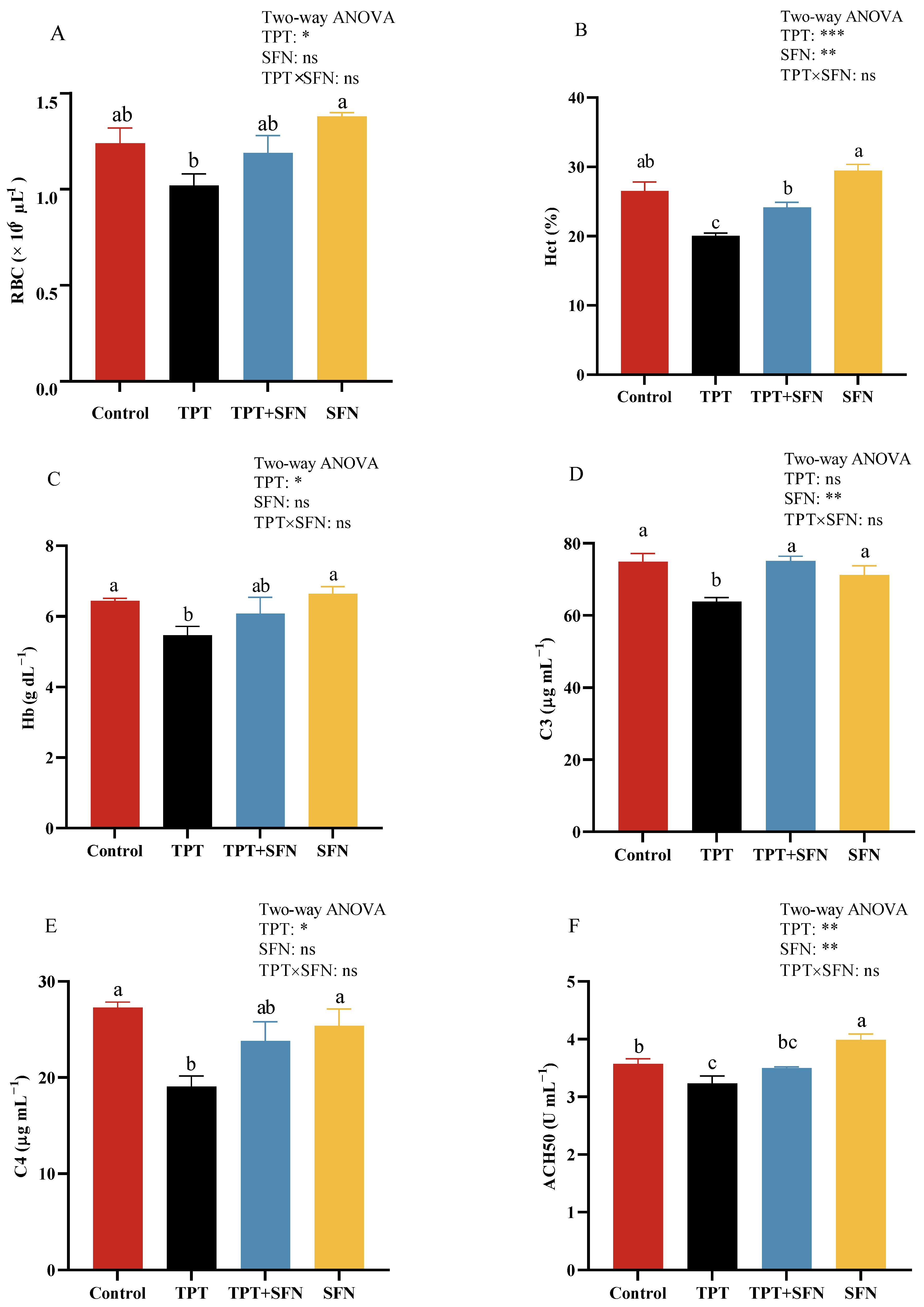
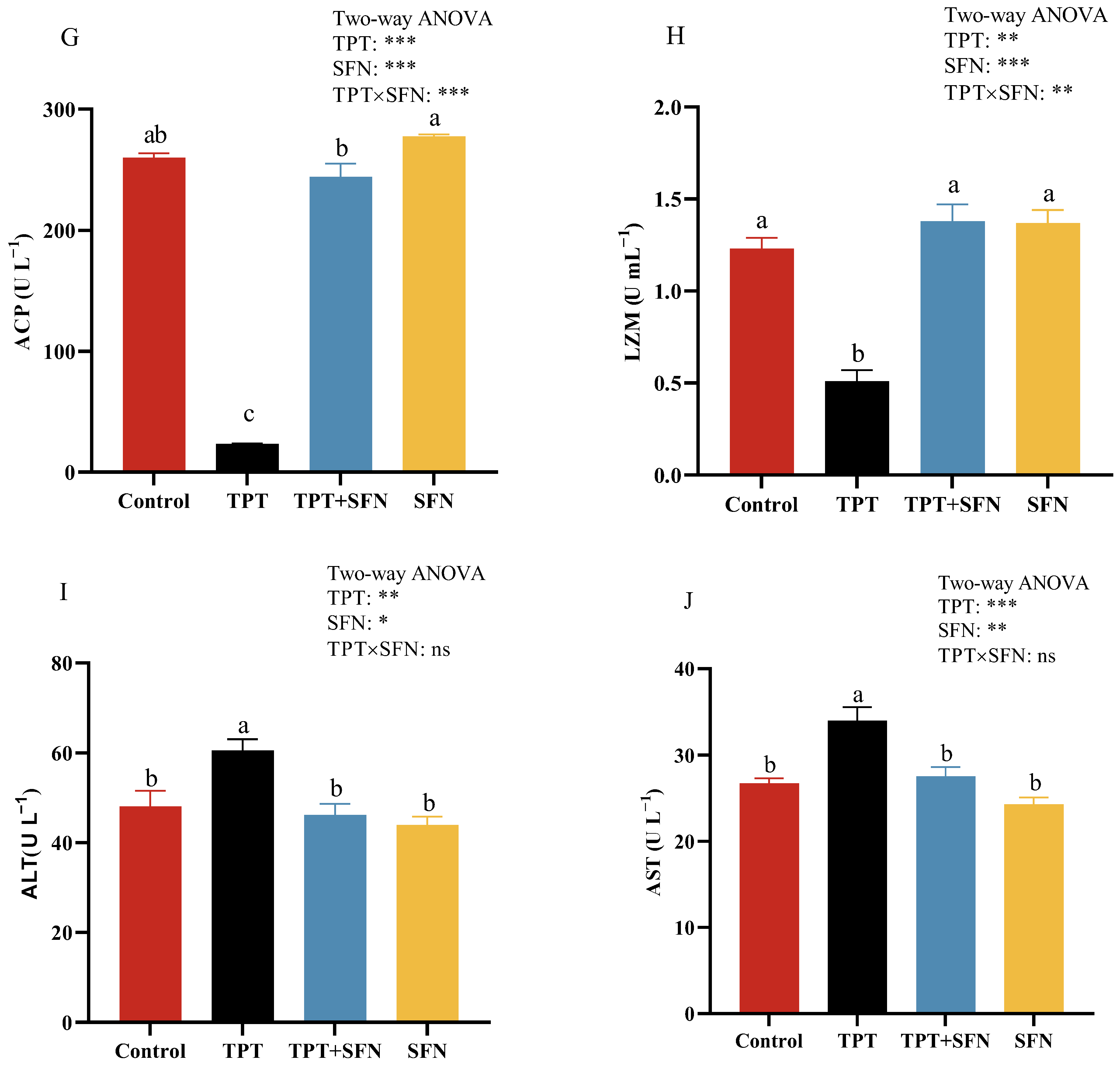
3.2. Effects of TPT and SFN on the Hematological and Plasma Immune Parameters of Cyprinus carpio haematopterus

As can be seen from Figure 1, the amounts of Hct and Hb, plasma C3 and C4, contents as well as ACH50, ACP, and LZM activities were significantly decreased (p < 0.05) in fish by TPT exposure compared with the control group, while plasma ALT and AST activities were notably higher (p < 0.05). However, treatment with SFN prevented a significant decrease in Hct amounts, plasma C3 content, as well as ACP and LZM activities that were induced by TPT exposure, but showed a decrease (p < 0.05) in plasma ALT and AST activities. Compared with the control group, plasma ACH50 activity was significantly higher in the SFN group (p < 0.05). Furthermore, there was an interaction between TPT and dietary SFN on plasma ACP and LZM activities (p < 0.05).

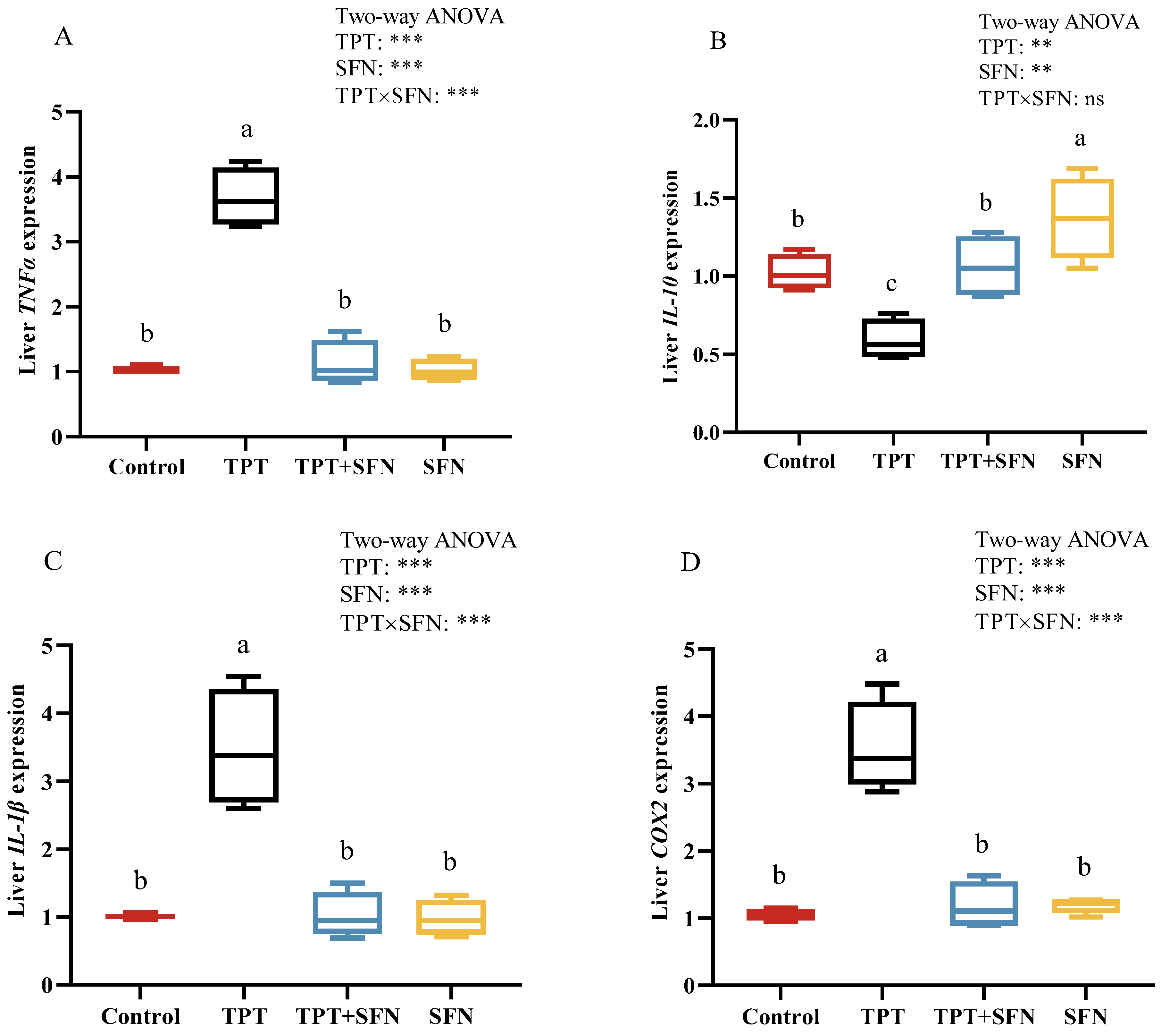
3.3. Effects of TPT and SFN on Liver Immune-Related Gene Expressions of Cyprinus carpio haematopterus

As seen in Figure 2, the mRNA expressions of liver IL-1β, TNFα, and COX2 were significantly increased (p < 0.05), while IL-10 mRNA expression decreased significantly (p < 0.05) in TPT-exposed fish compared to control. However, the addition of SFN significantly inhibited (p < 0.05) the mRNA expressions of IL-1β, TNFα, and COX2 and promoted the expression of IL-10 mRNA compared to the TPT-treated group. There was a significantly higher liver IL-10 mRNA expression in the SFN group than in the control group (p < 0.05). In addition, there was an interaction between TPT and dietary SFN on liver TNFα, IL-1β, and COX2 mRNA expressions (p < 0.05).

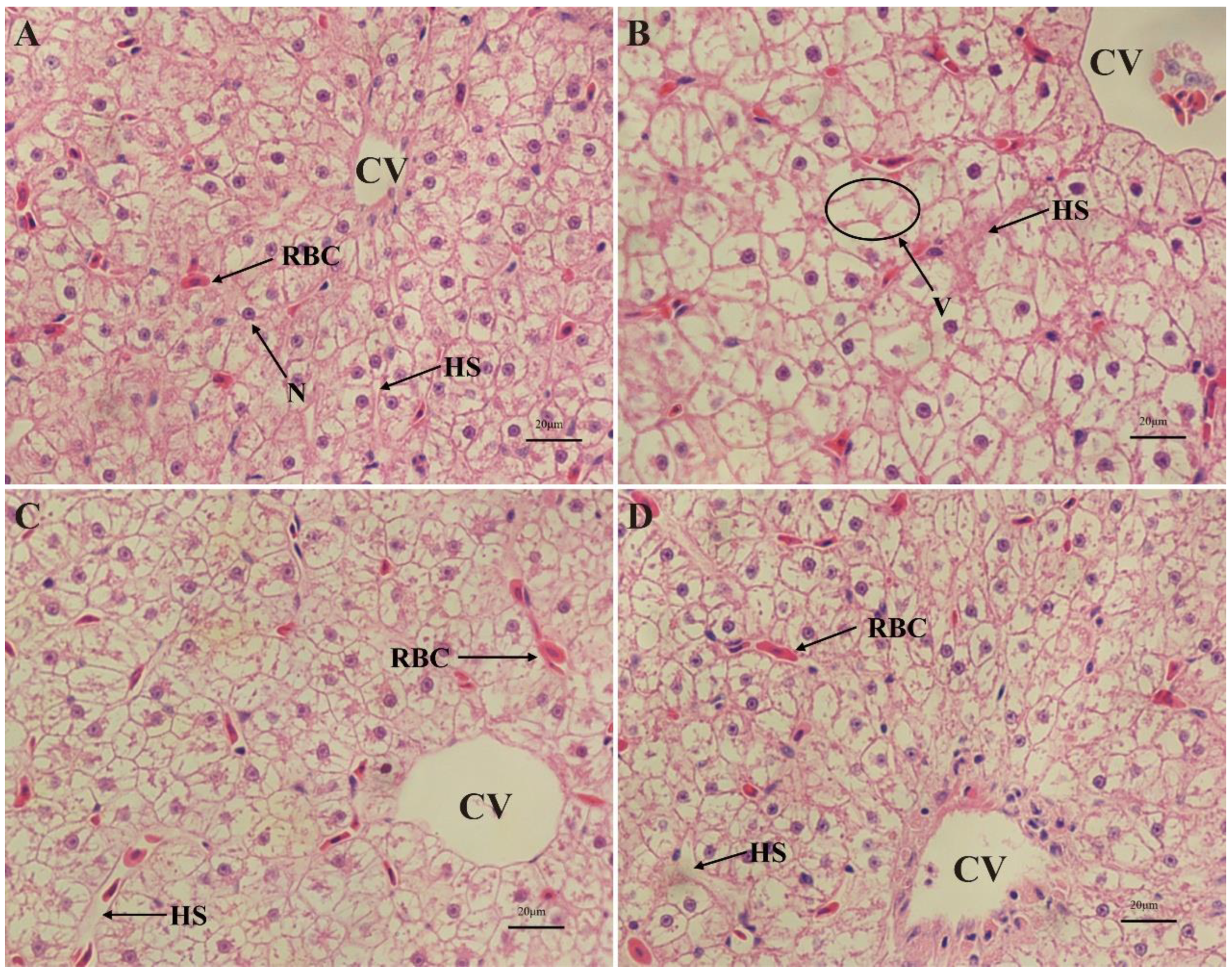
3.4. Liver Histological Examinations of Cyprinus carpio haematopterus

Liver histological examinations of Cyprinus carpio haematopterus were shown in Figure 3. No histopathological change was identified in the liver of the control group. The structure of the liver plate was clear and orderly arranged. The cytoplasm of the liver cell was uniform. The nucleus was regular and round, and it was located in the center of the cell. Hepatic sinusoids were morphologically normal and distributed between liver cells (Figure 3A). A higher occurrence rates of the hepatocyte hypertrophy, nuclear disappearance, and hepatocyte vacuolization was observed in the hepatocytes of fish exposed to TPT, and it was accompanied by the dilation of hepatic sinusoids (Figure 3B). However, liver damage caused by TPT was alleviated with the addition of SFN compared to the TPT group (Figure 3C). In addition, there were no obvious abnormalities in the basic structure of the liver in the SFN group (Figure 3D).

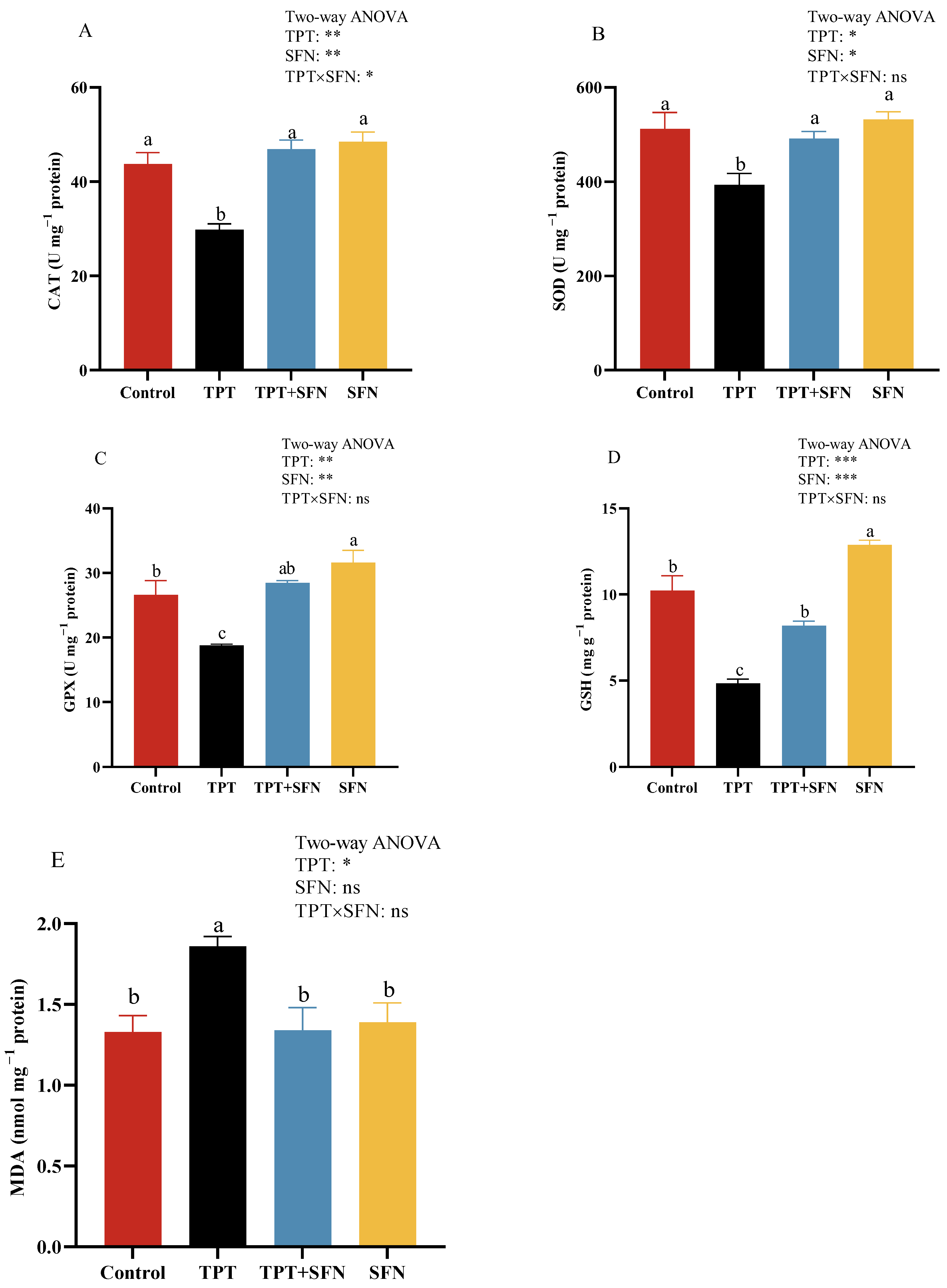
3.5. Effects of TPT and SFN on Liver Antioxidant Enzyme Activities of Cyprinus carpio haematopterus

Liver antioxidant capabilities of Cyprinus carpio haematopterus were shown in Figure 4. Fish in TPT group showed significant decreases (p < 0.05) in liver CAT, SOD, GPX activities, and GSH content, along with significant increases (p < 0.05) in MDA content compared to the control fish. However, SFN supplementation significantly increased (p < 0.05) CAT, SOD, GPX activities, and GSH content, along with significant decreasing (p < 0.05) MDA content compared to those of the TPT group. The SFN group showed significant elevations (p < 0.05) in liver GPX activity and GSH content compared to the control group. Furthermore, liver CAT activity was significantly affected (p < 0.05) by the interaction between TPT and dietary SFN.

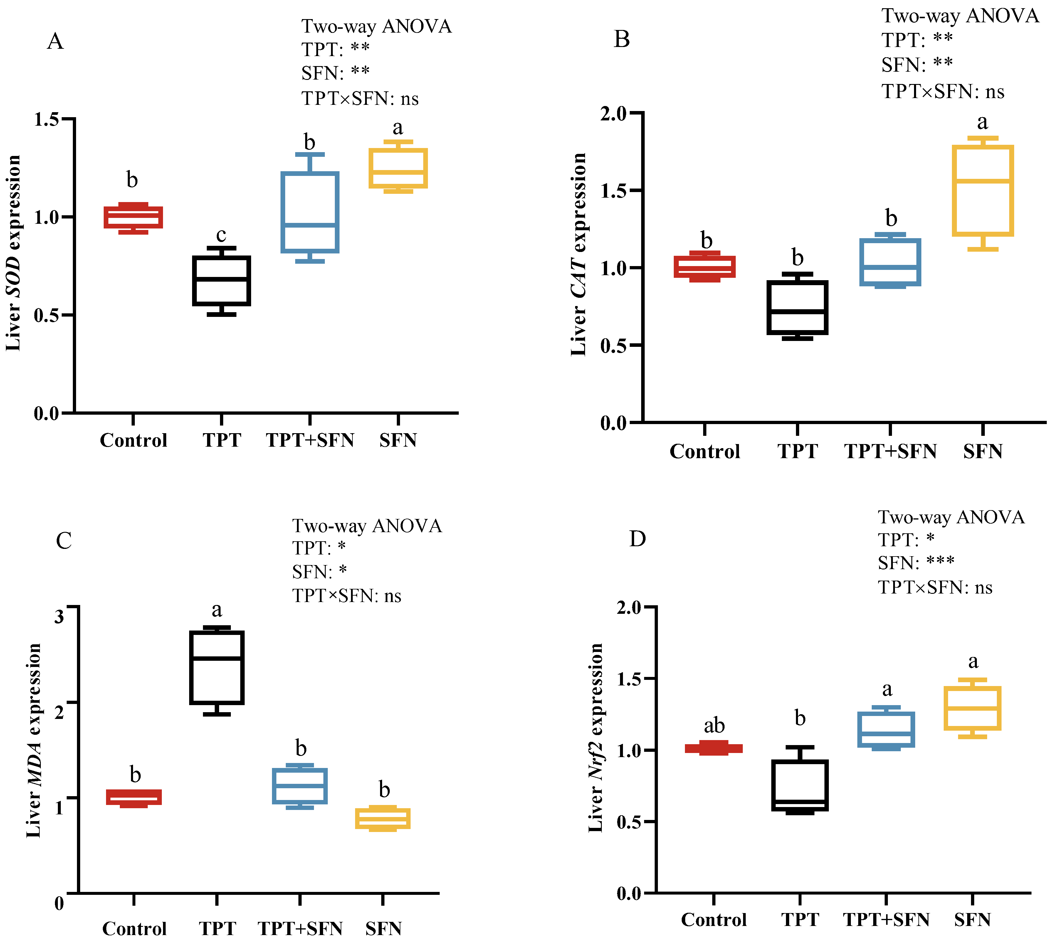
3.6. Effects of TPT and SFN on Liver Antioxidant Related Gene Expressions of Cyprinus carpio haematopterus

As presented in Figure 5. A significant decrease (p < 0.05) in SOD mRNA expression and a significant increase (p < 0.05) in MDA expression were observed in the livers of fish exposed to TPT compared to the control group. However, SOD and Nrf2 mRNA expressions were significantly higher and MDA mRNA expression was significantly lower after SFN application compared to the TPT group. The application of SFN alone presented significantly higher in SOD and CAT mRNA expressions compared to those in the control fish.

4. Discussion

In the present study, TPT exposure resulted in a remarkable decrease in final weight, WGR, SGR, and CF, but an increase in FCR and HSI of Cyprinus carpio haematopterus. This was not surprising, since TPT exposure could inhibit skeleton development and impair mobility and feeding capabilities [7]. This might inevitably result in slow growth and feed waste, with consequent decreased final weight, WGR, SGR, and CF, but increased FCR. The increase in HSI might be attributed to pathological hyperplasia of the liver, which grew larger to secrete more digestive fluid and digest feed better. This result was in accordance with previous studies on salamanders (Ambystoma barbourin) [30]. In addition, in contrast to TPT exposure alone, the supplementation of SFN in diets resulted in a remarkable increase in final weight, WGR, SGR, and CF, but a decrease in FCR. According to our previous studies, SFN could effectively improve growth performance parameters of Cyprinus carpio haematopterus [18]; this might be attributed to the effect of SFN on intestinal digestive enzyme activity and microbiota, and immune and antioxidant ability. Further in-depth studies were warranted to elucidate this.
Hematological parameters were typically used to determine the effects of feed additives on fish health. Data obtained from our study showed that TPT exposure could reduce the amounts of Hct and Hb. Indeed, TPT has a wide range of toxicological effects as a contaminant [12], and it may have an inhibitory effect on the hematopoietic function of the fish, thus slowing down the rate of cell renewal in the hematopoietic system. However, the addition of SFN to the diet resulted in a remarkable increase in Hct amount, as well as an increase in RBC and Hb amounts, although no significant difference was observed. The increases in RBC, Hct, and Hb could facilitate tissue oxygenation and elimination of carbon dioxide [31]. The results of the present study were in agreement with the results of previous studies, indicating that the plant extracts positively affect hematological parameters in fish [32]. However, no studies in the literature had reported the effect of SFN on hematological parameters. In fish, there was a key role for the generation of innate immune components (e.g., complement and lysozyme) in the regulation of the immune response. C3, C4, and lysozyme were essential immune components with non-selective effects against bacteria and antigens. With the same tendency as lysozyme, ACP was a component of macrophage lysosomes. Fish immune systems, especially innate immunity, which changed, could be used as an early indicator of a response to oxidative stress due to its sensitivity to chemicals [33]. In this study, TPT exposure reduced the levels of C3, C4, ACH50, ACP, and LZM, that is, immunosuppression occurred. This was not surprising, since exposure to TPT for long term might cause chronic immunotoxic effects [34]. Similar results were reported in common carp (Cyprinus carpio) [12], zebrafish (Danio rerio) [35], and gastropod abalone (Haliotis diversicolor) [36]. On the other hand, the addition of SFN showed a significant increase in immunological parameters, including plasma C3 content and ACP and LZM activities, confirming that dietary intake of SFN markedly affected some crucial defense molecules in plasma [37] and effectively protected against TPT-induced immunotoxicity in Cyprinus carpio haematopterus. This result was in accordance with our previous study [18].
TNF-α, IL-1β, and IL-10 are often used as markers of inflammatory response. In this study, there was an increase in mRNA expression of IL-1β and TNF-α with TPT treatment, but a decrease in the mRNA expressions of the anti-inflammatory cytokine IL-10 in the liver. This is further supported the fact that TPT gradually inhibited the occurrence of immune responses at the gene level [38]. COX2, as an important inflammatory mediator, is induced by a variety of stimuli, including cytokines. In our study, TPT exposure markedly increased the liver COX2 mRNA expression; the result had the same trend as TNF-α and IL-1β. This result was expected, since the expression level of COX2 in inflammatory cells increased rapidly and resulted in inflammatory responses and tissue damage when cells were stimulated by inflammation [39]. In addition, SFN supplementation efficiently alleviated the inflammatory response that was induced by TPT. This result confirmed the vital role of SFN in mediating inflammatory responses and immune function in animals [40]. Other studies on dietary plant extracts from Cyprinus carpio had observed similar results [41,42]. However, our previous study also had shown that the mRNA expressions of liver IL-1β and TNF-α both increased and then decreased with increasing dietary SFN levels from 0 to 20 mg kg−1 [18]. This might be explained by the ability of SFN to maintain the dynamic equilibrium of inflammatory factors in fish within a certain range of conditions.
Fish liver is correlated with nutrient deposition, biotransformation, metabolism, detoxification, and blood supply, and it was also one of the organs most affected by pollution. Changes in the liver may be useful as markers indicating prior exposure to environmental stressors. In this study, the liver of TPT-exposed fish illustrated hepatocyte hypertrophy, nuclear disappearance, hepatocyte vacuolization, and the dilation of hepatic sinusoids. Histopathological studies had shown that fish displayed compensatory reflexes such as dilation of hepatic sinusoids, hemorrhage, and intercellular vacuolization during exposure to toxicants [43]. Dilation of the hepatic sinusoids increased blood flow through tissue, which enabled speeding up the process of filtration to detoxify the fish [44]. Similarly, several studies had shown heavy metals in the environment could cause histopathological changes in the liver of fish [45,46]. On the contrary, the addition of SFN to the diet alleviated liver injury, with a reduced incidence of hepatocyte size, nuclear disappearance, and hepatocyte vacuolization with contraction of the hepatic sinusoids compared to the TPT group, which might be related to the function played by SFN. According to previous studies, SFN could improve antioxidant capacity and reduce immune stress in Cyprinus carpio haematopterus [18], while oxidative stress-associated molecular damages were eventually reflected in the impairments of the tissue structure [47].
Environmental pollution could lead to oxidative stress in biological systems. Oxidative stress was considered to play an essential role in many physiological and pathological phenomena in fish. In this study, TPT exposure resulted in a reduction in CAT, SOD, GPX activities, and GSH content, and an increase in MDA content in the liver of Cyprinus carpio haematopterus. Numerous studies have demonstrated the following: (1) GPX protected the organism mainly against the harmful effects of hydrogen peroxide; GSH is a widely available small molecule antioxidant found in cells, which could help the body to scavenge free radicals and peroxides [48]. (2) As the first line of defense against oxidative stress, the SOD-CAT system scavenged excess free radicals from the body and maintained a balance between the oxidative and antioxidant systems [18]. (3) MDA was a polyunsaturated fatty acid peroxidation product, mainly generated by free radicals, which could cause toxicity to the body and was a scientifically accepted oxidative stress indicator [49]. It also validated the fact that exposure to TPT could cause oxidative stress to the liver of fish, resulting in oxidative damage. The results were in line with previous studies on this species [50]. In contrast to TPT exposure alone, the incorporation of SFN into fish diets could increase CAT, SOD, GPX activities, and GSH content, but decrease MDA content. SFN was able to exert a protective effect because of its potential for antioxidant capacity; SFN reduced ROS production and enhances antioxidant enzyme activity in cells [51]. This might be attributed to the antioxidant property of sulfur in SFN, which could influence GSH metabolism, thus avoiding ROS accumulation and oxidative damage [52,53]. The results were in line with previous studies on Cyprinus carpio haematopterus [18,50], Litopenaeus Vannamei [15], and rats [54].
It has been shown that antioxidant parameters, such as SOD, CAT, and MDA in fish liver were positively related to their respective mRNA levels [55]. This was an important tool for investigating the effects of oxidative stress through gene mRNA expression. In this study, TPT exposure resulted in a remarkable decrease in hepatic SOD mRNA expression, but an increase in MDA mRNA expression of Cyprinus carpio haematopterus. We also found that TPT treatment decreased the mRNA expressions of liver CAT and Nrf2, although there was no significant difference. These genes confer protection against oxidative damage across various tissues, excluding MDA. This result was not surprising, since the reduced activity of antioxidant enzymes in TPT-treated fish might be partially attributed to the down-regulation of corresponding genes in liver tissue [9]. SFN supplementation partly prevented these alterations; this indicated that the addition of SFN was effective in restoring antioxidant indicator mRNA expression in TPT-exposed fish. According to previous studies, SFN could up-regulate the mRNA expression of Nrf2 in fish, as might consequently boost antioxidant capacity [56]. Nrf2 plays an important key role in the elimination of reactive oxygen through the up-regulation of the expression of antioxidant-related genes (e.g., GPX) to resist oxidative stress [57]. This was supported by the fact that SFN was an activator of Nrf2, and SFN exerts a powerful antioxidant capacity in activating the Nrf2 signaling pathway [58]. Although SFN shows promising antioxidant and immune-enhancing effects in the present study, it is crucial to acknowledge that further research is necessary to evaluate its long-term effectiveness.

5. Conclusions

In conclusion, this study revealed the protective role of SFN against the physiological toxicity of TPT in Cyprinus carpio haematopterus. The results obtained in this study suggested that TPT exposure inhibited the growth, immune and antioxidant capacity, and altered the liver tissue structure. SFN could relieve these effects by its growth-promoting, immune-enhancing, and antioxidant capabilities. Based on these results, we proposed that SFN was an excellent aquaculture feed additive for reducing the negative effects of TPT contamination.

Author Contributions

Conceptualization, B.W. and Q.Z.; Data curation, B.W. and J.M.; Project administration, B.W. and Q.Z.; Funding acquisition, B.W. and Q.Z.; Writing—original draft, B.W.; Formal analysis, J.M.; Validation, J.M.; Visualization, J.M.; Resources, C.Z., X.Y. and K.Z.; Writing—review and editing, C.Z.; Methodology, Y.W.; Investigation, L.Z.; Software, T.J.; Supervision, Q.Z. All authors have read and agreed to the published version of the manuscript.

Funding

This work was supported by Basic Research Business Fund Project of Henan Province (2024ZC114), and Earmarked Fund for CARS (CARS-45).

Institutional Review Board Statement

All animal handling procedures in this experiment had been approved by the Ethics Committee of Henan Academy of Fishery Sciences.

Informed Consent Statement

Not applicable.

Data Availability Statement

Data will be made available on request.

Conflicts of Interest

The authors declare no conflicts of interest.

References

  1. He, S.W.; Li, P.; Li, Z.H. Review on endocrine disrupting toxicity of triphenyltin from the perspective of species evolution: Aquatic, amphibious and mammalian. Chemosphere 2021, 269, 128711. [Google Scholar] [CrossRef]
  2. Antizar-Ladislao, B. Environmental levels, toxicity and human exposure to tributyltin (TBT)-contaminated marine environment. A review. Environ. Int. 2008, 34, 292–308. [Google Scholar] [CrossRef] [PubMed]
  3. Bettin, C.; Oehlmann, J.; Stroben, E. TBT-induced imposex in marine neogastropods is mediated by an increasing androgen level. Helgol. Meeresunters. 1996, 50, 299–317. [Google Scholar] [CrossRef]
  4. Sham, R.C.T.; Ho, K.K.Y.; Zhou, G.J.; Li, Y.; Wang, X.H.; Leung, K.M.Y. Occurrence and ecological risks of phenyltin compounds in the marine environment of Hong Kong. Mar. Pollut. Bull. 2020, 154, 111093. [Google Scholar] [CrossRef] [PubMed]
  5. Sham, R.C.T.; Tao, L.S.R.; Mak, Y.K.Y.; Yau, J.K.C.; Wai, T.C.; Ho, K.K.Y.; Zhou, G.J.; Li, Y.Y.; Wang, X.H.; Leung, K.M.Y. Occurrence and trophic magnification profile of triphenyltin compounds in marine mammals and their corresponding food webs. Environ. Int. 2020, 137, 105567. [Google Scholar] [CrossRef] [PubMed]
  6. Horie, Y.; Watanabe, H.; Takanobu, H.; Shigemoto, Y.; Yamagishi, T.; Iguchi, T.; Tatarazako, N. Effects of triphenyltin on reproduction in Japanese medaka (Oryzias latipes) across two generations. Aquat. Toxicol. 2017, 192, 16–23. [Google Scholar] [CrossRef] [PubMed]
  7. Hou, Y.; Cai, X.W.; Liang, Z.F.; Duan, D.D.; Diao, X.P.; Zhang, J.L. An integrative investigation of developmental toxicities induced by triphenyltin in a larval coral reef fish, Amphiprion ocellaris. Sci. Total Environ. 2023, 867, 161487. [Google Scholar] [CrossRef]
  8. Yao, F.; Li, Y.F.; Ru, H.J.; Wu, L.Y.; Xiao, Z.G.; Ni, Z.H.; Chen, D.Q.; Zhong, L.Q. Thyroid disruption and developmental toxicity caused by triphenyltin (TPT) in zebrafish embryos/larvae. Toxicol. Appl. Pharmacol. 2020, 394, 114957. [Google Scholar] [CrossRef] [PubMed]
  9. Zhang, C.N.; Wang, J.H.; Qi, Q.; Yang, L.; Sun, P.; Yuan, X.Y. Modulatory effect of fructooligosaccharide against triphenyltin-induced oxidative stress and immune suppression in goldfish (Carassius auratus). Ecotoxicol. Environ. Saf. 2021, 212, 111966. [Google Scholar] [CrossRef]
  10. Zhang, J.L.; Zhang, C.N.; Li, E.C.; Jin, M.M.; Huang, M.X.; Cui, W.; Lin, Y.Y.; Shi, Y.J. Triphenyltin exposure affects mating behaviors and attractiveness to females during mating in male guppies (Poecilia reticulata). Ecotoxicol. Environ. Saf. 2019, 169, 76–84. [Google Scholar] [CrossRef]
  11. Zhang, S.Q.; Li, P.; He, S.W.; Xing, S.Y.; Cao, Z.H.; Zhao, X.L.; Sun, C.C.; Li, Z.H. Combined effect of microplastic and triphenyltin: Insights from the gut-brain axis. Environ. Sci. Ecotechnol. 2023, 16, 100266. [Google Scholar] [CrossRef] [PubMed]
  12. Zhang, S.Q.; Li, P.; He, S.W.; Xing, S.Y.; Cao, Z.H.; Zhao, X.L.; Sun, C.C.; Li, Z.H. Assessing the ecotoxicity of combined exposure to triphenyltin and norfloxacin at environmental levels: A case study of immunotoxicity and metabolic regulation in carp (Cyprinus carpio). Chemosphere 2023, 313, 137381. [Google Scholar] [CrossRef] [PubMed]
  13. Zhang, J.L.; Zuo, Z.H.; He, C.Y.; Wu, D.; Chen, Y.X.; Wang, C.G. Inhibition of thyroidal status related to depression of testicular development in Sebastiscus marmoratus exposed to tributyltin. Aquat. Toxicol. 2009, 94, 62–67. [Google Scholar] [CrossRef] [PubMed]
  14. Abdel-Tawwab, M.; Hamed, H.S. Antagonistic effects of dietary guava (Psidium guajava) leaves extract on growth, hemato-biochemical, and immunity response of cypermethrin-intoxicated Nile tilapia, Oreochromis niloticus, fingerlings. Aquaculture 2020, 529, 735668. [Google Scholar] [CrossRef]
  15. Yang, S.P.; Huang, Y.X.; Chen, B.K.; Liu, H.L.; Huang, Y.C.; Cai, S.H.; Jian, J.C. Protective effects of sulphoraphane on oxidative damage caused by ammonia in litopenaeus vannamei. Aquac. Res. 2022, 53, 1197–1204. [Google Scholar] [CrossRef]
  16. Mahn, A.; Castillo, A. Potential of sulforaphane as a natural immune system enhancer: A review. Molecules 2021, 26, 752. [Google Scholar] [CrossRef]
  17. Zhao, F.F.; Zhang, J.L.; Chang, N. Epigenetic modification of Nrf2 by sulforaphane increases the antioxidative and anti-inflammatory capacity in a cellular model of Alzheimer’s disease. Eur. J. Pharmacol. 2018, 824, 1–10. [Google Scholar] [CrossRef]
  18. Wang, B.K.; Zhang, C.N.; Zhang, Q.; Zhang, L.; Jiang, X.Y.; Feng, J.X.; Yang, X.L.; Wang, Y.H. Can sulforaphane alter growth performance, innate immunity and antioxidant capability of common carp (Cyprinus carpio Huanghe var)? Aquac. Rep. 2023, 31, 101666. [Google Scholar] [CrossRef]
  19. Blaxhall, P.C.; Daisley, K.W. Routine haematological methods for use with fish blood. J. Fish Biol. 1973, 5, 771–781. [Google Scholar] [CrossRef]
  20. Montero, D.; Tort, L.M.; Izquierdo, S.; Robaina, L.; Vergara, J.M. Depletion of serum alternative complement pathway activity in gilthead seabream caused by a-tocopherol and n-3 HUFA dietary deficiencies. Fish Physiol. Biochem. 1998, 18, 399–407. [Google Scholar] [CrossRef]
  21. Tang, H.G.; Wu, T.X.; Zhao, Z.Y.; Pan, X.D. Effects of fish protein hydrolysate on growth performance and humoral immune response in large yellow croaker (Pseudosciaena crocea R.). J. Zhejiang Univ. Sci. 2008, 9, 684–690. [Google Scholar] [CrossRef] [PubMed]
  22. Tort, L.; Gómez, E.; Montero, D.; Sunyer, J.O. Serum haemolytic and agglutinating activity as indicators of fish immunocompetence: Their suitability in stress and dietary studies. Aquac. Int. 1996, 4, 31–41. [Google Scholar] [CrossRef]
  23. Obach, A.; Quentel, C.; Laurencin, F.B. Effects of alpha-tocopherol and dietary oxidized fish oil on the immune response of sea bass Dicentrarchus labrax. Dis. Aquat. Org. 1993, 15, 175–185. [Google Scholar] [CrossRef]
  24. Krajnović-Ozretić, M.; Ozretić, B. Estimation of the enzymes LDH, GOT and GPT in plasma of grey mullet Mugil auratus and their significance in liver intoxication. Dis. Aquat. Org. 1987, 3, 187–193. [Google Scholar] [CrossRef]
  25. Goth, L. A simple method for determination of serum catalase activity and revision of reference range. Clin. Chim. Acta 1991, 196, 143–151. [Google Scholar] [CrossRef]
  26. Placer, Z.A.; Cushman, L.L.; Johnson, B.C. Estimation of product of lipid peroxidation (malonyl dialdehyde) in biochemical systems. Anal. Biochem. 1966, 16, 359–364. [Google Scholar] [CrossRef]
  27. Marklund, S.; Marklund, G. Involvement of the superoxide anion radical in the autoxidation of pyrogallol and a convenient assay for superoxide dismutase. Eur. J. Biochem. 1974, 47, 469–474. [Google Scholar] [CrossRef]
  28. Lygren, B.; Hamre, K.; Waagbø, R. Effects of dietary pro- and antioxidants on some protective mechanisms and health parameters in Atlantic salmon. J. Aquat. Anim. Health 1999, 11, 211–221. [Google Scholar] [CrossRef]
  29. Livak, K.J.; Schmittgen, T.D. Analysis of relative gene expression data using real-time quantitative PCR and the 2−ΔΔCT method. Methods 2001, 25, 402–408. [Google Scholar] [CrossRef]
  30. Rehage, J.S.; Lynn, S.G.; Hammond, J.I.; Palmer, B.D.; Sih, A. Effects of larval exposure to triphenyltin on the survival, growth, and behavior of larval and juvenile Ambystoma barbouri salamanders. Environ. Toxicol. Chem. 2002, 21, 807–815. [Google Scholar] [CrossRef]
  31. Abdel-Tawwab, M.; Khattab, Y.A.E.; Ahmad, M.H.; Shalaby, A.M.E. Compensatory growth, feed utilization, whole-body composition, and hematological changes in starved Juvenile Nile tilapia, Oreochromis niloticus (L.). J. Appl. Aquac. 2006, 18, 17–36. [Google Scholar] [CrossRef]
  32. Jahazi, M.A.; Hoseinifar, S.H.; Jafari, V.; Hajimoradloo, A.; Doan, H.V.; Paolucci, M. Dietary supplementation of polyphenols positively affects the innate immune response, oxidative status, and growth performance of common carp, Cyprinus carpio L. Aquaculture 2020, 517, 734709. [Google Scholar] [CrossRef]
  33. Wang, B.K.; Wang, Y.H.; Jia, T.; Feng, J.X.; Qu, C.Y.; Wu, X.J.; Yang, X.L.; Zhang, Q. Changes in physiological responses and immunity of blunt snout bream Megalobrama amblycephala from transport stress. Fish Physiol. Biochem. 2022, 48, 1183–1192. [Google Scholar] [CrossRef]
  34. Nakayama, A.; Kurokawa, Y.; Harino, H.; Kawahara, E.; Miyadai, T.; Seikai, T.; Kawai, S. Effects of tributyltin on the immune system of Japanese flounder (Paralichthys olivaceus). Aquat. Toxicol. 2007, 83, 126–133. [Google Scholar] [CrossRef] [PubMed]
  35. Zhang, C.N.; Jiang, D.X.; Wang, J.H.; Qi, Q. The effects of TPT and dietary quercetin on growth, hepatic oxidative damage and apoptosis in zebrafish. Ecotoxicol. Environ. Saf. 2021, 224, 112697. [Google Scholar] [CrossRef]
  36. Gopalakrishnan, S.; Huang, W.B.; Wang, Q.W.; Wu, M.L.; Liu, J.; Wang, K.J. Effects of tributyltin and benzo[a]pyrene on the immune-associated activities of hemocytes and recovery responses in the gastropod abalone, Haliotis diversicolor. Comp. Biochem. Physiol. Part C Toxicol. Pharmacol. 2011, 154, 120–128. [Google Scholar] [CrossRef] [PubMed]
  37. Shih, Y.L.; Wu, L.Y.; Lee, C.H.; Chen, Y.L.; Hsueh, S.C.; Lu, H.F.; Liao, N.C.; Chung, J.G. Sulforaphane promotes immune responses in a WEHI-3–induced leukemia mouse model through enhanced phagocytosis of macrophages and natural killer cell activities in vivo. Mol. Med. Rep. 2016, 13, 4023–4029. [Google Scholar] [CrossRef]
  38. He, S.W.; Yu, D.D.; Li, P.; Zhang, M.; Xing, S.Y.; Sun, C.C.; Li, Z.H. Triphenyltin exposure causes changes in health-associated gut microbiome and metabolites in marine medaka. Environ. Pollut. 2021, 288, 117751. [Google Scholar] [CrossRef]
  39. Herseth, J.I.; Refsnes, M.; Låg, M.; Schwarze, P.E. Role of IL-1β and COX2 in silica-induced IL-6 release and loss of pneumocytes in co-cultures. Toxicol. Vitr. 2009, 23, 1342–1353. [Google Scholar] [CrossRef]
  40. Thejass, P.; Kuttan, G. Immunomodulatory activity of sulforaphane, a naturally occurring isothiocyanate from broccoli (brassica oleracea). Phytomedicine 2007, 14, 538–545. [Google Scholar] [CrossRef]
  41. Yousefi, M.; Shabunin, S.V.; Vatnikov, Y.A.; Kulikov, E.V.; Adineh, H.; Hamidi, M.K.; Hoseini, S.M. Effects of lavender (Lavandula angustifolia) extract inclusion in diet on growth performance, innate immunity, immune-related gene expression, and stress response of common carp, Cyprinus carpio. Aquaculture 2020, 515, 734588. [Google Scholar] [CrossRef]
  42. Zemheri-Navruz, F.; Acar, Ü.; Yılmaz, S. Dietary supplementation of olive leaf extract increases haematological, serum biochemical parameters and immune related genes expression level in common carp (Cyprinus carpio) juveniles. Fish Shellfish Immunol. 2019, 89, 672–676. [Google Scholar] [CrossRef] [PubMed]
  43. Sharma, K.; Sharma, P.; Dhiman, S.K.; Chadha, P.; Saini, H.S. Biochemical, genotoxic, histological and ultrastructural effects on liver and gills of fresh water fish Channa punctatus exposed to textile industry intermediate 2 ABS. Chemosphere 2022, 287, 132103. [Google Scholar] [CrossRef] [PubMed]
  44. Rodrigues, J.L.; Serpeloni, J.M.; Batista, B.L.; Souza, S.S.; Barbosa, F. Identification and distribution of mercury species in rat tissues following administration of thimerosal or methylmercury. Arch. Toxicol. 2010, 84, 891–896. [Google Scholar] [CrossRef]
  45. Javed, M.; Ahmad, I.; Usmani, N.; Ahmad, M. Studies on biomarkers of oxidative stress and associated genotoxicity and histopathology in Channa punctatus from heavy metal polluted canal. Chemosphere 2016, 151, 210–219. [Google Scholar] [CrossRef]
  46. Macêdo, A.K.S.; dos Santos, K.P.E.; Brighenti, L.S.; Windmöller, C.C.; Barbosa, F.A.R.; de Azambuja Ribeiro, R.I.M.; dos Santos, H.B.; Thomé, R.G. Histological and molecular changes in gill and liver of fish (Astyanax lacustris Lütken, 1875) exposed to water from the Doce basin after the rupture of a mining tailings dam in Mariana, MG, Brazil. Sci. Total Environ. 2020, 735, 139505. [Google Scholar] [CrossRef]
  47. Trivedi, S.P.; Ratn, A.; Awasthi, Y.; Kumar, M.; Trivedi, A. In vivo assessment of dichlorvos induced histological and biochemical impairments coupled with expression of p53 responsive apoptotic genes in the liver and kidney of fish, Channa punctatus (Bloch, 1793). Comp. Biochem. Physiol. Part C Toxicol. Pharmacol. 2021, 245, 109032. [Google Scholar] [CrossRef]
  48. Yildirim, Z.; Kilic, N. Effects of taurine and age on cerebellum antioxidant status and oxidative stress. Int. J. Gerontol. 2011, 5, 166–170. [Google Scholar] [CrossRef]
  49. Barbosa, M.L.; Menese, A.P.M.; Aguiar, R.P.S.; Sousa, J.M.C.; Cavalcante, A.A.C.; Maluf, S.W. Oxidative stress, antioxidant defense and depressive disorders: A systematic review of biochemical and molecular markers. Neurol. Psychiatry Brain Res. 2020, 36, 65–72. [Google Scholar] [CrossRef]
  50. Ma, J.S.; Wang, B.K.; Pu, C.C.; Chang, K.; Cheng, Y.F.; Sun, R.Y.; Qi, Q.; Xu, R.Y.; Chen, J.L.; Zhang, C.N. Protective effects of sulforaphane on inflammation, oxidative stress and intestinal dysbacteriosis induced by triphenyltin in Cyprinus carpio haematopterus. Fish Shellfish Immunol. 2023, 142, 109135. [Google Scholar] [CrossRef]
  51. Guerrero-Beltrán, C.E.; Calderón-Oliver, M.; Pedraza-Chaverri, J.; Chirino, Y.I. Protective effect of sulforaphane against oxidative stress: Recent advances. Exp. Toxicol. Pathol. 2012, 64, 503–508. [Google Scholar] [CrossRef] [PubMed]
  52. Brancaccio, M.; D’Argenio, G.; Lembo, V.; Palumbo, A.; Castellano, I. Antifibrotic Effect of Marine Ovothiol in an In Vivo Model of Liver Fibrosis. Oxid. Med. Cell. Longev. 2018, 2018, 5045734. [Google Scholar] [CrossRef]
  53. Milito, A.; Brancaccio, M.; D’Argenio, G.; Castellano, I. Natural Sulfur-Containing Compounds: An Alternative Therapeutic Strategy against Liver Fibrosis. Cells 2019, 8, 1356. [Google Scholar] [CrossRef] [PubMed]
  54. Chen, L.; Zhang, W.L.; Xie, D.Q.; Jia, W. Sulforaphane alleviates hepatic ischemia–reperfusion injury through promoting the activation of Nrf-2/HO-1 signaling. Transpl. Immunol. 2021, 68, 101439. [Google Scholar] [CrossRef]
  55. Jiang, W.D.; Zhou, X.Q.; Zhang, L.; Liu, Y.; Wu, P.; Jiang, J.; Kuang, S.Y.; Tang, L.; Tang, W.N.; Zhang, Y.A.; et al. Vitamin A deficiency impairs intestinal physical barrier function of fish. Fish Shellfish Immunol. 2019, 87, 546–558. [Google Scholar] [CrossRef] [PubMed]
  56. Lee, Y.J.; Lee, S.H. Sulforaphane induces antioxidative and antiproliferative responses by generating reactive oxygen species in human bronchial epithelial BEAS-2B cells. J. Korean Med. Sci. 2011, 26, 1474–1482. [Google Scholar] [CrossRef] [PubMed]
  57. Zhang, C.N.; Ma, J.S.; Wang, B.K.; Pu, C.C.; Chang, K.; Zhu, J.X.; Zhang, B.Y.; Li, J.J.; Qi, Q.; Xu, R.Y. Sulforaphane modulates some stress parameters in TPT-exposed Cyprinus carpio in relation to liver metabolome. Ecotoxicol. Environ. Saf. 2024, 284, 116882. [Google Scholar] [CrossRef]
  58. Cho, H.Y.; Miller-DeGraff, L.; Blankenship-Paris, T.; Wang, X.T.; Bell, D.A.; Lih, F.; Deterding, L.; Panduri, V.; Morgan, D.L.; Yamamoto, M.; et al. Sulforaphane enriched transcriptome of lung mitochondrial energy metabolism and provided pulmonary injury protection via Nrf2 in mice. Toxicol. Appl. Pharm. 2019, 364, 29–44. [Google Scholar] [CrossRef]
Figure 1. (AJ) Effects of TPT and SFN on the hematological and plasma immune parameters of Cyprinus carpio haematopterus. RBC, red blood cell; Hct, hematocrit; Hb, hemoglobin; C3, complement component 3; C4, complement component 4; ACH50, alternative complement pathway; ACP, acid phosphatase; LZM, lysozyme; AST, aspartate aminotransferase; ALT, alanine aminotransferase. * p < 0.05, ** p < 0.01, *** p < 0.001, ns, not significant. Values are expressed as means ± SEM. Bars assigned with different letters are significantly different (p < 0.05).
Antioxidants 13 01173 g001aAntioxidants 13 01173 g001b
Figure 2. (AD) Effects of TPT and SFN on liver immune related gene expressions of Cyprinus carpio haematopterus. TNF-α, tumor necrosis factor α; IL-10, interleukin 10; IL-1β, interleukin-1β; COX2, Cyclooxygenase 2. ** p < 0.01, *** p < 0.001, ns, not significant. Values are expressed means ± SEM. Bars assigned with different letters are significantly different (p < 0.05).
Antioxidants 13 01173 g002
Figure 3. (AD) Photomicrographs of liver tissue of Cyprinus carpio haematopterus (H&E staining, scale bars = 20 μm). (A), control group; (B), TPT group; (C), TPT + SFN group; (D), SFN group. CV, central vein; HS, hepatic sinusoid; N, nucleus; RBC, red blood cell; V, vacuolation.
Antioxidants 13 01173 g003
Figure 4. (AE) Effects of TPT and SFN on liver antioxidant enzyme activities of Cyprinus carpio haematopterus. CAT, catalase; SOD, superoxide dismutase; GPX, glutathione peroxidase; GSH, glutathione; MDA, malonaldehyde. * p < 0.05, ** p < 0.01, *** p < 0.001, ns, not significant. Values are expressed means ± SEM. Bars assigned with different letters are significantly different (p < 0.05).
Antioxidants 13 01173 g004
Figure 5. (AD) Effects of TPT and SFN on liver antioxidant-related gene expressions of Cyprinus carpio haematopterus. Nrf2, nuclear factor E2 related factor 2. * p < 0.05, ** p < 0.01, *** p < 0.001, ns, not significant. Values are expressed means ± SEM. Bars assigned with different letters are significantly different (p < 0.05).
Antioxidants 13 01173 g005
Table 1. Diet composition of Cyprinus carpio haematopterus.
Ingredients (%)
Fish meal5.00
Soybean meal32.00
Rapeseed meal15.00
Cottonseed meal15.00
Fish oil1.00
Soybean oil2.00
Wheat flour21.00
Wheat bran6.00
Ca(H2PO4)21.80
Premix 11.00
Salt0.20
Proximate analyses (% air-dry basis)
Dry matter89.73
Crude lipid4.04
Crude protein35.82
Crude ash9.45
1 Premixtures included vitamins (IU or mg kg−1) and minerals (g kg−1): VA, 900,000 IU; VB1, 320 mg; VB2, 1090 mg; VB5, 2000 mg; VB6, 500 mg; VB12, 1.6 mg; VC, 5000 mg; VD, 200,000 IU; VE, 4500 mg; VK3, 220 mg; Na2SeO3, 0.04 g; FeSO4·7H2O, 25 g; CuSO4·5H2O, 2.0 g; ZnSO4·7H2O, 22 g; MnSO4·4H2O, 7 g; CoCl2·6H2O, 0.1 g; KI, 0.026 g; choline, 60,000 mg; pantothenate, 1000 mg; folic acid, 165 mg.
Table 2. Sequence of primers for gene expression of Cyprinus carpio haematopterus.
GeneSequences of Primers (5′-3′)Accession Numbers
β-actinF: TTGACTTCCCTGACAGCTGTM24113.1
R: CGAATCCGGCTTTGCACATA
CATF: ACAATGCAATCTCCATCGGCXM_019068441.1
R: CGTCTTGAGGTGCATTCTGG
SODF: CACTGGCCTTACTCCTGGAAJX977106.1
R: GGTCCACCGTGAGTTTGATT
MDAF: GCTGCCAGTTGGGTGTTCTTTTADQ983598.1
R: CCATCCGTGACTCCAGTTATCAT
TNF-αF: GCACCGAAACGACGGAAGAAJ311800.2
R: ACGACCATGTTTCCCCCAC
IL-10F: GGAGGGCTTTCCAGTGAGACMG759384.1
R: TATCACACTTTGCCACCGCT
IL-1βF: CACCCGCTGGATTTGTCAGAAB757756.1
R: ATGGTTGAGGTGGTCAGTATGG
COX2F: TCCGGTATGTGCAAAGCCGGJX807772.1
R: CCGCAGATTTCAGAGCATTGTC
Nrf2F: CTCCCGAGACGAACAGAGAGMG759384.1
R: GGTGCTTGGACATCATCTCG
CAT, catalase; SOD, superoxide dismutase; MDA, malonaldehyde; TNF-α, tumor necrosis factor α; IL-10, interleukin 10; IL-1β, interleukin-1β; COX2, Cyclooxygenase 2; Nrf2, nuclear factor E2 related factor 2.
Table 3. Effects of TPT and SFN on the growth performance of Cyprinus carpio haematopterus.
ControlTPTTPT + SFNSFNTwo-Way ANOVA
TPTSFNInteraction
Wi (g)56.97 ± 0.8356.64 ± 0.8757.48 ± 0.8156.53 ± 1.00
Wf (g)110.47 ± 2.24 a98.04 ± 2.56 b112.26 ± 2.21 a111.28 ± 1.08 a****
WGR (%)93.93 ± 1.16 a73.23 ± 5.35 b95.30 ± 2.30 a96.88 ± 2.07 a*****
SGR (% day−1)1.18 ± 0.04 a0.98 ± 0.05 b1.19 ± 0.02 a1.21 ± 0.02 a****
FCR1.29 ± 0.07 b1.62 ± 0.08 a1.35 ± 0.02 b1.23 ± 0.07 b***ns
CF (%)2.31 ± 0.02 a1.95 ± 0.17 b2.32 ± 0.07 a2.47 ± 0.08 a**ns
HSI (%)1.49 ± 0.03 b1.91 ± 0.08 a1.67 ± 0.09 ab1.56 ± 0.04 b**ns*
VSI (%)5.54 ± 0.245.79 ± 0.235.60 ± 0.265.44 ±0.15nsnsns
TPT, Triphenyltin; SFN, Sulforaphane; Wi, the initial weight; Wf, the final weight; WGR, Weight gain rate; FCR, Feed conversion ratio; SGR, Specific growth rate; CF, Condition Factor; HSI, Hepatosomatic index; VSI, Viscera index. Values are expressed means ± SEM. * p < 0.05, ** p < 0.01, ns: not significant. Values in the same line with different superscripts are significantly different (p < 0.05).
Disclaimer/Publisher’s Note: The statements, opinions and data contained in all publications are solely those of the individual author(s) and contributor(s) and not of MDPI and/or the editor(s). MDPI and/or the editor(s) disclaim responsibility for any injury to people or property resulting from any ideas, methods, instructions or products referred to in the content.

