Article

Oxalis erythrorhiza Gillies ex Hooker et Arnott (Oxalidaceae): Chemical Analysis, Biological In Vitro and In Vivo Properties and Behavioral Effects

1
Instituto de Biotecnología-Instituto de Ciencias Básicas, Departamento de Ingeniería Agronómica, Universidad Nacional de San Juan (UNSJ), San Juan J5400ARL, Argentina
2
Consejo Nacional de Investigaciones Científicas y Técnicas (CONICET), Ciudad Autónoma de Buenos Aires C1425FQB, Argentina
3
Instituto de Farmacia, Facultad de Ciencias, Campus Isla Teja, Universidad Austral de Chile, Valdivia 5090000, Chile
4
Center for Interdisciplinary Studies on the Nervous System (CISNe), Universidad Austral de Chile, Valdivia 5090000, Chile
5
Laboratorio de Neurobiología, Instituto de Biología y Medicina Experimental (IBYME-CONICET), Ciudad Autónoma de Buenos Aires C1428ADN, Argentina
6
Instituto de Medicina y Biología Experimental de Cuyo Universidad Nacional de Cuyo (IMBECU), CCT CO NICET Mendoza, Mendoza M5500IRA, Argentina
*
Authors to whom correspondence should be addressed.
These authors contributed equally to this work.
Antioxidants 2024, 13(12), 1494; https://doi.org/10.3390/antiox13121494
Submission received: 6 November 2024 / Revised: 27 November 2024 / Accepted: 4 December 2024 / Published: 7 December 2024
(This article belongs to the Special Issue Antioxidant and Biological Properties of Plant Extracts—3rd Edition)

Abstract

:
In this work, a decoction (DOe) and a methanolic global extract (MGEOe), obtained with the aerial parts of Oxalis erythrorhiza Gillies ex Hooker et Arnott (Oxalidaceae), were evaluated. The high-resolution liquid chromatography in conjunction with electrospray ionization quadrupole time-of-flight mass spectrometry (UHPLC-ESI-QTOF-MS) analysis showed forty compounds in MGEOe and twenty-nine in DOe, including flavones, C-glycosyl flavones, isoflavones, fatty acids, terpenes, phenolic acids, and sterols. The antioxidant properties were evaluated by DPPH, TEAC, FRAP, and ILP assays. Both DOe and MGEOe showed stronger antioxidant activities. The anti-inflammatory effects were evaluated by COX inhibition method, where DOe demonstrated a significant inhibitory effect. The cytotoxic effects were evaluated in the tumoral HCT-116 and non-tumoral HBL-100 cell lines, revealing a selective action from DOe and MGOe on cancer cells. DOe was evaluated in an animal model of insulin resistance, which is characterized by alterations in glucose and lipid metabolism, as well as cognitive impairments, including anxiety-like behavior and memory deficits. Male SD rats received sucrose (10% w/v, SUC), a half dilution of DOe (5% w/v) with sucrose (HDOeS) or DOe with sucrose (DOeS) from PND21 to PND61. Then, anxiety-like behavior and spatial memory were assessed using the open field (OF), elevated plus maze (EPM) and the novel object location (NOL) tests, respectively. Serum parameters basal glycemia, total cholesterol (TC) and tryglicerides were measured using commercial kits. The lipid peroxidation was determined in homogenates of cerebral cortex, hippocampus and hypothalamus by TBAR assay. Only HDOeS exhibited lower anxiety-like behavior in OF and improved performance in NOL compared to SUC. Furthermore, DOeS showed reduced serum parameters, while HDOeS presented lower TC levels than SUC. No differences were observed on TBAR assay. The beneficial properties of these preparations could be attributed to the identified metabolites. These findings highlighted O. erythrorhiza as a potential source of compounds to improve human health; however, further research is required to elucidate its mechanisms of action.

Graphical Abstract

1. Introduction

Plants not only fulfill essential ecological roles but also serve as an abundant source of inspiration for innovations in green nanotechnology, biomedicine, and architecture. In particular, phytomedicine research holds significant value in the biomedical field, offering the potential to uncover novel therapeutic agents derived from natural sources [1]. For centuries, plants have been central to traditional medicine, providing bioactive compounds with diverse pharmacological properties, including anti-inflammatory, antioxidant, and anticancer effects. Phytomedicine bridges the gap between traditional knowledge and modern biomedical applications, enabling the discovery and development of safer, more cost-effective treatments. Furthermore, studying the mechanisms of action of plant-derived compounds can inspire innovative approaches to addressing complex health challenges, such as chronic diseases and drug resistance. Advancing research in this field is therefore essential for expanding the range of biomedical tools and enhancing global healthcare outcomes.
In the central Andean regions of the province of San Juan of Argentina, there is a significant number of plants used in popular medicine to treat several health disorders. The Andean cushion herb Oxalis erythrorhiza Gillies ex Hooker et Arnott (Oxalidaceae), commonly known as “boldo de la cordillera”, is employed in the treatment of hepatic and heart complaints even in the absence of scientific support [2]. This plant is a perennial shrub that forms very dense cushions, often exceeding 30 cm in diameter. Its rhizomes and woody stems are dark at the base, while its petioles, peduncles, and branches are reddish. These branches are covered by the stipules of the leaves, which consist of three narrow, obcordate, and hairy leaflets measuring 1 to 2 mm. The flowers are yellow and solitary, with five petals ranging from 6 to 8 mm, barely protruding above the foliage. The fruit is a small globose capsule, and its seeds are ovoid-asymmetric, measuring 1.2 mm in length, sharp, brown, and marked with interrupted transverse striations. This plant thrives in high mountain habitats, growing at altitudes between 1800 and 4000 m.a.s.l. on the eastern slopes of the Andes [3]. In the literature, there are few reports on the chemistry and biological activity of O. erythrorhiza. The hexane and dichloromethane (DCM) extracts of O. erythrorhiza showed activity against dermatophytes and bacteria [4]. So far, the literature only reports the isolation of embelin and alkyl phenols from DCM extracts [5]. Other reports state that extracts of Oxalidaceae family members produce antioxidant, cytotoxic and anti-inflammatory effects [6,7,8], improve parameters like basal glycemia (BG), total cholesterol (TC) and triglycerides (TG) [9,10,11] and produce beneficial effects on anxiety behavior and memory [12,13]. However, these latter biological properties of extracts and decoction are poorly explored in O. erythrorhiza. On the other hand, although various drugs are used to treat different health impairments, many produce side effects or lack therapeutic action in certain patients, and represent high costs for health systems. That is why the search for improved and low-cost medicinal treatments remains in force. Taking into account this aspect and the fact that the extracts and decoction properties of O. erythrorhiza are poorly explored, the aim of this work was to update the chemical profile, the antioxidant, anti-inflammatory and cytotoxic properties in vitro, along with the hypoglycemic, hypolipidemic and anxiolytic properties in vivo. This is the first study to investigate a decoction similar to that traditionally used by the local population, with the aim of supporting the medicinal application of this Andean species while also exploring its potential biological activities.

2. Materials and Methods

2.1. Chemicals

Ultra-pure water, with total organic carbon (TOC) levels below 5 µg/L, was sourced using an Arium 126 61316-RO purification system coupled with an Arium 611 UV unit (Sartorius, Goettingen, Germany). High-purity methanol (HPLC grade) and mass spectrometry-grade formic acid were supplied by J. T. Baker (Phillipsburg, NJ, USA), while HPLC-grade chloroform was provided by Merck (Santiago, Chile). Various drugs and HPLC standards, including gallic acid, quercetin, catechin, orientin, isoorientin, isovitexin, and rutin, among others, were acquired from Sigma-Aldrich Chem. Co. (St Louis, MO, USA) or Extrasynthèse (Genay, France).

2.2. Plant Material, Decoction (DOe) and Methanolic Extract (MGEOe)

The aerial parts of O. erythrorhiza were collected from the Bauchaceta Mountains in San Juan Province, Argentina, during the blossoming period (December–February 2022) at an altitude between 4000 and 4500 m.a.s.l. The entire plants were dried at room temperature (25 °C) and stored in conditions devoid of light and heat. A voucher specimen had been previously deposited in the herbarium of the Escuela de Química y Farmacia, Universidad de Chile (SQF), Santiago, Chile, under the reference number SQF 21009.
The DOe was prepared at 10% w/v with dried and milled leaves of the plant and water purified with PSA equipment. After 30 min of boiling, the decoctions were filtered and then stored at −20 °C until the animal treatments. Representative samples of each decoction (100 mL) were lyophilized with LA-B3 RIFICOR equipment (Buenos Aires, Argentina; yield: 1.13% w/w). The samples were stored in a freezer at −40 °C until the UHPLC-ESI-QTOF-MS analysis, phenolic and flavonoid quantification, and in vitro and in vivo assays.
The MGEOe was obtained by reflux extraction from the air-dried, ground aerial parts of the plant (1 kg) using methanol (3000 mL) in three separate 30 min intervals at 60 °C. The resulting mixtures were concentrated under reduced pressure using a Yamato rotary evaporator (RE-300-AW2, Santa Clara, CA, USA). The final MGEOe product (200 g, yielding 20% w/w relative to the initial dry material) was stored at −20 °C until it was analyzed via UHPLC–PDA–QTOF-MS, as well as for the quantification of phenols and flavonoids, and in vitro assays.

2.3. Ultra-High-Resolution Liquid Chromatography Analysis (UHPLC-ESI-QTOF-MS)

The separation and identification of secondary metabolites in DOe and MGEOe were performed following previous studies [14,15], utilizing a UHPLC-ESI-QTOF-MS system. This system included a UHPLC Ultimate 3000 RS with Chromeleon 6.8 software (Dionex GmbH, Idstein, Germany) and a Bruker maXis ESI-QTOF-MS. Mass spectrometry data were processed using Bruker Data Analysis 4.0 software (Bruker Daltonik GmbH, Bremen, Germany) and ACD Lab Spectrum Processor (New York, NY, USA), with sodium formate as the internal standard.

2.4. The Total Phenol and Flavonoid Content

The total phenol content and the flavonoid content were expressed as milligrams of gallic acid equivalents (GAE) or milligrams of quercetin equivalents (QE) per gram of DOe and MGEOe (mg GAE/g DOe or MEGOe), respectively [16]. The values were obtained in quadruplicate using a Multiskan FC Microplate Photometer (Thermo Scientific, Waltham, MA, USA). The results are expressed as mean ± SD.

2.5. In Vitro Studies

2.5.1. Antioxidant Activity

Radical Scavenging Capacity Assay of 2,2-Diphenyl-1-picrylhydrazyl (DPPH)

The free radical scavenging activity of DOe and MGEOe was assessed using the DPPH assay. The concentration required to achieve 50% radical scavenging (EC50) was determined from a graph plotting inhibition percentage at 517 nm against concentrations of DOe and MGEOe. Catechin (Sigma-Aldrich, ≥98%, St. Louis, MO, USA) served as the reference compound (EC50 4.1 μg/mL). Each test was conducted in quadruplicate [16].

Ferric-Reducing Antioxidant Power Assay (FRAP)

Results from the FRAP assay were calculated using linear regression based on the FRAP–Trolox calibration curve and are expressed as milligrams of Trolox equivalents per gram of DOe or MGEOe (mg Trolox/g DOe or MGEOe). All tests were conducted in quadruplicate [16].

Trolox Equivalent Antioxidant Activity Assay (TEAC)

The TEAC assay results were obtained through linear regression from a calibration curve created using Trolox concentrations ranging from 0 to 1 mM and are reported as milligrams of Trolox equivalents per gram of DOe and MGEOe (mg Trolox/g DOe or MGEOe). Each test was performed in quadruplicate [16].

Inhibition of Lipid Peroxidation (ILP) in Erythrocytes

The ILP assay in erythrocytes was performed following the previously described methodology [16], employing two concentrations of DOe and MGEOe (100 and 250 µg/mL). Catechin was employed as positive control (70% of ILP at100 µg/mL). The obtained results in quadruplicate were expressed as percentages of ILP.

2.5.2. Anti-Inflammatory Effects: Cyclooxygenase (COX) Inhibition Method

The inhibitory activity of DOe (18; 36 and 72 µg/mL) on COX-1 (ovine) and COX-2 (human) was determined using a commercial colorimetric kit 701050 (COX Colorimetric Inhibitor Screening Assay Kit, Cayman Chemical Co., Ann Arbor, MI, USA) following the manufacturer’s instructions, which involved measuring prostaglandin (PG) levels by ELISA. Diclofenac sodium (1–2 µg/mL) and etoricoxib COX-2 specific inhibitor (2.5–5.0 µg/mL) were used as a positive control. Once the buffer, hemin and enzymes were added to the background wells, 100% initial activity wells (Abs COX) and inhibitor wells, they were incubated for 5 min at 25 °C. Then, the colorimetric substrate was added to all the wells and arachidonic acid was quickly added. Finally, the solution was incubated for 2 min at 25 °C and measured for absorbance at 590 nm using a Multiskan FC Microplate Photometer (Thermo Scientific, Waltham, MA, USA). The results are expressed as mean ± SD. The obtained results were expressed as percentage of inhibition [17,18] of COX-1 and COX-2 enzymes, defined as:
% of Inhibition = ((A − B)/A) × 100
A = (Abs100% initial Act − Abs background); B = (Abs inhibitor − Abs background)

2.5.3. Antitumoral and Cytotoxic Assays

Cell Culture Procedure

Two different human cell lines were used: HCT-116, representative of colorectal cancer, and HBL-100, considered as non-tumoral epithelial cells. Both cell lines were obtained from the American Type Culture collection (ATCC, Manassas, VA, USA). The cells were cultured as a monolayer in a 37 °C humidified atmosphere enriched with 5% of CO2 in Dulbecco’s Modified Eagle Medium (DMEM, Gibco, Sigma-Aldrich Chem. Co. St Louis, MO, USA; pH 7.6), supplemented with 10% (v/v) fetal bovine serum (FBS, Internegocios, Mercedes, Buenos Aires Province, Argentina), 3.7 g/L NaHCO3 (Sigma-Aldrich, USA) and antibiotics (100 IU/mL penicillin and 100 µg/mL streptomycin, Gibco, Waltham, MA, USA).

Methyl Thiazolyl Tetrazolium (MTT) Assay

Cell proliferation was assessed by the MTT colorimetric assay as described previously [16]. Briefly, 3.5 × 10⁴ cells/100 µL were seeded in a 96-well microplate and let to attach overnight. Cells were treated with Doe (125, 250, 500, 1000 and 2000 µg/mL) and/or MGEOe (62.5, 125, 250, 500 and 1000 µg/mL) for 48 h. The stock solutions (100 mg/mL) of Doe and MGEOe were prepared in water or dimethyl sulfoxide (DMSO), respectively. The highest concentration of MGEOe was determined by DMSO toxicity. Solvent presence in concentrations higher than 1000 µg/mL resulted in intrinsic cytotoxicity. Thereafter, the medium was replaced with MTT solution (0.5 mg/mL) in DMEM lacking phenol red, FBS, and antibiotics. After 4 h incubation in the dark, the solution was removed, and 100 µL of DMSO per well was added to dissolve the formazan crystals. The optical density was measured at 570 nm with a Thermo Scientific Multiscan Elisa reader. The median effect dose (Dm), obtained with CompuSyn 1.0 software [19], was used as indicative of extract potency. All assays were performed three times in triplicate.

2.6. Experimental Procedure In Vivo

Adult male Sprague-Dawley rats (PND 21; n = 60) were maintained under standard laboratory conditions within a temperature- and humidity-controlled vivarium, adhering to a 12 h light/dark cycle with ad libitum access to food and water. All procedures related to animal care and use were conducted in accordance with the European Community Council Directive (86/609/EEC) and the National Institutes of Health Guide for the Care and Use of Laboratory Animals, and were approved by the ethical committee of IBYME (resolution number 021/2021) in CABA, Argentina. The animals were fed with a normal diet consisting of 3.3 kcal/g, with the following composition: protein (18.2%), carbohydrates (56.9%), lipids (3.9%), vitamins and minerals (3%), fiber (5%), and moisture (13%) (sourced from Gepsa feeds, Grupo Pilar SA, Buenos Aires Province, Argentina). The animal model was developed as described previously [20] with modifications. Briefly, from PND 21, the animals received water (CON, n = 10), sucrose 10% w/v (S, n = 10, commercial sugar, Ledesma, Ingenio Ledesma S.A.A.I, Buenos Aires, Argentina), a half dilution of Doe (5% w/v) alone (HDOe, n = 10) or with sucrose 10% w/v (HDOeS, n = 10), or Doe alone (n = 10) or with sucrose 10% w/v (DoeS, n = 10) for 40 days. The groups CON, HDOe and Doe were developed to evaluate the treatment effects in the normal condition. Their comparisons and comparisons between SUC and CON are in the Supplementary Materials. The body weight (BW) of the animals was recorded at the beginning (PND21) and at the end (PND61) of the treatments. The BW gain was calculated by subtracting the BW at PND21 from the BW at PND61. Beverage consumption was recorded every other day between 09:00 and 10:00 a.m. for 40 days of treatment, inside each group. At PND 61, all beverages were replaced by water. After one day, the animals were evaluated with a glucose tolerance test (GTT). The behavioral tests were performed between PND 63 and 69. After this period, the animals, fasted for 6 h, were rendered unconscious using CO2 and then decapitated. Blood was collected from the trunk to assess lipid parameters. The hypothalamus, hippocampus and cerebral cortex were dissected, frozen and stored at −80 °C [21,22] to determine the degree of lipid peroxidation by thiobarbituric acid reactive substances (TBAR) assay. The determination of BW gain, the beverage volume, the GTT, the serum parameters and TBAR were conducted on 7 animals from each group from two independent assays (n = 3 or 4/assay).

2.6.1. Determination of GTT and Serum Parameters

The BG was determined in a drop of blood, obtained from the tail vein of the 6 h fasted animal, using a test strip and the “One Touch Ultra” glucometer (Johnson & Johnson® Plaza, New Brunswick, NJ, USA). The GTT and the area under curve (AUC) were obtained as described previously [20]. The TC and TG levels were determined in the serum obtained from the trunk blood, as described previously [22,23].

2.6.2. TBAR Assay Method

The homogenates of hypothalamus, hippocampus and cerebral cortex were obtained following the protocol described previously [21,22]. The protein concentration was determined with the spectrophotometric method described by [24]. The extent of lipid peroxidation in these homogenates was evaluated by measuring the production of TBAR, which indicates the presence of malondialdehyde (MDA) in the tissue, according to the previously described protocol [20].

2.6.3. Behavioral Tests Procedure

All the tests were performed as described previously [20]. All devices were illuminated by a 42 W light positioned 100 cm above, directed towards the ceiling, ensuring uniform light intensity across all areas. The animals were placed in the testing room and allowed to acclimate for at least 15 min before testing. To eliminate olfactory cues, the devices were thoroughly cleaned with a 70% ethanol solution after each trial. All tests were recorded on video and analyzed using ANY-maze Video Tracking System version 7.44 (Stoelting Co., Wood Dale, IL, USA) by two observers who were blind to the treatments administered to the animals.

Open Field and Elevated Plus Maze Tests

The open field (OF) was digitally partitioned into zones: central, middle, and peripheral, while the elevated plus maze (EPM) was divided into central, open, and closed arms. The number of entries, distance traveled, and time spent in each zone were analyzed. The events of rearing [25] and grooming [26] were also analyzed. Additionally, the head dip events (when the animals protrude the head over the edge of an open arm) were determined in the elevated plus maze as a measure of risk assessment [27,28,29,30].

Novel Object Location Test

The novel object location test (NOL) consists of two trials. In the first trial (T1), the animal is placed in a device (a wooden box with an open top and dark interior) with two identical objects positioned equidistantly for 5 min. After a 2 h intertrial interval, the second trial (T2) is conducted, where the animal is placed back in the device with one object relocated to a new position (N), while the other remains in its original, familiar location (F). The animal explores the objects for 5 min. The time spent exploring each object is recorded and it is important to note that the animal should explore each object for 20 s to be considered for the analysis. The results are presented as the exploration ratio (ER), calculated as the time spent exploring object N divided by the total time spent exploring both objects [tN/(tN + tF)]. An ER of 0.50 or lower indicates either an equal preference for objects N and F or a preference for F, suggesting potential memory impairment. To ensure that the animals traveled the same distance within each trial, the number of virtual lines crossed was also analyzed.

2.6.4. Statistical Analysis

The results were analyzed with GraphPad Prism (GraphPad Software Inc., v.9, San Diego, CA, USA). The comparisons between two groups were conducted using Student t test, whereas the comparisons between three groups were assessed using one-way ANOVA followed by Duncan or Newman–Keuls post hoc tests. All results are expressed as mean ± standard deviation (SD). All differences were considered significant when p < 0.05.

3. Results

3.1. Metabolite Profiling: UHPLC-ESI-QTOF-MS Analysis of Doe and MGEOe

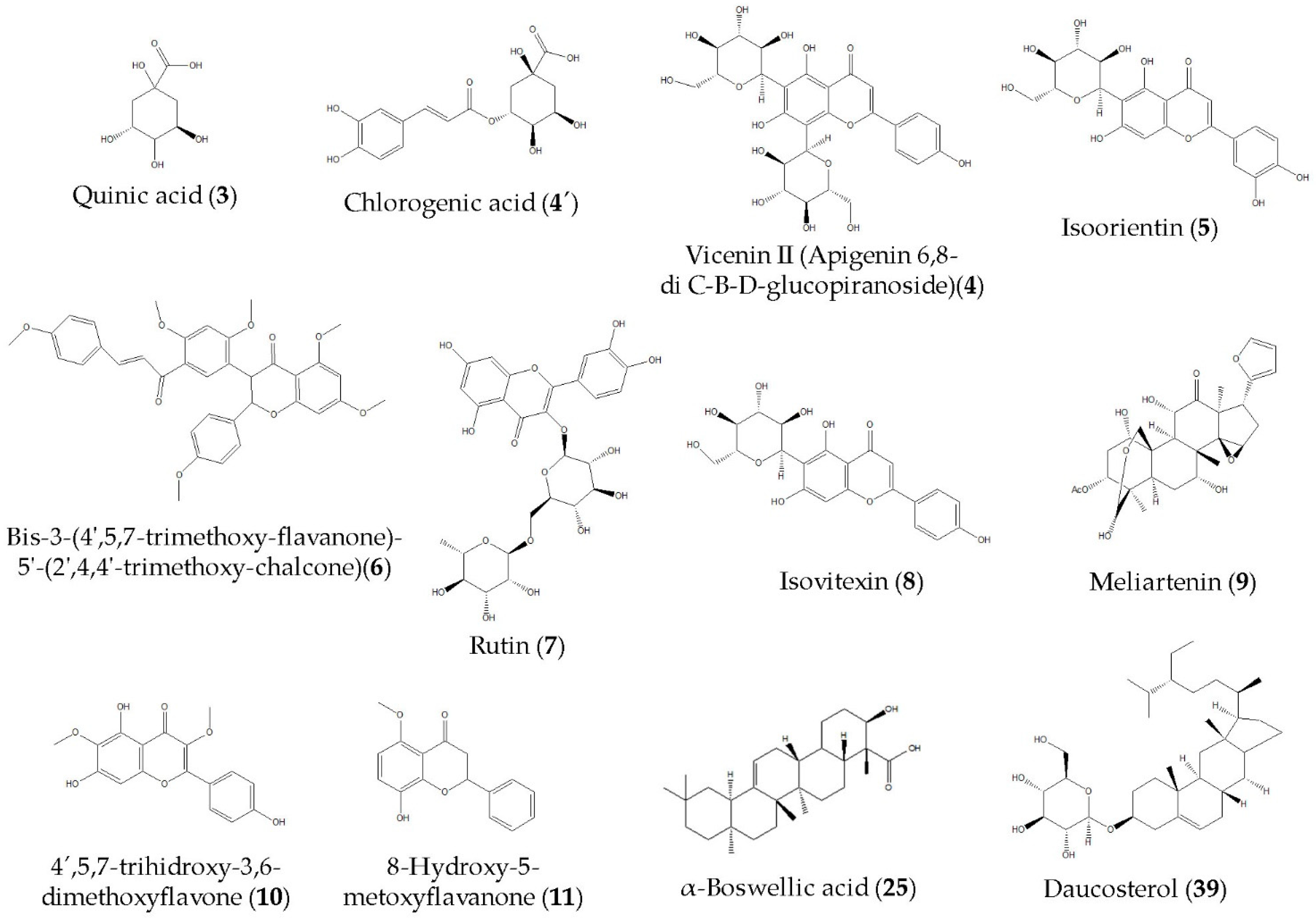
The exhaustive UHPLC-ESI-QTOF-MS analysis in negative mode revealed the presence of thirty-eight peaks in MGEOe, corresponding to forty compounds, and twenty-seven peaks corresponding to twenty-nine compounds in Doe. These included C-glycosyl flavones, flavones, aromatics, terpenes, sterols, furans, and several fatty acids; all of them are tentatively identified for the first time in O. erythrorhiza. The identification strategy implemented involved spiking experiments with available standards. Additionally, a comprehensive access strategy to several databases, including MONA mass spectrometry and Metaboscape, was employed, along with the use of specific software such as Data Analysis 4.0 (Bruker Daltonik GmbH, Bremen, Germany) and ACD Lab Spectrum Processor (Toronto, Canada). Figure 1 and Table 1 display the high-resolution UHPLC–PDA–QTOF analysis results for metabolite identification in Doe and MGEOe. Additionally, Figure 2 shows the chemical structures of some of the main compounds identified in Doe and MGEOe.

3.2. In Vitro Studies

3.2.1. Total Phenolic and Flavonoid Contents, Antioxidant Activity and Inhibition Enzyme COX of DOe and MGEOe

Both extracts exhibited high levels of total phenolics, measuring 69 mg GAE/g for DOe and 65 mg GAE/g for MGEOe. Also, the flavonoid contents were 43 mg QE/g DOe and 31 mg QE/g MGEOe (Table 2). Compared to catechin, quercetin and gallic acid (value range EC50: 1–10 µg/mL), the antioxidant activity in the DPPH assay was moderate for both extracts (DOe EC50: 70 µg/mL and MGEOe EC50: 62 µg/mL). DOe and MGEOe exhibited a significant ILP in erythrocytes, reaching 81–83%, respectively, at 250 μg DOe or MGEOe/mL (Table 2), compared to positive control catechin, which showed 70% of ILP at 100 µg/mL. Regarding enzyme inhibition of COX-1 and 2, DOe showed a good percentage of inhibition of COX-2 (86.97% at 72 µg/mL).

3.2.2. Cytotoxicity Study of DOe and MGEOe

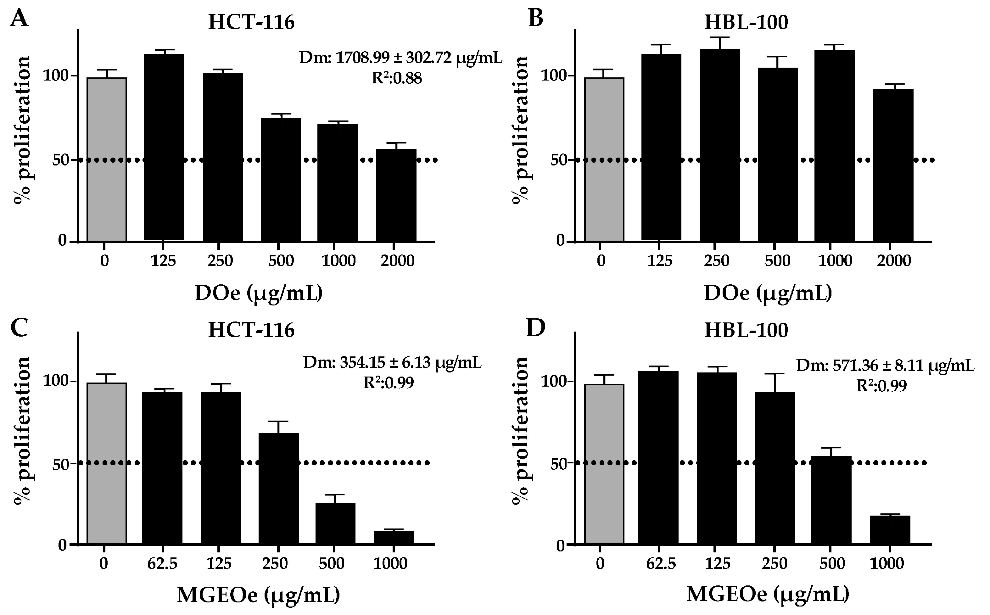
DOe produced a slight cytotoxic effect on HCT-116, but not on HBL-100 (Figure 3A,B). The dose–response effect against proliferation was present (R2 = 0.88), but the maximum dose was not enough to reach the Dm. The estimated Dm value was 1708.99 ± 302.72 µg/mL. In contrast, MGEOe induces marked cytotoxic effects in both cell lines. The dose–response effects against proliferation were present in both cell lines (R2 = 0.99). The Dm estimated was 354.15 ± 6.13 µg/mL for HCT-116 (Figure 3C) and 571.36 ± 8.11 µg/mL for HBL-100 (Figure 3D). The comparison between both Dm values revealed significant differences, revealing specific cytotoxic effects on HCT-116 (p = 0.002). The comparison between the extracts revealed that MGEOe showed higher potency than DOe on HCT-116 (p = 0.0015).

3.3. In Vivo Study

3.3.1. BW Gain and Average Beverage Volume

The analysis of BW gain did not show any significant differences between HDOeS, DOeS and SUC (Figure 4a). Regarding to the average beverage volume, HDOeS and DOeS drank less beverage than SUC (16.35% and 29.21%, respectively; p < 0.0001). Also, DOeS drank less beverage than HDOeS (15.39%; p < 0.0001 Figure 4b).

3.3.2. GTT and Serum Parameters

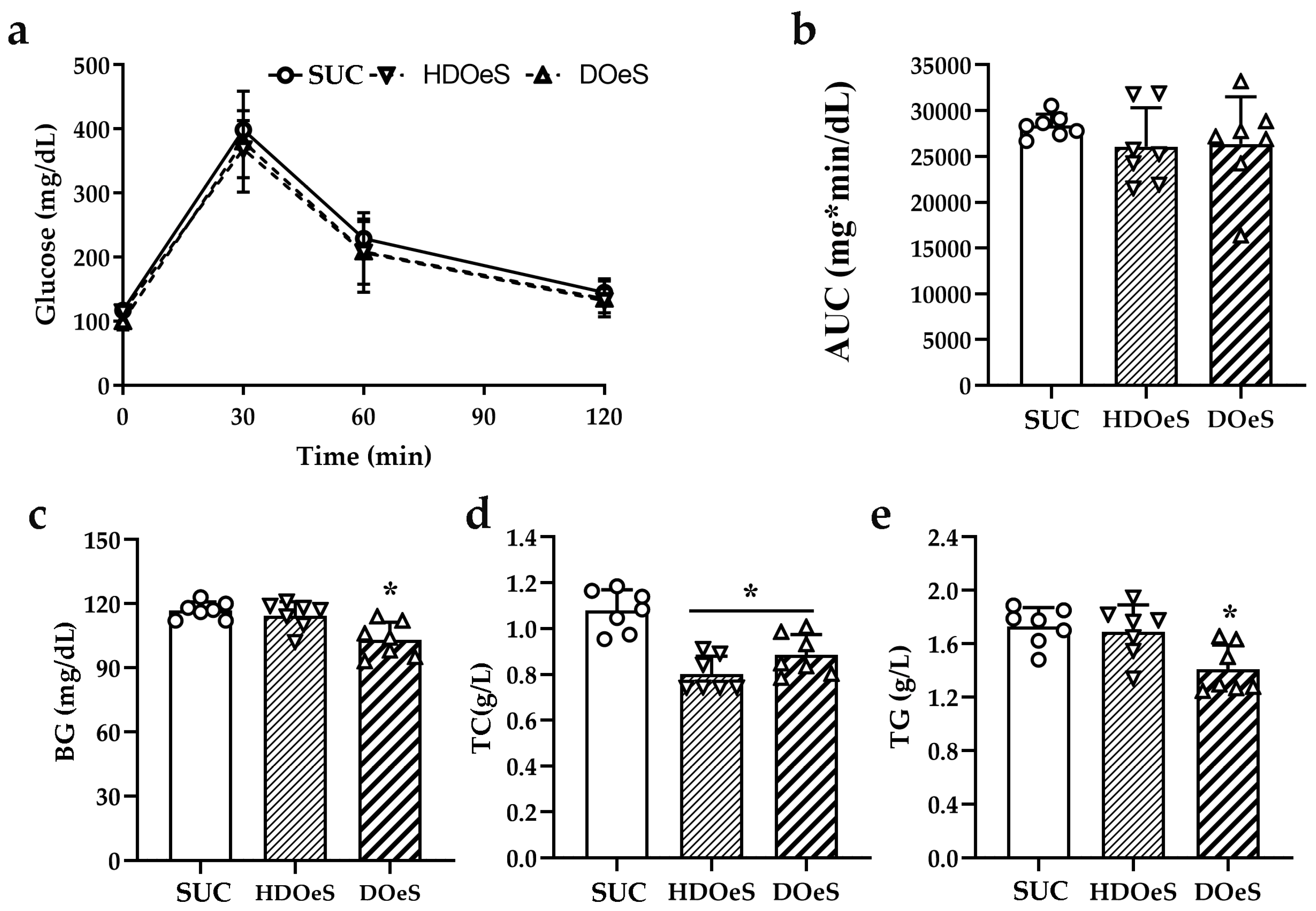
The analysis of AUC did not show significant differences between the groups (Figure 5a,b). No significant differences were observed in the levels of BG of HDOeS and SUC. In contrast, DOeS presented a decrease in BG levels compared to SUC (14.67%; p < 0.01) and HDOeS (12.86%; p < 0.001; Figure 5c). The levels of TC in HDOeS and DOeS were lower than those of SUC (17.44% and 11.69%, respectively; p < 0.05). Also, no significant differences were observed between HDOeS and DOeS (Figure 5d). The analysis of TG levels did not show significant differences between the HDOeS and SUC groups. However, DOeS presented lower levels than SUC and HDOeS (18.44% and 16.61%, respectively; p < 0.01; Figure 5e).

3.3.3. TBAR Assay

The analysis did not reveal significant differences between HDOeS and DOeS and SUC in any of the tissues evaluated (Table 3).

3.3.4. Behavioral Tests

OFT and EPM

In the OFT, the analysis of total distance and lines crossing did not show significant differences between the groups (FTotaldistance(2,27) = 0.646; p = 0.5322; FLines(2,27) = 0.822; p = 0.4504). In the zone 1, HDOeS entered more and spent more time than SUC (67.74%; p < 0.001 and 70.42%; p < 0.05, respectively, Figure 6a,b). However, the performance of DOeS was similar to SUC. Also, DOeS entered less and spent less time in the zone than HDOeS (50.77%; p < 0.001 and 43.57%; p < 0.05, respectively, Figure 6a,b). In zones 2 and 3, no significant differences were observed between the groups (Figure 6c,d). However, HDOeS spent less time in zone 2 (38.55%; p < 0.05) and more time in zone 3 (6.63%; p = 0.05) than HDOeS (Figure 6c,d). The analysis of the remaining parameters within each zone did not reveal significant differences between the groups (FDistancezone1(2,27) = 2.277; p = 0.1219; FEntrieszone2(2,27) = 2.388; p = 0.1110; FDistancezone2(2,27) = 2.185; p = 0.1319; FEntrieszone3(2,27) = 0.944; p = 0.4016 and FDistancezone3(2,27) = 2.193; p = 0.1311).
The analysis of grooming behavior did not indicate significant differences in either the number of events or the duration of the activity (FEvents(2,22) = 2.201; p = 0.1344 and FTime(2,22) = 0.081; p = 0.9222). It is important to note that only half of HDOeS performed the activity. Similarly, the analysis of rearing did not reveal significant differences in the number of events and/or time spent on the activity (FEvents(2,27) = 0.119; p = 0.8886 and FTime(2,27) = 2.154; p = 0.1356).
In the EPM, the analysis did not reveal significant differences between the groups in any of the parameters evaluated in closed (FEntries(2,27) = 2.536; p = 0.0979; FTime(2,27) = 0.712; p = 0.4996 and FDistance(2,27) = 0.588; p = 0.5625) and open arms (FEntries(2,27) = 3.218; p = 0.0558; FTime(2,27) = 2.312; p = 0.1183 and FDistance(2,27) = 2.312; p = 0.1183), and center (FEntries(2,27) = 2.815; p = 0.776; FTime(2,27) = 0.847; p = 0.4398 and FDistance(2,27) = 1.059; p = 0.3608). The activities of grooming and rearing were only observed in the closed arms. The analysis did not reveal significant differences in grooming (FEvents(2,22) = 0.691; p = 0.5114 and FTime(2,22) = 3.118; p = 0.0642) and rearing (FEvents(2,27) = 0.079; p = 0.9246 and FTime(2,27) = 1.755; p = 0.1921). It is important to note that not all the animals performed grooming (SUC n = 7; HDOeS n = 9 and DOeS n = 9). The analysis of head dip events did not show significant differences among the groups (FEvents(2,27) = 2.241; p = 0.1258).

NOL

The HDOeS group presented an increase in ER compared to SUC (47.03%; p = 0.0037). Also, no differences were observed in the amount of crossed lines in T1 and T2 and the time dedicated to exploring each object in T1 (Figure 7). It is important to note that the DOeS group was excluded from the analysis because only two animals explored both objects for the required duration during the trials.

4. Discussion

In this work, forty compounds and twenty-nine compounds were tentatively identified in MGEOe and DOe, respectively, including flavones (6, 7, 10 and 11), C-glycosyl flavones (4 and 5), isoflavone (8), fatty acids (2, 1418, 2026, 2835 and 37), terpenes (27, 38), phenolic acids (4′ and 19), aromatics (12 and 13), furane (9) and sterols (36 and 39), using UHPLC-ESI-QTOF-MS analysis in negative mode. These identifications represent the first report for O. erythrorhiza, providing an updated account of the chemical composition of this Andean species. Recent studies have reported the metabolite profiling of various medicinal plants and fruits collected in Argentina and Chile, utilizing two powerful techniques: UHPLC-ESI-OT-MS and UHPLC-ESI-QTOF-MS. These exhaustive analyses have significantly contributed to updating the chemical composition of all the Andean medicinal plants studied, providing a more comprehensive understanding of their bioactive compounds. On the other hand, it has allowed us to provide solid support for some of the evaluated activities by identifying the potential compounds responsible for the biological effects, including the strong antioxidant properties of the decoctions and polar extracts from most of the analyzed species [14,15,16,22,23,31,32,33,34,35].
In the last decade, there has been an increase in research focused on identifying new sources of antioxidant compounds derived from natural products, such as fruits and medicinal plants, in their various forms of consumption. These compounds have the potential to serve as therapeutic options for treating pathological conditions where free radicals play a recognized role [36]. In this study, antioxidant activity was evaluated using assays based on electron transfer mechanisms, including FCR, TEAC, FRAP, and DPPH assays, which can also be neutralized by hydrogen atom donation [36,37]. Among these methods, the DPPH assay is particularly notable for its strengths; it is simple, cost-effective, and rapid, with reproducible results that are comparable to other radical scavenging methods. It is highly sensitive and often shows good correlation with bioactive compounds such as phenols and flavonoids, typically yielding a regression factor R > 0.8. However, these strengths are counterbalanced by certain limitations, including its sensitivity to the presence of Lewis bases, which can affect DPPH absorbance. Additionally, the analysis must be conducted in the dark or protected from light to prevent absorbance decay. Moreover, the absence of DPPH free radicals in the human body limits its physiological relevance [36].
The in vitro studies of MGEOe and DOe revealed significant antioxidant activity, highlighting the effect on ILP process. This could be explained at least in part by the presence of chlorogenic acid (4′), isoorientin (5), rutin (7), isovitexin (8), 4′,5,7-trihydroxy-3,6-dimethoxyflavone (10), pinoleic acid (22), alpha-boswellic acid (25), and daucosterol (39) among others, which have been reported to have antioxidant activity [38,39,40,41,42,43,44,45,46,47,48,49]. However, the in vivo studies of HDOeS and DOeS did not reveal any significant effects on MDA levels in the tissues evaluated.
The Andean flora has been scarcely investigated for its cytotoxic properties. Recently, extracts and decoctions of two species growing in Argentina have demonstrated cytotoxic potential, supported in part by the comprehensive chemical characterization of these extracts and decoctions [16,35]. The cytotoxicity assays revealed that DOe produced a selective cytotoxic effect against a tumoral cell line, with no effects in non-tumoral cells, whereas MGEOe produced cytotoxic effects in both cell lines. Also, the effect of MGEOe was higher than that of DOe. These cytotoxic effects may be linked to the presence of vicentin II (4), chlorogenic acid (4′), isoorientin (5), rutin (7), meliartenin (9), alpha-boswellic acid (25), and daucosterol (39), which have been reported to have antitumoral properties [38,39,42,49,50,51,52,53,54,55,56].
The analysis of COX inhibition revealed that DOe produced a significant anti-inflammatory effect. This may be related to the presence of certain compounds tentatively identified in the UHPLC-MS analysis, including vicentin II (4), chlorogenic acid (4′), isoorientin (5), isovitexin(8), ricinoleic acid (18), pinoleic acid (22), alpha-boswellic acid (25), and daucosterol (39), which has reported anti-inflammatory properties [38,40,43,44,45,47,48,49,57,58,59,60,61,62].
On the other hand, the selected in vivo model of insulin resistance is characterized by alterations in glucose and lipid metabolism, oxidative state, as well as cognitive impairments, including anxiety-like behavior and memory deficits [20]. In this work, the administration of HDOeS and DOeS did not modify the BW gain. In contrast, several reports describe that compounds like vicentin II (4), chlorogenic acid (4′) and rutin (7), among others, could modify the BW gain [63,64,65,66,67]. Although these treatments did not modify the response to glucose overload, the animals presented a decrease in BG. This effect may be linked to the presence of compounds such as quinic acid (3), chlorogenic acid (4′) and bis-3-(4′,5,7-trimethoxy-flavanone)-5′-(2′,4,4′-trimethoxy-chalcone, (6), which can play vital functions in the process of regulating glucose metabolism [38,68,69]. Additionally, daucosterol (39) may inhibit key glucose metabolism enzymes, helping to maintain blood sugar levels within normal ranges [45]. Regarding the lipid parameters, the administration of the decoction reduces the levels of TC and TG. The hypocholesterolemic properties can be attributed to the combined action of flavonoids, fatty acids, phenolic compounds, and sesquiterpenes, all of which have hypolipidemic effects through several mechanisms [22,23,38,43,48,49,61,70,71,72].
In the evaluation of anxiety levels, only the administration of HDOeS produced an anxiolytic effect, which could be attributed to the action of compounds like chlorogenic acid (4′) [73], rutin (7) [74], isovitexin (8) [75], vicenin II (4) [76], ricinoleic acid isomer (20) [77], over different pathways. In addition, only the administration of HDOeS improved the spatial memory performance of the animals. This could be attributed to the content of chlorogenic acid (4′) [78], rutin (7) [79], vicentin II (4) [80], daucosterol (39) [81], among others, which are capable of improving the cognitive state through different mechanisms. In the animal model reported in this work, the variation in results of the doses employed could be attributed to the difference in the beverage intake. Furthermore, it is important to highlight that administering both doses of decoction to the control animals did not affect their well-being or the evaluated parameters (see Supplementary Materials).
Although the results are promising, additional research is needed to establish whether O. erythrorhiza can be regarded as a new source of phytocompounds. The demonstrated antioxidant, anti-tumoral, anti-inflammatory, hypoglycemic, hypocholesterolemic, anxiolytic, and memory-enhancing activities of O. erythrorhiza open the way for future studies, including the incorporation of its extracts into polymeric systems, which can preserve the integrity of drugs and biomacromolecules, such as nanoparticles [82,83].

5. Conclusions

In summary, the findings presented in this study provide an updated chemical profile of the Andean medicinal species O. erythrorhiza. Several of the reported compounds could explain, at least partially, the demonstrated antioxidant, anti-tumoral, anti-inflammatory, hypoglycemic, hypocholesterolemic, anxiolytic and memory improvement activities of O. erythrorhiza. While mechanistic and molecular assays are necessary to clarify the action pathways, the current study provides valuable information about this species that may be beneficial in addressing various health conditions. Additionally, to our knowledge, this is the first work that involves the study of O. erythrorhiza on in vitro and in vivo assays. Although the results are promising, additional research is needed to determine whether O. erythrorhiza can be regarded as a new source of beneficial phytocompounds relevant to human health.

Supplementary Materials

The following supporting information can be downloaded at: https://www.mdpi.com/article/10.3390/antiox13121494/s1, Gómez et al.: Table S1. BW gain (g) and average volume intake (mL/g rat); Table S2. AUC values from GTT; Table S3. Levels of BG (mg/dL), TC (g/L) and TG (g/L); Table S4. Levels of MDA (fmol/µg of protein); Table S5. OFT; Table S6. EPM, Table S7. NOL and Figure S1. MSn spectra of compounds detected by HR-MS in DOe and MGEOe (extracted using Bruker data analisis 4.0). Quinic acid (3); Rutin (7); Isovitexin (8); 8-hydroxy-5-methoxyflavanone (11); Diffractaic acid (13); Roccellaric acid (24) and Pristimerin (27).

Author Contributions

Conceptualization, M.R., A.T., M.J.S., C.G.-L., B.L. and H.C.; Data curation, M.R., M.S.K., J.G., M.J.S., A.T. and H.C.; Formal analysis, M.R., J.G., M.J.S., J.Z., M.P., B.L., C.G.-L., A.T. and H.C.; Funding acquisition, M.R., M.J.S., J.G., C.G.-L., A.T. and H.C.; Investigation, M.R., M.S.K., J.G., C.G.-L., M.J.S., A.T., B.L. and H.C.; Methodology, M.R., M.S.K., C.G.-L., M.J.S., A.T. and H.C.; Project administration, M.R., M.J.S., A.T. and H.C.; Resources, M.R., J.G., M.J.S., A.T. and H.C.; Software, M.R., J.G., M.J.S., A.T. and H.C.; Supervision, M.J.S., A.T. and H.C.; Validation, M.R., M.S.K., J.G., M.J.S., A.T. and H.C.; Visualization, M.R.; Writing—original draft, M.R., J.G., M.J.S., B.L., C.G.-L., A.T. and H.C.; Writing—review and editing, M.R., M.S.K., J.G., M.J.S., A.T. and H.C. All authors have read and agreed to the published version of the manuscript.

Funding

This research was supported by grants from Argentina and Chile: Agencia Nacional de Promoción Científica y Tecnológica (ANPCyT; grant numbers PICT-2019-0623 (M.R.) and PICT 2019-03791 (C.G.-L.)); Consejo Nacional de Investigaciones Científicas y Técnicas (CONICET, grant number PIP-0243 (H.C.)), Secretaría de Investigación, Internacionales y Posgrado—Universidad Nacional de Cuyo (SIIP-UNCuyo, grant number 06/J019-T1 (C.G.-L.)). Also, this research was funded by PROJOVI ((J.G.) Resolución Nº 1500-23-R and (PDTS) Resolución Nº 1499-23-R); CICITCA-UNSJ, Argentina. M.J.S. received financial support from Fondecyt (Grant 1220075) and Fondequip (EQM170172), Chile.

Institutional Review Board Statement

All procedures related to animal care and use were conducted in accordance with the European Community Council Directive (86/609/EEC) and the guidelines of the National Institutes of Health Guide for the Care and Use of Laboratory Animals. These procedures were also approved by the ethical committee of CICUAL, IBYME, in 2021, located in CABA, Argentina.

Informed Consent Statement

Not applicable.

Data Availability Statement

Data are contained within the article.

Acknowledgments

The authors express their gratitude to María Irantzu Sanz de Larrechea, HCP and Demand Generation Specialist at Johnson & Johnson, for providing the blood glucose test strips. They also thank Mónica B. Frungieri and Soledad P. Rossi from the Laboratory of Neuro-Immuno-Endocrinology at IBYME-CONICET for their technical assistance and guidance regarding TBARS. Additionally, they acknowledge the funding received from ANPCyT, CONICET, UNSJ (Argentina), and Fondecyt (Chile).

Conflicts of Interest

The authors declare no conflicts of interest.

References

  1. Barbinta-Patrascu, M.E.; Bita, B.; Negut, I. From Nature to Technology: Exploring the Potential of Plant-Based Materials and Modified Plants in Biomimetics, Bionics, and Green Innovations. Biomimetics 2024, 9, 390. [Google Scholar] [CrossRef] [PubMed]
  2. Bustos, D.A.; Tapia, A.A.; Feresin, G.E.; Ariza Espinar, L. Ethnopharmacobotanical survey of Bauchazeta district, San Juan Province, Argentina. Fitoterapia 1996, 67, 411–415. [Google Scholar]
  3. Lourteig, A. Oxalidaceae. In Flora Patagónica, Colección Científica; Correa, M.N., Ed.; Instituto Nacional de Tecnología Agropecuaria: Buenos Aires, Argentina, 1988; Volume 8, pp. 1–29. [Google Scholar]
  4. Feresin, G.E.; Tapia, A.; López, S.N.; Zacchino, S.A. Antimicrobial Activity of Plants Used in Traditional Medicine of San Juan Province, Argentine. J. Ethnopharmacol. 2001, 78, 103–107. [Google Scholar] [CrossRef] [PubMed]
  5. Feresin, G.E.; Tapia, A.; Sortino, M.; Zacchino, S.; Arias, A.R.D.; Inchausti, A.; Yaluff, G.; Rodriguez, J.; Theoduloz, C.; Schmeda-Hirschmann, G. Bioactive Alkyl Phenols and Embelin from Oxalis erythrorhiza. J. Ethnopharmacol. 2003, 88, 241–247. [Google Scholar] [CrossRef] [PubMed]
  6. Gudasi, S.; Gharge, S.; Koli, R.; Patil, K. Antioxidant properties and cytotoxic effects of Oxalis corniculata on human Hepatocarci-noma (Hep-G2) cell line: An in vitro and in silico evaluation. Futur. J. Pharm. Sci. 2023, 9, 25. [Google Scholar] [CrossRef]
  7. Gholipour, A.R.; Jafari, L.; Ramezanpour, M.; Evazalipour, M.; Chavoshi, M.; Yousefbeyk, F.; Kargar Moghaddam, S.J.; Yekta Kooshali, M.H.; Ramezanpour, N.; Daei, P.; et al. Apoptosis Effects of Oxalis corniculata L. Extract on Human MCF-7 Breast Cancer Cell Line. Galen Med. J. 2022, 11, e2484. [Google Scholar]
  8. Zhang, J.; Shen, W.; He, H. Exploring the action mechanism of Oxalis corniculata L. decoction in treating osteoarthritis utilizing liquid chromatography–mass spectrometry technology combined with network pharmacology. Medicine 2024, 103, e39515. [Google Scholar] [CrossRef]
  9. Jifar, W.W.; Debele, G.R.; Kanfe, S.G.; Mule, C.T. Evaluation of in vivo Antidiabetic, Antidyslipidemic and in vitro Anti-Oxidant Activity of Extract and Solvent Fractions of Discopodium penninervum Hoschst Leaf in Mice: Normoglycemic and Streptozocin-Induced Model. J. Exp. Pharmacol. 2022, 28, 317–330. [Google Scholar] [CrossRef]
  10. Kabach, I.; Bouchmaa, N.; Zouaoui, Z.; Ennoury, A.; El Asri, S.; Laabar, A.; Oumeslakht, L.; Cacciola, F.; El Majdoub, Y.O.; Mondello, L.; et al. Phytochemical Profile and Antioxidant Capacity, α-Amylase and α-Glucosidase Inhibitory Activities of Oxalis Pes-Caprae Extracts in Alloxan-Induced Diabetic Mice. Biomed. Pharmacother. 2023, 160, 114393. [Google Scholar] [CrossRef]
  11. Abhilash, P.A.; Nisha, P.; Prathapan, A.; Nampoothiri, S.V.; Lijo Cherian, O.; Sunitha, T.K.; Raghu, K.G. Cardioprotective Effects of Aqueous Extract of Oxalis Corniculata in Experimental Myocardial Infarction. Exp. Toxicol. Pathol. 2011, 63, 535–540. [Google Scholar] [CrossRef]
  12. Gupta, G.; Kazmi, I.; Afzal, M.; Rahman, M.; Anwar, F. Anxiolytic Effect of Oxalis Corniculata (Oxalidaceae) in Mice. Asian Pac. J. Trop. Dis. 2012, 2, S837–S840. [Google Scholar] [CrossRef]
  13. Aruna, K.; Rajeswari, P.D.R.; Sankar, S.R. The Effect of Oxalis Corniculata Extract against the Behavioral Changes Induced by 1-Methyl- 4-Phenyl-1,2,3,6-Tetrahydropyridine (MPTP) in Mice. J. Appl. Pharm. Sci. 2017, 7, 148–153. [Google Scholar]
  14. Manrique, S.; Gómez, J.; Piñeiro, M.; Sampietro, B.A.; Peschiutta, M.L.; Tapia, A.; Simirgiotis, M.J.; Lima, B. Zuccagnia punctata Cav., a Potential Environmentally Friendly and Sustainable Bionematicide for the Control of Argentinean Horticultural Crops. Plants 2023, 12, 4104. [Google Scholar] [CrossRef] [PubMed]
  15. Luna, L.; Simirgiotis, M.J.; Lima, B.; Bórquez, J.; Feresin, G.E.; Tapia, A. UHPLC-MS Metabolome Fingerprinting: The Isolation of Main Compounds and Antioxidant Activity of the Andean Species Tetraglochin ameghinoi (Speg.) Speg. Molecules 2018, 23, 793. [Google Scholar] [CrossRef] [PubMed]
  16. Gómez, J.; Simirgiotis, M.J.; Lima, B.; Paredes, J.D.; Villegas Gabutti, C.M.; Gamarra-Luques, C.; Bórquez, J.; Luna, L.; Wendel, G.H.; Maria, A.O.; et al. Antioxidant, gastroprotective, cytotoxic activities and UHPLC PDA-Q orbitrap mass spectrometry identification of metabolites in Baccharis grisebachii decoction. Molecules 2019, 24, 1085. [Google Scholar] [CrossRef] [PubMed]
  17. Kulmacz, R.J.; Lands, W.E. Requirements for Hydroperoxide by the Cyclooxygenase and Peroxidase Activities of Prostaglandin H Synthase. Prostaglandins 1983, 25, 531–540. [Google Scholar] [CrossRef]
  18. Torres Carro, R.; D’Almeida, R.E.; Isla, M.I.; Alberto, M.R. Antioxidant and Anti-Inflammatory Activities of Frankenia Triandra (J. Rémy) Extracts. S. Afr. J. Bot. 2016, 104, 208–214. [Google Scholar] [CrossRef]
  19. Chou, T.C.; Martin, N. CompuSyn for Dose–Effect Analysis and for Quantitation of Synergism and Antagonism. Software and Manual; ComboSyn: Paramus, NJ, USA, 2004. [Google Scholar]
  20. Rey, M.; Coirini, H.; Marchena, A.; González-Deniselle, M.C.; Kruse, M.S. Effects of metformin on behavioral alterations produced by chronic sucrose consumption in male rats. J. Neuroendocrinol. 2024, 36, e13362. [Google Scholar] [CrossRef]
  21. Rey, M.; Kruse, M.S.; Magrini-Huamán, R.N.; Coirini, H. High-Fat Diets and LXRs Expression in Rat Liver and Hypothalamus. Cell. Mol. Neurobiol. 2019, 39, 963–974. [Google Scholar] [CrossRef]
  22. Rey, M.; Kruse, M.S.; Magrini-Huamán, R.N.; Gómez, J.; Simirgiotis, M.J.; Tapia, A.; Feresin, G.E.; Coirini, H. Tessaria absinthioides (Hook. & Arn.) DC. (Asteraceae) Decoction Improves the Hypercholesterolemia and Alters the Expression of LXRs in Rat Liver and Hypothalamus. Metabolites 2021, 11, 579. [Google Scholar] [CrossRef]
  23. Rey, M.; Kruse, M.S.; Gómez, J.; Simirgiotis, M.J.; Tapia, A.; Coirini, H. Ultra-High-Resolution Liquid Chromatography Coupled with Electrospray Ionization Quadrupole Time-of-Flight Mass Spectrometry Analysis of Tessaria absinthioides (Hook. & Arn.) DC. (Asteraceae) and Antioxidant and Hypocholesterolemic Properties. Antioxidants 2024, 13, 50. [Google Scholar]
  24. Bradford, M.M. A rapid and sensitive method for the quantitation of microgram quantities of protein utilizing the principle of protein-dye binding. Anal. Biochem. 1976, 72, 248–254. [Google Scholar] [CrossRef] [PubMed]
  25. Sturman, O.; Germain, P.L.; Bohacek, J. Exploratory Rearing: A Context- and Stress-Sensitive Behavior Recorded in the Open-Field Test. Stress 2018, 21, 443–452. [Google Scholar] [CrossRef] [PubMed]
  26. Veloso, A.W.N.; Filgueiras, G.B.; Lorenzo, P.; Estanislau, C. Modulation of Grooming Behavior in Rats by Different Test Situations. Psychol. Neurosci. 2016, 9, 91–104. [Google Scholar] [CrossRef]
  27. Cruz, A.D.M.; Frei, F.; Graeff, F.G. Ethopharmacological analysis of rat behavior on the elevated plus-maze. Pharmacol. Biochem. Behav. 1994, 49, 171–176. [Google Scholar] [CrossRef]
  28. Rodgers, R.J.; Johnson, N.J. Factor analysis of spatiotemporal and ethological measures in the murine elevated plus-maze test of anxiety. Pharmacol. Biochem. Behav. 1995, 52, 297–303. [Google Scholar] [CrossRef]
  29. Espejo, E.F. Effects of weekly or daily exposure to the elevated plus-maze in male mice. Behav. Brain Res. 1997, 87, 233–238. [Google Scholar] [CrossRef]
  30. Holmes, A.; Rodgers, R.J. Responses of Swiss-Webster mice to repeated plus-maze experience: Further evidence for a qualitative shift in emotional state? Pharmacol. Biochem. Behav. 1998, 60, 473–488. [Google Scholar] [CrossRef]
  31. Gómez, J.; Simirgiotis, M.J.; Lima, B.; Gamarra-Luques, C.; Bórquez, J.; Caballero, D.; Feresin, G.E.; Tapia, A. UHPLC–Q/Orbitrap/MS/MS fingerprinting, free radical scavenging, and antimicrobial activity of Tessaria absinthiodes (Hook. &arn.) DC. (Asteraceae) lyophilized decoction from Argentina and Chile. Antioxidants 2019, 8, 593. [Google Scholar] [CrossRef]
  32. Gómez, J.; Simirgiotis, M.J.; Manrique, S.; Piñeiro, M.; Lima, B.; Bórquez, J.; Feresin, G.E.; Tapia, A. UHPLC-ESI-OT-MS Phenolics Profiling, Free Radical Scavenging, Antibacterial and Nematicidal Activities of “Yellow-Brown Resins” from Larrea spp. Antioxidants 2021, 10, 185. [Google Scholar] [CrossRef]
  33. Gómez, J.; Simirgiotis, M.J.; Manrique, S.; Lima, B.; Bórquez, J.; Feresin, G.E.; Tapia, A. UHPLC-HESI-OT-MS-MS biomolecules profiling, antioxidant and antibacterial activity of the “orange-yellow resin” from Zuccagnia punctata Cav. Antioxidants 2020, 9, 123. [Google Scholar] [CrossRef] [PubMed]
  34. Ramos, L.C.; Palacios, J.; Barrientos, R.E.; Gómez, J.; Castagnini, J.M.; Barba, F.J.; Tapia, A.; Paredes, A.; Cifuentes, F.; Simirgiotis, M.J. UHPLC-MS Phenolic Fingerprinting, Aorta Endothelium Relaxation Effect, Antioxidant, and Enzyme Inhibition Activities of Azara dentata Ruiz & Pav Berries. Foods 2023, 12, 643. [Google Scholar] [CrossRef] [PubMed]
  35. Persia, F.A.; Troncoso, M.E.; Rinaldini, E.; Simirgiotis, M.; Tapia, A.; Bórquez, J.; Mackern-Oberti, J.P.; Hapon, M.B.; Gamarra-Luques, C. UHPLC–Q/Orbitrap/MS/MS Fingerprinting and Antitumoral Effects of Prosopis Strombulifera (LAM.) BENTH. Queous Extract on Allograft Colorectal and Melanoma Cancer Models. Heliyon 2020, 6, e03353. [Google Scholar] [CrossRef] [PubMed]
  36. Bibi Sadeer, N.; Montesano, D.; Albrizio, S.; Zengin, G.; Mahomoodally, M.F. The versatility of antioxidant assays in food science and safety—Chemistry, applications, strengths, and limitations. Antioxidants 2020, 9, 709. [Google Scholar] [CrossRef] [PubMed]
  37. Huang, D.; Ou, B.; Prior, R.L. The chemistry behind antioxidant capacity assays. J. Agric. Food Chem. 2005, 53, 1841–1856. [Google Scholar] [CrossRef]
  38. Naveed, M.; Hejazi, V.; Abbas, M.; Kamboh, A.A.; Khan, G.J.; Shumzaid, M.; Xiao Hui, Z. Chlorogenic acid (CGA): A pharmacological review and call for further research. Biomed. Pharmacother. 2018, 97, 67–74. [Google Scholar] [CrossRef]
  39. Bender, O.; Atalay, A. Chapter 28—Polyphenol chlorogenic acid, antioxidant profile, and breast cancer. In Cancer, 2nd ed.; Academic Press: Cambridge, MA, USA, 2021; pp. 311–321. [Google Scholar]
  40. Liang, N.; Kitts, D.D. Role of chlorogenic acids in controlling oxidative and inflammatory stress conditions. Nutrients 2015, 8, 16. [Google Scholar] [CrossRef]
  41. Yuan, L.; Wang, J.; Wu, W.; Liu, Q.; Liu, X. Effect of isoorientin on intra-cellular antioxidant defense mechanisms in hepatoma and liver cell lines. Biomed. Pharmacother. 2016, 81, 356–362. [Google Scholar] [CrossRef]
  42. Ziqubu, K.; Dludla, P.V.; Joubert, E.; Muller, C.J.; Louw, J.; Tiano, L.; Mazibuko-Mbeje, S.E. Isoorientin: A dietary flavone with the potential to ameliorate diverse metabolic complications. Pharmacol. Res. 2020, 158, 104867. [Google Scholar] [CrossRef]
  43. Yuan, L.; Han, X.; Li, W.; Ren, D.; Yang, X. Isoorientin prevents hyperlipidemia and liver injury by regulating lipid metabolism, antioxidant capability, and inflammatory cytokine release in high-fructose-fed mice. J. Agric. Food Chem. 2016, 64, 2682–2689. [Google Scholar] [CrossRef]
  44. He, M.; Min, J.W.; Kong, W.L.; He, X.H.; Li, J.X.; Peng, B.W. A review on the pharmacological effects of vitexin and isovitexin. Fitoterapia 2016, 115, 74–85. [Google Scholar] [CrossRef] [PubMed]
  45. Hu, J.J.; Wang, H.; Pan, C.W.; Lin, M.X. Isovitexin alleviates liver injury induced by lipopolysaccharide/d-galactosamine by activating Nrf2 and inhibiting NF-?B activation. Microb. Pathog. 2018, 119, 86–92. [Google Scholar] [CrossRef] [PubMed]
  46. Durmaz, L. Antioxidant, antiepileptic, and anticholinergic properties of 4?, 5, 7-Trihydroxy-3, 6-dimethoxyflavone as natural phenolic compound: A toxicology approach. Toxin. Rev. 2021, 40, 292–299. [Google Scholar] [CrossRef]
  47. Zhang, Y.; Jia, J.; Ding, Y.; Ma, Y.; Shang, P.; Liu, T.; Wen, A. Alpha-boswellic acid protects against ethanol-induced gastric injury in rats: Involvement of nuclear factor erythroid-2-related factor 2/heme oxygenase-1 pathway. J. Pharm. Pharmacol. 2016, 68, 514–522. [Google Scholar] [CrossRef]
  48. Baker, E.J.; Miles, E.A.; Calder, P.C. A Review of the Functional Effects of Pine Nut Oil, Pinolenic Acid and Its Derivative Eicosatrienoic Acid and Their Potential Health Benefits. Prog. Lipid Res. 2021, 82, 101097. [Google Scholar] [CrossRef]
  49. El Omari, N.; Jaouadi, I.; Lahyaoui, M.; Benali, T.; Taha, D.; Bakrim, S.; El Menyiy, N.; El Kamari, F.; Zengin, G.; Bangar, S.P.; et al. Natural Sources, Pharmacological Properties, and Health Benefits of Daucosterol: Versatility of Actions. Appl. Sci. 2022, 12, 5779. [Google Scholar] [CrossRef]
  50. Ganeshpurkar, A.; Saluja, A.K. The pharmacological potential of rutin. Saudi Pharm. J. 2017, 25, 149–164. [Google Scholar] [CrossRef]
  51. Ghanbari-Movahed, M.; Shafiee, S.; Burcher, J.T.; Lagoa, R.; Farzaei, M.H.; Bishayee, A. Anticancer Potential of Apigenin and Isovitexin with Focus on Oncogenic Metabolism in Cancer Stem Cells. Metabolites 2023, 13, 404. [Google Scholar] [CrossRef]
  52. Joray, M.B.; Villafañez, F.; González, M.L.; Crespo, M.I.; Laiolo, J.; Palacios, S.M.; Carpinella, M.C. P53 tumor suppressor is required for efficient execution of the death program following treatment with a cytotoxic limonoid obtained from Melia azedarach. Food Chem. Toxicol. 2017, 109, 888–897. [Google Scholar] [CrossRef]
  53. Roy, N.K.; Deka, A.; Bordoloi, D.; Mishra, S.; Kumar, A.P.; Sethi, G.; Kunnumakkara, A.B. The potential role of boswellic acids in cancer prevention and treatment. Cancer Lett. 2016, 377, 74–86. [Google Scholar] [CrossRef]
  54. Zhao, C.; She, T.; Wang, L.; Su, Y.; Qu, L.; Gao, Y.; Xu, S.; Cai, S.; Shou, C. Daucosterol Inhibits Cancer Cell Proliferation by Inducing Autophagy through Reactive Oxygen Species-Dependent Manner. Life Sci. 2015, 137, 37–43. [Google Scholar] [CrossRef] [PubMed]
  55. Zeng, J.; Liu, X.; Li, X.; Zheng, Y.; Liu, B.; Xiao, Y. Daucosterol Inhibits the Proliferation, Migration, and Invasion of Hepatocellular Carcinoma Cells via Wnt/β-Catenin Signaling. Molecules 2017, 22, 862. [Google Scholar] [CrossRef] [PubMed]
  56. Zingue, S.; Gbaweng Yaya, A.J.; Michel, T.; Ndinteh, D.T.; Rutz, J.; Auberon, F.; Maxeiner, S.; Chun, F.K.H.; Tchinda, A.T.; Njamen, D.; et al. Bioguided Identification of Daucosterol, a Compound That Contributes to the Cytotoxicity Effects of Crateva Adansonii DC (Capparaceae) to Prostate Cancer Cells. J. Ethnopharmacol. 2020, 247, 112251. [Google Scholar] [CrossRef]
  57. Chen, Z.; Yang, Y.; Mi, S.; Fan, Q.; Sun, X.; Deng, B.; Wu, G.; Li, Y.; Zhou, Q.; Ruan, Z. Hepatoprotective Effect of Chlorogenic Acid against Chronic Liver Injury in Inflammatory Rats. J. Funct. Foods 2019, 62, 103540. [Google Scholar] [CrossRef]
  58. Lee, W.; Ku, S.K.; Bae, J.S. Vascular Barrier Protective Effects of Orientin and Isoorientin in LPS-Induced Inflammation in Vitro and in Vivo. Vascul. Pharmacol. 2014, 62, 3–14. [Google Scholar] [CrossRef]
  59. Li, Y.; Zhao, Y.; Tan, X.; Liu, J.; Zhi, Y.; Yi, L.; Bai, S.; Du, Q.; Li, Q.X.; Dong, Y. Isoorientin Inhibits Inflammation in Macrophages and Endotoxemia Mice by Regulating Glycogen Synthase Kinase 3β. Mediators Inflamm. 2020, 2020, 8704146. [Google Scholar] [CrossRef]
  60. Lin, C.M.; Huang, S.T.; Liang, Y.C.; Lin, M.S.; Shih, C.M.; Chang, Y.C.; Chen, T.Y.; Chen, C.T. Isovitexin Suppresses Lipopolysaccharide-Mediated Inducible Nitric Oxide Synthase through Inhibition of NF-Kappa B in Mouse Macrophages. Planta Med. 2005, 71, 748–753. [Google Scholar] [CrossRef]
  61. Takala, R.; Ramji, D.P.; Choy, E. The Beneficial Effects of PineNuts and Its Major Fatty Acid, Pinolenic Acid, on Inflammation and Metabolic Perturbations inInflammatory Disorders. Int. J. Mol. Sci. 2023, 24, 1171. [Google Scholar] [CrossRef] [PubMed]
  62. Vieira, C.; Evangelista, S.; Cirillo, R.; Lippi, A.; Maggi, C.A.; Manzini, S. Effect of ricinoleic acid in acute and subchronic experimental models of inflammation. Mediators Inflamm. 2000, 9, 223–228. [Google Scholar] [CrossRef]
  63. Seo, S.; Oh, S.; Shin, Y.; Jung, S.; Kim, Y. Reduction of body weight by rutin is associated with an increase of brown adipose tissue mitochondrial biogenesis in high-fat diet induced obese rat (LB430). FASEB J. 2014, 28, LB430. [Google Scholar] [CrossRef]
  64. Watanabe, T.; Kobayashi, S.; Yamaguchi, T.; Hibi, M.; Fukuhara, I.; Osaki, N. Coffee Abundant in Chlorogenic Acids Reduces Abdominal Fat in Overweight Adults: A Randomized, Double-Blind, Controlled Trial. Nutrients 2019, 16, 1617. [Google Scholar] [CrossRef] [PubMed]
  65. Yang, J.; Lee, J.; Kim, Y. Effect of Deglycosylated Rutin by Acid Hydrolysis on Obesity and Hyperlipidemia in High-Fat Diet-Induced Obese Mice. Nutrients 2020, 25, 1539. [Google Scholar] [CrossRef]
  66. Qiao, Y.; Zhang, Z.; Zhai, Y.; Yan, X.; Zhou, W.; Liu, H.; Guan, L.; Peng, L. Apigenin Alleviates Obesity-Associated Metabolic Syndrome by Regulating the Composition of the Gut Microbiome. Front. Microbiol. 2022, 12, 805827. [Google Scholar] [CrossRef]
  67. Kanchanasurakit, S.; Saokaew, S.; Phisalprapa, P.; Duangjai, A. Chlorogenic Acid in Green Bean Coffee on Body Weight: A Systematic Review and Meta-Analysis of Randomized Controlled Trials. Syst. Rev. 2023, 12, 163. [Google Scholar] [CrossRef]
  68. Heikkilä, E.; Hermant, A.; Thevenet, J.; Bermont, F.; Kulkarni, S.S.; Ratajczak, J.; Santo-Domingo, J.; Dioum, E.H.; Canto, C.; Barron, D.; et al. The plant product quinic acid activates Ca2+-dependent mitochondrial function and promotes insulin secretion from pancreatic beta cells. Br. J. Pharmacol. 2019, 176, 3250–3263. [Google Scholar] [CrossRef] [PubMed]
  69. Salehi, B.; Quispe, C.; Chamkhi, I.; El Omari, N.; Balahbib, A.; Sharifi-Rad, J.; Bouyahya, A.; Akram, M.; Iqbal, M.; Docea, A.O.; et al. Pharmacological Properties of Chalcones: A Review of Preclinical Including Molecular Mechanisms and Clinical Evidence. Front. Pharmacol. 2021, 11, 592654. [Google Scholar] [CrossRef]
  70. Meng, S.; Cao, J.; Feng, Q.; Peng, J.; Hu, Y. Roles of chlorogenic Acid on regulating glucose and lipids metabolism: A review. Evid. Based Complement. Alternat. Med. 2013, 2013, 801457. [Google Scholar] [CrossRef]
  71. Ding, L.; Zhang, L.; Wen, M.; Che, H.; Du, L.; Wang, J.; Xue, C.; Xu, J.; Wang, Y. Eicosapentaenoic acid-enriched phospholipids improve atherosclerosis by mediating cholesterol metabolism. J. Funct. Foods 2017, 32, 90–97. [Google Scholar] [CrossRef]
  72. Clandinin, M.T.; Cook, S.L.; Konard, S.D.; French, M.A. The effect of palmitic acid on lipoprotein cholesterol levels. Int. J. Food Sci. Nutr. 2000, 51 (Suppl. S1), S61–S71. [Google Scholar] [CrossRef]
  73. Bouayed, J.; Rammal, H.; Dicko, A.; Younos, C.; Soulimani, R. Chlorogenic acid, a polyphenol from Prunus domestica (Mirabelle), with coupled anxiolytic and antioxidant effects. J. Neurol. Sci. 2007, 262, 77–84. [Google Scholar] [CrossRef] [PubMed]
  74. Foudah, A.I.; Alqarni, M.H.; Alam, A.; Devi, S.; Salkini, M.A.; Alam, P. Rutin Improves Anxiety and Reserpine-Induced Depression in Rats. Molecules 2022, 27, 7313. [Google Scholar] [CrossRef] [PubMed]
  75. de Oliveira, D.D.; da Silva, C.P.; Iglesias, B.B.; Beleboni, R.O. Vitexin Possesses Anticonvulsant and Anxiolytic-Like Effects in Murine Animal Models. Front. Pharmacol. 2020, 11, 1181. [Google Scholar] [CrossRef] [PubMed]
  76. Salehi, B.; Venditti, A.; Sharifi-Rad, M.; Kręgiel, D.; Sharifi-Rad, J.; Durazzo, A.; Lucarini, M.; Santini, A.; Souto, E.B.; Novellino, E.; et al. The Therapeutic Potential of Apigenin. Int. J. Mol. Sci. 2019, 20, 1305. [Google Scholar] [CrossRef]
  77. Nisbett, K.E.; Vendruscolo, L.F.; Koob, G.F. Indulging Curiosity: Preliminary Evidence of an Anxiolytic-like Effect of Castor Oil and Ricinoleic Acid. Nutrients 2024, 16, 1527. [Google Scholar] [CrossRef]
  78. Munawaroh, F.; Arfian, N.; Saputri, L.A.A.W.S.; Kencana, S.M.S.; Sari, D.C.R. Chlorogenic acid may improve memory function and decrease inflamation of frontal lobe in diabetic rat. Med. J. Malays. 2023, 78, 476–483. [Google Scholar]
  79. Xu, P.X.; Wang, S.W.; Yu, X.L.; Su, Y.J.; Wang, T.; Zhou, W.W.; Zhang, H.; Wang, Y.J.; Liu, R.T. Rutin improves spatial memory in Alzheimer’s disease transgenic mice by reducing A? oligomer level and attenuating oxidative stress and neuroinflammation. Behav. Brain Res. 2014, 264, 173–180. [Google Scholar] [CrossRef]
  80. Olasehinde, T.A.; Olaokun, O.O. The Beneficial Role of Apigenin against Cognitive and Neurobehavioural Dysfunction: A Systematic Review of Preclinical Investigations. Biomedicines 2024, 12, 178. [Google Scholar] [CrossRef]
  81. Ji, Z.H.; Xu, Z.Q.; Zhao, H.; Yu, X.Y. Neuroprotective effect and mechanism of daucosterolpalmitate in ameliorating learning and memory impairment in a rat model of Alzheimer’s disease. Steroids 2017, 119, 31–35. [Google Scholar] [CrossRef] [PubMed]
  82. Cortez Tornello, P.R.; Feresin, G.E.; Tapia, A.; Dzieciuch, M.; Cuadrado, T.R.; Abraham, G.A. Effect of processing techniques on new poly (ε-caprolactone)-embelin microparticles of biomedical interest. Adv. Polym. Technol. 2018, 37, 1570–1580. [Google Scholar] [CrossRef]
  83. Wan, Z.; Gan, L.; Wang, W.N.; Jiang, Y. Rapid membrane surface functionalization with Ag nanoparticles via coupling electrospray and polymeric solvent bonding for enhanced antifouling and catalytic performance: Deposition and interfacial reaction mechanisms. J. Colloid Interface Sci. 2023, 639, 203–213. [Google Scholar] [CrossRef]
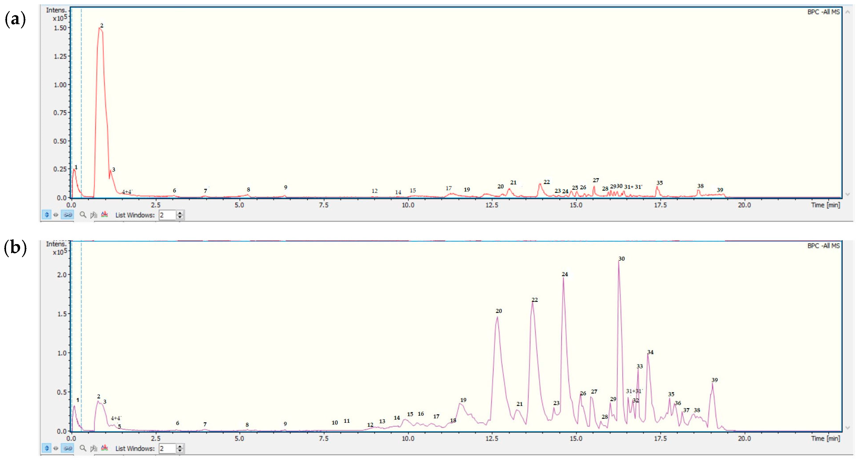
Figure 1. UHPLC Q-TOF-TIC chromatogram of DOe (a) and MGEOe (b). The peak numbers correspond to internal standard Na formiate (1); monohexosyldiglyceride (2); quinic acid (3); vicenin II (apigenin 6,8-di C-B-D-glucopiranoside) (4); chlorogenic acid (4′); luteolin-6-C-glucoside (isoorientin; (5); bis-3-(4′,5,7-trimethoxy-flavanone)-5′-(2′,4,4′-trimethoxy-chalcone) (6); rutin (7); isovitexin (8); meliartenin (9); 4′,5,7-trihydroxy-3,6-dimethoxyflavone (10); 8-hydroxy-5-methoxyflavanone (11); 17-hydroxylinolenic acid (12); diffractaic acid (13); 9-HPODE octadecenedioic acid (14); 9-hydroxy-10,12-octadecadienoic acid (15); 13-HPODE octadecenedioic acid (16); (9Z,12R)-12-hydroxyoctadec-9-enoic acid (17); ricinoleic acid (18); 5-(octadecyloxy) isophthalic acid (19); ricinoleic acid isomer (20); lesquerolic acid (21); pinolenic acid (22); 9-octadecenoic acid (Z)-,2-hydroxyethyl ester (23); roccellaric acid (24); alpha-boswellic acid (25); eicosapentaenoic acid (26); pristimerin (27); dodecanoic acid,2-ethylhexanoic acid, propane-1,2,3-triol (28); palmitic acid (29); FAHFA 18:1/2:0 hydroxy fatty acid ester: icos-10-enedioic acid (30); 22-oxodocosanoic acid (31); PI 34:21-phosphatidyl-1D-myo-inositol (31′); isopropyl linoleate (32); PI(16:0/13-HODE) hydroxyoctadecadienoic acid (33); muricatenol (34); 28-O-acetylbetulin-3-yl-β-D-glucopyranoside (35); 3β-O-acetyl-28-O-lup-20(29)-ene (36); 1-stearyl-2-cholesterylcarbonoyl-3-trityl glycerol (37); aipolic acid(38); daucosterol (39).
Antioxidants 13 01494 g001
Figure 2. Molecular structures of main compounds identified in DOe and MGEOe.
Antioxidants 13 01494 g002
Figure 3. HCT-116 and HBL-100 cell proliferation after 48 h treatment with DOe (A,B) or MGEOe (C,D) and their respective Dm values.
Antioxidants 13 01494 g003
Figure 4. BW gain (a) and average beverage volume (b) of SUC (empty circles), HDOeS (empty inverted triangles) and DOeS (empty triangles). Results are reported as mean ± SD from two independent assays (n = 7 animals per group). Significant differences were assessed using one-way ANOVA (FBWgain(2,18) = 0.464; p = 0.6359 and FBeverage(2,18) = 181.303; p < 0.0001) followed by Newman–Keuls post hoc test. * and a p < 0.05; * refers to SUC and a refers to HDOeS.
Antioxidants 13 01494 g004
Figure 5. Levels of glucose during the GTT (a) and its AUC values (b), levels of BG (c), TC (d) and TG (e) after 40 days of administration of SUC, HDOeS and DOeS. Results are expressed as mean ± SD from 2 independent assays (n = 7 animals/group). Significant differences were determined by one-way ANOVA (FAUC(2,18) = 1.348; p = 0.2847; FBG(2,18) = 7.603; p = 0.0040; FTC(2,18) = 5.864; p = 0.0109 and FTG(2,18) = 6.868; p = 0.0061) followed by Newman–Keuls post hoc test. p < 0.05, * refers to SUC.
Antioxidants 13 01494 g005
Figure 6. Entries in zone 1 (a), time spent in zones 1 (b), 2 (c) and 3 (d) in the OFT after 40 days of administration of SUC (empty circles), HDOeS (empty inverted triangles) and DOeS (empty triangles). Results are presented as mean ± SD from two independent assays (n = 10 animals per group). Significant differences were assessed using one-way ANOVA (FEntrieszone1(2,27) = 7.642; p = 0.0023; FTimezone1(2,27) = 4.165; p = 0.0265; FTimezone2(2,27) = 3.576; p = 0.0419 and FTimezone3(2,27) = 5.349; p = 0.0110) followed by Newman–Keuls post hoc test. *, a p < 0.05, * refers to SUC and a refers to HDOeS.
Antioxidants 13 01494 g006
Figure 7. ER (a), time dedicated to exploring objects N and F in T1 (b) and lines crossing in T1 and T2 (c) in NOL test after 40 days of administration of SUC (empty circles) and HDOeS (empty inverted triangles). Results are presented as mean ± SD from two independent assays (n = 7 animals per group). Significant differences were assessed using Student’s t-test (ER: p = 0.0037; time for object N: p = 0.6661 and for object F: p = 0.8597; and line crossing in T1: p= 0.6629 and in T2: p = 0.4890). p < 0.05, * refers to SUC.
Antioxidants 13 01494 g007
Table 1. UHPLC-PDA-Q-TOF identification of metabolites from DOe and MGEOe.
PeakTentative Identification[M-H]-Retention Time (min)Measured Mass (m/z)Molecular FormulaAccuracy (ppm)Metabolite TypeMS Ions (m/z)
1Naformiate
(internal standard)
NaC2H2O40.12112.9829112.98563.10A
2MonohexosyldiglycerideC12H18O120.78353.0786354.085917.08Fatty acid
3Quinic acidC7H12O60.84191.0563192.06362.26Acid93.0345
4Vicenin II (apigenin 6,8-di C-B-D-glucopiranoside)C27H30O151.20593.2185594.21992.45C-glycosil flavone353.1079
4′Chlorogenic acidC16H18O91.20353.0886354.09592.45Phenolic acid191.0513
5Luteolin-6-C-glucoside (Isoorientin)C21H20O111.50447.0953448.10253.93C-glycosil flavone429.1389
357.1038
327.0886
298.0861
6Bis-3-(4′,5,7-trimethoxy-flavanone)-5′-(2′,4,4′-trimethoxy-chalcone)C36H34O93.13609.2154610.21303.89Flavone447.1456
341.1492
112.9882
7RutinC27H30O164.42609.1477610.15504.67Flavone301.0333
300.0258
8IsovitexinC21H20O104.48431.1006432.10785.07Isoflavone311.0563
283.0608
9MeliarteninC28H36O106.25531.2313532.22363.05Furane486.2168
385.3007
104′,5,7-trihydroxy-3,6-dimethoxyflavoneC17H14O77.54329.0677330.07503.31Flavone311.1599
255.0120
118-hydroxy-5-methoxyflavanoneC16H14O47.93269.0817270.08944.06Flavone254.0580
1217-hydroxylinolenic acidC18H30O38.45293.2134294.22074.00Aromatic439.0996
311.1636
13Diffractaic acidC20H22O79.62373.1304374.13764.47Aromatic282.0879
135.0810
149-HPODE octadecenedioic acidC18H32O49.80311.223312.23050.80Fatty acid269.2072
257.2058
159-hydroxy-10,12-octadecadienoic acidC18H32O310.50295.2283296.23511.62Fatty acid269.2077
1613-HPODE octadecenedioic acid C18H32O410.56311.2235312.23083.49Fatty acid257.2058
17(9Z,12R)-12-hydroxyoctadec-9-enoic acidC18H34O311.52311.2235312.23085.76Fatty acid257.2057
18Ricinoleic acidC18H34O311.88297.2437298.25130.44Fatty acid253.5113
195-(octadecyloxy) isophthalic acidC26H41O511.57433.2866434.2960−21.04Phenolic acid
20Ricinoleic acid isomerC18H34O312.54297.2427298.25135.04Fatty acid283.0608
311.0561
21Lesquerolic acidC20H38O313.22325.2745326.27488.33Fatty acid299.2586
227.2255
22Pinolenic acidC18H30O214.23277.2184278.22574.04Fatty acid283.2152
239-octadecenoic acid (Z)-, 2-hydroxyethyl esterC20H38O314.31325.2767326.27485.74Fatty acid307.2263
299.2586
293.2044
24Roccellaric acidC19H34O414.62325.2396326.24642.70Fatty acid281.2495
25Alpha-boswellic acidC30H48O314.85455.3540456.36131.37Fatty acid227.2255
26Eicosapentaenoic acidC20H30O215.02301.2184302.22571.79Fatty acid251.2660
27PristimerinC30H40O415.02463.2860464.29331.44Terpene449.2673
433.2385
28Dodecanoic acid,2-ethylhexanoic acid,propane-1,2,3-triolC23H48O715.58435.3275436.3327−11.95Fatty acid255.2610
29Palmitic acidC16H32O216.13255.2338256.24113.29Fatty acid112.9879
30FAHFA 18:1/2:0
hydroxy fatty acid ester: icos-10-enedioic acid
C20H36O416.28339.2553340.26253.48Fatty acid281.2811
3122-oxodocosanoic acidC22H41O316.39353.3116354.306115.40Fatty acid339.2952
31′PI 34:21-phosphatidyl-1D-myo-inositolC43H79O13P16.39833.5264834.53379.47Fatty acid717.4680
533.4085
403.2977
323.2977
32Isopropyl linoleateC21H39O216.45323.2969324.29564.12Fatty acid275.0326
33PI(16:0/13-HODE)
hydroxyoctadecadienoic acid
C43H79O14P17.02849.5204850.52768.12Fatty acid353.3115
34MuricatenolC37H68O617.43607.4909607.4943−5.49Fatty acid599.4280
574.4127
409.3437
3528-O-acetylbetulin-3-yl-β-D-glucopyranosideC38H62O817.52645.4363646.4372−1.33Fatty acid607.4901
573.4062
363β-O-acetyl-28-O-lup-20(29)-eneC38H62O817.74645.4363645.4367−1.31Sterol409.3437
371-stearyl-2-cholesterylcarbonoyl-3-trityl glycerolC40H59O318.56586.4309587.4391−14.02Fatty acid281.2111
38Aipolic acidC32H52O518.51515.3647515.358911.09Terpene361.3160
39DaucosterolC35H60O619.12575.4261576.4317−9.66Sterol477.3778
Table 2. Total phenols, flavonoids, antioxidant activity and inhibition COX-2 assays of DOe and MGEOe.
Spectrophotometric Quantification of Compounds of Interest
Total phenols
(mg GAE/g extracts)
DOe69.24 ± 5.78
MGEOe65.45 ± 0.06
Flavonoids
(mg QE/g extracts)
DOe43.31 ± 4.13
MGEOe31.90 ± 0.05
Antioxidant and Inhibition COX-2 Assays
DPPH
(EC50 in µg extracts/mL)
DOe70.11 ± 0.07
MGEOe62.07 ± 1.74
FRAP
(mg TE/g of extracts)
DOe4.95 ± 0.08
MGEOe3.25 ± 0.06
TEAC
(mg TE/g of extracts)
DOe0.67 ± 0.05
MGEOe1.24 ± 0.05
Percentage ILP
(at 250 µg extract/mL)
DOe83.81 ± 3.59
MGEOe90.46 ± 0.03
Percentage inhibition COX-2
(at 72 µg/mL)
DOe86.97± 0.05
Table 3. Levels of MDA (fmol/µg of protein).
Cerebral CortexHippocampusHypothalamus
SUC40.45 ± 13.8234.67 ± 18.1160.24 ± 7.83
HDOeS44.39 ± 14.9532.26 ± 7.2746.64 ± 12.56
DOeS46.21 ± 17.8546.97 ± 10.6556.20 ± 9.13
Results are presented as mean ± SD from two independent assays (n = 7 animals per group). The comparison between HDOeS, DOeS and SUC was performed by one-way ANOVA (FCerebralcortex(2,18) = 0.248; p = 0.7829; FHippocampus(2,18) = 2643; p = 0.0985 and FHypothalamus(2,18) = 3.388; p = 0.0564).
Disclaimer/Publisher’s Note: The statements, opinions and data contained in all publications are solely those of the individual author(s) and contributor(s) and not of MDPI and/or the editor(s). MDPI and/or the editor(s) disclaim responsibility for any injury to people or property resulting from any ideas, methods, instructions or products referred to in the content.