Review

Redox Homeostasis and Nrf2-Regulated Mechanisms Are Relevant to Male Infertility

by
Cinzia Signorini
1,
Luciano Saso
2,*,
Somayyeh Ghareghomi
3,
Pelin Telkoparan-Akillilar
4,
Giulia Collodel
1 and
Elena Moretti
1
1
Department of Molecular and Developmental Medicine, University of Siena, 53100 Siena, Italy
2
Department of Physiology and Pharmacology “Vittorio Erspamer”, Sapienza University of Rome, 00185 Rome, Italy
3
Institute of Biochemistry and Biophysics, University of Tehran, Tehran 1417466191, Iran
4
Department of Medical Biology, Faculty of Medicine, Yuksek Ihtisas University, 6530 Ankara, Turkey
*
Author to whom correspondence should be addressed.
Antioxidants 2024, 13(2), 193; https://doi.org/10.3390/antiox13020193
Submission received: 29 December 2023 / Revised: 20 January 2024 / Accepted: 1 February 2024 / Published: 3 February 2024
(This article belongs to the Section ROS, RNS and RSS)

Abstract

:
Infertility represents a significant global health challenge, affecting more than 12% of couples worldwide, and most cases of infertility are caused by male factors. Several pathological pathways are implicated in male infertility. The main mechanisms involved are driven by the loss of reduction–oxidation (redox) homeostasis and the resulting oxidative damage as well as the chronic inflammatory process. Increased or severe oxidative stress leads to sperm plasma membrane and DNA oxidative damage, dysregulated RNA processing, and telomere destruction. The signaling pathways of these molecular events are also regulated by Nuclear factor-E2-related factor 2 (Nrf2). The causes of male infertility, the role of oxidative stress in male infertility and the Keap1-Nrf2 antioxidant pathway are reviewed. This review highlights the regulatory role of Nrf2 in the balance between oxidants and antioxidants as relevant mechanisms to male fertility. Nrf2 is involved in the regulation of spermatogenesis and sperm quality. Establishing a link between Nrf2 signaling pathways and the regulation of male fertility provides the basis for molecular modulation of inflammatory processes, reactive oxygen species generation, and the antioxidant molecular network, including the Nrf2-regulated antioxidant response, to improve male reproductive outcomes.

1. Introduction

Infertility is described as the inability to conceive after one year or more of orderly unprotected sexual intercourse. Available data estimates that 8–12% of couples worldwide face fertility problems [1]. Traditionally, it was thought that reproductive issues concern only women, mainly due to cultural beliefs, while nowadays, it is well recognized that fertility and, as a result, infertility are dependent on the couple. Infertility may be due to male factors, female factors, or a combination of them, or it can be defined as sine causa or idiopathic [2].
In half of the cases, there is a male infertility aspect independently or in grouping with a female aspect [3].
Oxidative stress (OS) is introduced as a common cause of male infertility by disrupting the structural and functional integrity of spermatozoa. Physiological level of reactive oxygen species (ROS) is essential for sperm capacitation and acrosomal reaction; in contrast, the pathological levels of ROS damage sperm quality [4]. DNA is one of the most oxidatively affected molecules; in particular, ROS overproduction can lead to sperm DNA fragmentation, caused by a mismatch of bases, loss of base, base modifications, DNA adducts and crosslink, pyrimidine dimers compromising natural and assisted fertilization outcomes [5,6].
Nuclear factor-E2-related factor 2 (Nrf2) is an endogenous antioxidant factor which controls a collection of genes to prevent redox imbalances in cells. Based on various studies, Nrf2 expression in sperm from patients affected by oligospermia was significantly lower than that in healthy men, signifying that Nrf2 was critical in spermatogenesis. Therefore, the modulatory effect of Nrf2 as a master antioxidant pathway is important in male fertility rate [7,8].
This review provides a summary of the pathophysiology of OS-induced harm in human sperm and its consequences for male fertility. The modulatory effect of the Nrf2 antioxidant signaling pathway on male fertility is then discussed.

A Brief General Overview of Male Infertility

Generally, causes of male infertility can be identified as congenital and acquired factors acting at pre-testicular, post-testicular or directly at the testicular level. Among congenital factors, anorchia, cryptorchidism and genetic abnormalities such as karyotype anomalies and chromosome microdeletions can be mentioned [9]. Acquired factors include testis trauma, testicular torsion, obstruction of the proximal or distal urogenital tract, varicocele, recurrent urogenital infections, and inflammations, hypogonadotropic hypogonadism or endocrine factors, exogenous and environmental factors such as smoking, alcohol, drug intake, occupational exposure to toxins, pollution [1].
It is noteworthy that the reason of about 30% [10] of cases of male infertility remains unclear and unexplained and it is ascribed to idiopathic infertility. Research in this case is mandatory.
Male gametes are produced in the seminiferous tubules by a process known as spermatogenesis [11] by which spermatogonia develop into mature spermatozoa with the contribution of different cell types, hormones, paracrine factors, and gene expression and epigenetics pathways. In addition to the germ line, spermatogenetic process is orchestrated by somatic cells. The peritubular myoid cells surround the seminiferous tubules and show contractile properties necessary to help the propulsion of testicular fluid for the transport of spermatozoa in the tubule. Leydig cells are present in the interstitial tissue and underpin the life-long androgen production. Both kinds of cells are target for hormones, cytokines, and growth factors, and they produce many substances necessary to support spermatogenesis. However, the real key driver of testis function is the Sertoli cells [12] that physically support and interact with all the germ lines in the seminiferous epithelium. They play a role in regulating androgen production, in arranging the blood–testis barriers, in cross-talking with the other somatic cells involved in spermatogenesis. This complex cell framework necessary to support a normal spermatogenesis can be target of OS, which impairs testicular and epididymal functions, and can affect the prostate and seminal vesicles, the major producers of seminal plasma.
Phenotypic manifestation of male infertility may range from the total absence of spermatozoa in the seminiferous tubules to different alterations of sperm quality. The World Health Organization (WHO) Laboratory Manual for the Examination and Processing of Human Semen [2] provides standard laboratory methods for semen analysis that are extensively used both in clinical practice and for research and allows internationally comparable results [13].
The semenogram is a fast, simple, and economical test that requires well-trained examiners and little equipment; however, its diagnostic and prognostic value for reproductive purposes is moderate, and evaluation of semen parameters such as sperm concentration, motility and morphology is not sufficient to predict reproductive success [14].
For this reason, there is a need to develop additional and advanced functional tests that complement and enrich the semenogram data providing information about sperm physiology [15] and mechanisms by which the different reproductive pathologies can affect male infertility.
These tests allow the examination of the individual “functional compartments” of the spermatozoon such as membrane, acrosome, chromatin, mitochondria and flagellum (Figure 1), the interaction of spermatozoon–oocyte and biomarkers found in the semen.
Llavanera et al. [15] recently reviewed the literature on some Omics, such as genomics and epigenomics, transcriptomics, and proteomics, and concluded that DNA structure and integrity can be considered as relevant molecular biomarkers that may help in the diagnosis and treatment of conditions causing semen quality disorders and fertilization failure. In addition, metabolomics and in particular ROS evaluation in spermatozoa and seminal plasma, total antioxidant capacity and oxidation–reduction potential have been proposed as well-characterized and promising molecular fertility biomarkers in semen with potential clinical application [15].

2. Male Infertility and Oxidative Stress

A disturbance in the signaling of redox network results in an imbalance in the redox steady state, leading to a disruption of homeostasis and the occurrence of OS. This condition favors the oxidative damages with respect to the antioxidant defenses; subsequently, it causes damage to biomolecules through oxidative reactions [16]. As oxidants, oxygen free radicals and additional ROS are known to be involved both in physiological and pathological (oxidative damage) mechanisms [17,18,19]. In sperm, ROS play a relevant role in regulating several intracellular pathways. Because ROS generation influences sperm membrane fluidity, activation of adenylate cyclase, and protein tyrosine phosphorylation, the normal physiological ROS levels mediate sperm capacitation, the acquisition of hyperactivated motility, the acrosome reaction, and the sperm–oocyte interaction [20,21].
High levels of ROS can cause infertility not only by lipid peroxidation or DNA oxidative damage, but also by inactivation of enzymes and oxidation of proteins in spermatozoa [22], consequently affecting sperm quality and impairing male fertility [23,24]. In human ejaculate, ROS originate mainly from immature spermatozoa and leukocytes [25,26,27].
Spermatozoa are particularly vulnerable to oxidative harm because of the abundant presence of polyunsaturated fatty acids (PUFA) in plasma membranes and the limited capacity to mend damage caused by ROS. Seminal plasma contains a significant amount of antioxidants such as glutathione peroxidase (GPx), catalase (CAT), and superoxide dismutase (SOD) [28] that help maintain the redox equilibrium under normal physiological circumstances; also, sperm cells contain antioxidants, but at extremely low concentrations [19]. Nevertheless, in some conditions, the production of ROS increases, overpowering the antioxidant activity, leading to OS [29].
ROS measurement in seminal fluid, which is an important element in the evaluation of male fertility, has also become an important target in preconception care [30].
Lipid peroxidation, which involves the oxidative damage of lipids, proceeds through three distinct phases (initiation, propagation and termination) along which free radicals are first produced, then quantitatively increased and finally neutralized by antioxidants or through reactions between free radicals [31]. In sperm, the lipid vulnerability to oxidative damage leads to the accumulation of lipid peroxides within spermatozoa, resulting in the formation of various decay end-products, including 4-hydroxynonenal, isoprostanes, and malondialdehyde (MDA). Such lipid peroxidation end-products serve as measurable indicators of OS [32]. There are many studies showing that lipid peroxidation of the sperm plasma membrane, as the main consequence of OS, is associated with poor sperm quality and infertility [33,34]. In sperm membranes, fatty acids with unstable bonds are vulnerable to oxidation by ROS.
In reproductive tissues, linoleic acid (LA), alpha-linolenic acid (ALA), their metabolites eicosapentaenoic acid (EPA), docosahexaenoic acid (DHA), and arachidonic acid (ARA), may influence reproductive function and fertility. In the sperm plasma membrane, lipid composition and specific types of PUFA (n-3 PUFA or n-6 PUFA) are related to membrane fluidity, flexibility, and receptor function. Ultimately, sperm membrane composition in PUFA highly regulates sperm membrane fusion at the time of sperm–oocyte interaction. In sperm, lipid composition and membrane lipid metabolism contribute to the formation of membrane microdomains involved in sperm motility, capacitation, and ability of sperm to penetrate the zona pellucida [35,36].
Dietary fats can influence the lipid composition of sperm cells, having harmful or beneficial consequences on male reproductive potential. An investigation is currently underway to explore the biochemical mechanisms that modulate sperm quality through diet [37,38,39,40].
Male infertility is a complex issue that can arise from various factors, including the interconnected pathophysiological processes of OS and inflammation.
These common mechanisms play a significant role in the development of male infertility, highlighting the need for further research in this area [41].
OS has been advocated in male infertility [42] associated with main pathologies altering the male reproductive system [43,44], bacterial infections, inflammation and leukocytospermia, environmental and lifestyle factors, and chronic/degenerative diseases [42,45,46,47]. In particular, in chronic prostatitis, OS plays an important role not only affecting male fertility, but also influencing the prostate cancer development. Large amounts of ROS and pro-inflammatory cytokines are continuously produced when inflammation persists. Consequently, this pathway triggers the activation of transcription factor nuclear factor-kappa B (NF-κB) and genes encoding for further production of pro-inflammatory cytokines, chemotactic factors, and growth factors [48].
Inflammation has been associated with many reproductive pathological conditions [49,50,51,52]. Increased levels of inflammatory cytokines, leukocyte counts that affect sperm quality and inflammation are also associated with sperm necrosis. It was recently observed that low sperm quality and sperm necrosis positively correlated with iron metabolism indices (ferritin, iron, transferrin evaluated in seminal plasma) in individuals with leukocytospermia and varicocele, both of which are characterized by the presence of inflammatory status and OS condition [53]. Altered iron metabolism is involved in ROS production [54,55,56,57] and drives chronic inflammation affecting male fertility [58,59]. Interestingly, several studies reported that ferroptosis, an iron-dependent programmed cell death, can affect spermatogenetic process leading to male infertility [60,61].
On this topic, it has been reported that pro-resolving lipid mediators, which are able to regulate the resolution of acute inflammation, are related to male infertility [62]. Interestingly, such pro-resolving lipid mediators are derived from PUFA. Mainly, pro-resolving lipid mediators of inflammation are biosynthesized from PUFA precursors, e.g., ARA, EPA, DHA, and docosapentaenoic acid [63].

3. Keap1-Nrf2 Antioxidant Pathway

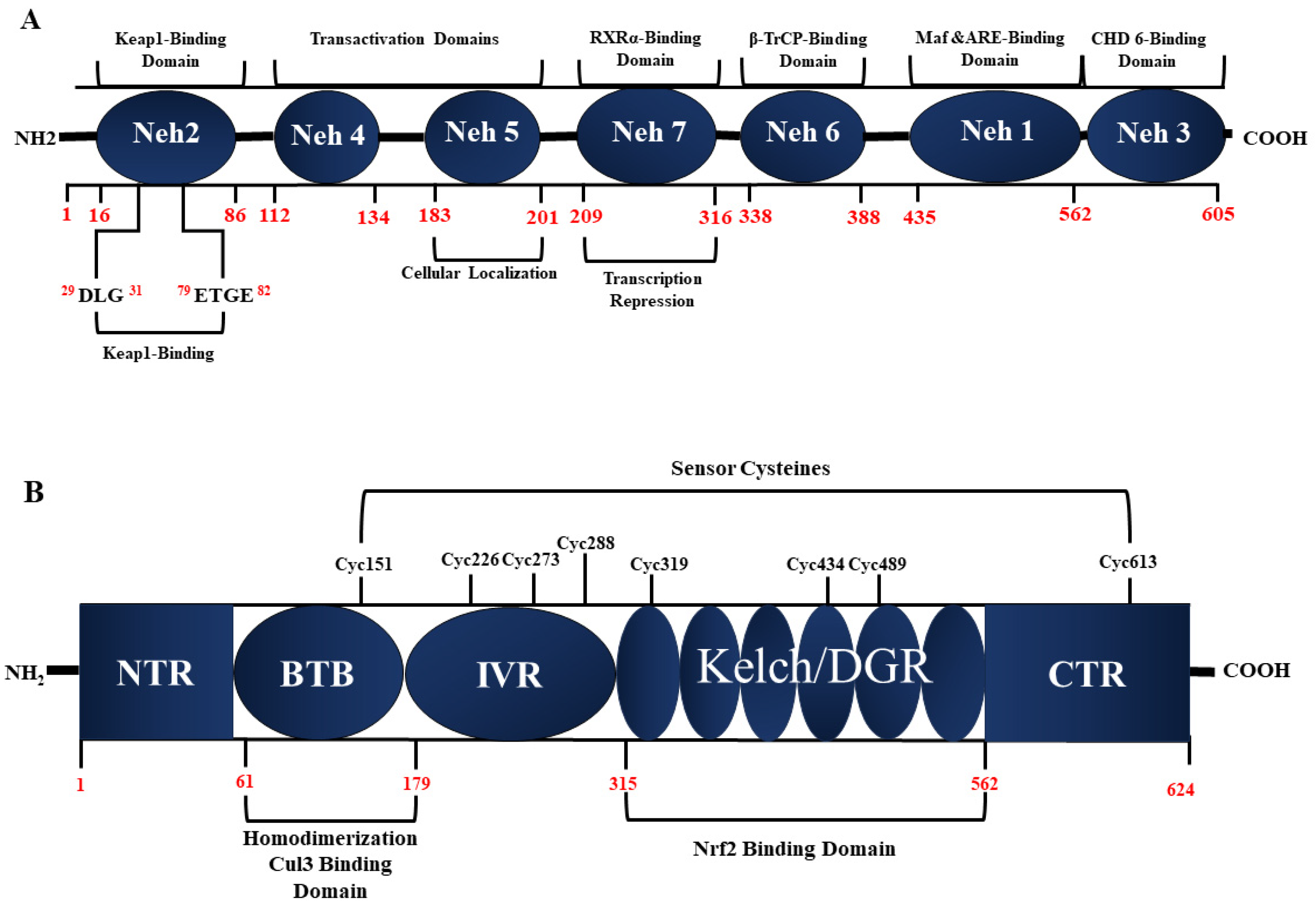
The Keap1-Nrf2 antioxidant pathway is a crucial cellular defense mechanism that plays a pivotal role in protecting organisms from oxidative stress and maintaining redox homeostasis. This intricate signaling pathway regulates the activation of transcription factor Nrf2 which, in turn, orchestrates the expression of several antioxidant and detoxification genes. The central element of this pathway is Kelch-like ECH associating protein 1 (Keap1). Under basal conditions, Keap1 directs Nrf2 for ubiquitin-dependent degradation, thereby repressing Nrf2-dependent gene expression. Under oxidative stress conditions, the Nrf2 level increases, leading to the activation of Nrf2-dependent gene expression [64]. The Nrf2 protein (Figure 2A), spanning 605 amino acids, is organized into seven highly conserved regions identified as Nrf2-ECH homology (Neh) domains [65]. These Neh domains (Neh1-7) play critical roles in regulating6 Nrf2’s stability and transcriptional activity (transactivation). Specifically, the Neh2 domain engages with two Keap1 molecules through the ETGE and DLG motifs, with the interaction and stability of Nrf2 being governed by Neh2.
Neh5 regulates the cellular localization of Nrf2, ensuring its proper positioning within the cell [66,67] The Neh1 domain, beyond controlling Nrf2 stability through its basic leucine zipper motif, facilitates the binding of Nrf2 to the antioxidant response element (ARE) sequence. This unique role in antioxidant responses induced by the pathway accentuates Neh1’s significance [68].
The Neh6 domain regulates Keap1-independent degradation of Nrf2, introducing an additional layer of complexity. The activation of ARE-dependent genes, post-chromatin remodeling, occurs following the interaction of Neh3 with the transcriptional co-activator CHD6, a DNA-binding protein with chromo-ATPase/helicase activity. Transcription activation domains Neh4 and Neh5 bind to the co-activator cAMP-responsive element-binding protein, facilitating Nrf2 transcription [69]. Lastly, the Neh7 domain, through interaction with retinoic X receptor α, can repress Nrf2 [70]. These sophisticated interactions and regulatory mechanisms within the various Neh domains underscore the complexity of the Keap1-Nrf2 antioxidant pathway, elucidating its pivotal role in cellular responses to oxidative stress.
Human Keap1, composed of 624 amino acid residues, boasts a molecular weight of approximately 70 kD [71]. As depicted in Figure 2B, this protein showcases five distinct domains: three broad complex–tramtrack–bric-a-brac (BTB) domains, one intervening region (IVR), and two glycine repeat domains (DGR). Each of these domains plays a pivotal role in inhibiting the activity of Nrf2 [72]. In a Keap1 homodimer, the DGR domains bind to different affinities with the DLG and ETGE domains in a single Nrf2 molecule. Under oxidative conditions, the separation of the DLG motif of Nrf2 from the DGR domain in Keap1 hinders Nrf2 degradation [73].
The IVR domain in Keap1 contains a consensus sequence of a nuclear export signal, crucial for Keap1’s cytoplasmic localization and its interaction with the Cul3 protein. Cul3, in conjunction with Roc1, constitutes the E3 ligase complex responsible for ubiquitinating Nrf2 [74,75]. Keap1 acts as a sensor for various stress stimuli, with numerous cysteine residues serving as critical sensing points. Notably, Cys273 and Cys288 are essential for Keap1 to regulate Nrf2 under both basal and stress conditions, while Cys151 is primarily required under stress conditions [76,77]. Other cysteine residues in Keap1, including Cys226, Cys434, and Cys613, play crucial roles in sensing specific toxins [73,78]. Modification of these cysteine residues inhibits the ubiquitin E3 ligase activity of the Keap1-Cul3 complex [79]. The intricacies of Keap1’s structural organization and the precise tuning of its cysteine residues underscore its role as a sophisticated cellular sensor and regulator in the Keap1-Nrf2 antioxidant pathway.
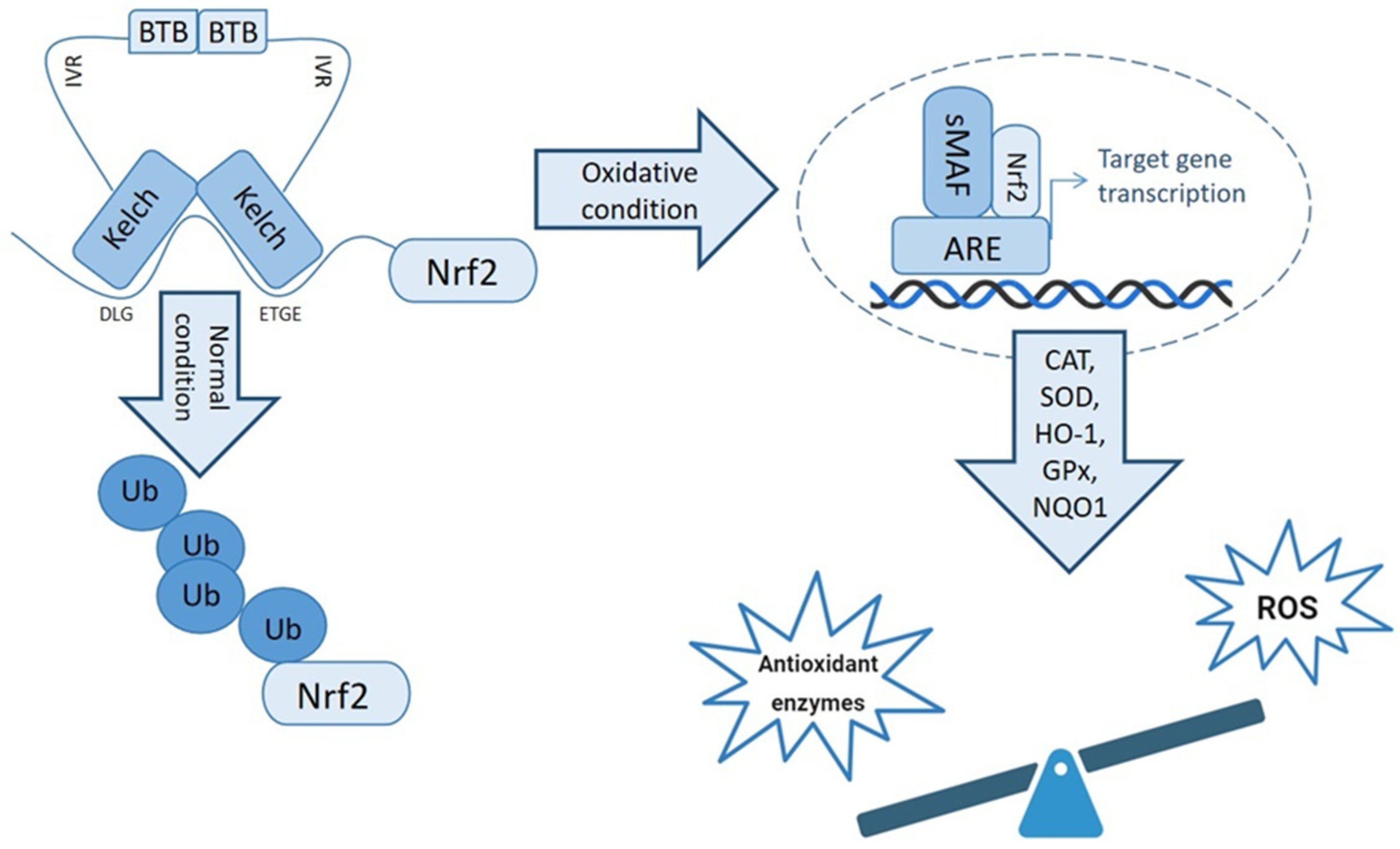
The induction of Nrf2 in response to OS leads to its release from Keap1, triggering its translocation to the nucleus. Upon reaching the nucleus, Nrf2 undergoes dimerization with small musculoaponeurotic fibrosarcoma oncogene homolog (Maf) proteins. This resulting Nrf2-Maf complex exhibits selective targeting towards genes possessing ARE in their regulatory regions. These genes constitute a diverse array of crucial components, including antioxidant enzymes, ABC transporters, and various stress-responsive proteins [79].
The Nrf2-Maf complex, selectively directed towards ARE-containing genes, plays a pivotal role in orchestrating a robust cellular defense against OS. Activation of the Nrf2 pathway induces the expression of a spectrum of antioxidant enzymes, including SOD, CAT, GPx, quinone oxidoreductase-1 (NQO1), and heme oxygenase-1 (HO-1). These enzymes collectively contribute to the modulation of oxidative conditions, neutralizing ROS and counteracting the detrimental effects of OS [80,81].
The Nrf2 antioxidant pathway, as summarized in Figure 3, serves as a critical mechanism in the cellular response to OS. By activating a repertoire of antioxidant enzymes and stress-responsive proteins, Nrf2 contributes significantly to the maintenance of cellular redox homeostasis and the prevention of oxidative damage. Understanding the intricacies of Nrf2-mediated responses provides valuable insights into potential therapeutic strategies for conditions associated with oxidative stress.

4. Role of Nrf2 in Male Fertility

Nrf2 appears to be a key regulator of the molecular mechanisms involved in male infertility. According to various studies, the Nrf2 function seems to play a beneficial role in safeguarding spermatogenesis against an excessive amount of ROS, thereby preserving specific sperm functions, including motility, which may otherwise be compromised under conditions of elevated ROS exposure [82].

4.1. Oxidative Stress and Nrf2-Related Gene Regulation

Several studies have shown a link between sperm abnormalities (function and/or morphology) and OS. Since Nrf2 is a transcription factor regulating the cellular response to stress conditions, its activity is involved in checking/regulating redox stress and inflammation. Thus, Nrf2 is a molecule of impressive relevance in male reproduction [83].
Since low Nrf2 levels as well as low antioxidant levels are associated with poor sperm quality, analyzing the transcripts (mRNA) of antioxidant genes is a fascinating issue. Interestingly, NRF2 gene expression was found to be associated with SOD activity and gene expression for SOD and CAT in human spermatozoa with low motility [84]. A gene expression profile related to OS and coordinated by the Nrf2/Keap1 system has been reported in rat and human testis and epididymis [85]. Functional nucleotide polymorphisms have been identified in the NRF2 promoter regions in idiopathic asthenozoospermic and oligoasthenozoospermic patients compared to the control group. These polymorphisms have also been related to altered levels of Nrf2 mRNA and/or decreased levels of antioxidant gene expression in spermatozoa [86]. In rat testis, OS results in the downregulation of the Nrf2 gene and to amplified testicular lipid peroxidation, germ cell death, and diminution of antioxidant levels [87].
Additionally, it was shown that reduced NRF2 expression in mouse testis and human spermatozoa is linked to dysfunction of spermatogenesis in case of diabetes mellitus and a smoking habit [88,89].
Diabetes is described as a condition of hyperglycemia that induces OS deleterious for male fertility. Indeed, in patients with diabetes, extremely high levels of hydroxyl radicals and advanced glycation end-products, induced by hyperglycemia, can trigger the triad: generation of ROS, OS and low sperm quality.
Even in this case, Nrf2 plays a pivotal role in diabetic testes because the autophagy mechanism induced by ROS activates a pathway that suppress Nrf2 activation worsening oxidative damage [90]. It is interesting to consider that an eventual upregulation of Nrf2-Keap1 pathway can increase the ratio nuclear Nrf2/cytosolic Nrf2 committing the transcription of SOD, CAT and GPx mRNAs.
Also, in obesity, a condition strongly linked to male infertility via OS, apoptosis, and glycolysis [91], the PKA/ERK/Nrf2 signaling axis mediates the improvement in fertility [92]. Moreover, the Keap1/Nrf2/ARE signaling pathway, which plays a key role in the OS response, was activated in reproductive system diseases [93], and enhanced Nrf2 expression was involved in the reduction in OS in patients with obesity [94].

4.2. Effects of Nrf2 Gene Deletion on Spermatogenesis

In 2010, Nakamura et al. [82] generated Nrf2 knockout (Nrf2−/−) mice by disrupting the gene through homologous recombination in embryonic stem cells. They observed that the absence of the transcription factor NRF2 caused a severe age-related spermatogenetic impairment. The effect of gene deletion was evident in mice by the age of 6 months in which sperm concentration decreased both in testis and epididymis, and the motility of epididymal spermatozoa was reduced with respect to those of wild-type mice. Concomitantly, the testes of Nrf2−/− male mice underwent an age-related process of declining in weights, indicating a process of degeneration. Furthermore, Nrf2−/− male mice were found to show elevated levels of testicular and epididymal lipid peroxidation, increased testicular germ cell apoptosis, and reduced antioxidant levels compared to wild-type animals [82]. This mouse model highlighted the crucial role played by transcription factor NRF2 in preventing oxidative disruption of spermatogenesis.

4.3. Thermoregulation Mechanisms of Nrf2 in Spermatogenesis

Nrf2 also appears to be involved in the thermoregulation mechanisms that regulate spermatogenesis. It is well known that spermatogenesis is influenced by temperature, and the elevation of testicular temperature can seriously affect mammalian spermatogenesis and the quality of semen [95] involving also somatic cells that are highly susceptible to elevated temperature [96]. Because of heat stress induced in mouse testes, the mRNA expression of Nrf2 increases together with the expression of different genes involved in redox homeostasis, promoting the expression of antioxidant genes [97].
He et al. [98] reported that in mice, Sertoli cells under heat stress activate the Keap1-Nrf2 pathway to prevent heat-induced oxidative damage.

4.4. Role of Nrf2 in Long-Term Storage of Sperm

By considering the supporting role of Nrf2 to spermatogenesis and sperm quality, Sadeghiani et al. [99] reported that pentoxifylline supplementation during freezing protocol and long-term storage protected mouse spermatogonial stem cell activating the antioxidant defense through the Nrf2/ARE signaling pathway. In addition, the transcription of NRF2, together with that referred to SOD1 and GPx1, was up-regulated in post-thawed buck semen after supplementation with antioxidants [100].
Jannatifar et al. [101] supplemented the cryopreservation medium with N-acetyl-cysteine (NAC) and alpha lipoic acid, used during a freezing protocol of semen samples of patients with asthenoteratozoospermia. They found that both antioxidants protected spermatozoa increasing the expression level of NRF2 gene and consequently the level of antioxidant enzymes, mainly CAT and SOD.
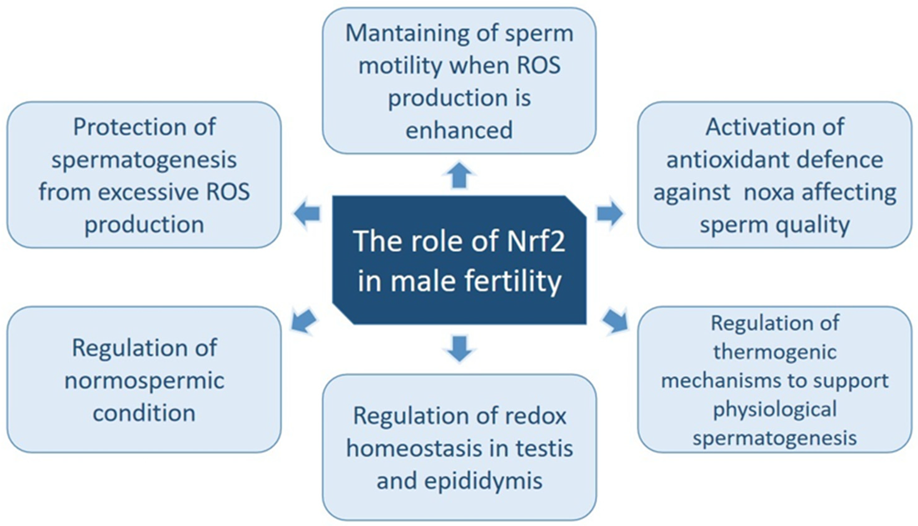
In Figure 4, the roles of Nrf2 in spermatogenesis and sperm quality are summarized.

5. Compounds Targeting Nrf2 and Influencing Male Fertility

Several studies conducted in animal models or in humans focused on the use of different compounds that influence the Nrf2 pathway negatively or positively. Based on the mentioned roles of Nrf2 in the regulation of OS, its targeting can display a remarkable effect on various aspects of cell development and fertility.
It is well known that therapeutic drugs, especially chemotherapeutics, can affect male fertility [102,103,104]. Cyclophosphamide (CYP) is one of the most successful anti-cancer agents ever synthesized. It is commonly utilized as a chemotherapeutic compound and in the conditioning regimens for blood and marrow transplantation [105]. Despite the anticancer properties of this agent, its metabolite led to increase in OS and apoptosis in the testes and male infertility. Maremanda et al. [106] reported that in rats, the treatment with CYP induced a decrease in Nrf2 levels in the testicular tissue. Furthermore, CYP therapy disrupted Nrf2 downstream pathway molecules such as HO-1 and SOD and the physiological antioxidant response. The concomitant administration of zinc improved the damages caused by CYP by modulating several mechanisms included a Nrf2-associated pathway [106].
Hydroxytyrosol (HT), one of the most common phytochemicals existing in oil and table olives, exerts strong antioxidant capacity and potential to demolish free radicals [107,108,109]. Recently, Fusco et al. [110] tested, in mice, a new compound Hidrox® based on HT as treatment for spermatogenetic damages induced by CYP. These authors found that the antioxidant capacities were not limited to the expression of type 2 detoxifying proteins such as SOD, CAT and GPx. Indeed, there are adaptive systems such as those involving HO-1, the expression of which is regulated by Nrf2.
Another tested compound, decursin, extracted from Angelica gigas Nakai roots, exerted an antioxidant activity involving Nrf2 in a cryptorchidism-induced infertility rat model. Cryptorchid rats treated with an A. gigas extract showed improved testis weight and sperm parameters in parallel with decreased OS and apoptosis and increased levels of Nrf2-related genes including SOD and HO-1 [111].
For this purpose, Arkali et al. [112] studied the protective effect of carvacrol, a phenolic monoterpene with antioxidant activity present in essential oils of aromatic plants, on diabetes-induced reproductive damage in male rats by evaluating the Nrf2/HO-1 pathway, NF-κB-mediated apoptosis and sperm parameters. They found that carvacrol significantly reduced MDA levels, Bax and NF-κB protein expression levels, and significantly increased Bcl-2, Nrf2/HO-1 levels and consequently GPx and CAT activities; this condition exerted a positive effect on sperm parameters. Other antioxidant compounds can target the Nrf2 pathway involved in redox homeostasis of male reproductive systems.
For example, treatment of rats exposed to cadmium [113] with piceatannol (PT; trans-3,4,3′,5′-tetrahydroxystilbene) present in various fruit and vegetables [114] protected the testis from alterations induced by the heavy metal. Spermatogenesis and steroidogenesis were improved, and concomitantly OS was reduced by targeting the Nrf2-Keap1 signaling pathway and its related genes.
Lycopene (LYC) is another effective antioxidant compound mostly present in tomatoes and other fruits with recognized health benefits [115]. Zhao et al. [116] demonstrated the beneficial effects of LYC in enhancing the expressions of Nrf2 and its downstream related genes and attenuating damages to Leydig cells of mice experimentally treated with di(2-ethylhexyl) phthalate, a plasticizer known to induce OS.
Melatonin, a hormone produced by the pineal gland, is involved in regulating biological timing. It is secreted at night in all species and is thereby associated with physiological events. This hormone, through its antioxidant properties, can modulate OS in various cells. In a mouse model of restraint stress, the administration of melatonin decreased the ROS level, amplified SOD and GSH activities, and downregulated iNOS and tumor necrosis factor-α (TNF-α) activities in testes. In addition, melatonin upregulated the expression of antioxidant proteins including Nrf2 and HO-1 with an improvement of spermatogenesis and sperm production [117].
In addition, melatonin inhibited the apoptosis of rooster Leydig cells in vitro by suppressing oxidative stress via AKT-Nrf2 pathway activation [118] and modulated Nrf2 activity to protect porcine pre-pubertal Sertoli cells from the abnormal H2O2 generation due to cadmium exposition [119].
Another tested antioxidant molecule is aucubin, extracted by Eucommia ulmoides and used as a protective treatment in mouse testis damaged by triptolide [120]. Aucubin, increasing the translocation of Nrf2 into the nucleus, triggered antioxidant enzymes in the testis, decreasing apoptotic process and protecting the blood–testis barrier integrity.
To the best of our knowledge, studies reporting data on the modulation of NRF2 male human reproduction are scant. Zhou et al. [121] treated in vitro spermatozoa from patients with asthenozoospermia with curcumin, a polyphenol with antioxidant activity, and observed that the mechanism of action by which curcumin exerted protective activity included the NRF2 pathway.
Additionally, as far as we know, only a couple of clinical trials in which the male subjects were treated with antioxidant compounds considered the effect on the NRF2 pathway. The treatment of infertile asthenozoospermic patients with NAC oral supplementation (600 mg, three times daily) improved sperm quality and decreased OS levels by enhancing NRF2 gene expression [122]. In a different clinical trial, Fallahi et al. [123] treated 60 infertile patients with date palm pollen (DPP, 400 mg/kg), a natural dietary food supplement rich in bioactive unsaturated fatty acid and flavonoid compounds [124]. DPP has a positive effect on reducing ROS levels, increasing sperm parameters and expression of NRF2, GPx4, SOD2, and CAT in infertile men.

6. Conclusions and Future Perspective

High levels of OS result in damage to sperm parameters and ultrastructure, and therefore might provide a common underlying etiology of male infertility.
OS is predominantly caused by lifestyle-related factors, the majority of which are modifiable.
Upregulation of some antioxidative pathways can reverse this effect and exert a remarkable effect on increasing male fertility. Therefore, in addition to the detailed investigation of the causes of infertility, the use of effective antioxidants to stimulate the master antioxidative pathway, including Nrf2 and modulation of the ROS levels, can be considered as a healthy treatment method to increase fertility rate in males.
Furthermore, the stimulation of antioxidant pathways, the support of Nrf2-regulated mechanisms in cryopreservation procedures, might represent concrete supplementary in vitro fertilization techniques in humans and animals.

Author Contributions

Conceptualization, C.S., E.M., G.C. and L.S.; writing—original draft preparation, C.S., E.M., L.S. and G.C.; writing—review and editing, C.S., E.M., G.C., L.S., S.G., P.T.-A. and G.C. All authors have read and agreed to the published version of the manuscript.

Funding

This research received no external funding.

Institutional Review Board Statement

Not applicable.

Informed Consent Statement

Not applicable.

Data Availability Statement

No new data were created in this study. Data availability sharing is not applicable.

Conflicts of Interest

The authors declare no conflicts of interest.

References

  1. Agarwal, A.; Baskaran, S.; Parekh, N.; Cho, C.L.; Henkel, R.; Vij, S.; Arafa, M.; Panner Selvam, M.K.; Shah, R. Male infertility. Lancet 2021, 397, 319–333. [Google Scholar] [CrossRef] [PubMed]
  2. World Health Organization. WHO Laboratory Manual for the Examination and Processing of Human Semen, 6th ed.; WHO Press: Geneva, Switzerland, 2021. [Google Scholar]
  3. Leslie, S.W.; Soon-Sutton, T.L.; Khan, M.A.B. Male infertility. In StatPearls; StatPearls Publishing: Treasure Island, FL, USA, 2022. [Google Scholar]
  4. Bisht, S.; Faiq, M.; Tolahunase, M.; Dada, R. Oxidative stress and male infertility. Nat. Rev. Urol. 2017, 14, 470–485. [Google Scholar] [CrossRef]
  5. Drevet, J.R.; Aitken, R.J. Oxidative Damage to Sperm DNA: Attack and Defense. Adv. Exp. Med. Biol. 2019, 1166, 107–117. [Google Scholar] [PubMed]
  6. Agarwal, A.; Majzoub, A.; Baskaran, S.; Panner Selvam, M.K.; Cho, C.L.; Henkel, R.; Finelli, R.; Leisegang, K.; Sengupta, P.; Barbarosie, C.; et al. Sperm DNA Fragmentation: A New Guideline for Clinicians. World J. Mens Health 2020, 38, 412–471. [Google Scholar] [CrossRef]
  7. Han, P.; Wang, X.; Zhou, T.; Cheng, J.; Wang, C.; Sun, F.; Zhao, X. Inhibition of ferroptosis attenuates oligospermia in male Nrf2 knockout mice. Free Radic. Biol. Med. 2022, 193, 421–429. [Google Scholar] [CrossRef] [PubMed]
  8. Ghareghomi, S.; Habibi-Rezaei, M.; Arese, M.; Saso, L.; Moosavi-Movahedi, A.A. Nrf2 modulation in breast cancer. Biomedicines 2022, 10, 2668. [Google Scholar] [CrossRef]
  9. Krausz, C. Male infertility: Pathogenesis and clinical diagnosis. Best Pract. Res. Clin. Endocrinol. Metab. 2011, 25, 271–285. [Google Scholar] [CrossRef]
  10. Corsini, C.; Boeri, L.; Candela, L.; Pozzi, E.; Belladelli, F.; Capogrosso, P.; Fallara, G.; Schifano, N.; Cignoli, D.; Ventimiglia, E.; et al. Is There a Relevant Clinical Impact in Differentiating Idiopathic versus Unexplained Male Infertility? World J Mens Health 2023, 41, 354–362. [Google Scholar] [CrossRef]
  11. Neto, F.T.; Bach, P.V.; Najari, B.B.; Li, P.S.; Goldstein, M. Spermatogenesis in humans and its affecting factors. Semin. Cell Dev. Biol. 2016, 59, 10–26. [Google Scholar] [CrossRef]
  12. O’Donnell, L.; Smith, L.B.; Rebourcet, D. Sertoli cells as key drivers of testis function. Semin. Cell Dev. Biol. 2022, 121, 2–9. [Google Scholar] [CrossRef]
  13. Björndahl, L.; Brown, J.K. The sixth edition of the WHO Laboratory Manual for the Examination and Processing of Human Semen: Ensuring quality and standardization in basic examination of human ejaculates. Fertil. Steril. 2022, 117, 246–251. [Google Scholar] [CrossRef] [PubMed]
  14. Dave, P.; Farber, N.; Vij, S. Conventional semen analysis and advanced sperm function tests in diagnosis and management of varicocele. Andrologia 2021, 53, e13629. [Google Scholar] [CrossRef] [PubMed]
  15. Llavanera, M.; Delgado-Bermúdez, A.; Ribas-Maynou, J.; Salas-Huetos, A.; Yeste, M. A systematic review identifying fertility biomarkers in semen: A clinical approach through Omics to diagnose male infertility. Fertil. Steril. 2022, 118, 291–313. [Google Scholar] [CrossRef] [PubMed]
  16. Sies, H. On the history of oxidative stress: Concept and some aspects of current development. Curr. Opin. Toxicol. 2018, 7, 122–126. [Google Scholar] [CrossRef]
  17. Halliwell, B.; Gutteridge, J.M.C. Free Radicals in Biology and Medicine, 5th ed.; Clarendon Press: Oxford, UK, 2015. [Google Scholar]
  18. Mannucci, A.; Argento, F.R.; Fini, E.; Coccia, M.E.; Taddei, N.; Becatti, M.; Fiorillo, C. The Impact of Oxidative Stress in Male Infertility. Front. Mol. Biosci. 2022, 8, 799294. [Google Scholar] [CrossRef] [PubMed]
  19. Moretti, E.; Signorini, C.; Corsaro, R.; Giamalidi, M.; Collodel, G. Human Sperm as an In Vitro Model to Assess the Efficacy of Antioxidant Supplements during Sperm Handling: A Narrative Review. Antioxidants 2023, 12, 1098. [Google Scholar] [CrossRef]
  20. Du Plessis, S.S.; Agarwal, A.; Halabi, J.; Tvrda, E. Contemporary evidence on the physiological role of reactive oxygen species in human sperm function. J. Assist. Reprod. Genet. 2015, 32, 509–520. [Google Scholar] [CrossRef]
  21. Takei, G.L.; Tourzani, D.A.; Paudel, B.; Visconti, P.E. Activation of cAMP-dependent phosphorylation pathways is independent of ROS production during mouse sperm capacitation. Mol. Reprod. Dev. 2021, 8, 544–557. [Google Scholar] [CrossRef]
  22. Barati, E.; Nikzad, H.; Karimian, M. Oxidative stress and male infertility: Current knowledge of pathophysiology and role of antioxidant therapy in disease management. Cell. Mol. Life Sci. 2020, 77, 93–113. [Google Scholar] [CrossRef]
  23. Tvrdá, E.; Kňažická, Z.; Bárdos, L.; Massányi, P.; Lukáč, N. Impact of oxidative stress on male fertility—A review. Acta Vet. Hung. 2011, 59, 465–484. [Google Scholar] [CrossRef]
  24. O’Flaherty, C. Protective role of peroxiredoxins against reactive oxygen species in neonatal rat testicular gonocytes. Antioxidants 2019, 9, 32. [Google Scholar] [CrossRef]
  25. Aitken, R.J.; Baker, H.W. Seminal leukocytes: Passengers, terrorists or good samaritans? Hum. Reprod. 1995, 10, 1736–1739. [Google Scholar] [CrossRef]
  26. Alvarez, J.G. Nurture vs nature: How can we optimize sperm quality? J. Androl. 2003, 24, 640–648. [Google Scholar] [CrossRef] [PubMed]
  27. Li, X.; Ni, M.; Xing, S.; Yu, Y.; Zhou, Y.; Yang, S.; Li, H.; Zhu, R.; Han, M. Reactive Oxygen Species Secreted by Leukocytes in Semen Induce Self-Expression of Interleukin-6 and Affect Sperm Quality. Am. J. Mens Health 2020, 14, 1557988320970053. [Google Scholar] [CrossRef]
  28. Micheli, L.; Cerretani, D.; Collodel, G.; Menchiari, A.; Moltoni, L.; Fiaschi, A.I.; Moretti, E. Evaluation of enzymatic and non-enzymatic antioxidants in seminal plasma of men with genitourinary infections, varicocele, and idiopathic infertility. Andrology 2016, 4, 456–464. [Google Scholar] [CrossRef]
  29. Henkel, R.R. Leukocytes and oxidative stress: Dilemma for sperm function and male fertility. Asian J. Androl. 2011, 13, 43. [Google Scholar] [CrossRef]
  30. Castleton, P.E.; Deluao, J.C.; Sharkey, D.J.; McPherson, N.O. Measuring reactive oxygen species in semen for male preconception care: A scientist perspective. Antioxidants 2022, 11, 264. [Google Scholar] [CrossRef] [PubMed]
  31. Gaschler, M.M.; Stockwell, B.R. Lipid peroxidation in cell death. Biochem. Biophys. Res. Commun. 2017, 482, 419–425. [Google Scholar] [CrossRef] [PubMed]
  32. Moretti, E.; Cerretani, D.; Noto, D.; Signorini, C.; Iacoponi, F.; Collodel, G. Relationship between semen IL-6, IL-33 and malondialdehyde generation in human seminal plasma and spermatozoa. Reprod. Sci. 2021, 28, 2136–2143. [Google Scholar] [CrossRef]
  33. Signorini, C.; Moretti, E.; Collodel, G. Role of isoprostanes in human male infertility. Syst. Biol. Reprod. Med. 2020, 66, 291–299. [Google Scholar] [CrossRef]
  34. Naderi Noreini, S.; Malmir, M.; Ghafarizadeh, A.; Faraji, T.; Bayat, R. Protective effect of L-carnitine on apoptosis, DNA fragmentation, membrane integrity and Lipid peroxidation of spermatozoa in the asthenoteratospermic men. Andrologia 2021, 53, e13932. [Google Scholar] [CrossRef]
  35. Wathes, D.C.; Abayasekara, D.R.E.; Aitken, R.J. Polyunsaturated fatty acids in male and female reproduction. Biol. Reprod. 2007, 77, 190–201. [Google Scholar] [CrossRef]
  36. delBarco-Trillo, J.; Roldan, E.R. Effects of metabolic rate and sperm competition on the fatty-acid composition of mammalian sperm. J. Evol. Biol. 2014, 27, 55–62. [Google Scholar] [CrossRef]
  37. Crisóstomo, L.; Videira, R.A.; Jarak, I.; Starčević, K.; Mašek, T.; Rato, L.; Raposo, J.F.; Batterham, R.L.; Oliveira, P.F.; Alves, M.G. Diet during early life defines testicular lipid content and sperm quality in adulthood. Am. J. Physiol. Endocrinol. Metab. 2020, 319, E1061–E1073. [Google Scholar] [CrossRef]
  38. Suliga, E.; Głuszek, S. The relationship between diet, energy balance and fertility in men. Int. J. Vitam. Nutr. Res. 2020, 90, 514–5262019. [Google Scholar] [CrossRef]
  39. Kljajic, M.; Hammadeh, M.E.; Wagenpfeil, G.; Baus, S.; Sklavounos, P.; Solomayer, E.F.; Kasoha, M. Impact of the vegan diet on sperm quality and sperm oxidative stress values: A preliminary study. J. Hum. Reprod. Sci. 2021, 14, 365. [Google Scholar] [CrossRef]
  40. Ferramosca, A.; Zara, V. Diet and male fertility: The impact of nutrients and antioxidants on sperm energetic metabolism. Int. J. Mol. Sci. 2022, 23, 2542. [Google Scholar] [CrossRef]
  41. Dutta, S.; Sengupta, P.; Slama, P.; Roychoudhury, S. Oxidative stress, testicular inflammatory pathways, and male reproduction. Int. J. Mol. Sci. 2021, 22, 10043. [Google Scholar] [CrossRef] [PubMed]
  42. Takalani, N.B.; Monageng, E.M.; Mohlala, K.; Monsees, T.K.; Henkel, R.; Opuwari, C.S. Role of oxidative stress in male infertility. Reprod. Fertil. 2023, 4, e230024. [Google Scholar] [CrossRef] [PubMed]
  43. Arya, D.; Balasinor, N.; Singh, D. Varicocoele-associated male infertility: Cellular and molecular perspectives of pathophysiology. Andrology 2022, 10, 1463–1483. [Google Scholar] [CrossRef] [PubMed]
  44. Lewis, S.E. Revisiting the impact of varicocele and its treatments on male fertility. Reprod. Biomed. Online 2022, 45, 1061–1063. [Google Scholar] [CrossRef]
  45. Bisconti, M.; Simon, J.F.; Grassi, S.; Leroy, B.; Martinet, B.; Arcolia, V.; Isachenko, V.; Hennebert, E. Influence of risk factors for male infertility on sperm protein composition. Int. J. Mol. Sci. 2021, 22, 13164. [Google Scholar] [CrossRef]
  46. Minas, A.; Fernandes, A.C.C.; Maciel Júnior, V.L.; Adami, L.; Intasqui, P.; Bertolla, R.P. Influence of physical activity on male fertility. Andrologia 2022, 54, e14433. [Google Scholar] [CrossRef]
  47. Kaur, P.; Rai, U.; Singh, R. Genotoxic Risks to Male Reproductive Health from Radiofrequency Radiation. Cells 2023, 12, 59456. [Google Scholar] [CrossRef]
  48. Paulis, G. Inflammatory mechanisms and oxidative stress in prostatitis: The possible role of antioxidant therapy. Res. Rep. Urol. 2018, 10, 75–87. [Google Scholar] [CrossRef] [PubMed]
  49. Agarwal, A.; Rana, M.; Qiu, E.; AlBunni, H.; Bui, A.D.; Henkel, R. Role of oxidative stress, infection and inflammation in male infertility. Andrologia 2018, 50, e13126. [Google Scholar] [CrossRef] [PubMed]
  50. Hærvig, K.K.; Kierkegaard, L.; Lund, R.; Bruunsgaard, H.; Osler, M.; Schmidt, L. Is male factor infertility associated with midlife low-grade inflammation? A population based study. Hum. Fertil. 2018, 21, 146–154. [Google Scholar] [CrossRef]
  51. Motrich, R.D.; Salazar, F.C.; Breser, M.L.; Mackern-Oberti, J.P.; Godoy, G.J.; Olivera, C.; Paira, D.A.; Rivero, V.E. Implications of prostate inflammation on male fertility. Andrologia 2018, 50, e13093. [Google Scholar] [CrossRef] [PubMed]
  52. Leisegang, K.; Sengupta, P.; Agarwal, A.; Henkel, R. Obesity and male infertility: Mechanisms and management. Andrologia 2021, 53, e13617. [Google Scholar] [CrossRef]
  53. Collodel, G.; Signorini, C.; Nerucci, F.; Gambera, L.; Iacoponi, F.; Moretti, E. Semen biochemical components in varicocele, leukocytospermia, and idiopathic infertility. Reprod. Sci. 2021, 28, 91–101. [Google Scholar] [CrossRef]
  54. Koppenol, W.H.; Hider, R. Iron and redox cycling. Do’s and don’ts. Free Radic. Biol. Med. 2019, 133, 3–10. [Google Scholar] [CrossRef] [PubMed]
  55. Li, J.; Cao, F.; Yin, H.L.; Huang, Z.J.; Lin, Z.T.; Mao, N.; Sun, B.; Wang, G. Ferroptosis: Past, present and future. Cell Death Dis. 2020, 11, 88. [Google Scholar] [CrossRef] [PubMed]
  56. Seiwert, N.; Adam, J.; Steinberg, P.; Wirtz, S.; Schwerdtle, T.; Adams-Quack, P.; Hövelmeyer, N.; Kaina, B.; Foersch, S.; Fahrer, J. Chronic intestinal inflammation drives colorectal tumor formation triggered by dietary heme iron in vivo. Arch. Toxicol. 2021, 95, 2507–2522. [Google Scholar] [CrossRef] [PubMed]
  57. Marques, O.; Weiss, G.; Muckenthaler, M.U. The role of iron in chronic inflammatory diseases: From mechanisms to treatment options in anemia of inflammation. Blood 2022, 140, 2011–2023. [Google Scholar] [CrossRef]
  58. Jing, T.; Wang, P.; Liu, Y.; Zhao, J.; Niu, X.; Wang, X. Pathological changes in Sertoli cells and dysregulation of divalent metal transporter 1 with iron responsive element in the testes of idiopathic azoospermia patients. Andrologia 2018, 50, e12878. [Google Scholar] [CrossRef]
  59. Ammar, O.; Houas, Z.; Mehdi, M. The association between iron, calcium, and oxidative stress in seminal plasma and sperm quality. Environ. Sci. Pollut. Res. Int. 2019, 26, 14097–14105. [Google Scholar] [CrossRef]
  60. Li, L.; Hao, Y.; Zhao, Y.; Wang, H.; Zhao, X.; Jiang, Y.; Gao, F. Ferroptosis is associated with oxygen-glucose deprivation/reoxygenation-induced Sertoli cell death. Int. J. Mol. Med. 2018, 41, 3051–3062. [Google Scholar] [CrossRef]
  61. Zhao, Y.; Zhang, H.; Cui, J.G.; Wang, J.X.; Chen, M.S.; Wang, H.R.; Li, X.N.; Li, J.L. Ferroptosis is critical for phthalates driving the blood-testis barrier dysfunction via targeting transferrin receptor. Redox Biol. 2023, 59, 102584. [Google Scholar] [CrossRef]
  62. Signorini, C.; Moretti, E.; Noto, D.; Micheli, L.; Ponchia, R.; Collodel, G. Fatty Acid Oxidation and Pro-Resolving Lipid Mediators Are Related to Male Infertility. Antioxidants 2022, 11, 107. [Google Scholar] [CrossRef] [PubMed]
  63. Chiang, N.; Serhan, C.N. Specialized pro-resolving mediator network: An update on production and actions. Essays Biochem. 2020, 64, 443–462. [Google Scholar] [PubMed]
  64. Lo, S.C.; Li, X.; Henzl, M.T.; Beamer, L.J.; Hannink, M. Structure of the Keap1: Nrf2 interface provides mechanistic insight into Nrf2 signaling. EMBO J. 2006, 25, 3605–3617. [Google Scholar] [CrossRef] [PubMed]
  65. Nam, L.B.; Keum, Y.S. Binding partners of NRF2: Functions and regulatory mechanisms. Arch. Biochem. Biophys. 2019, 678, 108184. [Google Scholar] [CrossRef] [PubMed]
  66. Namani, A.; Li, Y.; Wang, X.J.; Tang, X. Modulation of NRF2 signaling pathway by nuclear receptors: Implications for cancer. Biochim. Biophys. Acta. 2014, 1843, 1875–1885. [Google Scholar] [CrossRef] [PubMed]
  67. Krajka-Kuźniak, V.; Paluszczak, J.; Baer-Dubowska, W. The Nrf2-ARE signaling pathway: An update on its regulation and possible role in cancer prevention and treatment. Pharmacol. Rep. 2017, 69, 393–402. [Google Scholar] [CrossRef]
  68. Keum, Y.S.; Choi, B.Y. Molecular and chemical regulation of the Keap1-Nrf2 signaling pathway. Molecules 2014, 19, 10074–10089. [Google Scholar] [CrossRef]
  69. Xiang, M.; Namani, A.; Wu, S.; Wang, X. Nrf2: Bane or blessing in cancer? J. Cancer Res. Clin. Oncol. 2014, 140, 1251–1259. [Google Scholar] [CrossRef]
  70. Bai, X.; Chen, Y.; Hou, X.; Huang, M.; Jin, J. Emerging role of NRF2 in chemoresistance by regulating drug-metabolizing enzymes and efflux transporters. Drug Metab. Rev. 2016, 48, 541–567. [Google Scholar] [CrossRef]
  71. Wei, S.; Pei, Y.; Wang, Y.; Guan, H.; Huang, Y.; Xing, T.; Johnson, R.W.; Wang, D. Role of human Keap1 S53 and S293 residues in modulating the binding of Keap1 to Nrf2. Biochimie 2019, 158, 73–81. [Google Scholar] [CrossRef]
  72. Kang, M.I.; Kobayashi, A.; Wakabayashi, N.; Kim, S.G.; Yamamoto, M. Scaffolding of Keap1 to the actin cytoskeleton controls the function of Nrf2 as key regulator of cytoprotective phase 2 genes. Proc. Natl. Acad. Sci. USA 2004, 101, 2046–2051. [Google Scholar] [CrossRef]
  73. Canning, P.; Sorrell, F.J.; Bullock, A.N. Structural basis of Keap1 interactions with Nrf2. Free Radic. Biol. Med. 2015, 88, 101–107. [Google Scholar] [CrossRef]
  74. Furukawa, M.; Xiong, Y. BTB protein Keap1 targets antioxidant transcription factor Nrf2 for ubiquitination by the Cullin 3-Roc1 ligase. Mol. Cell. Biol. 2005, 25, 162–171. [Google Scholar] [CrossRef]
  75. Velichkova, M.; Hasson, T. Keap1 regulates the oxidation-sensitive shuttling of Nrf2 into and out of the nucleus via a Crm1-dependent nuclear export mechanism. Mol. Cell. Biol. 2005, 25, 4501–4513. [Google Scholar] [CrossRef]
  76. Levonen, A.L.; Landar, A.; Ramachandran, A.; Ceaser, E.K.; Dickinson, D.A.; Zanoni, G.; Morrow, J.D.; Darley-Usmar, V.M. Cellular mechanisms of redox cell signalling: Role of cysteine modification in controlling antioxidant defences in response to electrophilic lipid oxidation products. Biochem. J. 2004, 378, 373–382. [Google Scholar] [CrossRef]
  77. Zhang, D.D.; Hannink, M. Distinct cysteine residues in Keap1 are required for Keap1-dependent ubiquitination of Nrf2 and for stabilization of Nrf2 by chemopreventive agents and oxidative stress. Mol. Cell. Biol. 2003, 23, 8137–8151. [Google Scholar] [CrossRef]
  78. Fuse, Y.; Kobayashi, M. Conservation of the Keap1-Nrf2 system: An evolutionary journey through stressful space and time. Molecules 2017, 22, 436. [Google Scholar] [CrossRef]
  79. Suzuki, T.; Yamamoto, Y. Molecular basis of the Keap1–Nrf2 system. Free Radic. Biol. Med. 2015, 88, 93–100. [Google Scholar] [CrossRef]
  80. Zhao, L.; Yang, H.; Xu, M.; Wang, X.; Wang, C.; Lian, Y.; Mehmood, A.; Dai, H. Stevia residue extract ameliorates oxidative stress in d-galactose-induced aging mice via Akt/Nrf2/HO-1 pathway. J. Funct. Foods. 2019, 52, 587–595. [Google Scholar] [CrossRef]
  81. Ghareghomi, S.; Rahban, M.; Moosavi-Movahedi, Z.; Habibi-Rezaei, M.; Saso, L.; Moosavi-Movahedi, A.A. The potential role of curcumin in modulating the master antioxidant pathway in diabetic hypoxia-induced complications. Molecules 2021, 26, 7658. [Google Scholar] [CrossRef] [PubMed]
  82. Nakamura, B.N.; Lawson, G.; Chan, J.Y.; Banuelos, J.; Cortés, M.M.; Hoang, Y.D.; Ortiz, L.; Rau, B.A.; Luderer, U. Knockout of the transcription factor NRF2 disrupts spermatogenesis in an age-dependent manner. Free Radic. Biol. Med. 2010, 49, 1368–1379. [Google Scholar] [CrossRef] [PubMed]
  83. Rotimi, D.E.; Ojo, O.A.; Olaolu, T.D.; Adeyemi, O.S. Exploring Nrf2 as a therapeutic target in testicular dysfunction. Cell Tissue Res. 2022, 390, 23–33. [Google Scholar] [CrossRef] [PubMed]
  84. Chen, K.; Mai, Z.; Zhou, Y.; Gao, X.; Yu, B. Low NRF2 mRNA expression in spermatozoa from men with low sperm motility. Tohoku J. Exp. Med. 2012, 228, 259–266. [Google Scholar] [CrossRef]
  85. Wajda, A.; Łapczuk, J.; Grabowska, M.; Słojewski, M.; Laszczyńska, M.; Urasińska, E.; Droździk, M. Nuclear factor E2-related factor-2 (Nrf2) expression and regulation in male reproductive tract. Pharmacol. Rep. 2016, 68, 101–108. [Google Scholar] [CrossRef] [PubMed]
  86. Yu, B.; Lin, H.; Yang, L.; Chen, K.; Luo, H.; Liu, J.; Gao, X.; Xia, X.; Huang, Z. Genetic variation in the Nrf2 promoter associates with defective spermatogenesis in humans. J. Mol. Med. 2012, 90, 1333–1342. [Google Scholar] [CrossRef] [PubMed]
  87. Moreno-Fernandez, J.; Alférez, M.J.M.; López-Aliaga, I.; Diaz-Castro, J. Protective effects of fermented goat milk on genomic stability, oxidative stress and inflammatory signalling in testis during anaemia recovery. Sci. Rep. 2019, 9, 2232124. [Google Scholar] [CrossRef] [PubMed]
  88. Jiang, X.; Bai, Y.; Zhang, Z.; Xin, Y.; Cai, L. Protection by sulforaphane from type 1 diabetes-induced testicular apoptosis is associated with the up-regulation of Nrf2 expression and function. Toxicol. Appl. Pharmacol. 2014, 279, 198–210. [Google Scholar] [CrossRef] [PubMed]
  89. Aydos, O.S.; Yukselten, Y.; Aydos, D.; Sunguroglu, A.; Aydos, K. Relationship between functional Nrf2 gene promoter polymorphism and sperm DNA damage in male infertility. Syst. Biol. Reprod. Med. 2021, 67, 399–412. [Google Scholar] [CrossRef] [PubMed]
  90. Tian, Y.; Song, W.; Xu, D.; Chen, X.; Li, X.; Zhao, Y. Autophagy induced by ROS aggravates testis oxidative damage in diabetes via breaking the feedforward loop linking p62 and Nrf2. Oxid. Med. Cell. Longev. 2020, 2020, 7156579. [Google Scholar] [CrossRef] [PubMed]
  91. Han, J.; Zhao, C.; Guo, H.; Liu, T.; Li, Y.; Qi, Y.; Deussing, J.M.; Zhang, Y.; Tan, J.; Han, H.; et al. Obesity induces male mice infertility via oxidative stress, apoptosis, and glycolysis. Reproduction 2023, 166, 27–36. [Google Scholar] [CrossRef] [PubMed]
  92. Shan, W.; Lu, S.; Ou, B.; Feng, J.; Wang, Z.; Li, H.; Lu, X.; Ma, Y. PACAP ameliorates the fertility of obese mice through PAC1/PKA/ERK/Nrf2 signal axis. J. Endocrinol. 2021, 248, 337–354. [Google Scholar] [CrossRef]
  93. Agarwal, A.; Maldonado Rosas, I.; Anagnostopoulou, C.; Cannarella, R.; Boitrelle, F.; Munoz, L.V.; Finelli, R.; Durairajanayagam, D.; Henkel, R.; Saleh, R. Oxidative stress and assisted reproduction: A comprehensive review of its pathophysiological role and strategies for optimizing embryo culture environment. Antioxidants 2022, 11, 477. [Google Scholar] [CrossRef]
  94. Liu, Y.; Ding, Z. Obesity, a serious etiologic factor for male subfertility in modern society. Reproduction 2017, 154, R123–R131. [Google Scholar] [CrossRef]
  95. Gan, M.; Jing, Y.; Xie, Z.; Ma, J.; Chen, L.; Zhang, S.; Zhao, Y.; Niu, L.; Wang, Y.; Li, X.; et al. Potential Function of Testicular MicroRNAs in Heat-Stress-Induced Spermatogenesis Disorders. Int. J. Mol. Sci. 2023, 24, 8809. [Google Scholar] [CrossRef] [PubMed]
  96. Aldahhan, R.A.; Stanton, P.G. Heat stress response of somatic cells in the testis. Mol. Cell. Endocrinol. 2021, 527, 111216. [Google Scholar] [CrossRef] [PubMed]
  97. Li, Y.; Cao, Y.; Wang, F.; Li, C. Scrotal heat induced the Nrf2-driven antioxidant response during oxidative stress and apoptosis in the mouse testis. Acta Histochem. 2014, 116, 883–890. [Google Scholar] [CrossRef] [PubMed]
  98. He, C.; Sun, J.; Yang, D.; He, W.; Wang, J.; Qin, D.; Zhang, H.; Cai, H.; Liu, Y.; Li, N.; et al. Nrf2 activation mediates the protection of mouse Sertoli Cells damage under acute heat stress conditions. Theriogenology 2022, 177, 183–194. [Google Scholar] [CrossRef] [PubMed]
  99. Sadeghiani, G.; Khanehzad, M.; Sadighi Gilani, M.A.; Amidi, F.; Malekzadeh, M.; Rastegar, T. Evaluation of Nrf2/ARE signaling pathway in the presence of pentoxifylline as a cryoprotectant in mouse spermatogonial stem cells. Biopreserv. Biobank. 2023, 21, 294–307. [Google Scholar] [CrossRef] [PubMed]
  100. Ghanem, N.; Nasr, N.H.; Abu Elnaga, N.A.; Abou-Hashim, F.; Kamel, S.; Warda, M.; Dessouki, S.M.; AbdRabou, M.A.; Mehaisen, G.M.K. Molecular and physiochemical evaluation of buck semen cryopreserved with antioxidants. Reprod. Domest. Anim. 2023, 58, 813–822. [Google Scholar] [CrossRef] [PubMed]
  101. Jannatifar, R.; Asa, E.; Sahraei, S.S.; Verdi, A.; Piroozmanesh, H. N-acetyl-l-cysteine and alpha lipoic acid are protective supplement on human sperm parameters in cryopreservation of asthenoteratozoospermia patients. Andrologia 2022, 54, e14612. [Google Scholar] [CrossRef]
  102. Clark, I.; Brougham, M.F.H.; Spears, N.; Mitchell, R.T. The impact of vincristine on testicular development and function in childhood cancer. Hum. Reprod. Update 2023, 29, 233–245. [Google Scholar] [CrossRef]
  103. Ghafouri-Fard, S.; Shoorei, H.; Abak, A.; Seify, M.; Mohaqiq, M.; Keshmir, F.; Taheri, M.; Ayatollahi, S.A. Effects of chemotherapeutic agents on male germ cells and possible ameliorating impact of antioxidants. Biomed. Pharmacother. 2021, 142, 112040. [Google Scholar] [CrossRef]
  104. Zakhem, G.A.; Motosko, C.C.; Mu, E.W.; Ho, R.S. Infertility and teratogenicity after paternal exposure to systemic dermatologic medications: A systematic review. J. Am. Acad. Dermatol. 2019, 80, 957–969. [Google Scholar] [CrossRef] [PubMed]
  105. Emadi, A.; Jones, R.J.; Brodsky, R.A. Cyclophosphamide and cancer: Golden anniversary. Nat. Rev. Clin. Oncol. 2009, 6, 638–647. [Google Scholar] [CrossRef]
  106. Maremanda, K.P.; Khan, S.; Jena, G. Zinc protects cyclophosphamide-induced testicular damage in rat: Involvement of metallothionein, tesmin and Nrf2. Biochem. Biophys. Res. Commun. 2014, 445, 591–596. [Google Scholar] [CrossRef]
  107. Robles-Almazan, M.; Pulido-Moran, M.; Moreno-Fernandez, J.; Ramirez-Tortosa, C.; Rodriguez-Garcia, C.; Quiles, J.L.; Ramirez-Tortosa, M. Hydroxytyrosol: Bioavailability, toxicity, and clinical applications. Food Res. Int. 2018, 105, 654–667. [Google Scholar] [CrossRef] [PubMed]
  108. Peyrol, J.; Riva, C.; Amiot, M.J. Hydroxytyrosol in the prevention of the metabolic syndrome and related disorders. Nutrients 2017, 9, 306. [Google Scholar] [CrossRef] [PubMed]
  109. Rodríguez-Morató, J.; Xicota, L.; Fitó, M.; Farré, M.; Dierssen, M.; de la Torre, R. Potential role of olive oil phenolic compounds in the prevention of neurodegenerative diseases. Molecules 2015, 20, 4655–4680. [Google Scholar] [CrossRef] [PubMed]
  110. Fusco, R.; Trovato Salinaro, A.; Siracusa, R.; D’Amico, R.; Impellizzeri, D.; Scuto, M.; Ontario, M.L.; Crea, R.; Cordaro, M.; Cuzzocrea, S.; et al. Hidrox® counteracts cyclophosphamide-induced male infertility through NRF2 pathways in a mouse model. Antioxidants 2021, 10, 778. [Google Scholar] [CrossRef] [PubMed]
  111. Bae, W.J.; Ha, U.S.; Choi, J.B.; Kim, K.S.; Kim, S.J.; Cho, H.J.; Hong, S.H.; Lee, J.Y.; Wang, Z.; Hwang, S.Y.; et al. Protective effect of decursin extracted from Angelica gigas in male infertility via Nrf2/HO-1 signaling pathway. Oxid. Med. Cell Longev. 2016, 2016, 5901098. [Google Scholar] [CrossRef]
  112. Arkali, G.; Aksakal, M.; Kaya, Ş.Ö. Protective effects of carvacrol against diabetes-induced reproductive damage in male rats: Modulation of Nrf2/HO-1 signalling pathway and inhibition of Nf-kB-mediated testicular apoptosis and inflammation. Andrologia 2021, 53, e13899. [Google Scholar] [CrossRef]
  113. Shi, X.; Fu, L. Piceatannol inhibits oxidative stress through modification of Nrf2-signaling pathway in testes and attenuates spermatogenesis and steroidogenesis in rats exposed to cadmium during adulthood. Drug Des. Dev. Ther. 2019, 13, 2811–2824. [Google Scholar] [CrossRef]
  114. Hu, W.H.; Dai, D.K.; Zheng, B.Z.; Duan, R.; Dong, T.T.; Qin, Q.W.; Tsim, K.W. Piceatannol, a natural analog of resveratrol, exerts anti-angiogenic efficiencies by blockage of vascular endothelial growth factor binding to its receptor. Molecules 2020, 25, 3769. [Google Scholar] [CrossRef]
  115. Rajput, S.A.; Liang, S.J.; Wang, X.Q.; Yan, H.C. Lycopene protects intestinal epithelium from deoxynivalenol-induced oxidative damage via regulating Keap1/Nrf2 Signaling. Antioxidants 2021, 10, 1493. [Google Scholar] [CrossRef]
  116. Zhao, Y.; Li, M.Z.; Shen, Y.; Lin, J.; Wang, H.R.; Talukder, M.; Li, J.L. Lycopene prevents DEHP-induced Leydig cell damage with the Nrf2 antioxidant signaling pathway in mice. J. Agric. Food Chem. 2019, 68, 2031–2040. [Google Scholar] [CrossRef]
  117. Guo, Y.; Sun, J.; Li, T.; Zhang, Q.; Bu, S.; Wang, Q.; Lai, D. Melatonin ameliorates restraint stress-induced oxidative stress and apoptosis in testicular cells via NF-κB/iNOS and Nrf2/HO-1 signaling pathway. Sci. Rep. 2017, 7, 9599. [Google Scholar] [CrossRef]
  118. Dong, Y.; Zhao, J.; Zhu, Q.; Liu, H.; Wang, J.; Lu, W. Melatonin inhibits the apoptosis of rooster Leydig cells by suppressing oxidative stress via AKT-Nrf2 pathway activation. Free Radic. Biol. Med. 2020, 160, 1–12. [Google Scholar] [CrossRef]
  119. Bartolini, D.; Arato, I.; Mancuso, F.; Giustarini, D.; Bellucci, C.; Vacca, C.; Aglietti, M.C.; Stabile, A.M.; Rossi, R.; Cruciani, G.; et al. Melatonin modulates Nrf2 activity to protect porcine pre-pubertal Sertoli cells from the abnormal H2O2 generation and reductive stress effects of cadmium. J. Pineal Res. 2022, 73, e12806. [Google Scholar] [CrossRef]
  120. Ma, B.; Zhang, J.; Zhu, Z.; Bao, X.; Zhang, M.; Ren, C.; Zhang, Q. Aucubin, a natural iridoid glucoside, attenuates oxidative stress-induced testis injury by inhibiting JNK and CHOP activation via Nrf2 up-regulation. Phytomedicine 2019, 64, 153057. [Google Scholar] [CrossRef] [PubMed]
  121. Zhou, Q.; Wu, X.; Liu, Y.; Wang, X.; Ling, X.; Ge, H.; Zhang, J. Curcumin improves asthenozoospermia by inhibiting reactive oxygen species reproduction through nuclear factor erythroid 2-related factor 2 activation. Andrologia 2020, 52, e13491. [Google Scholar] [CrossRef] [PubMed]
  122. Jannatifar, R.; Parivar, K.; Hayati Roodbari, N.; Nasr-Esfahani, M.H. The effect of N-acetyl-cysteine on NRF2 antioxidant gene expression in asthenoteratozoospermia men: A clinical trial study. Int. J. Fertil. Steril. 2020, 14, 171. [Google Scholar] [PubMed]
  123. Fallahi, S.; Rajaei, M.; Hesam, M.J.; Koolivand, M.; Malekzadeh, K. The effect of Phoenix dactylifera pollen on the expression of NRF2, SOD2, CAT, and GPX4 genes, and sperm parameters of fertile and infertile men: A controlled clinical trial. Int. J. Reprod. Biomed. 2021, 19, 545. [Google Scholar] [CrossRef] [PubMed]
  124. El-Kholy, W.M.; Soliman, T.N.; Darwish, A.M.G. Evaluation of date palm pollen (Phoenix dactylifera L.) encapsulation, impact on the nutritional and functional properties of fortified yoghurt. PLoS ONE 2019, 14, e0222789. [Google Scholar] [CrossRef] [PubMed]
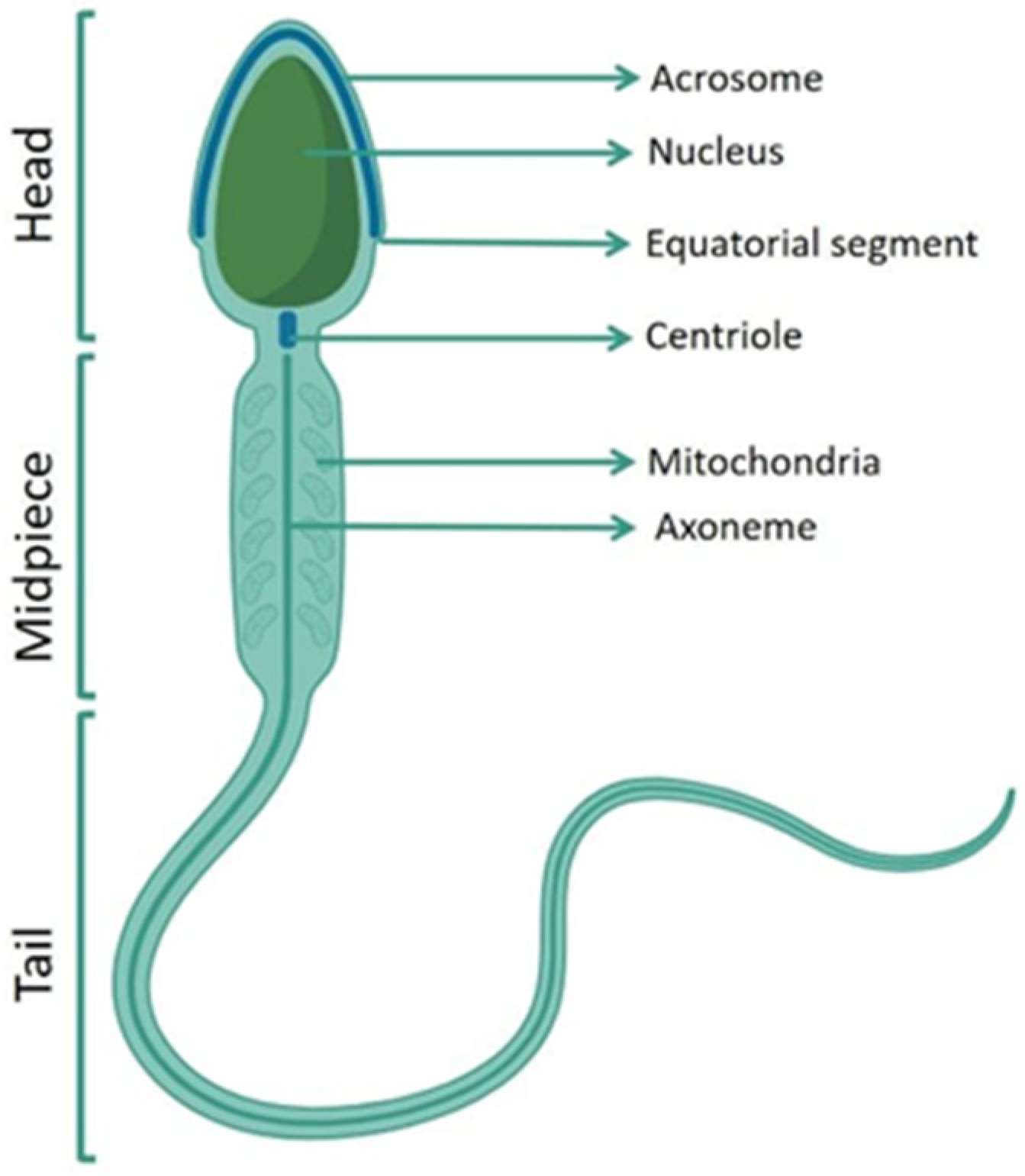
Figure 1. A schematic human spermatozoon. Human spermatozoon comprises three main sections: head, midpiece, and tail. The structures that characterize each region are indicated. The acrosome is located in the apical part of the nucleus and contains lytic enzymes; the nucleus is characterized by highly condensed chromatin; the equatorial segment is located at the end of acrosome. From the centriole the axoneme originates, the mitochondria surround the axoneme in the midpiece.
Antioxidants 13 00193 g001
Figure 2. The structural domains of Nrf2 (A) and Keap1 (B). (A): Nrf2 comprises seven Nrf2-ECH homeodomains (Neh). For each Neh domain, the specific activity is indicated. (B): Keap1 has five domains, including NTR (N-terminal region), BTB (Bric a brac, tramtrack and broad complex), IVR (intervening region), Kelch (Kelch or DGR domains) and CTR (C-terminal region). The specific role is indicated for each region. The amino acid residues for each domain are indicated in red.
Antioxidants 13 00193 g002
Figure 3. The Nrf2 antioxidative pathway. Activation of the Nrf2 transcription factor in oxidative conditions led to upregulation of Nrf2-related gene expression and reduction in reactive oxygen species (ROS). In normal conditions, ubiquitination of Nrf2 leads to proteasomal degradation. ARE, antioxidant response element; Nrf2, nuclear erythroid-factor-2; SOD, Superoxide Dismutase; CAT, Catalase; GPx, Glutathione Peroxidase, NQO1, quinone oxidoreductase-1; HO-1, heme oxygenase-1; sMAF, small musculoaponeurotic fibrosarcoma oncogene homolog, Ub: ubiquitin.
Antioxidants 13 00193 g003
Figure 4. Nrf2 roles in male fertility. Nrf2 is a regulator of multiple mechanisms involved in the adequate production and maturation of sperm. The figure summarizes the main roles of Nrf2 in the regulation of male fertility. Further details and bibliographical references are given in Section 4.
Antioxidants 13 00193 g004
Disclaimer/Publisher’s Note: The statements, opinions and data contained in all publications are solely those of the individual author(s) and contributor(s) and not of MDPI and/or the editor(s). MDPI and/or the editor(s) disclaim responsibility for any injury to people or property resulting from any ideas, methods, instructions or products referred to in the content.