Article

Senegenin Attenuates Pulmonary Fibrosis by Inhibiting Oxidative-Stress-Induced Epithelial Cell Senescence through Activation of the Sirt1/Pgc-1α Signaling Pathway

1
Xiangya Nursing School, Central South University, Changsha 410013, China
2
Xiangya School of Medicine, Central South University, Changsha 410013, China
*
Authors to whom correspondence should be addressed.
These authors contributed equally to this work.
Antioxidants 2024, 13(6), 675; https://doi.org/10.3390/antiox13060675
Submission received: 16 April 2024 / Revised: 27 May 2024 / Accepted: 28 May 2024 / Published: 31 May 2024

Abstract

:
Idiopathic pulmonary fibrosis is a fatal interstitial lung disease for which effective drug therapies are lacking. Senegenin, an effective active compound from the traditional Chinese herb Polygala tenuifolia Willd, has been shown to have a wide range of pharmacological effects. In this study, we investigated the therapeutic effects of senegenin on pulmonary fibrosis and their associated mechanisms of action. We found that senegenin inhibited the senescence of epithelial cells and thus exerted anti-pulmonary-fibrosis effects by inhibiting oxidative stress. In addition, we found that senegenin promoted the expression of Sirt1 and Pgc-1α and that the antioxidative and antisenescent effects of senegenin were suppressed by specific silencing of the Sirt1 and Pgc genes, respectively. Moreover, the senegenin-induced effects of antioxidation, antisenescence of epithelial cells, and antifibrosis were inhibited by treatment with Sirt1 inhibitors in vivo. Thus, the Sirt1/Pgc-1α pathway exerts its antifibrotic effect on lung fibrosis by mediating the antioxidative and antisenescent effects of senegenin.

1. Introduction

Idiopathic pulmonary fibrosis (IPF) is a chronic progressive interstitial lung disease, and its incidence and prevalence are increasing globally with an aging population [1,2]. Currently, there is a lack of specific drugs that can effectively treat IPF. Nintedanib and pirfenidone, the only two medicines approved by the Food and Drug Administration for the clinical treatment of IPF, cannot reverse pulmonary fibrosis and have multiple side effects [3,4]. Therefore, research on the pathogenesis of IPF and the development of specific medicines for its treatment are of great significance.
Cellular senescence is usually defined as irreversible cell cycle arrest associated with changes in cellular morphology, secretory profiles, and epigenetic alterations [5,6]. Senescent cells experience irreversible replication arrest, apoptosis resistance, increased reactive oxygen species production, and increased abundance of senescence-associated secretory phenotypes, all of which affect normal cellular physiological functions [7]. Cellular senescence has been shown to be a key pathogenic mechanism in a variety of chronic diseases, and inhibition of cellular senescence can effectively inhibit the progression of these diseases [8]. In particular, epithelial cell senescence has been a research hot spot in recent years, and inhibition of epithelial cell senescence has also been shown to be effective in treating the progression of multiple chronic respiratory and urologic diseases [9,10,11,12].
Although the etiology and pathogenesis of IPF are currently unknown, an increasing number of studies have shown that alveolar epithelial cell senescence promotes the development of IPF, whereas its inhibition effectively suppresses the progression of pulmonary fibrosis. This may be related to the fact that the key secretory and regenerative roles of senescent alveolar epithelial cells in the alveoli are compromised, leading to impairment of their role in maintaining lung homeostasis [13]. Multiple antiaging mechanisms in the body play an important role in inhibiting the progression of IPF due to the senescence of epithelial cells. Among them, Sirtuin proteins including Sirt1, Sirt3, and Sirt6 have been shown to play key roles; however, their detailed mechanisms have yet to be explored [14,15,16].
Senegenin, a major bioactive component isolated from the roots of Polygala tenuifolia Willd [17], has been shown to have a wide range of pharmacological effects, including attenuating acute lung injury, improving cognitive impairment, and promoting the repair of spinal cord injury [18,19,20]. However, its use in the treatment of pulmonary fibrosis has not been studied. In the present study, we investigated for the first time the anti-pulmonary-fibrosis effect of senegenin and elucidated the mechanism of its anti-pulmonary-fibrosis effect from the perspective of inhibiting epithelial cell senescence. The results further enrich the pharmacological application of senegenin and provide novel insights into the pharmacological treatment of idiopathic pulmonary fibrosis.

2. Materials and Methods

2.1. Animal Experiment

Eight–week–old male C57BL/6 mice without underlying diseases were housed in a pathogen–free environment at the animal center of Central South University. After 1 week of acclimatization feeding, the mice were anesthetized with sodium pentobarbital. A lung fibrosis model was constructed via intratracheal injection of 50 μL of bleomycin (3 mg/kg; Nippon Kayaku, Tokyo, Japan).
From a total of 110 mice, 10 were randomly selected as controls, and the remaining mice were used for bleomycin modeling. Control mice were injected intraperitoneally with the solvent on days 15–28 after tracheal injection of 50 μL of saline. Meanwhile, on the 14th day after the tracheal injection of bleomycin, the model mice were stratified into four groups, each with the same number of mice according to their body weights: a lung fibrosis model group, which was injected intraperitoneally with the solvent on days 15–28; a high-dose group, which was injected intraperitoneally with 20 mg/kg of senegenin (Solarbio, Beijing, China) on days 15–28; a medium-dose group, which was injected intraperitoneally with 5 mg/kg of senegenin on days 15–28; and a low-dose group, which was injected intraperitoneally with 1 mg/kg of senegenin on days 15–28. The mice were euthanized on day 29, and the lung tissues were obtained.
From a total of 100 mice, 10 were randomly selected as controls and the remaining were used for modeling. The control mice were treated as described above. On the 14th day after the tracheal injection of bleomycin, the successfully constructed mouse models were divided into three groups, with the same number of mice according to body weight: a lung fibrosis model group and high-dose group, whose mice were treated as described above; and an EX527 group, whose mice were pretreated for 30 min with an intraperitoneal injection of 5 mg/kg of EX527 (Selleck Chemicals, Houston, TX, USA) on days 15–28, followed by intraperitoneal injection of 20 mg/kg of senegenin. This study was carried out in accordance with the laboratory animal welfare and ethical principles. The experimental protocol was approved by the Laboratory Animal Welfare and Ethical Committee of Central South University.
We formulated the in vivo injection of senegenin and EX527 according to the solvent formulation recommended by Selleck Chemicals. We first solubilized senegenin or EX527 with DMSO and then added PEG300, Tween80, and saline sequentially, resulting in a final ratio of 5%:40%:5%:50% (V:V:V:V:V) for DMSO, PEG300, Tween80, and saline, respectively.

2.2. Hematoxylin–Eosin (HE) Staining, Masson Staining, and Ashcroft Score

Mouse lung tissue was fixed with 4% paraformaldehyde, embedded in paraffin, and cut into sections. After deparaffinization, the sections were stained with HE (Pinuofei Biological, Wuhan, China) and Masson stain (Pinuofei Biological) and finally dehydrated and sealed. The Ashcroft scoring method based on Ashcroft et al. was used to score the fibrosis level of each HE-stained section, and then the mean value was calculated [21]. Assessment was performed by three researchers.

2.3. Determination of Hydroxyproline in Lung Tissue

A homogenate of mouse lung tissue was prepared using a hydroxyproline assay kit (Nanjing Jiancheng Biotechnology Institute, Nanjing, China) according to the manufacturer’s instructions and incubated at room temperature. After centrifugation, the absorbance value of the supernatant at 550 nm was measured, and the hydroxyproline level was calculated according to the absorbance value.

2.4. Immunofluorescence Staining

Immunofluorescence was performed using a tyramide signal amplification (TSA) Immunofluorescence Kit (Aifang Biological, Changsha, China) according to the manufacturer’s instructions.
For the immunofluorescence staining of lung tissue, the sections were deparaffinized to repair the antigen and block the endogenous peroxidase and the nonspecific antigen. The sections were incubated with anti-P21 antibody (Proteintech, Wuhan, China) overnight. The next day, the sections were incubated with a horseradish-peroxidase-(HRP)-conjugated secondary antibody, followed by incubation with a TSA fluorescent dye. The above steps were repeated, with modifications of anti-SPC antibody (Proteintech) incubation and nuclear staining with DAPI dye. The slides were sealed, and images were captured using a fluorescence microscope.
For cellular immunofluorescence staining, the cells were fixed with 4% paraformaldehyde, blocked for nonspecific antigens after membrane rupture, incubated with anti-P21 antibody overnight, and then incubated with HRP-conjugated secondary antibody. Incubation with TSA fluorescent dye reactive solution was then performed, followed by nuclear staining with DAPI dye. The slides were blocked, and images were captured using a fluorescence microscope.

2.5. ROS Staining

For ROS staining of lung tissue, frozen sections of lung tissue were heated to room temperature and were circled using an immunohistochemical pen, after which a fluorescent brightening agent was added. The tissues were incubated with ROS dye, which was followed by nuclear staining with DAPI. The sections were sealed, and images were captured via fluorescence microscopy.
For cellular ROS staining, cells were incubated with ROS fluorescent probe (Beyotime Biotechnology, Shanghai, China) at 37 °C for 30 min and then washed twice with DMEM. Images were then captured via fluorescence microscopy, and ROS levels were measured using flow cytometry.

2.6. Determination of Malondialdehyde (MDA) and Glutathione (GSH) Content and Total Superoxide Dismutase (SOD) Activity in Lung Tissue

Total SOD activity and MDA and GSH contents were measured using an MDA, GSH, and SOD assay kit (Nanjing Jiancheng Biotechnology Institute) according to the manufacturer’s instructions and with reference to our previous research method. The mouse lung tissue was made into tissue homogenate, and each reagent in the kit was sequentially added. The corresponding absorbance values were measured using each enzyme marker and were calculated according to the instructions.

2.7. RNA Extraction and Real-Time Quantitative Polymerase Chain Reaction (qPCR)

RNA was extracted from cells and lung tissues with TRIzol (Thermo Fisher Scientific, Waltham, MA, USA) and reverse transcribed to cDNA using a reverse transcription kit (Thermo Fisher Scientific). The primer sequences (Sangon Biotech, Shanghai, China) are listed in Table 1.

2.8. Western Blotting

Mouse lung tissues or cells were extracted using radioimmunoprecipitation assay lysate containing phenylmethylsulfonyl fluoride, phosphatase inhibitor, and protease inhibitor (Solarbio), and the total protein concentration was determined using bicinchoninic acid assay kits (CWBIO, Taizhou, China). The proteins were subjected to sodium dodecyl sulfate–polyacrylamide gel electrophoresis and were then transferred to polyvinylidene fluoride membranes. The membranes were incubated with 5% skimmed milk, which was followed by incubation with β-actin, α-SMA, Pgc-1α, and Sirt1 antibodies (Proteintech) and with collagen I, p21, and p16 antibodies (Abcam, Cambridge, UK). The membranes were then incubated with HRP-labeled goat anti-rabbit immunoglobulin G monoclonal antibody (Proteintech) or HRP-labeled goat anti-mouse immunoglobulin G monoclonal antibody (Proteintech) at room temperature and washed with Tris-buffered saline with Tween 20 before protein expression was visualized with an ECL developer.

2.9. Cell Culture and Hydrogen–Peroxide–Induced Epithelial Cell Senescence

Mouse lung epithelial cells (MLE-12 cells) were cultured in a humidified incubator at 37 °C with 5% CO2. Induction of cellular senescence was performed as described in several previous studies. The cells were seeded in plates and incubated with medium containing 200 µΜ of hydrogen peroxide (Sigma, St. Louis, MO, USA) for 2 h, which was then changed to normal medium for 48 h.

2.10. Cell Viability Assay

Cell viability was determined using Cell Counting Kit-8 (CCK8; Elabscience, Wuhan, China) according to the manufacturer’s instructions. The cells were seeded in 96-well plates and incubated with different concentrations of senegenin for 48 h. The cells were washed with phosphate-buffered saline, treated with CCK-8 reagent, and incubated in an incubator at 37 °C. Finally, the absorbance values at 450 nm were measured.

2.11. SiRNA–Specific Gene Silencing

Cells were seeded in well plates and transfected upon reaching 50% confluency. Transfection complexes were prepared by mixing transfection reagents (Rebobio, Guangzhou, China) with SiRNAs (control and Sirt1 SiRNAs from Santa Cruz Biotech (Santa Cruz, CA, USA); Pgc-1α SiRNA from Sangon Biotech). The cells were incubated at room temperature for 15 min and were washed twice with DMEM, and the transfection complexes were then mixed with complete medium without penicillin–streptomycin mixture and added to the well plates before subsequent interventions.

2.12. Statistical Analysis

All data were analyzed using Graphpad Prism 8.3.1. All data are expressed as the mean ± standard deviation. Comparisons between two measures were performed using the t-test. Comparisons between multiple measures were performed using one-way analysis of variance followed by Tukey’s test. Statistical significance was set at a p-value < 0.05.

3. Results

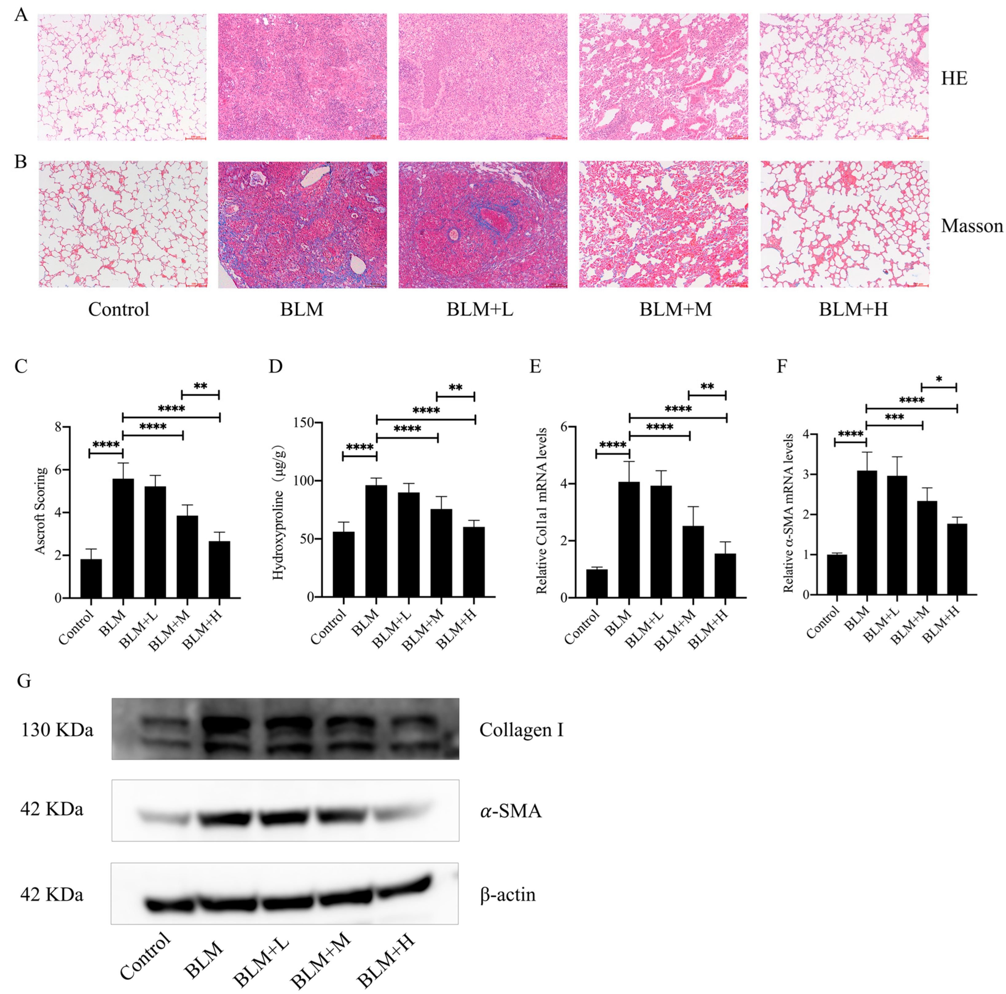
3.1. Senegenin Inhibited Bleomycin-Induced Pulmonary Fibrosis in Mice

To investigate the direct antifibrotic effect of senegenin, we constructed a mouse lung fibrosis model using bleomycin and administered low, medium, and high doses of senegenin intraperitoneally on days 15–28 after establishment of the model. HE and Masson staining showed that medium and high doses of senegenin effectively improved the lung tissue structure and reduced extracellular matrix deposition in lung tissues of mice with lung fibrosis (Figure 1A,B). The Ashcroft score and hydroxyproline determination showed that medium and high doses of senegenin effectively improved the overall pathological changes and collagen deposition in the lungs of mice with pulmonary fibrosis (Figure 1C,D). In addition, medium and high doses of senegenin effectively reduced the mRNA and protein expression levels of fibrosis markers type I collagen and α-SMA (Figure 1E–G). These results suggest that medium and high doses of senegenin effectively reduce bleomycin-induced pulmonary fibrosis, with high doses having a more significant effect.

3.2. Senegenin Reduced Oxidative Stress Levels in the Lung Tissues of Pulmonary Fibrosis Mice

Senegenin has been shown to have antioxidative effects [22]. To investigate whether it exerts an antioxidative effect in addition to its antifibrotic effect, we examined the levels of ROS in the lung tissues of different groups of mice and found that medium and high doses of senegenin significantly reduced the levels of ROS in the lung tissues of mice with pulmonary fibrosis (Figure 2A). Meanwhile, high and medium doses of senegenin effectively reduced the levels of MDA and increased the levels of GSH and total SOD activity in the lung tissues of mice with pulmonary fibrosis (Figure 2B–D), with the high dose exhibiting a more pronounced effect. This suggests that senegenin also exerts antioxidative effects along with antifibrotic effects, and its anti–pulmonary-fibrosis effects may also be related to their antioxidative effects.

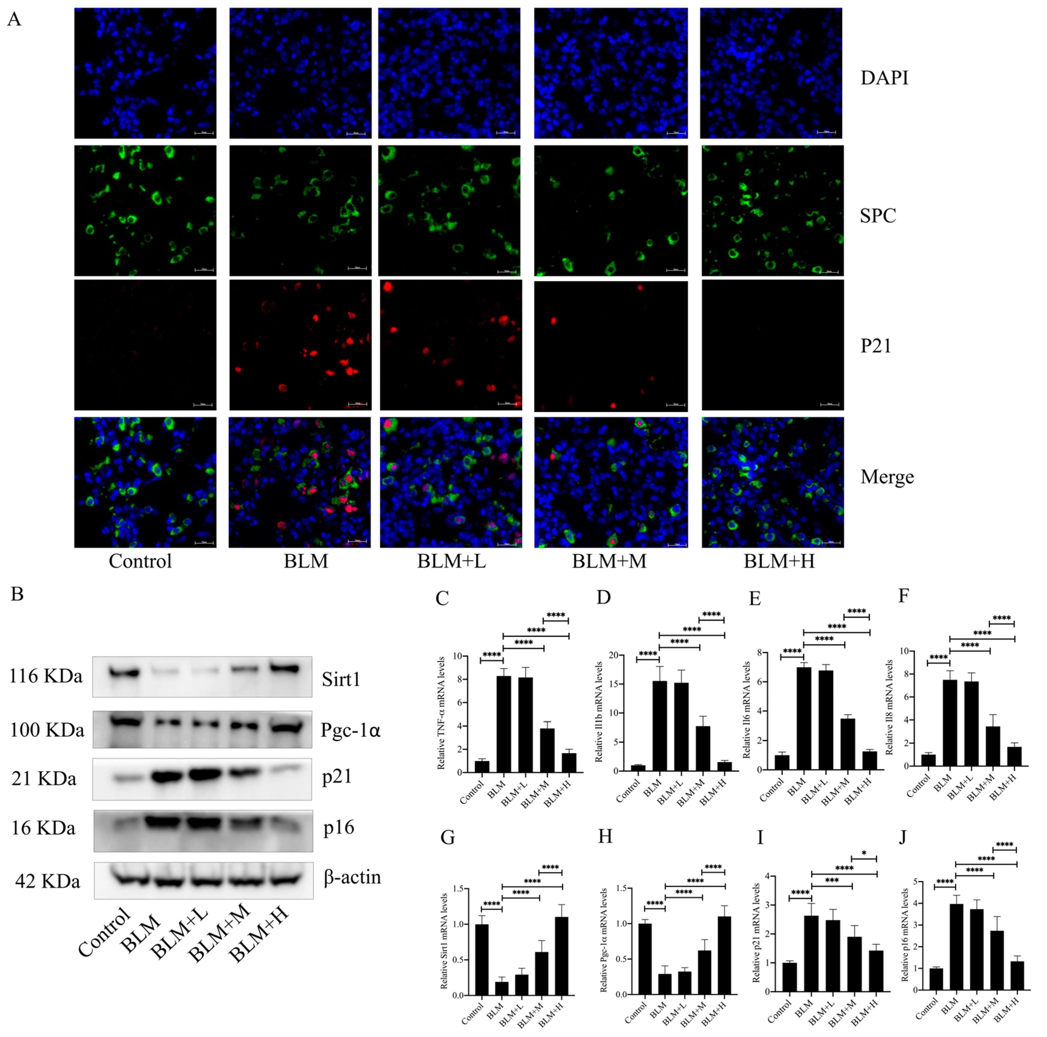
3.3. Senegenin Inhibited Epithelial Cell Senescence in Lung Tissues of Pulmonary Fibrosis Mice

Although the specific pathogenesis of IPF is currently unknown, epithelial cell senescence has been shown to be an important pathological change in the development of the disease. Oxidative stress is one of the important factors leading to epithelial senescence, whereas inhibition of epithelial cell senescence inhibits the progression of IPF [23]. In the results described above, we found that senegenin exerted its antifibrotic effect along with its antioxidative effect. We then investigated whether the antifibrotic effect of senegenin is related to its antisenescent effect on epithelial cells. The results showed that medium and high doses of senegenin significantly reduced epithelial cell senescence (Figure 3A) and the levels of four senescence-associated secretory phenotype (SASP) secretions, namely TNF-α, IL1-β, IL-6, and IL-8, in lung tissues of mice with pulmonary fibrosis (Figure 3C–F). Medium and high doses of senegenin also significantly reduced the protein and mRNA expression levels of the senescence markers p21 and p16 (Figure 3B,I–J). Sirt1 plays a key role in exerting antiaging effects in the body, and activation of the Sirt1/Pgc-1α pathway has been demonstrated to be an important pathway by which Sirt1 exerts its antiaging effects [24]. To investigate whether the mechanism of the antisenescence of epithelial cells exerted by senegenin is related to the activation of the Sirt1/Pgc-1α pathway, we examined the expression levels of Sirt1 and Pgc-1α in mouse lung tissues. The results showed that medium and high doses of senegenin significantly increased the protein and mRNA expression levels of Sirt1 and Pgc-1α in the lung tissues of pulmonary fibrosis mice (Figure 3B,G–H). The above results indicated that senegenin exerts an antisenescent effect, which might be related to its activation of the Sirt1/Pgc-1α pathway, and that this effect might also be related to its antifibrotic effect.

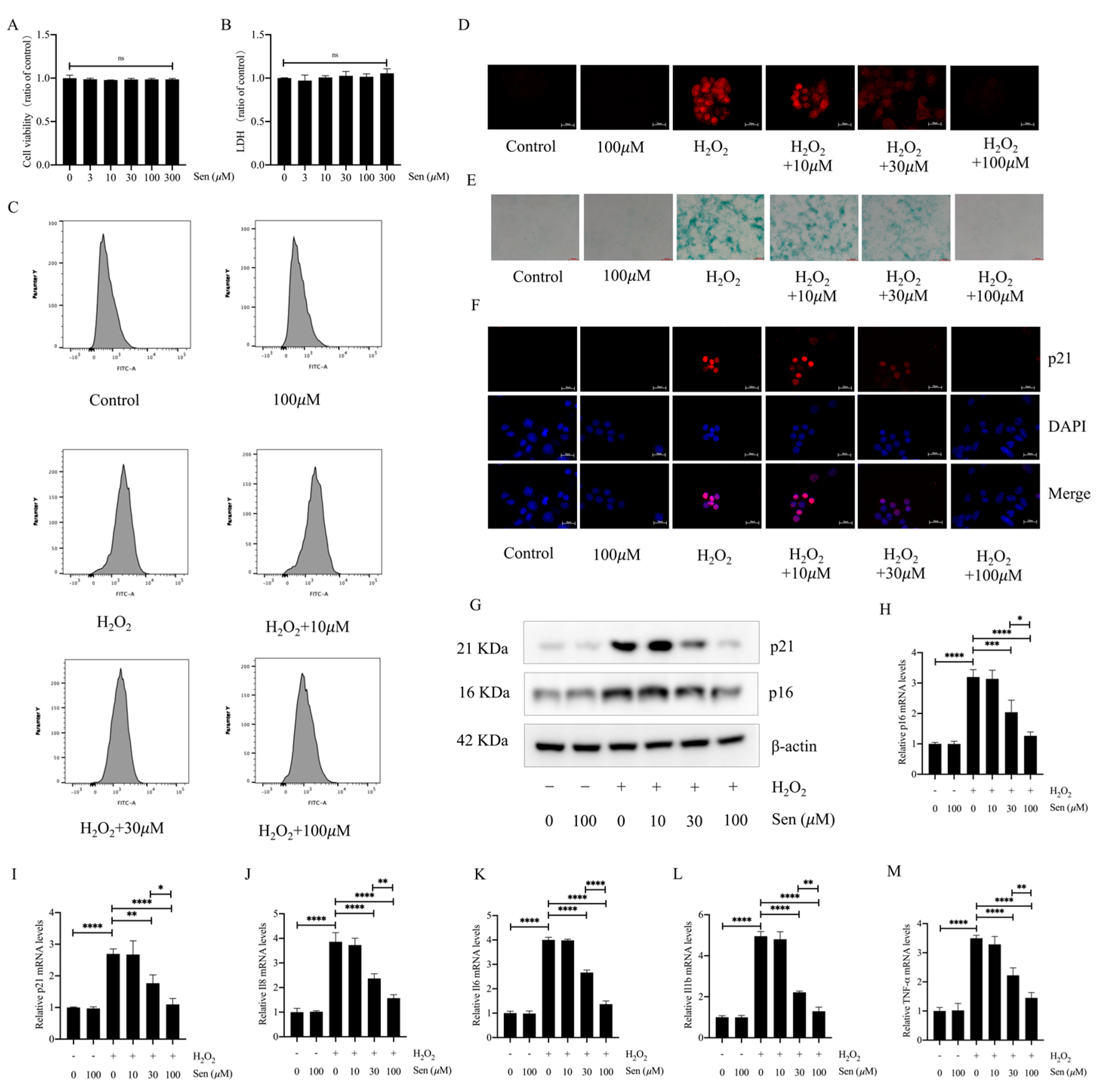
3.4. Senegenin Inhibited Hydrogen-Peroxide-Induced Epithelial Cell Senescence

To further investigate whether the antifibrotic effect of senegenin is related to its antisenescent effect, we explored the effect of senegenin on hydrogen-peroxide-induced epithelial cell senescence. We first examined the effect of different concentrations of senegenin on the viability and supernatant lactate dehydrogenase (LDH) content of lung epithelial cells to determine the concentration to be used, and the results showed that concentrations of 0–300 μΜ did not significantly affect epithelial cell viability (Figure 4A,B). Oxidative stress is one of the main factors leading to cellular senescence, and the level of ROS increases significantly during cellular senescence [25]. In the results described above, we found that senegenin significantly reduced the level of oxidative stress in lung tissues of mice with pulmonary fibrosis. We similarly found that 30 µΜ versus 100 µΜ of senegenin significantly reduced the level of ROS during hydrogen-peroxide-induced senescence of epithelial cells (Figure 4C,D). In addition, staining of cellular senescence-associated β-galactosidase and measurement of the expression levels of p21, p16, TNF-α, IL1-β, IL-6, and IL-8 revealed that 30 µΜ versus 100 µΜ of senegenin significantly reduced the expression levels of these indicators (Figure 4E–M). Thus, senegenin significantly inhibits hydrogen-peroxide-induced epithelial cell senescence, which may also be the cellular mechanism for its anti-pulmonary-fibrosis effect.

3.5. The Sirt1/Pgc-1α Pathway Mediated the Antisenescent Effect of Senegenin

To further investigate the mechanism of antisenescence of epithelial cells by senegenin, we examined its effects on Sirt1 and Pgc-1α, considering that senegenin significantly increased Sirt1 and Pgc-1α expression in lung tissues of pulmonary fibrosis mice and that the Sirt1/Pgc-1α pathway plays an important role in antiaging. The results showed that 30 and 100 µΜ of senegenin significantly increased the protein and mRNA expression levels of Sirt1 and Pgc-1α (Figure 5A–C). Silencing of the Sirt1 and Pgc genes with siRNA resulted in the inhibition of the effects of senegenin on the ROS and β-galactosidase content and p16, p21, TNFα, IL1β, IL6, and IL8 expression levels during hydrogen-peroxide-induced epithelial cellular senescence (Figure 5D–M). Thus, the inhibitory effect of senegenin on epithelial cell senescence was mediated via the Sirt1/Pgc-1α pathway.

3.6. Sirt1 Mediated the In Vivo Antifibrotic Effects of Senegenin

To further validate the role of Sirt1 in the anti–pulmonary fibrosis activity of senegenin in vivo, we observed the effects of senegenin on pulmonary fibrosis mice administered with a Sirt1-specific inhibitor EX527 and subsequently treated with a high dose of senegenin. The results showed that EX527 inhibited the effects of senegenin on the disordered lung tissue outcome with extracellular matrix overdeposition in pulmonary fibrosis mice (Figure 6A–D). EX527 also inhibited the effects of senegenin on the reduction of α-SMA and type I collagen levels in the lung tissue of pulmonary fibrosis mice (Figure 6E–G). These results indicate that EX527 significantly inhibits the anti-pulmonary-fibrosis effects of senegenin, suggesting that the in vivo antifibrotic effects of senegenin are mediated by Sirt1.

3.7. Sirt1 Mediated the In Vivo Antioxidative Effect of Senegenin

To investigate whether Sirt1 also mediated the in vivo antioxidative effects of senegenin, mouse lung tissues were stained for ROS, and we found that EX527 inhibited the ROS-lowering effect of senegenin in lung tissues of pulmonary fibrosis mice (Figure 7A). Similarly, EX527 also inhibited the reduction of MDA and the elevation of GSH levels and total SOD activity by senegenin in the lung tissues of pulmonary fibrosis mice (Figure 7B–D). These results suggest that Sirt1 also mediates the in vivo antioxidative effects of senegenin.

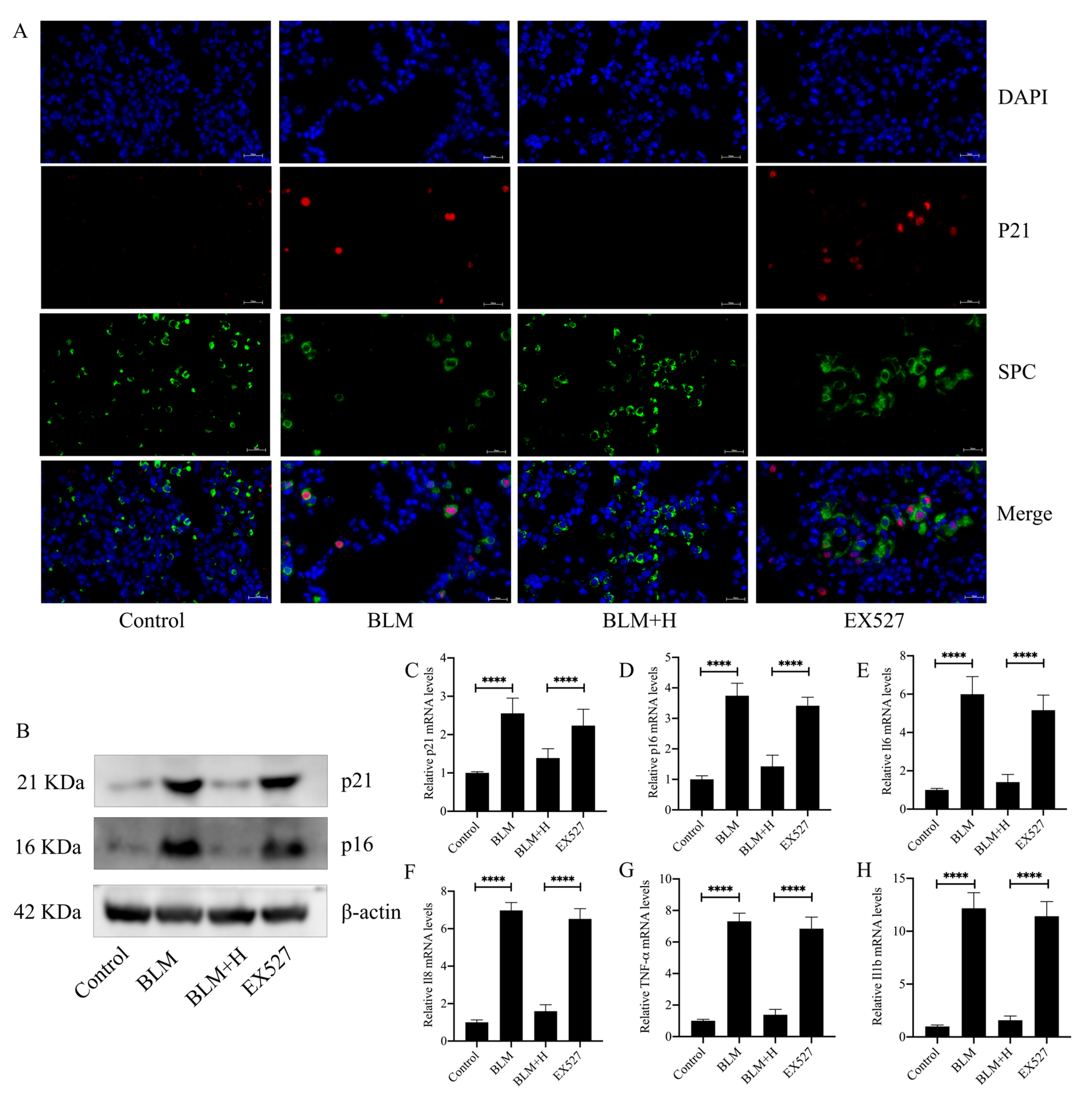
3.8. Sirt1 Mediated the Antisenescent Effect of Senegenin In Vivo

To further characterize the Sirt1-mediated antifibrotic effects in relation to its antisenescent effects on epithelial cells, we examined the effect of EX527 on indicators related to epithelial cell senescence. The results showed that EX527 inhibited the role of senegenin in inhibiting epithelial cell senescence (Figure 8A). Similarly, the inhibitory effects of senegenin on the expression of p21 and p16 in lung tissues of pulmonary fibrosis mice, along with the inhibitory effects of secretion of various SASPs, were inhibited by EX527 (Figure 8B–H). These results demonstrate that Sirt1 mediates the antisenescent effect of senegenin on epithelial cells and further shows that the antisenescent effect of senegenin mediates its antifibrotic effect.

4. Discussion

In the present study, we demonstrated, for the first time, that senegenin inhibited alveolar epithelial cell senescence induced by oxidative stress through the activation of the Sirt1/Pgc-1α pathway, thereby exerting an antifibrotic effect on lungs. Activation of the Sirt1/Pgc-1α signaling pathway has been shown to exert antifibrotic effects through effects on the FOXO3 pathway, the PI3K/AKT pathway, and the Nrf2 pathway [26,27,28], and thus these pathways may also be relevant to the antifibrotic effects exerted by senegenin. In addition, activation of the Sirt1/Pgc-1α signaling pathway has been shown to exert anti-oxidative stress effect, ameliorate mitochondrial dysfunction, and inhibit apoptosis, which are all closely related to the development of pulmonary fibrosis [24,29,30]. Thus, the ability of senegenin to exert antifibrotic effects on lung fibrosis through the activation of the Sirt1/Pgc-1α signaling pathway may also be related to its modulation of oxidative stress, mitochondrial dysfunction, and apoptosis. All of the above deserve to be further explored in subsequent studies.
Oxidative stress has an important role in cellular senescence and the development of IPF [23,31,32,33]. For one, ROS may drive cellular senescence through DNA damage and the activation of DNA-damage-response-associated pathways, and the senescence of different cells, including epithelial cells, leads to the progression of idiopathic pulmonary fibrosis. For another, mitochondrial dysfunction occurring in senescent cells leads to more ROS production, which may further contribute to the progression of cellular senescence and fibrosis [34]. We investigated the anti-pulmonary-fibrosis effects of senegenin and the underlying molecular mechanisms using a mouse lung fibrosis model and a stress-induced alveolar epithelial premature senescence model, which were constructed using bleomycin and hydrogen peroxide, respectively, and the results showed that senegenin exerted antioxidative and antisenescent effects both in vivo and in vitro. Considering the important role of oxidative stress in alveolar epithelial cell senescence and in idiopathic pulmonary fibrosis, we postulate that the anti-pulmonary-fibrosis effect of senegenin may be closely related to its inhibition of oxidative-stress-induced epithelial cell senescence. Studies have confirmed that the antioxidative effect of senegenin is involved in their neuroprotective effects in the treatment of Parkinson’s disease [35], and whether the therapeutic effects of senegenin on Parkinson’s and other disorders are also related to their anticellular senescence effects warrants further exploration.
Sirtuin proteins are a class of proteins with nicotinamide adenine dinucleotide (NAD+)-dependent deacetylase activity or ADP-ribosyltransferase activity, and seven Sirtuin proteins (from Sirt1 to Sirt7) have been identified in mammals, of which Sirt1 is the most extensively studied [36]. Sirtuin proteins have a wide range of functions, among which the role of Sirt1 in antiaging has been widely validated [37]. Owing to its antiaging effects, Sirt1 is used as a key target for the treatment of a variety of diseases, including idiopathic pulmonary fibrosis, tumors, osteoporosis, and atherosclerosis [38,39,40,41]. In the present study, we found that senegenin significantly increased the expression level of Sirt1 in the lung tissues of pulmonary fibrosis mice and alveolar epithelial cells in vivo and in vitro, respectively. Moreover, the antifibrotic and antisenescent effects of senegenin were suppressed in vivo by treatment with the Sirt1 inhibitor EX527, and the antioxidative and antisenescent effects of senegenin were suppressed in vitro by silencing the Sirt1 gene using siRNA, which suggest that Sirt1 is a key target for senegenin in exerting antifibrotic and antisenescent effects. In addition to Sirt1, other proteins of the Sirtuin family such as Sirt3 and Sirt6 play important roles in antiaging and anti-pulmonary fibrosis [16,42]. Whether senegenin, while exerting a role in promoting Sirt1 expression, also affects the expression of other Sirtuin proteins warrants further exploration.
Pgc-1α is a key mediator of mitochondrial energy metabolism and has been shown to be a key regulator of cellular responses to a variety of physiological stimuli [43,44,45]. The Sirt1/Pgc-1α axis exerts a positive effect in various diseases by inhibiting cellular senescence [46,47,48]. However, the role of Pgc-1α in pulmonary fibrosis is currently unknown. When we explored the effects of senegenin on Sirt1 in vivo and in vitro, we similarly found that senegenin promoted the expression of Pgc-1α and that the senegenin-induced antioxidative and antisenescent effects on alveolar epithelial cell were suppressed upon specific silencing of Pgc-1α. This suggests that the Sirt1/Pgc-1α pathway mediates the antioxidative and antisenescent effects of senegenin. In addition, Pgc-1α is closely related to the function of the mitochondria, which play an important role in cellular senescence; however, we did not test the effects of senegenin on mitochondrial function in this study. Our subsequent studies should examine the effects of Sirt1/Pgc-1α on the mitochondrial function of epithelial cells.
In addition to antioxidative effects, senegenin has also been reported to have antiapoptotic, anti–inflammatory, and anti-injury pharmacological effects [49,50,51], and whether these pharmacological effects are related to its antioxidative and antisenescent effects warrants further exploration. The development of pulmonary fibrosis involves a variety of pathological mechanisms, which also include apoptosis, inflammation, cellular senescence, and oxidative stress injury. In our study, we demonstrated that senegenin exerts its anti–pulmonary fibrosis effect through its anti-cellular senescence effect, and whether the anti–pulmonary fibrosis effect of senegenin is also related to the other pharmacological effects mentioned above deserves to be further explored. The present study showed that senegenin inhibits oxidative-stress-induced epithelial cell senescence through the Sirt1/Pgc-1α pathway, thus exerting an antifibrotic effect on lung fibrosis, but whether senegenin has synergistic or other effects with drugs commonly used for the treatment of IPF, including pirfenidone and nintedanib, were not explored in this study. It is important to explore these interactions, which will provide a better scientific basis for the clinical application of senegenin in the future, and we will further explore the relevant elements in future studies. This study is the first to propose the Sirt1/Pgc-1α pathway as a novel drug target for the treatment of pulmonary fibrosis by inhibiting alveolar epithelial cell senescence. This proposition has important theoretical value, as it reveals a novel molecular mechanism for the treatment of pulmonary fibrosis. Furthermore, from a clinical perspective, targeting the Sirt1/Pgc-1α pathway may enable the development of innovative therapeutic strategies for the treatment of pulmonary fibrosis, which could help address the critical medical need for pulmonary fibrosis therapy.

Author Contributions

Conceptualization, Q.Z. and W.L.; methodology, Q.Z. and X.S.; software, Z.H.; validation, Q.Z., Y.L. and M.L.; formal analysis, B.W.; investigation, Q.Z.; resources, X.S. and S.T.; data curation, Y.L.; writing—original draft preparation, Q.Z. and Y.L.; writing—review and editing, W.L., X.H. and Q.Z.; visualization, M.S. and Z.L.; supervision, W.L. and S.T.; project administration, W.L. and S.T.; funding acquisition, Y.L., X.H., M.S., W.L. and S.T. All authors have read and agreed to the published version of the manuscript.

Funding

This research was funded by National Key Research and Development Program of China (grant number: 2022YFC3601603), the National Natural Science Foundation of China (grant number: 81500056, 82100084, and 82170853), the Natural Science Foundation of Hunan Province (grant number: 2020JJ4773, 2021JJ30899, and 2022JJ40293), The science and technology innovation program of Hunan province (grant number: 2022WZ1033), and the Graduate Student Research and Innovation Program of Central South University (grant number: CX20230307).

Institutional Review Board Statement

This study was conducted in accordance with the welfare and ethical principles of laboratory animals. The experimental protocol was approved by the Laboratory Animal Welfare and Ethical Committee of Central South University (IACUC number: CSU-2023-0330).

Data Availability Statement

Any necessary data from this study can be obtained by contacting the corresponding authors.

Acknowledgments

The authors thank all those who contributed to this article.

Conflicts of Interest

The authors declare no conflicts of interest.

References

  1. Lederer, D.J.; Martinez, F.J. Idiopathic Pulmonary Fibrosis. N. Engl. J. Med. 2018, 378, 1811–1823. [Google Scholar] [CrossRef] [PubMed]
  2. Martinez, F.J.; Collard, H.R.; Pardo, A.; Raghu, G.; Richeldi, L.; Selman, M.; Swigris, J.J.; Taniguchi, H.; Wells, A.U. Idiopathic pulmonary fibrosis. Nat. Rev. Dis. Prim. 2017, 3, 17074. [Google Scholar] [CrossRef] [PubMed]
  3. Richeldi, L.; Collard, H.R.; Jones, M.G. Idiopathic pulmonary fibrosis. Lancet 2017, 389, 1941–1952. [Google Scholar] [CrossRef] [PubMed]
  4. Raghu, G.; Collard, H.R.; Egan, J.J.; Martinez, F.J.; Behr, J.; Brown, K.K.; Colby, T.V.; Cordier, J.F.; Flaherty, K.R.; Lasky, J.A.; et al. An official ATS/ERS/JRS/ALAT statement: Idiopathic pulmonary fibrosis: Evidence-based guidelines for diagnosis and management. Am. J. Respir. Crit. Care Med. 2011, 183, 788–824. [Google Scholar] [CrossRef] [PubMed] [PubMed Central]
  5. van Deursen, J.M. The role of senescent cells in ageing. Nature 2014, 509, 439–446. [Google Scholar] [CrossRef] [PubMed] [PubMed Central]
  6. Gorgoulis, V.; Adams, P.D.; Alimonti, A.; Bennett, D.C.; Bischof, O.; Bishop, C.; Campisi, J.; Collado, M.; Evangelou, K.; Ferbeyre, G.; et al. Cellular Senescence: Defining a Path Forward. Cell 2019, 179, 813–827. [Google Scholar] [CrossRef] [PubMed]
  7. Tchkonia, T.; Zhu, Y.; van Deursen, J.; Campisi, J.; Kirkland, J.L. Cellular senescence and the senescent secretory phenotype: Therapeutic opportunities. J. Clin. Investig. 2013, 123, 966–972. [Google Scholar] [CrossRef] [PubMed] [PubMed Central]
  8. Zhang, L.; Pitcher, L.E.; Yousefzadeh, M.J.; Niedernhofer, L.J.; Robbins, P.D.; Zhu, Y. Cellular senescence: A key therapeutic target in aging and diseases. J. Clin. Investig. 2022, 132, e158450. [Google Scholar] [CrossRef] [PubMed] [PubMed Central]
  9. Tian, Y.; Li, H.; Qiu, T.; Dai, J.; Zhang, Y.; Chen, J.; Cai, H. Loss of PTEN induces lung fibrosis via alveolar epithelial cell senescence depending on NF-κB activation. Aging Cell 2019, 18, e12858. [Google Scholar] [CrossRef] [PubMed] [PubMed Central]
  10. Sarker, R.S.J.; Conlon, T.M.; Morrone, C.; Srivastava, B.; Konyalilar, N.; Verleden, S.E.; Bayram, H.; Fehrenbach, H.; Yildirim, A. CARM1 regulates senescence during airway epithelial cell injury in COPD pathogenesis. Am. J. Physiol. Cell. Mol. Physiol. 2019, 317, L602–L614. [Google Scholar] [CrossRef] [PubMed]
  11. Ni, J.Y.; Wang, X.; Xie, H.Y.; Yang, N.H.; Li, J.Y.; Sun, X.A.; Guo, H.J.; Zhou, L.; Zhang, W.; Liu, J.; et al. Deubiquitinating enzyme USP11 promotes renal tubular cell senescence and fibrosis via inhibiting the ubiquitin degradation of TGF-β receptor II. Acta Pharmacol. Sin. 2023, 44, 584–595. [Google Scholar] [CrossRef] [PubMed] [PubMed Central]
  12. Shen, S.; Ji, C.; Wei, K. Cellular Senescence and Regulated Cell Death of Tubular Epithelial Cells in Diabetic Kidney Disease. Front. Endocrinol. 2022, 13, 924299. [Google Scholar] [CrossRef] [PubMed] [PubMed Central]
  13. Yao, C.; Guan, X.; Carraro, G.; Parimon, T.; Liu, X.; Huang, G.; Mulay, A.; Soukiasian, H.J.; David, G.; Weigt, S.S.; et al. Senescence of Alveolar Type 2 Cells Drives Progressive Pulmonary Fibrosis. Am. J. Respir. Crit. Care Med. 2021, 203, 707–717, Erratum in Am. J. Respir. Crit. Care Med. 2021, 204, 113. [Google Scholar] [CrossRef] [PubMed] [PubMed Central]
  14. Liang, J.; Huang, G.; Liu, X.; Taghavifar, F.; Liu, N.; Wang, Y.; Deng, N.; Yao, C.; Xie, T.; Kulur, V.; et al. The ZIP8/SIRT1 axis regulates alveolar progenitor cell renewal in aging and idiopathic pulmonary fibrosis. J. Clin. Investig. 2022, 132, e157338. [Google Scholar] [CrossRef] [PubMed] [PubMed Central]
  15. Cheresh, P.; Kim, S.J.; Jablonski, R.; Watanabe, S.; Lu, Z.; Chi, M.; Helmin, K.A.; Gius, D.; Budinger, G.R.S.; Kamp, D.W. SIRT3 Overexpression Ameliorates Asbestos-Induced Pulmonary Fibrosis, mt-DNA Damage, and Lung Fibrogenic Monocyte Recruitment. Int. J. Mol. Sci. 2021, 22, 6856. [Google Scholar] [CrossRef] [PubMed] [PubMed Central]
  16. Minagawa, S.; Araya, J.; Numata, T.; Nojiri, S.; Hara, H.; Yumino, Y.; Kawaishi, M.; Odaka, M.; Morikawa, T.; Nishimura, S.L.; et al. Accelerated epithelial cell senescence in IPF and the inhibitory role of SIRT6 in TGF-β-induced senescence of human bronchial epithelial cells. Am. J. Physiol.-Lung Cell. Mol. Physiol. 2011, 300, L391–L401. [Google Scholar] [CrossRef] [PubMed] [PubMed Central]
  17. Chen, Z.; Yang, Y.; Han, Y.; Wang, X. Neuroprotective Effects and Mechanisms of Senegenin, an Effective Compound Originated from the Roots of Polygala Tenuifolia. Front. Pharmacol. 2022, 13, 937333. [Google Scholar] [CrossRef] [PubMed] [PubMed Central]
  18. Liu, C.H.; Zhang, W.D.; Wang, J.J.; Feng, S.D. Senegenin Ameliorate Acute Lung Injury Through Reduction of Oxidative Stress and Inhibition of Inflammation in Cecal Ligation and Puncture-Induced Sepsis Rats. Inflammation 2016, 39, 900–906. [Google Scholar] [CrossRef] [PubMed]
  19. Xie, W.; Yang, Y.; Gu, X.; Zheng, Y.; Sun, Y.E.; Liang, Y.; Bo, J.; Ma, Z. Senegenin attenuates hepatic ischemia-reperfusion induced cognitive dysfunction by increasing hippocampal NR2B expression in rats. PLoS ONE 2012, 7, e45575. [Google Scholar] [CrossRef] [PubMed] [PubMed Central]
  20. Zhang, S.Q.; Wu, M.F.; Gu, R.; Liu, J.B.; Li, Y.; Jiang, J.L. Senegenin inhibits neuronal apoptosis after spinal cord contusion injury. Neural Regen. Res. 2016, 11, 657–663. [Google Scholar] [CrossRef] [PubMed] [PubMed Central]
  21. Ashcroft, T.; Simpson, J.M.; Timbrell, V. Simple method of estimating severity of pulmonary fibrosis on a numerical scale. J. Clin. Pathol. 1988, 41, 467–470. [Google Scholar] [CrossRef] [PubMed] [PubMed Central]
  22. Zhang, H.; Zhou, W.; Li, J.; Qiu, Z.; Wang, X.; Xu, H.; Wang, H.; Lu, D.; Qi, R. Senegenin Rescues PC12 Cells with Oxidative Damage Through Inhibition of Ferroptosis. Mol. Neurobiol. 2022, 59, 6983–6992. [Google Scholar] [CrossRef] [PubMed]
  23. Otoupalova, E.; Smith, S.; Cheng, G.; Thannickal, V.J. Oxidative Stress in Pulmonary Fibrosis. Compr. Physiol. 2020, 10, 509–547. [Google Scholar] [CrossRef] [PubMed]
  24. Wu, N.; Jin, W.; Zhao, Y.; Wang, H.; He, S.; Zhang, W.; Zhou, J. Sulfated Fucogalactan from Laminaria Japonica Ameliorates β-Cell Failure by Attenuating Mitochondrial Dysfunction via SIRT1–PGC1-α Signaling Pathway Activation. Front. Endocrinol. 2022, 13, 881256. [Google Scholar] [CrossRef] [PubMed] [PubMed Central]
  25. Papaconstantinou, J. The Role of Signaling Pathways of Inflammation and Oxidative Stress in Development of Senescence and Aging Phenotypes in Cardiovascular Disease. Cells 2019, 8, 1383. [Google Scholar] [CrossRef] [PubMed] [PubMed Central]
  26. Ho, J.H.; Baskaran, R.; Wang, M.F.; Yang, H.S.; Lo, Y.H.; Mohammedsaleh, Z.M.; Lin, W.T. Bioactive Peptides and Exercise Modulate the AMPK/SIRT1/PGC-1α/FOXO3 Pathway as a Therapeutic Approach for Hypertensive Rats. Pharmaceuticals 2022, 15, 819. [Google Scholar] [CrossRef] [PubMed] [PubMed Central]
  27. Jia, D.; Hou, L.; Lv, Y.; Xi, L.; Tian, Z. Postinfarction exercise training alleviates cardiac dysfunction and adverse remodeling via mitochondrial biogenesis and SIRT1/PGC-1α/PI3K/Akt signaling. J. Cell. Physiol. 2019, 234, 23705–23718. [Google Scholar] [CrossRef] [PubMed]
  28. Han, B.; Li, S.; Lv, Y.; Yang, D.; Li, J.; Yang, Q.; Wu, P.; Lv, Z.; Zhang, Z. Dietary melatonin attenuates chromium-induced lung injury via activating the Sirt1/Pgc-1α/Nrf2 pathway. Food Funct. 2019, 10, 5555–5565. [Google Scholar] [CrossRef] [PubMed]
  29. Niu, X.; Di, W.; Zhang, Z.; Li, N.; Qiu, Z.; Shi, W.; Lei, W.; Tang, J.; Yang, Y.; Xu, B.; et al. Activation of ITLN-1 attenuates oxidative stress injury via activating SIRT1/PGC1-α signaling in neuroblastoma cells. J. Cell. Physiol. 2024, 239, e31144. [Google Scholar] [CrossRef] [PubMed]
  30. Li, K.; Gao, L.; Zhou, S.; Ma, Y.R.; Xiao, X.; Jiang, Q.; Kang, Z.H.; Liu, M.L.; Liu, T.X. Erythropoietin promotes energy metabolism to improve LPS-induced injury in HK-2 cells via SIRT1/PGC1-α pathway. Mol. Cell. Biochem. 2023, 478, 651–663. [Google Scholar] [CrossRef] [PubMed]
  31. Beckman, K.B.; Ames, B.N. The free radical theory of aging matures. Physiol. Rev. 1998, 78, 547–581. [Google Scholar] [CrossRef] [PubMed]
  32. Muller, M. Cellular senescence: Molecular mechanisms, in vivo significance, and redox considerations. Antioxid. Redox Signal. 2009, 11, 59–98. [Google Scholar] [CrossRef] [PubMed]
  33. Liao, C.Y.; Kennedy, B.K. SIRT6, oxidative stress, and aging. Cell Res. 2016, 26, 143–144. [Google Scholar] [CrossRef] [PubMed] [PubMed Central]
  34. Miwa, S.; Kashyap, S.; Chini, E.; von Zglinicki, T. Mitochondrial dysfunction in cell senescence and aging. J. Clin. Investig. 2022, 132, e158447. [Google Scholar] [CrossRef] [PubMed] [PubMed Central]
  35. Fan, Z.; Liang, Z.; Yang, H.; Pan, Y.; Zheng, Y.; Wang, X. Tenuigenin protects dopaminergic neurons from inflammation via suppressing NLRP3 inflammasome activation in microglia. J. Neuroinflamm. 2017, 14, 256. [Google Scholar] [CrossRef] [PubMed] [PubMed Central]
  36. Houtkooper, R.H.; Pirinen, E.; Auwerx, J. Sirtuins as regulators of metabolism and healthspan. Nat. Rev. Mol. Cell Biol. 2012, 13, 225–238. [Google Scholar] [CrossRef] [PubMed] [PubMed Central]
  37. Chen, C.; Zhou, M.; Ge, Y.; Wang, X. SIRT1 and aging related signaling pathways. Mech. Ageing Dev. 2020, 187, 111215. [Google Scholar] [CrossRef] [PubMed]
  38. Zhang, Y.; Huang, W.; Zheng, Z.; Wang, W.; Yuan, Y.; Hong, Q.; Lin, J.; Li, X.; Meng, Y. Cigarette smoke-inactivated SIRT1 promotes autophagy-dependent senescence of alveolar epithelial type 2 cells to induce pulmonary fibrosis. Free. Radic. Biol. Med. 2021, 166, 116–127. [Google Scholar] [CrossRef] [PubMed]
  39. Alves-Fernandes, D.K.; Jasiulionis, M.G. The Role of SIRT1 on DNA Damage Response and Epigenetic Alterations in Cancer. Int. J. Mol. Sci. 2019, 20, 3153. [Google Scholar] [CrossRef] [PubMed] [PubMed Central]
  40. Zhou, D.; Ran, Y.; Yu, R.; Liu, G.; Ran, D.; Liu, Z. SIRT1 regulates osteoblast senescence through SOD2 acetylation and mitochondrial dysfunction in the progression of Osteoporosis caused by Cadmium exposure. Chem.-Biol. Interact. 2023, 382, 110632. [Google Scholar] [CrossRef] [PubMed]
  41. Karnewar, S.; Pulipaka, S.; Katta, S.; Panuganti, D.; Neeli, P.K.; Thennati, R.; Jerald, M.K.; Kotamraju, S. Mitochondria-targeted esculetin mitigates atherosclerosis in the setting of aging via the modulation of SIRT1-mediated vascular cell senescence and mitochondrial function in Apoe−/− mice. Atherosclerosis 2022, 356, 28–40. [Google Scholar] [CrossRef] [PubMed]
  42. Li, N.; Xiong, R.; Li, G.; Wang, B.; Geng, Q. PM2.5 contributed to pulmonary epithelial senescence and ferroptosis by regulating USP3-SIRT3-P53 axis. Free Radic. Biol. Med. 2023, 205, 291–304. [Google Scholar] [CrossRef] [PubMed]
  43. Chen, L.; Qin, Y.; Liu, B.; Gao, M.; Li, A.; Li, X.; Gong, G. PGC-1α-Mediated Mitochondrial Quality Control: Molecular Mechanisms and Implications for Heart Failure. Front. Cell Dev. Biol. 2022, 10, 871357. [Google Scholar] [CrossRef] [PubMed] [PubMed Central]
  44. Huss, J.M.; Kelly, D.P. Mitochondrial energy metabolism in heart failure: A question of balance. J. Clin. Investig. 2005, 115, 547–555. [Google Scholar] [CrossRef] [PubMed] [PubMed Central]
  45. Aggarwal, R.; Potel, K.N.; McFalls, E.O.; Butterick, T.A.; Kelly, R.F. Novel Therapeutic Approaches Enhance PGC1-alpha to Reduce Oxidant Stress-Inflammatory Signaling and Improve Functional Recovery in Hibernating Myocardium. Antioxidants 2022, 11, 2155. [Google Scholar] [CrossRef] [PubMed] [PubMed Central]
  46. Lee, J.J.; Ng, S.C.; Hsu, J.Y.; Liu, H.; Chen, C.J.; Huang, C.Y.; Kuo, W.W. Galangin Reverses H2O2-Induced Dermal Fibroblast Senescence via SIRT1-PGC-1α/Nrf2 Signaling. Int. J. Mol. Sci. 2022, 23, 1387. [Google Scholar] [CrossRef] [PubMed] [PubMed Central]
  47. Zhu, N.; Xu, M.H.; Li, Y. Bioactive Oligopeptides from Ginseng (Panax ginseng Meyer) Suppress Oxidative Stress-Induced Senescence in Fibroblasts via NAD+/SIRT1/PGC-1α Signaling Pathway. Nutrients 2022, 14, 5289. [Google Scholar] [CrossRef] [PubMed] [PubMed Central]
  48. Yu, D.; Zhao, X.Y.; Meng, Q.P.; Teng, D.; Deng, K.; Lin, N. Resveratrol activates the SIRT1/PGC-1 pathway in mice to improve synaptic-related cognitive impairment after TBI. Brain Res. 2022, 1796, 148109. [Google Scholar] [CrossRef] [PubMed]
  49. Li, X.; Zhao, Y.; Liu, P.; Zhu, X.; Chen, M.; Wang, H.; Lu, D.; Qi, R. Senegenin Inhibits Hypoxia/Reoxygenation-Induced Neuronal Apoptosis by Upregulating RhoGDIα. Mol. Neurobiol. 2015, 52, 1561–1571. [Google Scholar] [CrossRef] [PubMed]
  50. Yu, B.; Qiao, J.; Shen, Y.; Li, L. Protective effects of tenuigenin on Staphylococcus aureus-induced pneumonia in mice. Microb. Pathog. 2017, 110, 385–389. [Google Scholar] [CrossRef] [PubMed]
  51. Xu, Y.; Jiang, Y.; Gai, W.; Yu, B. Protective role of tenuigenin on sepsis-induced acute kidney injury in mice. Exp. Ther. Med. 2017, 14, 5051–5056. [Google Scholar] [CrossRef] [PubMed] [PubMed Central]
Figure 1. Senegenin improved the pathological changes in lung tissue morphology and structure and reduced the levels of lung fibrosis markers in mice with pulmonary fibrosis. High (20 mg/kg), medium (5 mg/kg), and low (1 mg/kg) doses of senegenin were intraperitoneally injected into mice on days 15–28 after tracheal injection of bleomycin to examine the therapeutic effects of senegenin on pulmonary fibrosis. (A,B) HE and Masson staining were used to assess the changes in lung tissue structure and extracellular matrix in mice. (C) The Ashcroft scoring method was used to comprehensively analyze the degree of lung fibrosis in each group of mice. (D) Biochemical assay was used to determine the hydroxyproline content in mouse lung tissues. (E,F) qPCR was used to analyze the mRNA expression levels of ACTA2 and Col1a1 in lung tissues. (G) Western blotting was used to determine the levels of α-SMA and type I collagen in mouse lung tissues. Control—control group; BLM—lung fibrosis model group; BLM + L—low-dose treatment group; BLM + M—medium-dose treatment group; BLM + H—high-dose treatment group. (Data are expressed as the mean ± standard deviation; n = 8, * p < 0.05; ** p < 0.01; *** p < 0.001; **** p < 0.0001).
Antioxidants 13 00675 g001
Figure 2. Senegenin reduced ROS and MDA levels and increased GSH levels and total SOD activity in lung tissues of mice with pulmonary fibrosis. (A) DHE staining was used to detect ROS (red) levels in mouse lung tissues. (BD) Biochemical assays were used to detect MDA and GSH levels and SOD activity. Control—control group; BLM—lung fibrosis model group; BLM + L—low-dose treatment group; BLM + M—medium-dose treatment group; BLM + H—high-dose treatment group. (Data are expressed as the mean ± standard deviation; n = 8, ** p < 0.01; **** p < 0.0001).
Antioxidants 13 00675 g002
Figure 3. Senegenin reduced epithelial cell senescence; decreased the expression levels of TNF-α, IL1-β, IL-6, IL-8, p21, and p16; and increased the expression levels of Sirt1 and Pgc-1α in lung tissues of mice with pulmonary fibrosis. (A) Immunofluorescence staining of SPC (green), P21 (red) and DAPI (blue) was used to detect the number of senescent epithelial cells in mouse lung tissues. (B) Western blotting was used to detect the protein expression levels of Sirt1, Pgc-1α, p21, and p16 in mouse lung tissues. (CJ) qPCR was used to detect the mRNA expression levels of TNF-α, IL1-β, IL-6, IL-8, p21, p16, Sirt1, and Pgc-1α in mouse lung tissues. Control—control group; BLM—lung fibrosis model group; BLM + L—low-dose treatment group; BLM + M—medium-dose treatment group; BLM + H—high-dose treatment group. (Data are expressed as the mean ± standard deviation; n = 8, * p < 0.05; *** p < 0.001; **** p < 0.0001).
Antioxidants 13 00675 g003
Figure 4. Senegenin decreased the levels of ROS and β-galactosidase in epithelial cells and reduced the expression levels of TNF-α, IL1-β, IL-6, IL-8, p21, and p16 in lung tissues of mice with pulmonary fibrosis. (A) Cell viability was detected using CCK8 assay. (B) Biochemical assays were used to detect cell supernatant LDH content. (C) Flow cytometry was used to detect ROS content in epithelial cells incubated with ROS probes. (D) Fluorescence microscopy was used to detect ROS (red) content in epithelial cells incubated with ROS probes. (E) β–galactosidase staining was performed to detect the expression level of β-galactosidase. (F) Immunofluorescence was used to detect the expression level of cellular p21 (red) and DAPI (blue). (G) Western blotting was used to detect the protein expression levels of p21 and p16. (H,I) qPCR was used to detect the mRNA expression levels of p21 and p16. (JM) qPCR was used to detect the mRNA expression levels of TNF-α, IL1-β, IL-6, and IL-8. (Data are expressed as the mean ± standard deviation, and all experiments were repeated independently at least thrice; * p < 0.05; ** p < 0.01; *** p < 0.001; **** p < 0.0001).
Antioxidants 13 00675 g004
Figure 5. Senegenin increased the expression levels of Sirt1 and Pgc-1α, and silencing of each gene inhibited the effects of senegenin on ROS, β-galactosidase, p21, p16, TNF-alpha, IL1-β, IL-6, and IL-8. (A) Western blotting was used to detect the protein expression levels of Sirt1 and Pgc-1α. (B,C) qPCR was used to detect the mRNA expression levels of Sirt1 and Pgc-1α. (D) Fluorescence microscopy was used to detect ROS (red) levels in epithelial cells incubated with ROS probes. (E) Biochemical assay was used to detect β-galactosidase content in cells. (FI) qPCR was used to detect the mRNA expression levels of TNF-α, IL1-β, IL-6, and IL-8. (J) Immunofluorescence was used to detect the expression level of p21 (red) and DAPI (blue). (K) Western blotting was used to detect the protein expression levels of p21 and p16. (L,M) qPCR was used to detect the mRNA expression levels of p21 and p16. (Data are expressed as the mean ± standard deviation, and all experiments were repeated independently at least thrice; **** p < 0.0001).
Antioxidants 13 00675 g005
Figure 6. Sirt1 inhibitor EX527 inhibited the effect of senegenin on lung-fibrosis-related indices. EX527 (5 mg/kg) was intraperitoneally injected 30 min prior to high-dose senegenin treatment in pulmonary fibrosis mice modeled using bleomycin, and the results were compared with those in high–dose senegenin–treated pulmonary fibrosis mice. (A,B) HE and Masson staining were used to assess the changes in lung tissue structure and extracellular matrix in mice. (C) The Ashcroft score was used to comprehensively analyze the degree of lung fibrosis in each group of mice. (D) Biochemical assay was used to detect the hydroxyproline content in mouse lung tissues. (E,F) qPCR was used to analyze the mRNA expression levels of ACTA2 and Col1a1 in lung tissues. (G) Western blotting was used to detect the levels of α-SMA and type I collagen in mouse lung tissues. Control—control group; BLM—lung fibrosis model group; BLM + H—high-dose treatment group; EX527—group treated with EX527 before high-dose treatment. (Data are expressed as the mean ± standard deviation; n = 8; **** p < 0.0001).
Antioxidants 13 00675 g006
Figure 7. EX527 inhibited the effects of senegenin on ROS, MDA, and GSH levels and SOD activity in lung tissues of pulmonary fibrosis mice. (A) DHE staining was used to detect ROS (red) levels in the lung tissues of mice in each group. (BD) Biochemical assays were used to detect MDA and GSH levels and SOD activity. Control—control group; BLM—lung fibrosis model group; BLM + H—high-dose treatment group; EX527—group treated with EX527 before high-dose treatment. (Data are expressed as the mean ± standard deviation; n = 8; **** p < 0.0001).
Antioxidants 13 00675 g007
Figure 8. EX527 inhibited the effect of senegenin on epithelial cell senescence and on the expression of p21, p16, TNF-α, IL1-β, IL-6, and IL-8 in lung tissues of pulmonary fibrosis mice. (A) Immunofluorescence staining of SPC (green), P21 (red) and DAPI (blue) was used to detect the number of senescent epithelial cells in mouse lung tissues. (B) Western blotting was used to detect the protein expression levels of p21 and p16 in mouse lung tissues. (CH) qPCR was used to detect the expression levels of p21, p16, TNF-α, IL1-β, IL-6, and IL-8 in mouse lung tissues. Control—control group; BLM—lung fibrosis model group; BLM + H—high-dose treatment group; EX527—group treated with EX527 before high-dose treatment. (Data are expressed as the mean ± standard deviation; n = 8, **** p < 0.0001).
Antioxidants 13 00675 g008
Table 1. The primer sequence.
Gene NameForward [5′–3′]Reverse [5′–3′]
Mouse collagen IGAGCGGAGAGTACTGGATCGGCTTCTTTTCCTTGGGGTTC
Mouse α-SMATGGCTATTCAGGCTGTGCTGTCCAATCTCACGCTCGGCAGTAGT
Mouse β-ActinGTGCTATGTTGCTCTAGACTTCGATGCCACAGGATTCCATACC
Mouse p21CCTGGTGATGTCCGACCTGTTCCGAAGTCAAAGTTCCACCGTTCTC
Mouse p16AAGAGCGGGGACATCAAGACATCAAAGACCACCCAGCGGAACAC
Mouse Pgc-1αTTCGCTGCTCTTGAGAATGGATATACTCGTCTGAGTTGGTATCTAGGTCTG
Mouse Sirt1GTGGCAGTAACAGTGACAGTGGTCCAGATCCTCCAGCACATTCG
Mouse TNF-αGTGCCTATGTCTCAGCCTCTTCTCTGGTTTGTGAGTGTGAGGGTCTG
Mouse IL-1βCTCGCAGCAGCACATCAACAAGCCACGGGAAAGACACAGGTAGC
Mouse IL-6TTCTTGGGACTGATGCTGGTGACGTGGTATCCTCTGTGAAGTCTCCTC
Mouse IL-8CTCCTGCTGGCTGTCCTTAACCTGGGACTGCTATCACTTCCTTTCTG
Disclaimer/Publisher’s Note: The statements, opinions and data contained in all publications are solely those of the individual author(s) and contributor(s) and not of MDPI and/or the editor(s). MDPI and/or the editor(s) disclaim responsibility for any injury to people or property resulting from any ideas, methods, instructions or products referred to in the content.

