Article

Screening of Mediterranean Plant-Derived Extracts for Antioxidant Effect in Cell-Free and Human Cell Line Models

by
Giuseppe Argentino
1,2,*,†,
Edoardo Giuseppe Di Leo
1,2,†,
Chiara Stranieri
1,
Stefano Negri
2,3,
Mauro Commisso
2,3,
Flavia Guzzo
2,3,
Anna Maria Fratta Pasini
1,2,
Annalisa Castagna
1 and
Simonetta Friso
1,2
1
Department of Medicine, University of Verona, 37134 Verona, Italy
2
National Biodiversity Future Center (NBFC), 90133 Palermo, Italy
3
Department of Biotechnology, University of Verona, 37134 Verona, Italy
*
Author to whom correspondence should be addressed.
These authors contributed equally to this work.
Antioxidants 2025, 14(10), 1217; https://doi.org/10.3390/antiox14101217
Submission received: 29 August 2025 / Revised: 22 September 2025 / Accepted: 6 October 2025 / Published: 9 October 2025
(This article belongs to the Section Natural and Synthetic Antioxidants)

Abstract

Oxidative stress plays a critical role in the development of various chronic diseases, leading to major health problems worldwide. There has been increasing interest in using natural antioxidants as complementary agents for maintaining redox homeostasis and assuring a healthy lifestyle. This study aimed to systematically screen the antioxidant potential and cytotoxicity profiles of 19 plant-derived extracts using both a cell-free Fenton reaction-based assay and human monocytic THP-1 cells in vitro. The radical-scavenging capacity varied markedly among the extracts, with Acalypha virginica Linnaeus (ACALYPHA), Acorus calamus Linnaeus (ACORUS), Actinidia deliciosa (A.Chev.) C.F. Liang & A.R. Ferguson (ACTINIDIA), and Heuchera sanguinea Pursh (HEUCHERA) demonstrating strong activity in the chemical assay. In the cellular model, 15 extracts significantly reduced intracellular reactive oxygen species (ROS) levels without inducing cytotoxicity at effective concentrations. Notably, Acalypha virginica Linnaeus (ACALYPHA), Actinidia deliciosa (A.Chev.) C.F. Liang & A.R. Ferguson (ACTINIDIA), Dianthus superbus Linnaeus subsp. superbus (DIANTHUS), Succisa pratensis Moench (SUCCISA), and Typha laxmannii Lepech (TYPHA) exhibited consistent antioxidant efficacy across multiple doses. At higher concentrations, all extracts triggered apoptosis and/or necrosis, emphasizing the importance of defining safe ranges. These findings provide a comprehensive comparative analysis of Mediterranean plant-based natural antioxidants obtained by an in vitro approach. The selected plant extracts could be considered as promising candidates for the development of strategies targeting oxidative stress-related disorders. Further investigations considering the specific phytochemical composition of each extract and in vivo validation are needed to confirm their efficacy and safety.

1. Introduction

Reactive oxygen species (ROS), including superoxide anion (O2), hydrogen peroxide (H2O2), nitric oxide (NO), and hydroxyl radicals (•OH), are highly reactive molecules involved in numerous physiological and pathological processes [1,2,3,4]. While essential for signaling, immune homeostasis, proliferation, tissue repair, and stress adaptation [5,6,7], excessive ROS disrupt redox balance, impair regenerative processes, and trigger regulated cell death pathways such as apoptosis, necrosis, ferroptosis, and proptosis [8,9,10,11,12,13]. Thus, maintaining ROS at optimal levels is crucial for cellular integrity.
Organisms regulate ROS through complex antioxidant networks [14]. Enzymatic antioxidants—superoxide dismutase (SOD), catalase (CAT), and glutathione peroxidase (GPx)—act synergistically with non-enzymatic scavengers such as vitamins C and E, β-carotene, coenzyme Q10, selenium, and zinc to preserve redox balance without eliminating ROS completely [15,16]. This modulation supports vital cellular functions while preventing oxidative damage.
Chronic oxidative stress contributes to cardiovascular diseases, diabetes, neurodegeneration, and cancer [17,18]. ROS-induced damage affects proteins, lipids, and nucleic acids, promoting inflammation, mitochondrial dysfunction, and cell death [17,18,19,20,21,22]. Neurons are especially vulnerable due to their high oxygen consumption and limited regeneration capacity [17,18,19,20,21]. Accordingly, antioxidant strategies targeting ROS offer promising avenues for prevention and therapeutic intervention across a range of chronic diseases.
Plants represent a prolific and sustainable source of bioactive compounds with considerable pharmaceutical potential. Land plants are capable of synthesizing an extensive array of metabolites, commonly known as phytochemicals, with diverse and complex chemical structures and functions. The ecological interactions in which plants are embedded often drive the biosynthesis of these compounds, which can subsequently be extracted, isolated, and purified using both conventional and innovative methodologies [23]. While the selection of plant materials for research and development is frequently guided by availability, this empirical strategy proves especially fruitful in regions characterized by high biodiversity and endemism, where chemical diversity often reflects the ecological richness of the source organisms. This approach is grounded in the theory that secondary metabolites that evolved to serve specific ecological purposes, such as signaling and protection, may interact beneficially with human biological systems, owing to the evolutionary conservation of core biochemical architectures [24]. Phytochemicals are generally classified into two broad categories: primary constituents and secondary (specialized) metabolites. Primary plant constituents such as sugars, amino acids, proteins, chlorophyll, and nucleic acids are essential for metabolism and structural integrity. In contrast, specialized metabolites (including, for instance, alkaloids, flavonoids, terpenes, and phenolic compounds) are not directly involved in primary metabolism but are crucial for plant defense and environmental adaptation (e.g., against herbivores, pathogens, ultraviolet radiation, and other stressors). Many of these compounds display notable antioxidant properties. While not essential for human physiology, phytochemicals have shown diverse health-promoting effects, including antioxidant, anti-inflammatory, antimicrobial, anticancer, and neuroprotective activities [18,25]. Their ability to scavenge free radicals and modulate redox-sensitive pathways highlights their potential in preventing and managing oxidative stress-related diseases such as cardiovascular and neurodegenerative disorders, and cancer [26,27]. Consequently, the growing scientific interest in phytochemicals spans across disciplines, from pharmacology to nutrition science, highlighting their potential not only as therapeutic agents but also as functional ingredients in preventive healthcare and as basic diet constituents for a healthy lifestyle [28].
Based on the growing interest in the antioxidant properties of phytochemicals, the aim of this study is to identify species with notable antioxidant activity among plant species collected within the scope of the “National Biodiversity Future Center (NBFC)” project (https://www.nbfc.it/en). The species explored for this investigation represent a broad and diverse selection of plants, encompassing different ecological origins, including wild, medicinal, aromatic, industrial, and urban biomass sources, as well as a wide biochemical potential. These species were chosen with a view to maximize the coverage of different plant families, capture distinct metabolomic profiles, and prioritize extracts enriched in rare or underexplored compounds. This approach ensures that the species studied reflect the diversity of Italian flora and possess the potential to identify novel plant-derived compounds with antioxidant activity. Through this targeted analysis, the project also aims to provide a comprehensive resource on bioactive phytochemicals and explore their potential applications in promoting human health, preventing oxidative stress-related disorders, and supporting the sustainable and biodiversity-conscious utilization of Mediterranean plant resources.

2. Materials and Methods

2.1. Selection of Plant Species, Extract Preparation, and Phytochemical Characterization

The selection of plant species was based on the official checklist of the Italian flora, available through the Portal of Italian Flora (available at: https://dryades.units.it/floritaly/index.php, accessed on 1 November 2022), which includes both native and non-native species, comprising naturalized and non-naturalized taxa. For this project, we selected 19 species starting from an initial selection of 720, which was then further refined to 200, based on NBFC criteria (available at: https://www.nbfc.it/en, accessed on 1 November 2022). The species included in this study were Acalypha virginica Linnaeus (ACALYPHA; Euphorbiaceae), Acorus calamus Linnaeus (ACORUS; Acoraceae), Actinidia deliciosa (A.Chev.) C.F. Liang & A.R. Ferguson (ACTINIDIA; Actinidiaceae), Adenophora liliifolia (L.) Gaudin (ADENOPHORA; Campanulaceae), Akebia quinata (Thunberg ex Houttuyn) Decaisne (AKEBIA; Lardizabalaceae), Allium lusitanicum (L.) Mertens (ALLIUM; Amaryllidaceae), Althaea officinalis Linnaeus (ALTHAEA; Malvaceae), Aquilegia atrata Wilhelm Daniel Joseph Koch (AQUILEGIA; Ranunculaceae), Ceratophyllum demersum Linnaeus (CERATOPHYLLUM; Ceratophyllaceae), Cistus monspeliensis Linnaeus (CISTUS; Cistaceae), Dianthus superbus Linnaeus subsp. superbus (DIANTHUS; Caryophyllaceae), Empetrum hermaphroditum Hagerup (EMPETRUM; Ericaceae), Eryngium maritimum Linnaeus (ERYNGIUM; Apiaceae), Heuchera sanguinea Pursh (HEUCHERA; Saxifragaceae), Iris pseudacorus Linnaeus (IRIS; Iridaceae), Petasites paradoxus (Retzius) Baumgarten (PETASITES; Asteraceae), Salvia pratensis Linnaeus (SALVIA; Lamiaceae), Succisa pratensis Moench (SUCCISA; Dipsacaceae), and Typha laxmannii Lepech (TYPHA; Typhaceae). Plant material (leaves and herbaceous stems) from each species was collected during the vegetative growth phase. Extracts were obtained using previously established protocols [29,30]. In brief, 1 g of frozen powdered plant material was extracted with 10 volumes (w/v) of LC-MS grade methanol, vortexed for 30 s, and sonicated on ice in a 40 kHz ultrasonic bath (Soltec, Milano, Italy) for 10 min. Following centrifugation, the extract was diluted with LC-MS-grade water and injected into a UPLC-ESI-HRMS system for phytochemical profiling with the instruments and methods previously described [31,32]. Briefly, extract separation and phytochemical detection were performed via RP C18 chromatography followed by MS analysis performed in FAST-DDA mode in negative and positive ionization [31,32]. Metabolite annotation was supported by the comparison of three orthogonal parameters (m/z and isotopic distribution, retention time, and fragmentation pattern) with an in-house library of authentic standard compounds and with the information available in public MS databases or in the literature.
Other aliquots of the plant extracts obtained were dried with a speed-vac system (Heto-Holten; Frederiksborg, Denmark) and re-solubilized in an equal volume of ethanol, compatible with the following cell assays.

2.2. Fenton Reaction

The Fenton reaction was performed as previously described by Wardman and Candeias [33] with slight modifications to assess the ability of plant extracts to modulate or trigger intrinsic free radical production without the mediation of the cellular “system”. In brief, ammonium iron (II) sulfate (Merck KGaA, Darmstadt, Germany) was used as a reference standard for the reaction titration at a concentration of 3.55 µM. Prior to use, ammonium iron (II) sulfate was oxidized in air and stored in small aliquots at −80 °C. Different preparations containing ammonium iron (II) sulfate were combined with increasing concentrations of plant extract (1–100 µg/mL). Following the addition of ascorbic acid (20 µM) (Merck KGaA) and the fluorescent dye dihydrorhodamine 123 (50 µM) (Merck KGaA), all samples were monitored every 3 min for 60 min. Each sample was analyzed in triplicate using a fluorescence plate reader (Fluoroskan Ascent, Thermo Electron Corporation, Vantaa, Finland; excitation at 485 nm, emission at 520 nm).

2.3. Cell Cultures

This study was conducted using three distinct human cell lines: human monocytic leukemia cells (THP-1; AddexBio, San Diego, CA, USA), human umbilical vein endothelial cells (HUVECs; ScienCell Research Laboratories, Carlsbad, CA, USA), and human primary small intestinal epithelial cells (HIECs; Cell Biologics, Chicago, IL, USA). THP-1 cells were cultured in RPMI 1640 medium (AddexBio) supplemented with 10% fetal bovine serum (FBS; Sigma-Aldrich, St. Louis, MO, USA), 1% penicillin–streptomycin solution, 0.1% amphotericin B, 0.1% tylosin, and 0.5% heparin, all obtained from Thermo Fisher Scientific (Waltham, MA, USA). HUVECs were maintained in endothelial cell medium (ScienCell Research Laboratories) supplemented with 5% FBS, an endothelial cell growth supplement (ScienCell Research Laboratories), and 1% penicillin–streptomycin. HIECs were cultured in human epithelial cell medium (Cell Biologics) with the addition of 5% FBS, a proprietary epithelial cell medium supplement (Cell Biologics), and 1% penicillin–streptomycin. All cell lines were incubated at 37 °C in a humidified atmosphere containing 5% CO2 and subcultured using Accutase (Millipore, Billerica, MA, USA). For non-immortalized cell lines, including HUVECs and HIECs, experimental procedures were conducted using cells between passages 4 and 10 to maintain consistency and ensure cell viability.

2.4. Cell Viability Assay

To identify a plant extract concentration range that did not elicit apoptotic or necrotic effects in cultured cells, the PE Annexin V Apoptosis Detection Kit I (AnnV; BD Pharmingen/BD Biosciences, San Jose, CA, USA) was employed following the manufacturer’s protocol. Cells were seeded in 24-well plates at a density of 1 × 10⁶ cells/mL and exposed to increasing concentrations of plant extract for 16–18 h. Following incubation, cell viability and the extent of apoptosis were assessed via flow cytometry using a FACSCelesta (BD Biosciences, Franklin Lakes, NJ, USA).

2.5. Intracellular ROS Measurement

The intracellular accumulation of ROS was quantified using the CellROX™ Deep Red Flow Cytometry Assay Kit (Molecular Probes/Thermo Fisher Scientific, Waltham, MA, USA), following the protocol provided by the manufacturer. Cells were seeded in 24-well plates at a density of 5 × 10⁵ cells/mL and treated overnight with the selected plant extracts. Post-incubation, cells were rinsed with phosphate-buffered saline (PBS) and exposed to 200 µM tert-butyl hydroperoxide (TBHP; Sigma-Aldrich) for 45 min to serve as a positive control for oxidative stress. Subsequently, cells were stained with 500 nM CellROX™ Deep Red reagent to detect ROS production and analyzed by flow cytometry. All nineteen plant extracts were initially screened in THP-1 cells. Based on their ROS-modulatory effects, the three most active extracts were then further tested in HUVEC and HIEC cell lines.

2.6. Statistical Analysis

The data were analyzed using GraphPad Prism software (Version 10, GraphPad Software Inc., La Jolla, CA, USA) for statistical evaluation. The results are presented as the mean ± standard deviation (SD) of oxidative stress percentages from at least three independent experiments, each including three biological replicates and three technical replicates, with the positive control set at 100% oxidative stress. Statistical comparisons were performed using one-way analysis of variance (ANOVA), with significance determined at p < 0.05.

3. Results

3.1. Phytochemical Characterization

An untargeted metabolomic analysis was performed in order to elucidate the major phytochemicals characterizing the extracts of the plant species under investigation. As expected, the species exhibited very diversified phytocomplexes, with distinct classes of metabolites accumulating in each extract (Table 1 and Supplementary Files S1 and S2).

3.2. Cell-Free Radical-Scavenging Activity

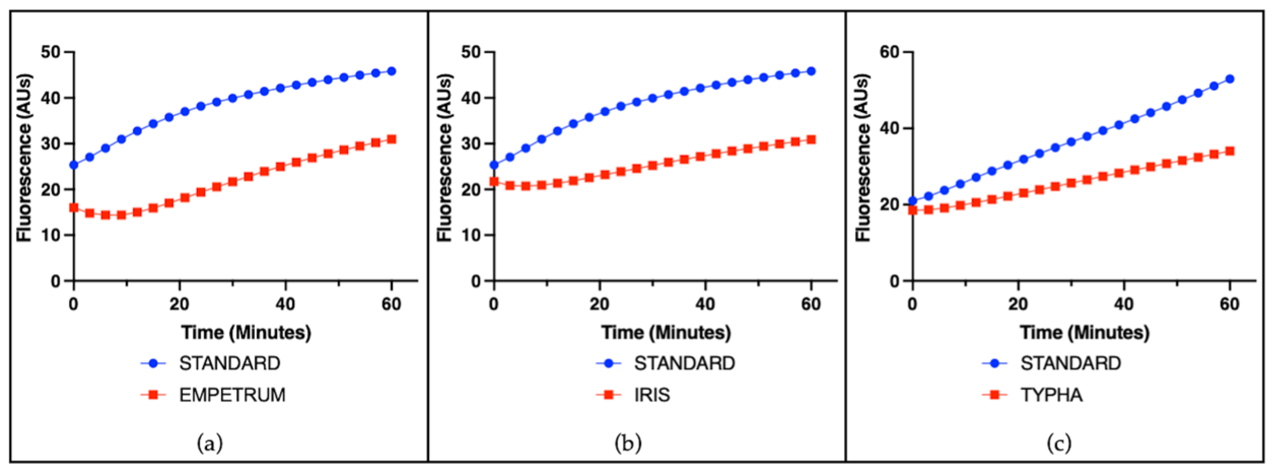
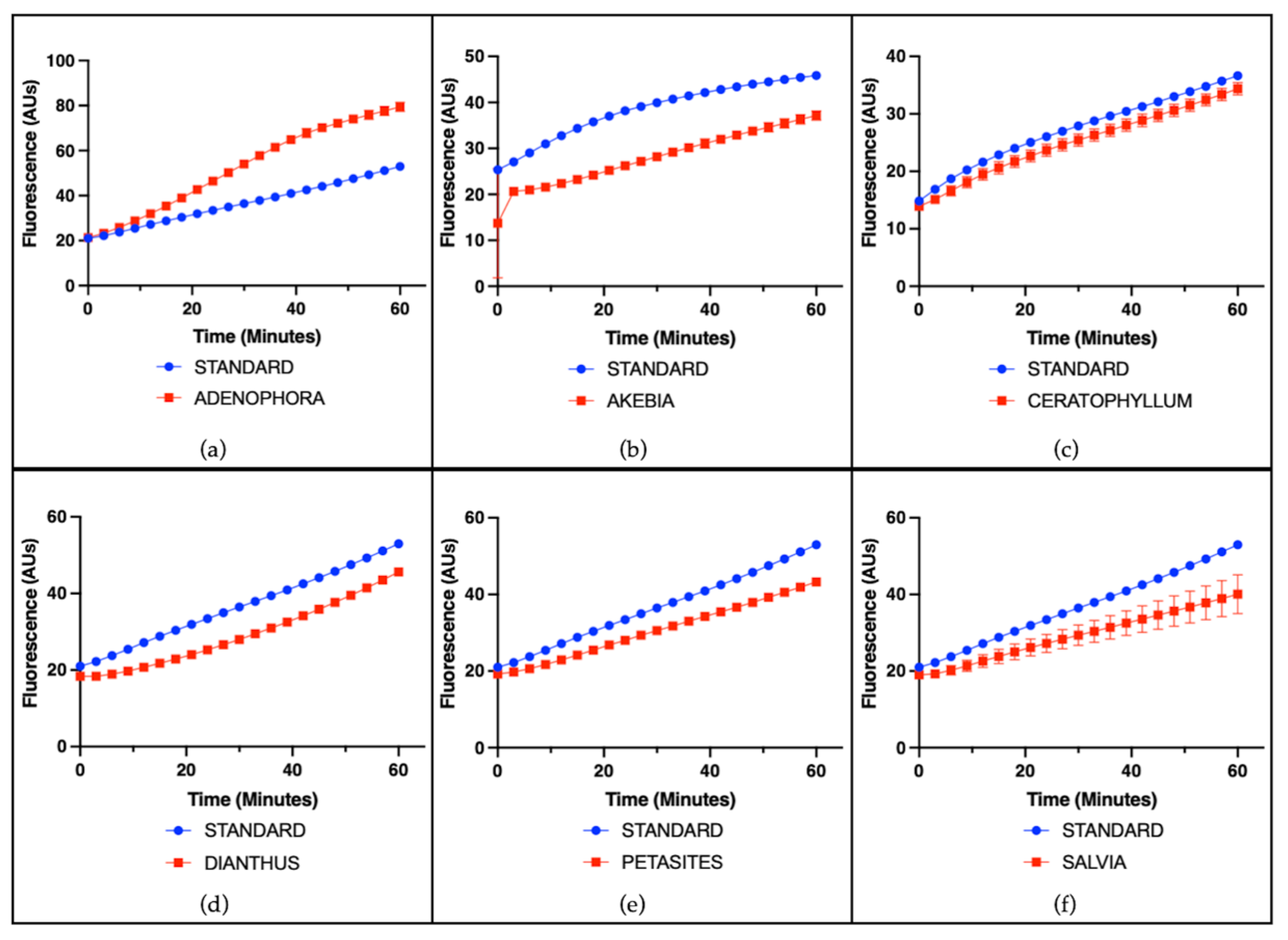
The radical-scavenging capacity of single plant extracts was evaluated using a cell-free assay based on the ascorbate-driven Fenton reaction, which enables the assessment of intrinsic antioxidant activity independently of cellular components. A representative example of the results for all species is shown using a concentration of 25 µg/mL. Among the tested extracts, ACALYPHA, ACTINIDIA, CISTUS, and HEUCHERA exhibited high radical-scavenging activity (Figure 1), reflecting a strong intrinsic antioxidant potential. EMPETRUM, IRIS, and TYPHA showed moderate scavenging capacity (Figure 2), indicating a partial efficacy in neutralizing free radicals under the applied conditions. In contrast, ADENOPHORA, AKEBIA, CERATOPHYLLUM, DIANTHUS, PETASITES, and SALVIA demonstrated only low activity levels (Figure 3), suggesting a limited antioxidant profile in this experimental setting. The remaining extracts did not display any significant radical-scavenging effect (Figure 4), suggesting the absence of active constituents capable of counteracting ROS generation in this non-cellular system.

3.3. Effect of Plant Extract on Cell Viability

The AnnV assay was used to assess cell viability and to determine the appropriate concentration range for subsequent antioxidant activity analyses. Since the data indicated that all extracts, ranging from 0.5 to 25 µg/mL medium, did not induce apoptosis and/or necrosis in THP-1 cells, the subsequent experiments were performed using this concentration range. However, cytotoxicity was observed at a concentration of 250 µg/mL (Supplementary Figure S1a). Non-immortalized cells exhibited cytotoxic responses at the following concentrations: 25 µg/mL in HUVECs (Supplementary Figure S1b) and 150 µg/mL in HIECs (Supplementary Figure S1c).

3.4. Evaluation of Antioxidant Activity

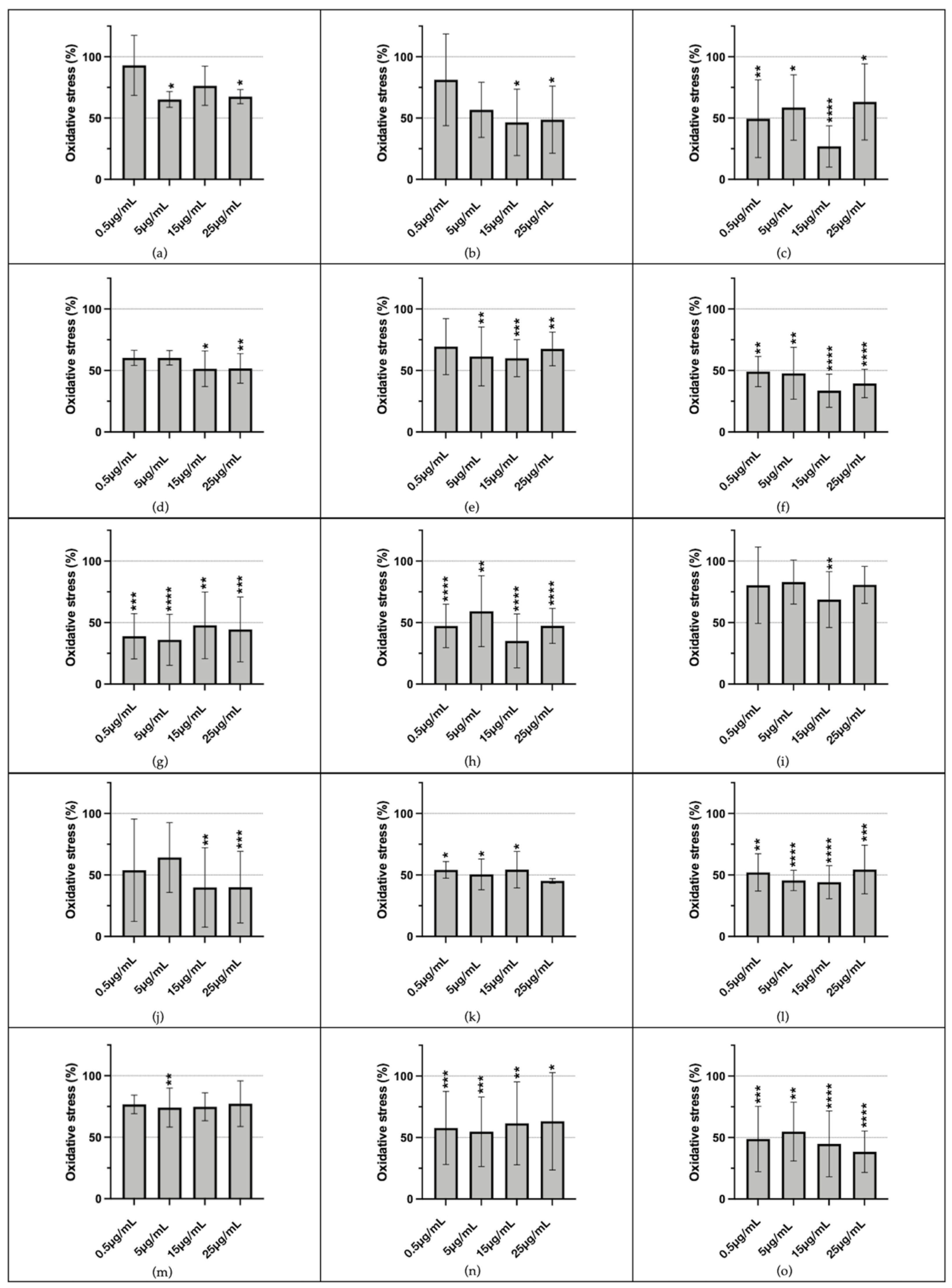
The assessment of the antioxidant potential of the tested plant-derived extracts in THP-1 cells revealed promising effects. Among the 19 extracts analyzed, only four (ACORUS, CERATOPHYLLUM, CISTUS, and HEUCHERA) did not induce a statistically significant reduction in intracellular ROS production. The remaining extracts exhibited varying degrees of antioxidant activity at different concentrations (Figure 5), as detailed in Supplementary Table S1.
The antioxidant effect in primary non-immortalized cells (HUVECs and HIECs), given the results in THP-1 cells, was limited to three extracts (ALTHEA, AQUILEGIA, and ERYNGIUM), and only ERYNGIUM at 100 µg/mL induced a statistically significant decrease in ROS production (5.94% ± 3.51, p = 0.005) (Supplementary Figure S2).

4. Discussion

In this study, we evaluated the antioxidant potential of a series of plant extracts using two complementary approaches: a cell-free system based on the Fenton reaction and a cellular model employing human monocytic THP-1 cells, human umbilical vein endothelial cells, and human primary small intestinal epithelial cells. The results highlighted the ability to neutralize ROS, to a significant extent, of several extracts, with variability depending on the assay system and the tested concentration.
The untargeted metabolomic analysis revealed a wide spectrum of secondary metabolites with well-documented biological activities, which may plausibly explain the bioactivities observed in the extracts. For instance, flavonoids (flavonols, flavones, C-glycosylated flavonoids), detected in ACORUS, AQUILEGIA, SUCCISA, and EMPETRUM, are recognized for their strong antioxidant and anti-inflammatory potential, often acting through the modulation of signaling pathways such as NF-κB and MAPK, inhibition of pro-inflammatory enzymes (COX-2, iNOS), and suppression of cytokine release (IL-6, TNF-α) [34]. Proanthocyanidins, highly abundant in ACTINIDIA and TYPHA, are well known for their antioxidant, antimicrobial, and cardioprotective effects, as well as their ability to prevent lipid peroxidation and support cellular integrity [33]. Pentacyclic triterpenoids (oleanane, ursane, taraxastane types), detected in AKEBIA, ACTINIDIA, ERYNGIUM, and DIANTHUS, are reported to exert anti-inflammatory and cytoprotective effects, partly by downregulating pro-inflammatory mediators (NO, TNF-α, IL-6, IL-1β) and inhibiting NF-κB nuclear translocation [35]. Similarly, steroidal saponins, as observed in ALLIUM, are widely associated with immunomodulatory, antifungal, and cholesterol-lowering properties, thereby possibly contributing synergistically to the bioactivity profiles [36]. Phenolic acids and hydrolysable tannins (gallic acid derivatives, ellagitannins), characterizing ACALYPHA, CISTUS, and HEUCHERA, are also well established for their potent radical-scavenging capacity and antimicrobial potential, which may play a central role in the extracts’ antioxidant activities. In Salvia, the detection of rosmarinic acid and related phenylethanoids is particularly relevant, as these compounds are strongly linked to anti-inflammatory and neuroprotective mechanisms [37]. It is also noteworthy that certain species, such as ADENOPHORA and PETASITES, were found to contain pyrrolidine and pyrrolizidine alkaloids. While such alkaloids may possess pharmacological activity, the toxicological implications, particularly hepatotoxicity and genotoxicity, are also well documented, suggesting that their role in the overall bioactivity of these extracts requires careful evaluation [38].
In the cell-free system, extracts such as ACALYPHA, ACTINIDIA, CISTUS, and HEUCHERA exhibited strong radical-scavenging activity, suggesting a substantial intrinsic antioxidant capacity. Previous studies have shown that polyphenols and flavonoids are major contributors to the scavenging activity observed in many plant extracts [39]. These findings are in line with reports that flavonoids such as luteolin and anthocyanins can reduce oxidative stress and inflammation in diverse cell types, including pulmonary cells, red blood cells, and endothelial cells, suggesting a broad-spectrum protective role against ROS-mediated tissue damage [40,41,42]. Other extracts, including EMPETRUM, IRIS, and TYPHA, demonstrated moderate activity, whereas several others did not significantly inhibit radical generation.
Subsequently, we tested these observations in a cellular context, and we were able to confirm that multiple extracts retained antioxidant efficacy under more biologically complex conditions. In THP-1 cells, extracts such as ACALYPHA, ACTINIDIA, DIANTHUS, SUCCISA, and TYPHA significantly reduced intracellular ROS levels at non-cytotoxic concentrations. This is consistent with previous findings suggesting that specific plant extracts can modulate intracellular redox homeostasis [43]. Similar protective effects have been documented in studies where flavonoids mitigated oxidative stress in COPD (chronic obstructive pulmonary disease) models, RBCs exposed to oxidative agents, and diabetic endothelial cells, highlighting the translational potential of these natural antioxidants across multiple disease contexts [40,41,42].
These results are particularly relevant considering accumulating evidence that natural antioxidants derived from medicinal plants may offer effective, low-toxicity strategies for addressing oxidative stress-mediated pathologies [44]. The ability of plant extracts to act at the cellular level supports their translational potential in future therapeutic applications. For instance, luteolin in COPD models was shown to modulate TRPV1/SIRT6 and CYP2A13/NRF2 signaling pathways, reducing ROS production and inflammation [40]. Anthocyanin-enriched fractions protected RBCs from oxidative stress by preserving band 3 function and reducing lipid peroxidation [42]. Moreover, flavonoids have been observed to counteract endothelial dysfunction in diabetes through ROS scavenging and modulation of nitric oxide availability [41].
Cytotoxicity assessment by an AnnV assay revealed that, at high concentrations (250 µg/mL), all extracts induced apoptosis and/or necrosis in THP-1 cells. Notably, primary non-immortalized cells, such as HUVECs and HIECs, exhibited cytotoxic responses at even lower concentrations (25 µg/mL and 150 µg/mL, respectively), underscoring the importance of carefully defining the concentration range in the therapeutic window [45]. Despite some cytotoxicity, antioxidant effects were detectable at relatively low doses. Comparing these effective in vitro concentrations with the known bioactivity and realistic dietary intake of the main phytochemicals in these extracts could provide further insight into their translational potential. However, defining a precise therapeutic window is not possible from this initial screening, which was intended to identify promising candidates for further investigations.
Biologically, the capacity to reduce intracellular ROS levels in monocytes like THP-1 cells is of particular interest, as oxidative stress is a known promoter of immune dysfunction and chronic inflammatory diseases [46], especially atherosclerosis. THP-1 cells are in fact a model of atherosclerosis because they mimic the behavior of foam cells, the leading type of cells in the progression of atherosclerotic lesions.
Indeed, the link between oxidative imbalance and chronic inflammation, neurodegeneration, and cardiovascular damage is well established, making it essential to discover compounds capable of modulating redox homeostasis effectively and safely. Our findings contribute to this growing area of research and support the exploration of plant-derived molecules as adjuvants or leads in pharmaceutical development. These observations align with evidence from animal studies where plant-derived antioxidants such as citric acid reduced LPS-induced oxidative stress and tissue damage in the brain and liver [47].
Identifying effective and safe plant extracts could open new therapeutic avenues for the management of redox-related disorders such as atherosclerosis, diabetes, and neurodegenerative diseases. Moreover, in vivo and in vitro studies have demonstrated that flavonoids and anthocyanins not only reduce ROS but also modulate key signaling pathways (e.g., NRF2, SIRT6, TRPV1) involved in oxidative stress responses, inflammation, and apoptosis, reinforcing their potential as multi-target agents [40,41,42].
Furthermore, in vitro studies on neuronal models have shown that several plant extracts can attenuate oxidative stress-induced apoptosis, pointing to their neuroprotective effects and further underscoring the clinical relevance of identifying such compounds [48]. These insights are consistent with the ROS-reducing capabilities observed in this study, which may be extended to various disease models in future investigations.
However, this study has some limitations. First, antioxidant activity was assessed under acute conditions and in monoculture systems; thus, long-term effects and interactions within complex microenvironments remain to be investigated [49]. Secondly, the challenge in dealing with such different and rich phytocomplexes hinders the attribution of the observed effects to specific phytochemicals.
To address these aspects, fractionation of the plant extracts to test specific groups of phytochemicals from the original phytocomplex might represent a useful approach for identifying the molecule/molecules responsible for the observed bioactivities [50]. Additionally, it will be crucial to explore molecular mechanisms, such as the activation of the Nrf2/ARE pathway, a key player in cellular antioxidant responses [51]. This approach mirrors studies where modulation of NRF2 and SIRT6 signaling by flavonoids and anthocyanins was associated with reduced oxidative stress and cellular protection [40,41,42].
Finally, future studies using animal models of chronic oxidative stress and inflammation will be essential to deepen our knowledge on the potential of these extracts in specific, physiologically relevant settings.

5. Conclusions

This study systematically investigated the antioxidant potential of various plant extracts using both a cell-free Fenton reaction-based assay and a human monocytic THP-1 cell model. The findings demonstrate that several extracts, particularly ACALYPHA, ACTINIDIA, DIANTHUS, SUCCISA, and TYPHA, all characterized by very different phytocomplexes, possess significant radical-scavenging and intracellular ROS-reducing activities, without causing cytotoxicity at effective concentrations.
By contributing to broadening the knowledge in the growing field related to the function of natural antioxidants, the findings of this study highlight the potential of novel Mediterranean plant extracts as potential candidates for the development of health-promoting strategies, eventually useful against oxidative stress-related diseases.
Nevertheless, further research is indeed warranted to elucidate their phytochemical composition, molecular mechanisms of action, safety profiles, and bioavailability in vivo.
Importantly, the dual approach adopted in this study, combining a biochemical assay and a cellular model, allowed for a more comprehensive evaluation of antioxidant efficacy under both simplified and biologically relevant conditions. Moreover, the identification of active extracts among species native or naturalized within the Italian flora emphasizes the untapped pharmacological potential of regional biodiversity. By focusing on plant-derived antioxidants with both efficacy and acceptable cytotoxicity profiles, this research contributes valuable knowledge to the field of redox biology and supports the role of phytochemicals in future human health-preserving policies.

6. Patents

A patent application related to the antioxidant plant extracts screened in this study has been filed and is currently under review.

Supplementary Materials

The following supporting information can be downloaded at: https://www.mdpi.com/article/10.3390/antiox14101217/s1, Table S1: Antioxidant activity of plant extract in THP-1 cells at concentration showing statistically significant effects; Figure S1: Example of the effect of a plant extract on THP-1 cells (a), HUVECs (b), and HIECs (c); Figure S2: Antioxidant potential of ERYNGIUM extracts in HIECs; Supplementary File S1: UPLC-ESI-HRMS chromatograms recorded in negative ionization mode (ESI-) of the plant extracts; Supplementary File S2: LC-MS features of the phytochemicals identified in the plant extracts.

Author Contributions

Conceptualization, G.A., C.S., and A.C.; Methodology, G.A., C.S., and A.C.; Validation, G.A. and E.G.D.L.; Formal Analysis, G.A., E.G.D.L., and C.S.; Investigation, G.A., E.G.D.L., C.S., S.N., and M.C.; Resources, F.G.; Writing—Original Draft, G.A.; Writing—Review and Editing, E.G.D.L., S.N., A.M.F.P., A.C., and S.F.; Visualization, G.A., S.N., and M.C.; Supervision, A.M.F.P. and S.F. All authors have read and agreed to the published version of the manuscript.

Funding

Project funded under the National Recovery and Resilience Plan (NRRP), Mission 4 Component 2 Investment 1.4—Call for tender No. 3138 of 16 December 2021, rectified by Decree n.3175 of 18 December 2021 of the Italian Ministry of University and Research funded by the European Union—NextGenerationEU; Award Number: Project code CN_00000033, Concession Decree No. 1034 of 17 June 2022 adopted by the Italian Ministry of University and Research, CUP B33C22000660001, Project title “National Biodiversity Future Center—NBFC”.

Institutional Review Board Statement

Not applicable. The study was conducted using established human cell lines and did not involve human participants or animals.

Informed Consent Statement

Not applicable. The study did not involve human participants.

Data Availability Statement

The data presented in this study are available on request from the corresponding author. The data are not publicly available due to ongoing research activities and a pending patent application.

Acknowledgments

This study was performed, in part, in the LURM (Laboratorio Universitario di Ricerca Medica) Research Center, University of Verona, Italy.

Conflicts of Interest

The authors declare no conflicts of interest.

Abbreviations

The following abbreviations are used in this manuscript:
ROSreactive oxygen species
SODsuperoxide dismutase
CATcatalase
GPxglutathione peroxidase
ACALYPHAAcalypha virginica Linnaeus
ACORUSAcorus calamus Linnaeus
ACTINIDIAActinidia deliciosa (A.Chev.) C.F. Liang & A.R. Ferguson
ADENOPHORAAdenophora liliifolia (L.) Gaudin
AKEBIAAkebia quinata (Thunberg ex Houttuyn) Decaisne
ALLIUMAllium lusitanicum (L.) Mertens
ALTHAEAAlthaea officinalis Linnaeus
AQUILEGIAAquilegia atrata Wilhelm Daniel Joseph Koch
CERATOPHYLLUMCeratophyllum demersum Linnaeus
CISTUSCistus monspeliensis Linnaeus
DIANTHUSDianthus superbus Linnaeus subsp. superbus
EMPETRUMEmpetrum hermaphroditum Hagerup
ERYNGIUMEryngium maritimum Linnaeus
HEUCHERAHeuchera sanguinea Pursh
IRISIris pseudacorus Linnaeus (synonim Limniris pseudacorus)
PETASITESPetasites paradoxus (Retzius) Baumgarten
SALVIASalvia pratensis Linnaeus
SUCCISASuccisa pratensis Moench
TYPHATypha laxmannii Lepech
THP-1human monocytic leukemia cells
HUVECshuman umbilical vein endothelial cells
HIECshuman primary small intestinal epithelial cells
FBSfetal bovine serum
AnnVAnnexin V
TBHPtert-butyl hydroperoxide
UPLCultra-pressure liquid chromatography
ESIelectro-spray ionization
HRMSHigh-resolution mass spectrometry
COPDchronic obstructive pulmonary disease

References

  1. Ahmed, S.A.; Alahmadi, Y.M.; Abdou, Y.A. The Impact of Serum Levels of Reactive Oxygen and Nitrogen Species on the Disease Severity of COVID-19. Int. J. Mol. Sci. 2023, 24, 8973. [Google Scholar] [CrossRef]
  2. Ghosh, S.; Roy, P.; Prasad, S.; Mugesh, G. A GPx-Mimetic Copper Vanadate Nanozyme Mediates the Release of Nitric Oxide from S-Nitrosothiols. Faraday Discuss. 2022, 234, 284–303. [Google Scholar] [CrossRef]
  3. Sies, H.; Jones, D.P. Reactive Oxygen Species (ROS) as Pleiotropic Physiological Signalling Agents. Nat. Rev. Mol. Cell Biol. 2020, 21, 363–383. [Google Scholar] [CrossRef]
  4. Szechyńska-Hebda, M.; Ghalami, R.Z.; Kamran, M.; Van Breusegem, F.; Karpiński, S. To Be or Not to Be? Are Reactive Oxygen Species, Antioxidants, and Stress Signalling Universal Determinants of Life or Death? Cells 2022, 11, 4105. [Google Scholar] [CrossRef] [PubMed]
  5. Holmström, K.M.; Finkel, T. Cellular Mechanisms and Physiological Consequences of Redox-Dependent Signalling. Nat. Rev. Mol. Cell Biol. 2014, 15, 411–421. [Google Scholar] [CrossRef] [PubMed]
  6. Tsesmelis, K.; Maity-Kumar, G.; Croner, D.; Sprissler, J.; Tsesmelis, M.; Hein, T.; Baumann, B.; Wirth, T. Accelerated Aging in Mice with Astrocytic Redox Imbalance as a Consequence of SOD2 Deletion. Aging Cell 2023, 22. [Google Scholar] [CrossRef] [PubMed]
  7. Lin, S.; Ye, C.; Lin, Z.; Huang, L.; Li, D. Recent Progress of Near-Infrared Fluorescent Probes in the Determination of Reactive Oxygen Species for Disease Diagnosis. Talanta 2024, 268. [Google Scholar] [CrossRef]
  8. Lam, C.R.I.; Tan, M.J.; Tan, S.H.; Tang, M.B.Y.; Cheung, P.C.F.; Tan, N.S. TAK1 Regulates SCF Expression to Modulate PKBα Activity That Protects Keratinocytes from ROS-Induced Apoptosis. Cell Death Differ. 2011, 18, 1120–1129. [Google Scholar] [CrossRef]
  9. Chang, M.; Nguyen, T.T. Strategy for Treatment of Infected Diabetic Foot Ulcers. Acc. Chem. Res. 2021, 54, 1080–1093. [Google Scholar] [CrossRef]
  10. Dong, Y.; Wang, Z. ROS-Scavenging Materials for Skin Wound Healing: Advancements and Applications. Front. Bioeng. Biotechnol. 2023, 11, 1304835. [Google Scholar] [CrossRef]
  11. Wang, Y.; Wei, Z.; Pan, K.; Li, J.; Chen, Q. The Function and Mechanism of Ferroptosis in Cancer. Apoptosis 2020, 25, 786–798. [Google Scholar] [CrossRef]
  12. Evavold, C.L.; Hafner-Bratkovič, I.; Devant, P.; D’Andrea, J.M.; Ngwa, E.M.; Boršić, E.; Doench, J.G.; LaFleur, M.W.; Sharpe, A.H.; Thiagarajah, J.R.; et al. Control of Gasdermin D Oligomerization and Pyroptosis by the Ragulator-Rag-mTORC1 Pathway. Cell 2021, 184, 4495–4511.e19. [Google Scholar] [CrossRef]
  13. Zhang, L.; Song, A.; Yang, Q.C.; Li, S.J.; Wang, S.; Wan, S.C.; Sun, J.; Kwok, R.T.K.; Lam, J.W.Y.; Deng, H.; et al. Integration of AIEgens into Covalent Organic Frameworks for Pyroptosis and Ferroptosis Primed Cancer Immunotherapy. Nat. Commun. 2023, 14, 5355. [Google Scholar] [CrossRef]
  14. Alfadda, A.A.; Sallam, R.M. Reactive Oxygen Species in Health and Disease. J. Biomed. Biotechnol. 2012, 2012, 936486. [Google Scholar] [CrossRef] [PubMed]
  15. Wang, Y.; Branicky, R.; Noë, A.; Hekimi, S. Superoxide Dismutases: Dual Roles in Controlling ROS Damage and Regulating ROS Signaling. J. Cell Biol. 2018, 217, 1915–1928. [Google Scholar] [CrossRef] [PubMed]
  16. He, L.; He, T.; Farrar, S.; Ji, L.; Liu, T.; Ma, X. Antioxidants Maintain Cellular Redox Homeostasis by Elimination of Reactive Oxygen Species. Cell. Physiol. Biochem. 2017, 44, 532–553. [Google Scholar] [CrossRef] [PubMed]
  17. Jomova, K.; Raptova, R.; Alomar, S.Y.; Alwasel, S.H.; Nepovimova, E.; Kuca, K.; Valko, M. Reactive Oxygen Species, Toxicity, Oxidative Stress, and Antioxidants: Chronic Diseases and Aging. Arch. Toxicol. 2023, 97, 2499–2574. [Google Scholar] [CrossRef]
  18. Zhang, Y.J.; Gan, R.Y.; Li, S.; Zhou, Y.; Li, A.N.; Xu, D.P.; Li, H.B.; Kitts, D.D. Antioxidant Phytochemicals for the Prevention and Treatment of Chronic Diseases. Molecules 2015, 20, 21138–21156. [Google Scholar] [CrossRef]
  19. Dubois-deruy, E.; Peugnet, V.; Turkieh, A.; Pinet, F. Oxidative Stress in Cardiovascular Diseases. Antioxidants 2020, 9, 864. [Google Scholar] [CrossRef]
  20. Weinberg Sibony, R.; Segev, O.; Dor, S.; Raz, I. Overview of Oxidative Stress and Inflammation in Diabetes. J. Diabetes 2024, 16, e70014. [Google Scholar] [CrossRef]
  21. Houldsworth, A. Role of Oxidative Stress in Neurodegenerative Disorders: A Review of Reactive Oxygen Species and Prevention by Antioxidants. Brain Commun. 2024, 6. [Google Scholar] [CrossRef]
  22. Hayes, J.D.; Dinkova-Kostova, A.T.; Tew, K.D. Oxidative Stress in Cancer. Cancer Cell 2020, 38, 167–197. [Google Scholar] [CrossRef]
  23. Bergonzi, M.C.; Heard, C.M.; Garcia-Pardo, J. Bioactive Molecules from Plants: Discovery and Pharmaceutical Applications. Pharmaceutics 2022, 14, 2116. [Google Scholar] [CrossRef]
  24. Atanasov, A.G.; Waltenberger, B.; Pferschy-Wenzig, E.M.; Linder, T.; Wawrosch, C.; Uhrin, P.; Temml, V.; Wang, L.; Schwaiger, S.; Heiss, E.H.; et al. Discovery and Resupply of Pharmacologically Active Plant-Derived Natural Products: A Review. Biotechnol. Adv. 2015, 33, 1582–1614. [Google Scholar] [CrossRef] [PubMed]
  25. Lobo, V.; Patil, A.; Phatak, A.; Chandra, N. Free Radicals, Antioxidants and Functional Foods: Impact on Human Health. Pharmacogn. Rev. 2010, 4, 118–126. [Google Scholar] [CrossRef] [PubMed]
  26. Sasidharan, S.; Chen, Y.; Saravanan, D.; Sundram, K.M.; Latha, L.Y. Extraction, isolation and characterization of bioactive compounds from plants’ extracts. Afr. J. Tradit. Complement. Altern. Med. 2011, 8, 1–10. [Google Scholar] [CrossRef] [PubMed]
  27. Muscolo, A.; Mariateresa, O.; Giulio, T.; Mariateresa, R. Oxidative Stress: The Role of Antioxidant Phytochemicals in the Prevention and Treatment of Diseases. Int. J. Mol. Sci. 2024, 25, 3264. [Google Scholar] [CrossRef]
  28. World Health Organization Healthy Diet. 2020. Available online: https://www.Who.Int/News-Room/Fact-Sheets/Detail/Healthy-Diet (accessed on 28 August 2025).
  29. Giammona, A.; Commisso, M.; Bonanomi, M.; Remedia, S.; Avesani, L.; Porro, D.; Gaglio, D.; Bertoli, G.; Lo Dico, A. A Novel Strategy for Glioblastoma Treatment by Natural Bioactive Molecules Showed a Highly Effective Anti-Cancer Potential. Nutrients 2024, 16, 2389. [Google Scholar] [CrossRef]
  30. De Soricellis, G.; Rinaldi, F.; Tengattini, S.; Temporini, C.; Negri, S.; Capelli, D.; Montanari, R.; Cena, H.; Salerno, S.; Massolini, G.; et al. Development of an Analytical Platform for the Affinity Screening of Natural Extracts by SEC-MS towards PPARα and PPARγ Receptors. Anal. Chim. Acta 2024, 1309, 342666. [Google Scholar] [CrossRef]
  31. Zorzi, G.; Gambini, S.; Negri, S.; Guzzo, F.; Commisso, M. Untargeted Metabolomics Analysis of the Orchid Species Oncidium Sotoanum Reveals the Presence of Rare Bioactive C-Diglycosylated Chrysin Derivatives. Plants 2023, 12, 655. [Google Scholar] [CrossRef]
  32. Commisso, M.; Negri, S.; Bianconi, M.; Gambini, S.; Avesani, S.; Ceoldo, S.; Avesani, L.; Guzzo, F. Untargeted and Targeted Metabolomics and Tryptophan Decarboxylase in Vivo Characterization Provide Novel Insight on the Development of Kiwifruits (Actinidia deliciosa). Int. J. Mol. Sci. 2019, 20, 897. [Google Scholar] [CrossRef] [PubMed]
  33. Mannino, G.; Chinigò, G.; Serio, G.; Genova, T.; Gentile, C.; Munaron, L.; Bertea, C.M. Proanthocyanidins and Where to Find Them: A Meta-Analytic Approach to Investigate Their Chemistry, Biosynthesis, Distribution, and Effect on Human Health. Antioxidants 2021, 10, 1229. [Google Scholar] [CrossRef] [PubMed]
  34. Jomova, K.; Alomar, S.Y.; Valko, R.; Liska, J.; Nepovimova, E.; Kuca, K.; Valko, M. Flavonoids and Their Role in Oxidative Stress, Inflammation, and Human Diseases. Chem.-Biol. Interact. 2025, 413, 111489. [Google Scholar] [CrossRef] [PubMed]
  35. Yue, L.; Luo, J.; Zhao, C.; Zhao, J.; Ye, J.; He, K.; Zou, J. Oleanane Triterpenoids with C-14 Carboxyl Group from Astilbe Grandis Inhibited LPS-Induced Macrophages Activation by Suppressing the NF-κB Signaling Pathway. Front. Pharmacol. 2024, 15, 1413876. [Google Scholar] [CrossRef]
  36. Sparg, S.G.; Light, M.E.; Van Staden, J. Biological Activities and Distribution of Plant Saponins. J. Ethnopharmacol. 2004, 94, 219–243. [Google Scholar] [CrossRef]
  37. Sampol, B.; Bota, J.; Riera, D.; Medrano, H.; Flexas, J. Analysis of the Virus-induced Inhibition of Photosynthesis in Malmsey Grapevines. New Phytol. 2003, 160, 403–412. [Google Scholar] [CrossRef]
  38. Moreira, R.; Pereira, D.M.; Valentão, P.; Andrade, P.B. Pyrrolizidine Alkaloids: Chemistry, Pharmacology, Toxicology and Food Safety. Int. J. Mol. Sci. 2018, 19, 1668. [Google Scholar] [CrossRef]
  39. Heim, K.E.; Tagliaferro, A.R.; Bobilya, D.J. Reviews: Current topics Flavonoid Antioxidants: Chemistry, Metabolism and Structure-Activity Relationships. J. Nutr. Biochem. 2002, 13, 572–584. [Google Scholar] [CrossRef]
  40. Zhou, L.; Jian, T.; Wan, Y.; Huang, R.; Fang, H.; Wang, Y.; Liang, C.; Ding, X.; Chen, J. Luteolin Alleviates Oxidative Stress in Chronic Obstructive Pulmonary Disease Induced by Cigarette Smoke via Modulation of the TRPV1 and CYP2A13/NRF2 Signaling Pathways. Int. J. Mol. Sci. 2023, 25, 369. [Google Scholar] [CrossRef]
  41. Dou, B.; Zhu, Y.; Sun, M.; Wang, L.; Tang, Y.; Tian, S.; Wang, F. Mechanisms of Flavonoids and Their Derivatives in Endothelial Dysfunction Induced by Oxidative Stress in Diabetes. Molecules 2024, 29, 3265. [Google Scholar] [CrossRef]
  42. Remigante, A.; Spinelli, S.; Patanè, G.T.; Barreca, D.; Straface, E.; Gambardella, L.; Bozzuto, G.; Caruso, D.; Falliti, G.; Dossena, S.; et al. AAPH-Induced Oxidative Damage Reduced Anion Exchanger 1 (SLC4A1/AE1) Activity in Human Red Blood Cells: Protective Effect of an Anthocyanin-Rich Extract. Front. Physiol. 2023, 14, 1303815. [Google Scholar] [CrossRef]
  43. Poljsak, B.; Šuput, D.; Milisav, I. Achieving the Balance between ROS and Antioxidants: When to Use the Synthetic Antioxidants. Oxidative Med. Cell. Longev. 2013, 2013, 956792. [Google Scholar] [CrossRef] [PubMed]
  44. Zafar, F.; Asif, H.M.; Shaheen, G.; Ghauri, A.O.; Rajpoot, S.R.; Tasleem, M.W.; Shamim, T.; Hadi, F.; Noor, R.; Ali, T.; et al. A Comprehensive Review on Medicinal Plants Possessing Antioxidant Potential. Clin. Exp. Pharmacol. Physiol. 2023, 50, 205–217. [Google Scholar] [CrossRef] [PubMed]
  45. Mosmann T Rapid Colorimetric Assay for Cellular Growth and Survival: Application to Proliferation and Cytotoxicity Assays. J. Immunol. Methods 1983, 65, 55–63. [CrossRef] [PubMed]
  46. Mittal, M.; Siddiqui, M.R.; Tran, K.; Reddy, S.P.; Malik, A.B. Reactive Oxygen Species in Inflammation and Tissue Injury. Antioxid. Redox Signal. 2014, 20, 1126–1167. [Google Scholar] [CrossRef]
  47. Abdel-Salam, O.M.E.; Youness, E.R.; Mohammed, N.A.; Morsy, S.M.Y.; Omara, E.A.; Sleem, A.A. Citric Acid Effects on Brain and Liver Oxidative Stress in Lipopolysaccharide-Treated Mice. J. Med. Food 2014, 17, 588–598. [Google Scholar] [CrossRef]
  48. May, N.; de Sousa Alves Neri, J.L.; Clunas, H.; Shi, J.; Parkes, E.; Dongol, A.; Wang, Z.; Jimenez Naranjo, C.; Yu, Y.; Huang, X.F.; et al. Investigating the Therapeutic Potential of Plants and Plant-Based Medicines: Relevance to Antioxidant and Neuroprotective Effects. Nutrients 2023, 15, 3912. [Google Scholar] [CrossRef]
  49. Valko, M.; Leibfritz, D.; Moncol, J.; Cronin, M.T.D.; Mazur, M.; Telser, J. Free Radicals and Antioxidants in Normal Physiological Functions and Human Disease. Int. J. Biochem. Cell Biol. 2007, 39, 44–84. [Google Scholar] [CrossRef]
  50. Pandey, K.B.; Rizvi, S.I. Plant Polyphenols as Dietary Antioxidants in Human Health and Disease. Oxidative Med. Cell. Longev. 2009, 2, 270–278. [Google Scholar] [CrossRef]
  51. Ma, Q. Role of Nrf2 in Oxidative Stress and Toxicity. Annu. Rev. Pharmacol. Toxicol. 2013, 53, 401–426. [Google Scholar] [CrossRef]
Figure 1. Radical-scavenging activity of plant extracts. Representative fluorescence (arbitrary units) over time induced by the standard iron (II) and by individual plant extracts (25 μg/mL) in the ascorbate-driven Fenton reaction after 60 min. Strong radical-scavenging (red line) activity was observed for (a) ACALYPHA, (b) ACTINIDIA, (c) CISTUS, and (d) HEUCHERA.
Antioxidants 14 01217 g001
Figure 2. Radical-scavenging activity of plant extracts. Representative fluorescence (arbitrary units) over time induced by the standard iron (II) and by individual plant extracts (25 μg/mL) in the ascorbate-driven Fenton reaction after 60 min. Moderate activity (red line) was observed for (a) EMPETRUM, (b) IRIS, and (c) TYPHA.
Antioxidants 14 01217 g002
Figure 3. Radical-scavenging activity of plant extracts. Representative fluorescence (arbitrary units) over time induced by the standard iron (II) and by individual plant extracts (25 μg/mL) on the ascorbate-driven Fenton reaction after 60 min. Low activity (red line) was observed for (a) ADENOPHORA, (b) AKEBIA, (c) CERATOPHYLLUM, (d) DIANTHUS, (e) PETASITES, and (f) SALVIA.
Antioxidants 14 01217 g003
Figure 4. Radical-scavenging activity of plant extracts. Representative fluorescence (arbitrary units) over time induced by the standard iron (II) and by individual plant extracts (25 μg/mL) on the ascorbate-driven Fenton reaction after 60 min. No relevant activity was recorded for (a) ACORUS, (b) ALLIUM, (c) ALTHEA, (d) AQUILEGIA, or (e) ERYNGIUM.
Antioxidants 14 01217 g004
Figure 5. Antioxidant potential of plant extracts in THP-1 cells. Significant antioxidant effects, indicated by a reduction in intracellular ROS levels compared to the positive control, were observed in (a) ACALYPHA at 5 and 25 µg/mL; (b) ACTINIDIA at 15 and 25 µg/mL; (c) ADENOPHORA at 0.5, 5, 15, and 25 µg/mL; (d) AKEBIA at 15 and 25 µg/mL; (e) ALLIUM at 5, 15, and 25 µg/mL; (f) ALTHEA at 0.5, 5, 15, and 25 µg/mL; (g) AQUILEGIA at 0.5, 5, 15, and 25 µg/mL; (h) DIANTHUS at 0.5, 5, 15, and 25 µg/mL; (i) EMPETRUM at 15 µg/mL; (j) ERYNGIUM at 15 and 25 µg/mL; (k) IRIS at 0.5, 5, and 15 µg/mL; (l) PETASITES at 0.5, 5, 15, and 25 µg/mL; (m) SALVIA at 5 µg/mL; (n) SUCCISA at 0.5, 5, 15, and 25 µg/mL; and (o) TYPHA at 0.5, 5, 15, and 25 µg/mL (* p < 0.05, ** p < 0.01, *** p < 0.001, **** p < 0.0001).
Antioxidants 14 01217 g005
Table 1. Plant species and characterizing metabolite classes of the phytochemicals detected in the methanolic extracts following untargeted metabolomic analysis.
SpeciesCharacterizing Metabolites
ACALYPHAgallic acid derivatives, hydrolysable tannins
ACORUSC-glycosylated flavonoids
ACTINIDIAproanthocyanidins, flavonols, ursane/taraxastane triterpenoids
ADENOPHORAflavonoids, pyrrolidine alkaloids
AKEBIAflavonols, oleanane triterpenoids
ALLIUMsteroidal saponins
ALTHAEAnorisoprenoids sulfoglycosides, hypoleatin derivatives
AQUILEGIAswertisin derivatives, cycloartane triterpenoids
CERATOPHYLLUMhydroxycinnamates, flavonoids
CISTUSellagitannins, flavonols/methoxylated flavonols, labdane diterpenoids
DIANTHUSflavonoids, oleanane triterpenoids
EMPETRUMquercetin derivatives, flavanones, dihydrochalcones
ERYNGIUMflavonols, triterpenoids
HEUCHERAgallotannins
IRISxanthones, flavones
PETASITEShydroxycinnamate esters, otonecine-type pyrrolizidine alkaloids
SALVIArosmarinic acid and related phenylethanoid/phenylpropanoid esters
SUCCISAiridoids, flavones
TYPHAproanthocyanidins, flavonols
Disclaimer/Publisher’s Note: The statements, opinions and data contained in all publications are solely those of the individual author(s) and contributor(s) and not of MDPI and/or the editor(s). MDPI and/or the editor(s) disclaim responsibility for any injury to people or property resulting from any ideas, methods, instructions or products referred to in the content.