Article

BMPO-OOH Spin-Adduct as a Model for Study of Decomposition of Organic Hydroperoxides and the Effects of Sulfide/Selenite Derivatives. An EPR Spin-Trapping Approach

1
Department of Molecular Physiology, Institute of Clinical and Translational Research, Biomedical Research Center, Slovak Academy of Sciences, Dúbravská cesta 9, 84505 Bratislava, Slovakia
2
Faculty of Chemical and Food Technology, Institute of Physical Chemistry and Chemical Physics, Slovak University of Technology in Bratislava, Radlinského 9, 81237 Bratislava, Slovakia
3
Department of Genetics, Cancer Research Institute, Biomedical Research Center, Slovak Academy of Sciences, Dúbravská cesta 9, 84505 Bratislava, Slovakia
*
Author to whom correspondence should be addressed.
Antioxidants 2020, 9(10), 918; https://doi.org/10.3390/antiox9100918
Submission received: 7 August 2020 / Revised: 7 September 2020 / Accepted: 24 September 2020 / Published: 26 September 2020
(This article belongs to the Section ROS, RNS and RSS)

Abstract

:
Lipid hydroperoxides play an important role in various pathophysiological processes. Therefore, a simple model for organic hydroperoxides could be helpful to monitor the biologic effects of endogenous and exogenous compounds. The electron paramagnetic resonance (EPR) spin-trapping technique is a useful method to study superoxide (O2•−) and hydroxyl radicals. The aim of our work was to use EPR with the spin trap 5-tert-butoxycarbonyl-5-methyl-1-pyrroline-N-oxide (BMPO), which, by trapping O2•− produces relatively stable BMPO-OOH spin-adduct, a valuable model for organic hydroperoxides. We used this experimental setup to investigate the effects of selected sulfur/selenium compounds on BMPO-OOH and to evaluate the antioxidant potential of these compounds. Second, using the simulation of time-dependent individual BMPO adducts in the experimental EPR spectra, the ratio of BMPO-OH/BMPO-OOH—which is proportional to the transformation/decomposition of BMPO-OOH—was evaluated. The order of potency of the studied compounds to alter BMPO-OOH concentration estimated from the time-dependent BMPO-OH/BMPO-OOH ratio was as follows: Na2S4 > Na2S4/SeO32− > H2S/SeO32− > Na2S2 ~Na2S2/SeO32− ~H2S > SeO32− ~SeO42− ~control. In conclusion, the presented approach of the EPR measurement of the time-dependent ratio of BMPO-OH/BMPO-OOH could be useful to study the impact of compounds to influence the transformation of BMPO-OOH.

1. Introduction

Exogenously added and endogenously produced hydrogen sulfide (H2S) and polysulfides affect many physiological and pathologic processes [1,2,3,4]. They modulate oxidative stress by reacting with reactive oxygen and nitrogen species [1,5,6,7]. Selenium (Se) is an essential trace element for humans, with multiple and complex effects on health, having antioxidant properties due to its presence in 25 selenoproteins in the form of selenocysteine amino acid [8]. Se compounds and H2S are present in living organisms and either alone or in combination interact with reactive oxygen species (ROS) [1,9,10,11,12]. In our previous work, we found that products of the sulfide/selenite (H2S/SeO32−) interaction scavenge superoxide-derived radicals, cleave plasmid DNA (pDNA) and modulate tonus of isolated rat aorta and blood pressure [13]. However in this work, using the procedure of addition of KO2 as a source of (O2•−) into the mixture of the compounds (H2S, SeO32−) with the spin-trapping agent 5-tert-butoxycarbonyl-5-methyl-1-pyrroline N-oxide (BMPO) it was not clear which components of the electron paramagnetic resonance (EPR) spectra resulted from the compounds/O2•− or compounds/BMPO-OOH interactions. To solve this issue, a new experimental strategy for the interaction of the compounds with BMPO-OOH only is herein presented.
Lipid peroxidation is a process under which oxidants such as free radicals attack lipids containing carbon-carbon double bond(s), leading to the radical chain reactions. It has an important role in cell biology and in various pathophysiological processes in which lipid hydroperoxides display a crucial function [14]. Importantly, many pathophysiological states can be regulated by the modulation of lipid peroxidation induced by exogenous compounds. Therefore, simple models of organic hydroperoxide could be useful for studying the effects of various compounds. EPR spin-trapping technique using cyclic nitrone BMPO is a reliable method to study O2•− and hydroxyl radicals (HO) [15,16,17].
The spin-trapping agent BMPO in aqueous solutions represents a racemic mixture of two enantiomers [S (−) and R (+)], and the previous detailed study evidenced that starting with racemate, as well as with the individual enantiomers, the reaction with O2•− resulted in the identical EPR spectrum representing two signals of diastereoisomers at the same ratio (trans and cis with respect to the tert-butoxycarbonyl group) (Scheme 1). The EPR spectra of BMPO-OOH diastereoisomers are characterized with the similar nitrogen splittings and slightly different β-hydrogen splittings [18,19,20]. Analogously, the experimental EPR spectra of BMPO-OH were interpreted considering the superposition of individual signals of two diastereoisomers with the different hyperfine coupling constants [15,21] (Scheme 1).
Therefore, the aim of our work was to use EPR with BMPO—which in the presence of O2•− forms a relatively stable BMPO-OOH and can serve as a model for organic hydroperoxide—to study the effects of Na2Sn (n = 1, 2, 4) and Na2SeOn (n = 3, 4) on their own or their mixture Na2Sn/Na2SeOn. This approach enables the comparison of the relative potential of the investigated compounds to affect the ROOH bond and to eliminate radicals formed during its decomposition. Since 10% DMSO (v/v) is used in the studied system, the procedure is useful for evaluating antioxidant potency of compounds insoluble in water.

2. Materials and Methods

2.1. Chemicals

Stock solutions of sodium selenite (Na2SeO3, 10 or 40 mmol L−1, Sigma-Aldrich 214485, Saint Louis, MO, USA) and sodium selenate (Na2SeO4, 10 mmol L−1, Sigma-Aldrich S0882, Saint Louis, MO, USA) were prepared freshly in deionized H2O, stored at 23 °C and used within 5 h. Na2SeO3 dissociates in solution to yield mostly H2SeO3 at acidic pH, HSeO3 at neutral pH and SeO32− at alkaline pH. For simplicity, the terms SeO32− and SeO42− are employed as representative expression to encompass the total mixture of different (de)protonation states. Spin-trapping agent 5-tert-butoxycarbonyl-5-methyl-1-pyrroline-N-oxide (BMPO, 100 mmol L−1, DoJindo B568-10, Munich, Germany) was dissolved in deionized H2O, stored at −80 °C and used after thawing. Na2S (100 mmol L−1) as a source of H2S and polysulfides, sodium disulfide (Na2S2, 10 mmol L−1) and sodium tetrasulfide (Na2S4, 10 mmol L−1) (Dojindo SB01, SB02 and SB04, respectively, Munich, Germany) were prepared in argon-bubbled deionized H2O, aliquoted, stored at −80 °C and thawed just before use [5]. Na2S dissociates in aqueous solution and reacts with H+ to yield H2S, HS and a trace of S2. We use the term H2S to describe the total mixture of H2S, HS and S2− forms. Similarly, Na2S2 and Na2S4 dissociate in aqueous solution yielding Sn2−, HSn and traces of H2Sn (n = 2 and 4). For simplicity, we use the terms Na2S2 and Na2S4. For EPR samples, buffer consisting of 50 mmol L−1 sodium phosphate (pH 7.4, 37 °C) and 100 µmol L−1 diethylenetriaminepentaacetic acid (DTPA) was used. Saturated KO2/DMSO solution was prepared by the addition of powdered KO2 (Sigma-Aldrich 278904, Steinheim, Germany) into the anhydrous DMSO (1.42 mg mL−1; 23 ± 1 °C; theoretically 20 mmol L−1 KO2), vortexed for 2 min, sonicated for 20 s and let for 1 h to settle down the undissolved KO2 powder. When an aliquot of KO2/DMSO was taken from the bottom part of the stock solution and added into the phosphate buffer, the spectral intensity of BMPO-OOH was 2- to 4-fold higher than that of the aliquot taken from the upper part [5,13]. For EPR study we used the aliquots of the saturated stock KO2/DMSO obtained from the upper part of the stock solution. The intensity of BMPO-OOH EPR spectra was reproducible when the KO2/DMSO stock solution was incubated at 23 ± 1 °C and used within ~4 h.

2.2. EPR Study of the BMPO-Adducts

A modified protocol from our previous study was used [13]. First, BMPO-OOH was prepared by the addition of the saturated KO2/DMSO solution (10% v/v DMSO/final buffer) into the BMPO (30 mmol L−1 final concentration, 37 °C) diluted in the phosphate buffer. The BMPO/KO2 sample was mixed for 10 s and then the studied compounds, H2Sn, SeOn2− or H2Sn/SeOn2− mixtures, were added. The sample was mixed again for 5 s and transferred to a standard cavity aqueous EPR flat cell (WG 808-Q, Wilmad-LabGlass, Vineland, NJ, USA). The first EPR spectrum was recorded 100 s after the addition of the compounds. The sets of the individual EPR spectra of the BMPO spin-adducts were recorded as 15 sequential scans, each 42 s, with a total acquisition time of 11 min. Each experiment was repeated at least twice. EPR spectra of the BMPO spin-adducts were measured on a Bruker EMX spectrometer (Rheinstetten, Germany), X-band ~9.4 GHz, 335.15 mT central field, 8 mT scan range, 20 mW microwave power, 0.1 mT modulation amplitude, 42 s sweep time, 20.48 ms time constant and 20.48 ms conversion time at 37 °C. To compare the relative potency of the compounds to decrease overall trapped radical concentration, a second integral of the total EPR spectra intensity of the BMPO-adducts was evaluated. To obtain the BMPO-OH/BMPO-OOH radical proportion, the simulated spectra ratio was calculated using EasySpin program working on MatLab platform [22].

2.3. Plasmid DNA Cleavage
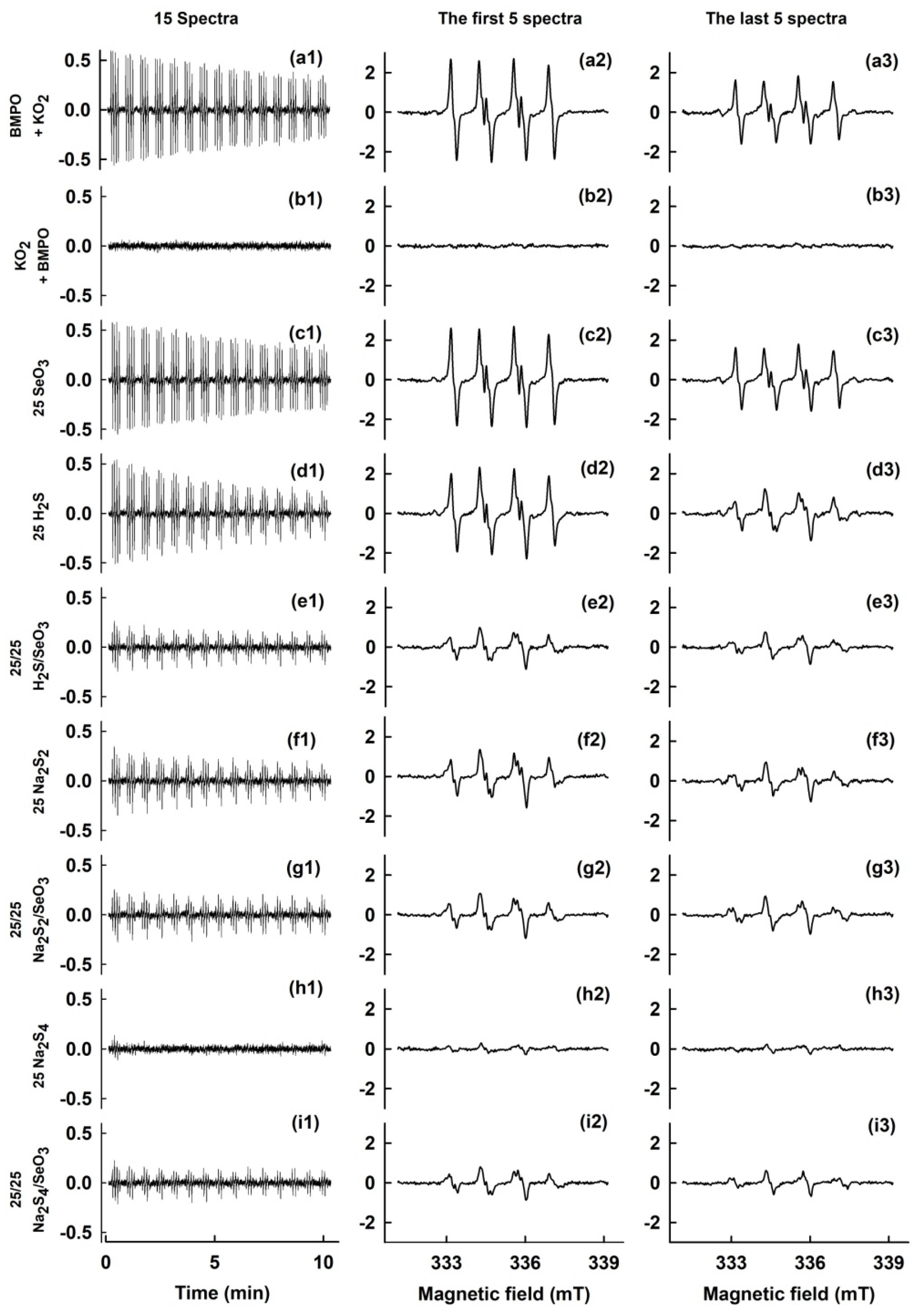
A pDNA cleavage assay with the use of pBR322 plasmid (New England BioLabs, Inc., N3033 L, Ipswich, MA, USA) was performed as reported previously [13]. In this assay, all samples contained 0.2 μg pDNA in the sodium phosphate buffer (25 mmol L−1 sodium phosphate, 50 μmol L−1 DTPA, pH 7.4). FeCl2 (150 µmol L−1) was used in control experiments. Powdered KO2 was dissolved by 4 mmol L−1 BMPO in the buffer containing 10% DMSO (v/v), vortexed for 10 s and 10 µL of the mixture was added into 10 µL solution of pDNA. The resulting mixtures were incubated for 30 min at 37 °C. After incubation, the reaction mixtures were subjected to 0.6% agarose gel electrophoresis. Integrated densities of all pBR322 forms in each lane were quantified using the Image Studio analysis software (LI-COR Biotechnology, Bad Homburg, Germany) to estimate pDNA-cleavage efficiency.

3. Results

3.1. BMPO-OOH as a Model Hydroperoxide and the Effects of Na2Sn/Na2SeOn

KO2 in DMSO dissociates to K+ and relatively stable O2•−, but its solubility in DMSO is extremely low [23,24]. EPR spectra after the addition of KO2 into the BMPO-buffered solution showed signals of two conformers of the BMPO-OOH adduct, as a result of O2•− trapping by BMPO (Figure 1a1–a3). The BMPO-OOH signal was relatively stable and slowly decreased (t1/2 ~23 min) in accordance with [15,16,18]. However, when KO2 was added into the buffer first, followed by the addition of BMPO 10 s later no radical was trapped by BMPO, i.e., the EPR spectrum was not observed (Figure 1b1–b3). The results confirmed a short life time of O2•− in water solution (t1/2 ~1–10 µs). Therefore, we used the interaction of O2•− with BMPO, producing the BMPO-OOH spin-adduct, as a model of organic hydroperoxides to study the effects of Na2Sn/Na2SeOn. The following sequence of the compounds was used to prepare the sample: KO2 was added into BMPO-buffered solution, resulting in the formation of BMPO-OOH and then the studied compounds were added 10 s later.
Information that can be obtained from the EPR spectra after the interaction of BMPO-OOH with compounds added into the system is as follows: First, it is an effect of compounds on the total integral EPR intensity of the BMPO-adducts obtained as a second integral of EPR spectra. Second, from the simulation of individual BMPO-adducts of the experimental EPR spectra it may be possible to obtain the changes in their relative concentrations (BMPO-OOH and BMPO-OH), thereby reflecting the effect of compounds.
The presence of SeO32− (25 µmol L−1) did not influence the rate of BMPO-OOH decomposition (Figure 1c1). From the similar shape of the first five cumulative spectra (Figure 1c2) and the last five accumulated spectra (Figure 1c3), it is assumed that there was no interaction between SeO32− and BMPO-OOH. The addition of Na2S (25 µmol L−1; H2S) slightly increased the rate of BMPO-OOH decay (Figure 1d1), as an indication of scavenging/interaction of the BMPO-adducts by/with H2S. Since the shape of the last five accumulated spectra (Figure 1d3) was different from the first five spectra (Figure 1d2), it can be concluded that H2S affects the BMPO-OOH spin-adduct. The details on the interaction are described in the next section. The H2S/SeO32− (25/25 µmol L−1/µmol L−1) mixture decreased BMPO-OOH concentration during 100 s after the sample preparation (before EPR measurement started) and later the BMPO-adducts concentration was approximately constant (Figure 1e1). However, spectra were different (Figure 1e2,e3) from control BMPO-OOH (Figure 1a2,a3), indicating interaction of H2S/SeO32− with BMPO-OOH. Similar effect was detected when polysulfide Na2S2 (25 µmol L−1) and the Na2S2/SeO32− (25/25 µmol L−1/µmol L−1) mixture was used (Figure 1f1–f3,g1–g3). The effects of polysulfide Na2S4 (25 µmol L−1) was the most pronounced, it diminished BMPO-OOH concentration during 100 s, before EPR measurement started (Figure 1h1–h3). However, its effect to scavenge the BMPO-adducts decreased when the Na2S4/SeO32− (25/25 µmol L−1/µmol L−1) mixture was used (Figure 1i1). The spectra (Figure 1i2,i3) revealed pronounced interaction with BMPO-OOH.
When SeO42 was used instead of SeO32−, it did not influence the effects of Na2S, Na2S2 and Na2S4, indicating no formation of redox active products of the Na2Sn/SeO42 interaction (Figure 2).

3.2. Simulation of BMPO-Adducts Spectra in the Presence of Na2Sn/Na2SeO3

The studied compounds changed shapes of EPR spectra of the BMPO-adducts, indicating the superposition of signals corresponding to the generation of individual BMPO-adducts. Therefore, we analyzed all accumulated spectra by simulation. Figure 3 shows representative examples of the simulation of the sixth to tenth accumulated spectra only. The results showed that the best fit was obtained when the hyperfine coupling constants for two conformers of BMPO-OOH and two of BMPO-hydroxyl radical (BMPO-OH) adducts, along with those of BMPO-adduct with carbon-centered radical (BMPO-CR) were inserted in spin-Hamiltonian calculations. The simulated spectra shown in Figure 3 were calculated using the hyperfine coupling constants elucidated from the experimental spectra (Table 1).
For simplicity, the relative concentration of two conformers BMPO-OH(1) and BMPO-OH(2) were summed up and described as BMPO-OH. Analogously, BMPO-OOH(1) and BMPO-OOH(2) were summed up and described as BMPO-OOH. The time dependence of such evaluated BMPO-adducts EPR integral intensity is shown in Figure 4. In this figure, the absolute integral of individual BMPO-adducts is depended besides sample composition also on time. For example, the first to fifth accumulated spectra of control integral intensity of BMPO-OOH spectra (circle; measured 1.7–5.2 min after sample preparation; see Figure 4a tick 1–5) has value 100 (r.u.) and the integrals of BMPO-OH (triangle) and BMPO-CR (cross) components of the sample are close to zero. The same control sample is measured 5.2–8.7 min after sample preparation (sixth to tenth accumulated spectra; see Figure 4a tick 6–10) and shows decrease to 73.5 (r.u.) for BMPO-OOH component, whereas integral of BMPO-OH is close to zero and BMPO-CR increased to 4.2.
In controls and samples with SeO32−, similar concentration of radicals and a slow decay of the BMPO-OOH component was seen, indicating no interaction of SeO32− with BMPO-OOH (Figure 4a,b). The addition of Na2S increased the rate of BMPO-OOH decay (Figure 4c), whereas slightly increased the concentration of BMPO-OH. The decay was several times pronounced when the H2S/SeO32− mixture was used (Figure 4d). Na2S2 and Na2S4 alone and their mixture with SeO32− significantly decreased the number of radicals and increased the rate of BMPO-OOH decay (Figure 4e–h), whereas slightly increased concentration of BMPO-OH with exception of Na2S4, where radical concentration was close to zero (Figure 4g). It is noticed that spin adducts concentration in the case of Na2S4/SeO32− was noticeably higher compared to Na2S4 alone (Figure 4g vs. Figure 4h). The total integral EPR intensity of all components (Figure 4) revealed that the order of potential to transform/scavenge the BMPO-adducts was Na2S4 > Na2S2 > H2S (Figure 4c,e,g) and H2S/SeO32− > H2S (Figure 4c,d), Na2S2 ~Na2S2/SeO32 (Figure 4e,f), but Na2S4 > Na2S4/SeO32− (Figure 4g,h).
In order to compare the relative ratio of components of the BMPO-adducts the sum of the radicals at each time was normalized to 100% (Figure 5). In controls and samples with SeO32−, the major BMPO-OOH component was relatively constant over the time with minor BMPO-CR component, indicating no decomposition of BMPO-OOH by SeO32− (Figure 5a,b). H2S and the H2S/SeO32 mixture decreased BMPO-OOH component and increased BMPO-OH over the time (Figure 5c,d), suggesting time-dependent transformation/decomposition of the model hydroperoxide BMPO-OOH to BMPO-OH. Similar qualitative results were observed when Na2S2 alone or in the combination with SeO32− was used (Figure 5e,f). Na2S4 alone significantly decreased BMPO-OOH and increased the BMPO-OH component (Figure 5g). The Na2S4/SeO32 mixture had similar, but slightly lower effects compared to Na2S4 (Figure 5h). The order of the compounds potency to cleave BMPO-OOH, estimated from the time-dependent BMPO-OH/BMPO-OOH ratio was: Na2S4 > Na2S4/SeO32 > H2S/SeO32 > Na2S2 ~Na2S2/SeO32 ~H2S > SeO32 ~control.

3.3. Cleavage of Plasmid DNA

Since Na2S interacted with BMPO-OOH (Figure 4 and Figure 5), it was of interest to know whether radical species, which can damage DNA, were formed during the interaction. The procedure for EPR experiments was modified to observe pDNA cleavage. In the control experiment, Fe2+ (150 µmol L−1) cleaved pDNA. However, when the reaction buffer contained 10% (v/v) DMSO, Fe2+ did not cleave pDNA. Cleaving of pDNA was observed neither in the presence of BMPO/KO2 mixture in buffer with 10% (v/v) DMSO, nor when Na2S was added into BMPO/KO2/DMSO (Figure 6). From the experiments with Fe2+ it is evident that DMSO interfered with the assay. This is supported also by information that DMSO is a scavenger of HO radicals [13,25]. Therefore, we did not precede with the pDNA experiments.

4. Discussion

In our previous study, when KO2 as a source of O2•− was added into the mixture of H2S/BMPO or H2S/SeO32−/BMPO, the concentration of trapped radicals changed and a superposition of several individual BMPO-adducts was detected [13]. It was not clear which components of the spectra resulted from the compounds/O2•− or from the compounds/BMPO-OOH interaction. Therefore, in the present work we used a different approach, which allowed us to study interaction of the compounds with a model of organic hydroperoxide BMPO-OOH. This approach is based on the short lifetime of O2•− in water solution and relatively stable BMPO-OOH to which the studied compounds were added.
This approach allowed the study of the effects of compounds on decomposition/transformation of the organic hydroperoxide BMPO-OOH to the ratio of BMPO-OH/BMPO-OOH, which was detected by EPR. To elaborate this, we performed a detailed simulation of the experimental EPR spectra obtaining the hyperfine coupling constants of the individual BMPO-adducts under these experimental conditions, along with their relative concentrations. From the comparison of the experimental and the simulated EPR spectra (Figure 3), it can be concluded that the BMPO-adducts spectra were very well simulated by the hyperfine coupling constants (Table 1), based on the presence of two conformers, BMPO-OH(1) and BMPO-OH(2), BMPO-OOH(1) and BMPO-OOH(2) and BMPO-CR. The hyperfine coupling constants were clearly indicated from the spectra simulations (Table 1) and were comparable to the published BMPO-adducts. The relative intensity of BMPO-OOH decreased slowly over time and was comparable, but not identical, to the reported values under physiological conditions without DMSO [15,16,18]. It is likely that the BMPO-CR component resulted from the trapping carbon-centered radical, originating from DMSO. From the time dependence of the integral EPR intensity (Figure 4 and Figure 5) it was possible to evaluate the time-dependent effects of the compounds on the total BMPO-adduct concentration and the BMPO-OH/BMPO-OOH ratio, which is suggested to be proportional to the organic hydroperoxide BMPO-OOH transformation/decomposition. The presence of SeO32− potentiated the decrease of integral intensity of spin-adducts induced by Na2S, had no significant effect on Na2S2 potency, but decreased the potency of Na2S4 (Figure 4). These results indicate that the interaction of SeO32− with Na2Sn, which leads to the formation of redox radical species significantly depends on the number of S atoms.
The order of ability to decrease the total integral intensity of the BMPO-adducts (Figure 4) of H2S/SeO32 > H2S or Na2S4 > H2S was similar to the order when KO2 was added to the mixtures of the BMPO/compounds or DEPMPO/compounds [5,13]. This indicates that in the case when KO2 was added to the mixtures of spin trap/compounds, the compounds affected mostly BMPO-OOH or DEPMPO-OH radicals.
A comparison of the time-dependent ratio of BMPO-OH/BMPO-OOH (Figure 5) revealed that the increase of the BMPO-OH spectral component was at the expense of the BMPO-OOH component, supporting the concept of decomposition/transformation of organic hydroperoxide. Interactions of H2S and polysulfides with radical species are complex [4,7] and more studies are needed to explain the most profound potential of Na2S4 > Na2S2 or effects of SeO32 to decrease the potency of Na2S4 to cleave BMPO-OOH.
In conclusion, BMPO-OOH was demonstrated to be a helpful model of organic hydroperoxide. The presented approach of EPR spectra measurement and analysis of the time-dependent ratio of the BMPO-OH/BMPO-OOH spin-adducts utilizing the spectra simulation could be useful to study potential of compounds to transform/decompose BMPO-OOH. Using this approach, the impact of sulfide derivatives (Na2Sn) alone or in the combination with SeO32 to transform/decompose BMPO-OOH was detected and compared. Since 10% DMSO (v/v) is used in the studied system, the procedure is useful for evaluating antioxidant potency of compounds insoluble in water.

Author Contributions

K.O. conceived, initiated and coordinated the study; K.O. and V.B. designed research; V.B., K.O., M.G. and A.M. performed EPR experiments and analyzed data; A.M. and M.C. performed pDNA cleavage experiments and analyzed data; V.B. simulated EPR spectra; K.O. wrote the paper; V.B., M.C. and L.T. contributed to analyze data and manuscript writing. All authors have read and agreed to the published version of the manuscript.

Funding

This work was supported by the Slovak Research and Development Agency (Grant Numbers APVV-19-0154 to K.O. and APVV-17-0384 to M.C.), the Scientific Grant Agency of the Slovak Republic (Grant Numbers VEGA 1/0026/18 to V.B., 2/0079/19 to M.G., 2/0053/19 to M.C. and 2/0014/17 to K.O.).

Conflicts of Interest

The authors declare no conflict of interest.

References

  1. Wang, R. Physiological implications of hydrogen sulfide: A whiff exploration that blossomed. Physiol. Rev. 2012, 92, 791–896. [Google Scholar] [CrossRef] [Green Version]
  2. Tomasova, L.; Konopelski, P.; Ufnal, M. Gut bacteria and hydrogen sulfide: The new old players in circulatory system homeostasis. Molecules 2016, 21, 1558. [Google Scholar] [CrossRef]
  3. Cacanyiova, S.; Berenyiova, A.; Balis, P.; Kristek, F.; Grman, M.; Ondrias, K.; Breza, J.; Breza, J., Jr. Nitroso-sulfide coupled signaling triggers specific vasoactive effects in the intrarenal arteries of patients with arterial hypertension. J. Physiol. Pharmacol. 2017, 68, 527–538. [Google Scholar]
  4. Fukuto, J.M.; Ignarro, L.J.; Nagy, P.; Wink, D.A.; Kevil, C.G.; Feelisch, M.; Cortese-Krott, M.M.; Bianco, C.L.; Kumagai, Y.; Hobbs, A.J.; et al. Biological hydropersulfides and related polysulfides—A new concept and perspective in redox biology. FEBS Lett. 2018, 592, 2140–2152. [Google Scholar] [CrossRef] [Green Version]
  5. Misak, A.; Grman, M.; Bacova, Z.; Rezuchova, I.; Hudecova, S.; Ondriasova, E.; Krizanova, O.; Brezova, V.; Chovanec, M.; Ondrias, K. Polysulfides and products of H2S/S-nitrosoglutathione in comparison to H2S, glutathione and antioxidant Trolox are potent scavengers of superoxide anion radical and produce hydroxyl radical by decomposition of H2O2. Nitric Oxide 2018, 76, 136–151. [Google Scholar] [CrossRef]
  6. Eghbal, M.A.; Pennefather, P.S.; O’Brien, P.J. H2S cytotoxicity mechanism involves reactive oxygen species formation and mitochondrial depolarisation. Toxicology 2004, 203, 69–76. [Google Scholar] [CrossRef] [PubMed]
  7. Liu, H.; Radford, M.N.; Yang, C.T.; Chen, W.; Xian, M. Inorganic hydrogen polysulfides: Chemistry, chemical biology and detection. Br. J. Pharmacol. 2019, 176, 616–627. [Google Scholar] [CrossRef] [Green Version]
  8. Fairweather-Tait, S.J.; Bao, Y.; Broadley, M.R.; Collings, R.; Ford, D.; Hesketh, J.E.; Hurst, R. Selenium in human health and disease. Antioxid. Redox Signal. 2011, 14, 1337–1383. [Google Scholar] [CrossRef] [PubMed]
  9. Jablonska, E.; Vinceti, M. Selenium and human health: Witnessing a Copernican revolution? J. Environ. Sci. Health C Environ. Carcinog. Ecotoxicol. Rev. 2015, 33, 328–368. [Google Scholar] [CrossRef] [PubMed]
  10. Misak, A.; Kurakova, L.; Goffa, E.; Brezova, V.; Grman, M.; Ondriasova, E.; Chovanec, M.; Ondrias, K. Sulfide (Na₂S) and Polysulfide (Na₂S₂) interacting with doxycycline produce/scavenge superoxide and hydroxyl radicals and induce/inhibit DNA cleavage. Molecules 2019, 24, 1148. [Google Scholar] [CrossRef] [Green Version]
  11. Kharma, A.; Grman, M.; Misak, A.; Domínguez-Álvarez, E.; Nasim, M.J.; Ondrias, K.; Chovanec, M.; Jacob, C. Inorganic Polysulfides and related reactive sulfur–selenium species from the perspective of chemistry. Molecules 2019, 24, 1359. [Google Scholar] [CrossRef] [Green Version]
  12. Kharma, A.; Misak, A.; Grman, M.; Brezova, V.; Kurakova, L.; Baráth, P.; Jacob, C.; Chovanec, M.; Ondrias, K.; Domínguez-Álvarez, E. Release of reactive selenium species from phthalic selenoanhydride in the presence of hydrogen sulfide and glutathione with implications for cancer research. New J. Chem. 2019, 43, 11771–11783. [Google Scholar] [CrossRef] [Green Version]
  13. Grman, M.; Misak, A.; Kurakova, L.; Brezova, V.; Cacanyiova, S.; Berenyiova, A.; Balis, P.; Tomasova, L.; Kharma, A.; Domínguez-Álvarez, E.; et al. Products of sulfide/selenite interaction possess antioxidant properties, scavenge superoxide-derived radicals, react with DNA, and modulate blood pressure and tension of isolated thoracic aorta. Oxidative Med. Cell. Longev. 2019, 2019, 9847650. [Google Scholar] [CrossRef] [PubMed] [Green Version]
  14. Ingold, I.; Berndt, C.; Schmitt, S.; Doll, S.; Poschmann, G.; Buday, K.; Roveri, A.; Peng, X.; Porto Freitas, F.; Seibt, T.; et al. Selenium utilization by GPX4 is required to prevent hydroperoxide-induced ferroptosis. Cell 2018, 172, 409–422.e421. [Google Scholar] [CrossRef] [PubMed] [Green Version]
  15. Zhao, H.; Joseph, J.; Zhang, H.; Karoui, H.; Kalyanaraman, B. Synthesis and biochemical applications of a solid cyclic nitrone spin trap: A relatively superior trap for detecting superoxide anions and glutathiyl radicals. Free Radic. Biol. Med. 2001, 31, 599–606. [Google Scholar] [CrossRef]
  16. Bézière, N.; Hardy, M.; Poulhès, F.; Karoui, H.; Tordo, P.; Ouari, O.; Frapart, Y.M.; Rockenbauer, A.; Boucher, J.L.; Mansuy, D.; et al. Metabolic stability of superoxide adducts derived from newly developed cyclic nitrone spin traps. Free Radic. Biol. Med. 2014, 67, 150–158. [Google Scholar] [CrossRef] [Green Version]
  17. Suzen, S.; Gurer-Orhan, H.; Saso, L. Detection of reactive oxygen and nitrogen species by Electron Paramagnetic Resonance (EPR) technique. Molecules 2017, 22, 181. [Google Scholar] [CrossRef]
  18. Tsai, P.; Marra, J.M.; Pou, S.; Bowman, M.K.; Rosen, G.M. Is there stereoselectivity in spin trapping superoxide by 5-tert-butoxycarbonyl-5-methyl-1-pyrroline N-oxide? J. Org. Chem. 2005, 70, 7093–7097. [Google Scholar] [CrossRef]
  19. Tsai, P.; Ichikawa, K.; Mailer, C.; Pou, S.; Halpern, H.J.; Robinson, B.H.; Nielsen, R.; Rosen, G.M. Esters of 5-carboxyl-5-methyl-1-pyrroline N-oxide: A family of spin traps for superoxide. J. Org. Chem. 2003, 68, 7811–7817. [Google Scholar] [CrossRef]
  20. Stolze, K.; Udilova, N.; Rosenau, T.; Hofinger, A.; Nohl, H. Synthesis and characterization of EMPO-derived 5,5-disubstituted 1-pyrroline N-oxides as spin traps forming exceptionally stable superioxide spin adducts. Biol. Chem. 2003, 384, 493–500. [Google Scholar] [CrossRef]
  21. Chang, J.; Taylor, R.D.; Davidson, R.A.; Sharmah, A.; Guo, T. Electron paramagnetic resonance spectroscopy investigation of radical production by gold nanoparticles in aqueous solutions under X-ray irradiation. J. Phys. Chem. A 2016, 120, 2815–2823. [Google Scholar] [CrossRef] [PubMed]
  22. Stoll, S.; Schweiger, A. EasySpin, a comprehensive software package for spectral simulation and analysis in EPR. J. Magn. Reson. 2006, 178, 42–55. [Google Scholar] [CrossRef] [PubMed]
  23. Merritt, M.V.; Sawyer, D.T. Electrochemical studies of the reactivity of superoxide ion with several alkyl halides in dimethyl sulfoxide. J. Org. Chem. 1970, 35, 2157–2159. [Google Scholar] [CrossRef]
  24. Hayyan, M.; Hashim, M.A.; AlNashef, I.M. Superoxide ion: Generation and chemical implications. Chem. Rev. 2016, 116, 3029–3085. [Google Scholar] [CrossRef] [PubMed] [Green Version]
  25. Noda, M.; Ma, Y.; Yoshikawa, Y.; Imanaka, T.; Mori, T.; Furuta, M.; Tsuruyama, T.; Yoshikawa, K. A single-molecule assessment of the protective effect of DMSO against DNA double-strand breaks induced by photo-and γ-ray-irradiation, and freezing. Sci. Rep. 2017, 7, 8557. [Google Scholar] [CrossRef] [PubMed]
Scheme 1. 5-tert-butoxycarbonyl-5-methyl-1-pyrroline-N-oxide (BMPO) spin trap enantiomers and illustration of the diastereoisomeric spin-adducts generation in BMPO reactions with O2•− and HO. Nuclei included in the simulation of the experimental EPR spectra of the BMPO-OOH and BMPO-OH adducts are marked in red.
Antioxidants 09 00918 sch001
Figure 1. Electron paramagnetic resonance (EPR) spectra of BMPO-OOH modulated by Na2S, Na2S2, Na2S4 and their interaction with SeO32. (a1i1) Collection of 15 EPR spectra arranged back-to-back of the BMPO-adducts, each 42 s, with starting acquisition 100 s after sample preparation; (a2i2) first to fifth accumulated spectra; (a3i3) last five accumulated spectra; (a1a3) representative control EPR spectra from 2–3 measurements of BMPO-OOH after saturated KO2/DMSO solution (final 10% v/v DMSO) was added to 30 mmol L−1 BMPO in the buffer consisting 50 mmol L−1 sodium phosphate buffer and 0.1 mmol L−1 DTPA (pH 7.4, 37 °C); spectra of BMPO-OOH after the addition of (c1c3) 25 µmol L−1 SeO32−, (d1d3) 25 µmol L−1 Na2S, (e1e3) 25/25 (in µmol L−1) Na2S/SeO32−, (f1f3) 25 µmol L−1 Na2S2, (g1g3) 25/25 (in µmol L−1) Na2S2/SeO32−, (h1h3) 25 µmol L−1 Na2S4 and (i1i3) 25/25 (in µmol L−1) Na2S4/SeO32−. In the case of (b1b3) spectra, 30 mmol L−1 BMPO was added 10 s after KO2/DMSO was mixed with the buffer. The intensities of the (a1i1) time-dependent EPR spectra and (a2i2, a3i3) detailed spectra are comparable; they were measured under identical EPR spectrometer settings.
Antioxidants 09 00918 g001
Figure 2. EPR spectra of BMPO-OOH modulated by the mixture of Na2S, Na2S2 and Na2S4 with SeO42−. Representative EPR spectra from 2–3 measurements of BMPO-OOH obtained in KO2/DMSO solution (final 10% v/v DMSO) in the presence of 30 mmol L−1 BMPO prepared in buffer consisting 50 mmol L−1 sodium phosphate buffer and 0.1 mmol L−1 DTPA (pH 7.4, 37 °C), after the addition of (a1a3) 25 µmol L−1 SeO42−, (b1b3) 25/25 (in µmol L−1) Na2S/SeO42−, (c1c3) 25/25 (in µmol L−1) Na2S2/SeO42− and (d1d3) 25/25 (in µmol L−1) Na2S4/SeO42−. Sets of individual EPR spectra of the BMPO-adducts were recorded as described in legend to Figure 1. The intensities of the (a1d1) time-dependent EPR spectra and (a2d2, a3d3) detailed spectra are comparable, they were measured under identical EPR spectrometer settings.
Antioxidants 09 00918 g002
Figure 3. Representative normalized experimental EPR spectra of the BMPO-adducts along with their simulation using the hyperfine coupling constants summarized in Table 1. Experimental spectra of the sixth to tenth accumulated spectra are shown only (blue); simulated spectra are red. (a) Control 30 mmol L−1 BMPO + KO2 and after addition of (b) 25 µmol L−1 SeO32−, (c) 25 µmol L−1 H2S, (d) 25/25 (in µmol L−1) H2S/SeO32−, (e) 25 µmol L−1 Na2S2, (f) 25/25 (in µmol L−1) Na2S2/SeO32−, (g) 25 µmol L−1 Na2S4 and (h) 25/25 (in µmol L−1) Na2S4/SeO32−.
Antioxidants 09 00918 g003
Figure 4. Comparison of integral EPR intensity of individual BMPO-adducts elucidated from the simulation of experimental EPR spectra. The first to fifth accumulated spectra (1–5; 1.7–5.2 min after sample preparation; see Figure 1), the sixth to tenth accumulated spectra (6–10; 5.2–8.7 min after sample preparation) and the eleventh to fifteen accumulated spectra (11–15; 8.7–12.2 min after sample preparation; see Figure 1) Spectral components: BMPO-OOH (blue), BMPO-OH (red) and BMPO-CR (green); n = 2–3. (a) Control 30 mmol L−1 BMPO + KO2 and after addition of (b) 25 µmol L−1 SeO32−, (c) 25 µmol L−1 H2S, (d) 25/25 (in µmol L−1) H2S/SeO32−, (e) 25 µmol L–1 Na2S2, (f) 25/25 (in µmol L–1) Na2S2/SeO32−, (g) 25 µmol L–1 Na2S4 and (h) 25/25 (in µmol L−1) Na2S4/SeO32−. Data are presented as the means ± SEM.
Antioxidants 09 00918 g004
Figure 5. Ratio of normalized integral EPR intensity of individual BMPO-adducts evaluated from experimental spectra simulation. Data calculated from Figure 4. For explanation of (a–h) and figure description, see legend to Figure 4.
Antioxidants 09 00918 g005
Figure 6. Effect of BMPO/KO2 on pDNA cleavage in the absence and presence of Na2S. Representative gel (a) and column graph (b) indicating control (black) and the effects of Fe2+ (150 µmol L−1 FeCl2) in 25 mmol L−1 sodium phosphate buffer and 50 µmol L−1 DTPA (pH 7.4) without (dark) and with 10% (v/v) DMSO (gray) and increasing concentrations of Na2S on pDNA cleavage in the phosphate buffer containing 10% DMSO (v/v). The band at the bottom corresponds to the circular supercoiled form of pDNA and the less intense band appearing above in the case of Fe2+ (dark column) represents the linear form of pDNA. The top band corresponds to the nicked circular form of pDNA. Values are the means ± SEM, n = 3.
Antioxidants 09 00918 g006
Table 1. Hyperfine coupling constants of the BMPO spin-adducts elucidated from the simulations of experimental spectra measured in the buffer solutions containing KO2 and 10% DMSO (v/v). BMPO-OOH and BMPO-OH were simulated considering presence of two conformers [15,18,19].
BMPO-AdductaN, mTaHβ, mTaHγ, mT
BMPO-OH(1)1.423 ± 0.0111.541 ± 0.0140.078 ± 0.011
BMPO-OH(2)1.365 ± 0.0381.248 ± 0.0360.073 ± 0.015
BMPO-OOH(1)1.339 ± 0.0021.186 ± 0.007
BMPO-OOH(2)1.334 ± 0.0030.958 ± 0.007
BMPO-CR1.5282.221

