Review

Plant-RNA in Extracellular Vesicles: The Secret of Cross-Kingdom Communication

by
Ornella Urzì
,
Roberta Gasparro
,
Nima Rabienezhad Ganji
,
Riccardo Alessandro
* and
Stefania Raimondo
*
Dipartimento di Biomedicina, Neuroscienze e Diagnostica Avanzata, Università degli Studi di Palermo, 90133 Palermo, Italy
*
Authors to whom correspondence should be addressed.
These authors contributed equally to this work.
Membranes 2022, 12(4), 352; https://doi.org/10.3390/membranes12040352
Submission received: 21 February 2022 / Revised: 18 March 2022 / Accepted: 21 March 2022 / Published: 23 March 2022
(This article belongs to the Special Issue Cell Membrane Vesicles)

Abstract

:
The release of extracellular vesicles (EVs) is a common language, used by living organisms from different kingdoms as a means of communication between them. Extracellular vesicles are lipoproteic particles that contain many biomolecules, such as proteins, nucleic acids, and lipids. The primary role of EVs is to convey information to the recipient cells, affecting their function. Plant-derived extracellular vesicles (PDEVs) can be isolated from several plant species, and the study of their biological properties is becoming an essential starting point to study cross-kingdom communication, especially between plants and mammalians. Furthermore, the presence of microRNAs (miRNAs) in PDEVs represents an interesting aspect for understanding how PDEVs can target the mammalian genes involved in pathological conditions such as cancer, inflammation, and oxidative stress. In particular, this review focuses on the history of PDEVs, from their discovery, to purification from various matrices, and on the functional role of PDEV-RNAs in cross-kingdom interactions. It is worth noting that miRNAs packaged in PDEVs can be key modulators of human gene expression, representing potential therapeutic agents.

1. Introduction

The constant communication between all living organisms, such as plants, bacteria, and animals, attracts the scientific interest of the biomedical community. In general, lipids, proteins, and nucleic acids can be transferred through extracellular vesicles (EVs), which are shuttles involved in cell-to-cell communication.
Extracellular vesicles are lipoproteic particles released by organisms belonging to different kingdoms, and research in recent years has indicated they are mainly involved in cross-kingdom interaction.
The content of EVs consists of various biomolecules, such as proteins, nucleic acids, sugars, and lipids. The main role of EVs is to transfer information to the receiving cells, thereby influencing their functions [1]. Among EVs, different subtypes can be recognized; the International Society for Extracellular Vesicles periodically publishes new guidelines for EV research (MISEV guidelines), and in the latest recommendation, EV subtypes are distinguished based on their physical properties, such as dimension and density (small EVs or medium/large EVs), the biochemical composition (CD63+/CD81+—EVs, Annexin A5-stained EVs, etc.), and on the cell of origin [1]. The discovery of the presence of both messenger and small noncoding RNAs inside mammalian EVs in 2007 revolutionized the field of cell–cell communication [2]. Many studies correlated the ability of EVs to modulate the functional properties of target cells with their RNA cargo [3,4,5]. RNAs contained in EVs, once internalized, can trigger molecular and phenotypic changes in the recipient cell. EV-mRNAs can be translated into functional proteins, while EV-ncRNAs can engage complex networks of interactions that regulate gene expression [6]. For instance, the EVs released by multiple myeloma cells are enriched in some miRNAs that can affect the expression of genes involved in osteogenic differentiation in mesenchymal stem cells. Among them, miR-129-5p downregulates the expression of SP1, a positive regulator of osteogenic differentiation, and alkaline phosphatase in mesenchymal stem cells; thus, inhibiting osteoblastic differentiation [3]. RNAs delivered by EVs are also involved in pre-metastatic niche formation. Conigliaro et al. demonstrated that EVs released by CD90+ liver cancer cells carry lncRNA H19, which promotes tube formation and the cell–cell interaction of endothelial cells; thus, favoring angiogenesis [7]. EV-RNAs play a key role, not only in pathological, but also in physiological, processes; understanding their mechanisms of action could lead to new therapeutic strategies. Li and colleagues demonstrated the beneficial effects of EVs isolated from M2 microglia cells in mice with ischemic stroke. They found that M2 microglial EVs were enriched in miR-124, which suppresses astrocytes proliferation through the inhibition of the STAT3 pathway; thus, reducing glial scar formation [8].
Nowadays, it is known that extracellular vesicles are also released by plants. Although the guidelines provided in MISEV [1] are also valid for those studying vesicles within the plant kingdom, the growing interest in plant-derived extracellular vesicles (PDEVs) requires a more detailed standardization, recently discussed by Pinedo and colleagues [9]. First, the nomenclature of EVs isolated from plants should be revised. PDEVs in the literature are named using different terms, such as nanovesicles [10], nanoparticles [11], microvesicles [12], exosomes [13], and exosome-like vesicles [14]. However, information on their biogenesis is still lacking, thereby complicating the establishment of a clear nomenclature. Pinedo et al. suggested using the term ‘plant EVs’ only for particles isolated from the apoplastic fluid, while for vesicles obtained from plant tissues after disruptive processes, they recommend the term ‘plant-derived nanovesicles’, since the origin of the obtained vesicles is dubious [9]. In this review, we will use the term ‘PDEVs’ to refer to both EVs from apoplastic fluid and those obtained by destructive methods. In addition to the appropriate nomenclature, isolation methods and the identification of specific markers are critical for PDEVs standardization; these points will be discussed in the subsequent paragraph.
The information contained in this review article aims to summarize the history of the research on plant-derived extracellular vesicles, as well as to report evidence on their role in cross-kingdom interaction. Our manuscript will focus on the PDEV-RNA component. The PubMed database was used as the source for the retrieval of the literature included in this work.
The figure below (Figure 1) represents a chronological summary of the discovery of the EVs in the plant kingdom that will be discussed in this review.

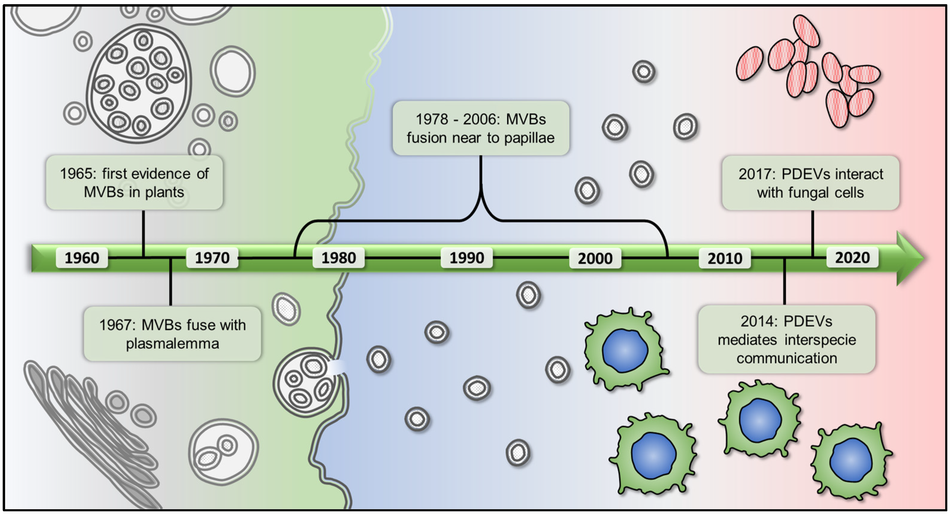
2. The Story of Plant EVs, from Their Discovery to the Purification from Different Matrices

Similarly to other living organisms, plants produce extracellular vesicles (EVs), and the discovery of plant-derived extracellular vesicles can be attributed to Jensen, who in 1965 found multivesicular bodies (MVBs), and, thus, intraluminal vesicles, in cotton [15]. Two years later, Halperin and Jensen analyzed the ultrastructure of clumps in suspension cell cultures of wild carrot and found the presence of numerous MVBs able to fuse with the plasmalemma and release their contents into the wall space [16]. In 1978, Politis and Goodman observed membrane-bound vesicles in the microfibrillar material forming the cell wall apposition of tobacco leaves inoculated with non-pathogenic Pseudomonas pisi [17]. Similarly, other studies found that, during pathogen attacks, plant cells have MVBs that are observed in various states of fusion with the plasmalemma. Most of these fusion events occur near the papillae; extracellular defense structures that block pathogen entry into the cell [18,19,20]. PDEVs were isolated from the apoplastic fluid for the first time in 2009 by Regente et al. [21]. The initial studies suggested that EVs in plants contributed to the development of early defense structures, in response to pathogens; however, their biological functions remained obscure for a long time. Some years later, it was found that PDEVs can mediate interspecies communication [22,23,24], for instance after oral administration grapes and grapefruit-derived EVs were taken up by intestinal macrophages in mice and exerted anti-inflammatory and antioxidant effects [22]. Cross-kingdom interaction also includes fungi; in 2017, Regente and colleagues demonstrated that PDEVs isolated from sunflower can interact with fungal cells, inhibiting fungal growth and causing cell death [25]. This observation was supported by the study of Rutter and Innes, who demonstrated that PDEVs secretion was enhanced in infected Arabidopsis thaliana leaves, contributing to innate immunity [26].
In the figure below, we have summarized the results of the first studies on the functional role of PDEVs (Figure 2).
The interest of the scientific community in PDEVs increased when the presence of microRNAs able to bind target genes in human cells was demonstrated [27]. Moreover, recently the mechanism of small RNA sorting into PDEVs was partially elucidated by He et al., who described the involvement of RNA binding proteins, such as Argonaute 1, RNA helicases, and Annexins, in this process [28]. We will analyze the functional role of PDEV-RNA in the following paragraph.
As mentioned previously, one of the main challenges in the field of PDEVs remains the standardization of the isolation method, and consequently the nature of isolated EVs [9]. PDEVs have been isolated from the tissues, organs, apoplastic fluid, and juice of several plant species, such as lemon [10,29,30,31,32,33], grapefruit [22,27,30,31,34,35,36,37,38], tomato [27,39,40,41,42], and Arabidopsis [26,28,43,44,45,46,47,48] (Figure 3). Only a minority of studies have purified EVs from the extracellular apoplastic fluid [21,25,26]; most of the works were conducted using other methods to isolate PDEVs, which mainly consisted of organ and tissues disruption. The first technique ensures the recovery of most EVs, while the second method causes the contamination of the sample with intracellular compartment and artificial nanoparticles [9].
Although new techniques for PDEVs isolation have emerged in the last year, such as immunocapture purification [28] and the aqueous two-phase system [49], differential centrifugation remains the most widely used method [40,46,50]. Some groups adopted isolation protocols similar to those used for mammalian EVs. Briefly, the starting material is squeezed, and the obtained juice is subjected to multiple steps of centrifugation: low-speed centrifugation (500–3000× g for about 10 min), intermediate speed centrifugation (2000–10,000× g for about 30 min), and ultracentrifugation (100,000–150,000× g for 1.5–2 h) to obtain a PDEV pellet. However, since ultracentrifugation also sediments other vesicles, proteins, and protein/RNA aggregates; while, some protocols add a subsequent density gradient ultracentrifugation using iodixanol or sucrose/deuterium oxide to separate PDEVs from contaminants [50,51,52]. On the other hand, Regente and other researchers have found that PDEVs can pellet at a lower speed, such as 40,000× g [21,26,53].
Once isolated, PDEVs should be appropriately characterized using various methods, including nanoparticle tracking, flow-cytometry, transmission electron microscopy, and other forms of electron microscopy. The PDEV cargo is complex and heterogeneous, for this reason, the best methods for studying their content are based on omics analysis. In the last years, several studies were carried out to characterize the proteomic profile of EVs isolated from different plant species [10,21,26,30,42,54]; these findings could help to identify possible PDEVs markers. Among the proteins identified in PDEVs, the most represented are heat shock protein 70 (HSP70), S-adenosyl-homocysteinase, and glyceraldehyde 3 phosphate dehydrogenase, which were found in EVs isolated from olive pollen [55], Nicotiana benthamiana [56], Arabidopsis thaliana [26], and sunflower [25]; these proteins could be candidates as PDEVs markers, even if further studies are needed to confirm their presence in EVs isolated from the majority of plants. Other protein families well represented in PDEVs are that of aquaporins, present in citrus- [10,30,52] and grape-derived EVs [56]; and annexins, found in citrus- [10,30], sunflower- [21], and Arabidopsis-derived EVs [26]. Besides proteins, the lipid characterization of PDEVs has attracted the interest of the scientific community, since this component seems to be strictly correlated to their biological functions [23]. The main lipid species identified in PDEVs are phosphatidic acid (PA), found in EVs from sunflower [21], grape [56], ginger [23], Uvae-ursi folium, Craterostigma plantagineum, and Zingiberis rhizome [57]; phosphatidylethanolamine (PE), described in EVs-derived from grapefruit [58], grape [56], Craterostigma plantagineum, and Zingiberis rhizome [57]; and phosphatidylcholine (PC), present in grapefruit- [58] and Craterostigma plantagineum-EVs [57]. Moreover, PDEV cargo includes several metabolites, such as sulforaphane [59], shogaol [60], and flavonoids [36,56,58], which could explain the PDEV-mediated beneficial roles in plant–mammalian interactions. On the other hand, a recent study published by Stanly and colleagues showed that both micro-and nano-vesicles isolated from strawberry carry functional allergens, including Fra a 1, Fra a 3, and Fra a 4 [51]; thus, demonstrating for the first time that PDEVs can also transport this type of molecule.
PDEVs also contain different species of RNA; this topic and the correlated studies will be discussed in the following paragraphs.

3. Functional Role of PDEV-RNAs in Cross-Kingdom Interactions

Discoveries correlated to plant-derived extracellular vesicles are becoming an essential starting point to study cross-kingdom communication; as a consequence, several studies have been carried out to analyze the interaction between plant vesicles and mammalian targets (Figure 4).

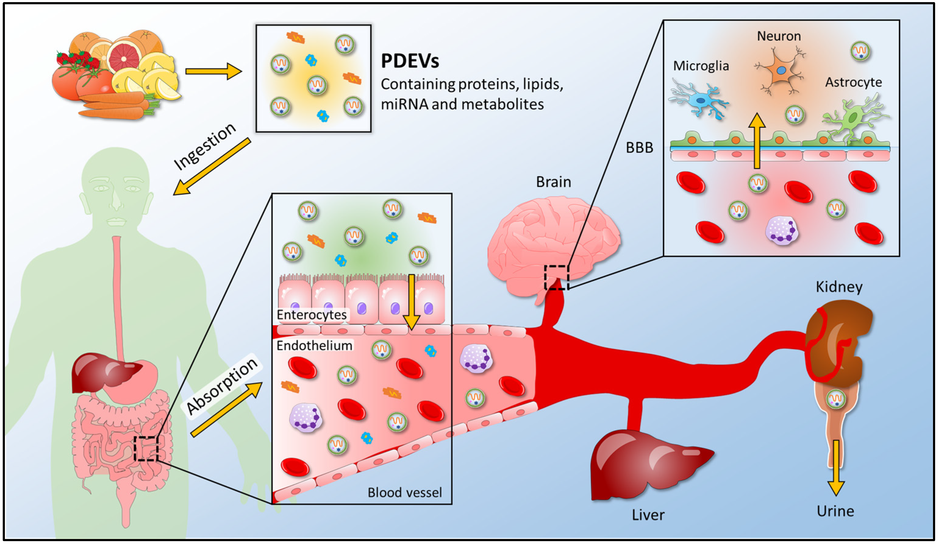
3.1. Functional Roles of Plant Extracellular Vesicles in Plant-Mammalian Communication

Plant-derived extracellular vesicles are also known for their specific proprieties, having a low immunological risk, and above all for their higher bioavailability. In terms of safety, plants do not have any zoonotic or human pathogens. Higher bioavailability is one of their most interesting features in cross-kingdom interaction: components packed into extracellular vesicles, including miRNAs, are protected by the lipid layer, in order to avoid their degradation. Consequently, cross-kingdom interaction paves the way for new applications of plant-derived extracellular vesicles and other findings on the regulation of mammalian targets as mediated by plants’ miRNAs. PDEVs are also known for their anti-inflammatory and anti-cancer properties. For example, EVs derived from grape [56], grapefruit [58], ginger [61], ginseng [62], and mulberry bark [63] have been reported for their effects in treating colitis, inflammation, and cancer. Ginseng is also used to enhance neurogenesis: Xu et al. reported the efficacy of extracellular vesicles derived from ginseng in transferring active nucleic acids to stem cells. In particular, miRNAs packed into EVs derived from ginseng can induce the neural differentiation of BMSCs. Ginseng-EVs upregulate PI3K signaling and induce the activation of neural differentiation in vitro. In vivo, ginseng-EVs promote neural restoration through upregulating PI3K signaling, increase nerve regeneration by promoting neurotrophin expression, and influence the Ras/Erk pathways. In addition, it was reported that mtr-miR-159a, a miRNA packed into ginger-EVs, can upregulate the PI3K signaling pathway [13]. Furthermore, Sundaram et al. recently reported that oral administration of garlic-EVs resolves brain inflammation and obesity in mice under a high-fat diet. These EVs are taken up by microglial cells, and inhibit brain inflammation via IDO1-mediated AHR pathway and c-Myc-mediated c-GAS/STING inflammatory pathway [64]. Cao et al. demonstrated, for the first time, that EVs isolated from the roots of Panax ginseng can induce M1-like macrophage polarization via the Toll-like receptor 4/myeloid differentiation antigen 88 signaling pathway and enhance the production of total reactive oxygen species (ROS), to induce apoptosis of mouse melanoma cells. Consequently, it was found that ginseng-derived EVs can inhibit tumor growth in mice [62]. Furthermore, another interesting study reported that leaf-derived extracellular vesicles from Dendropanax morbifera can inhibit tyrosinase activity and also reduce melanin content in melanoma cells, representing a candidate for the development of new anti-melanogenic agents [65]. Kim et al. demonstrated in 2020 that Dendropanax Morbifera-EVs exerts cytotoxic effects on malignant breast and skin tumor cells, without affecting normal cells, while Pinus densiflora-EVs are cytotoxic toward malignant skin tumor cells, but not toward normal cells. These results indicate that EVs derived from plant sap are selectively cytotoxic against tumor cells [66]. Recently, it was found that EVs isolated from apple possess anti-inflammatory properties, since they can downregulate the expression of pro-inflammatory cytokines, such as IL-8 and IL-1β, in human macrophages. The anti-inflammatory activities of apple-EVs could be explained by their ability to upregulate some miRNAs in target cells, such as miR-146a-5p, which is involved in NF-κB (Nuclear Factor kappa B) regulation [67].
Interestingly, Zhang [68] and others [69,70] highlighted for the first time that the microRNAs (miRNAs) present in plant extracts, introduced by diet, can control gene expression in human cells. Indeed, miRNAs have emerged as novel signaling molecules, to mediate intercellular communication. MiRNAs are small ∼21–22 nt non-coding RNAs, which are known to be regulators of essential biological processes in animals and plants. In 2012, it was found that food-derived miRNAs can provide cross-kingdom regulation. In particular, plant-derived miRNA can regulate gene expression with complementarity to their target mRNA, even if the mammalian mRNAs only possess partial homology to the targets [71]. Despite this, several studies focus on the role of the exogenous plant’s miRNAs in the gene expression of mammalian cells regulation. Interesting findings regarding plants’ extracellular vesicles concern the presence of small RNAs, especially miRNAs, which can represent a new class of cross-kingdom modulators. Recent evidence demonstrated that plants’ miRNAs may be considered a new class of micronutrients responsible for the medical properties of plants. Generally, plant miRNAs are involved in gene targeting, and the literature reports their beneficial role in some pathological conditions such as cancer. For example, plant miR-159 is correlated with a decrease in breast cancer incidence and progression. In particular, TCF7 is a mammalian target for plant miR-159, and it has shown the anti-proliferative function of miR-159 in breast cancer cells; demonstrating that a plant miRNA can influence cancer cell growth. Furthermore, a synthetic mimic of miR-159 can inhibit proliferation by targeting the TCF7 that encodes a Wnt signaling transcription factor, leading to a decrease in MYC protein levels [72]. Moreover, miR-58 derived from salvia has an effect on the reduction of Akt/mTOR (mammalian target of rapamycin) signaling, also facilitating autophagy [73]. It was also recently reported that miR-167e-5p represses intestinal cell proliferation by targeting β-catenin, and that the natural oeu-sRNAs decrease the protein expression of miR-34a mRNA targets, reducing proliferation and increasing apoptosis in different tumor cells [74]. However, in other studies, the intake of plant miRNAs through the diet has been challenged [75,76,77]. Snow et al. selected three highly expressed plant miRNAs (miR-156a, miR-159a, and miR-169a) and analyzed their levels in the plasma of healthy subjects after the intake of fruits containing these miRNAs. However, the plant miRNAs were undetectable in the plasma of healthy subjects. Similar data were obtained in mice: the authors fed mice with a vegetarian diet, but they did not detect any significant increase in plant miRNAs expression, both in the plasma and organs of mice [75]. These observations were supported by another study in which pigtailed macaques were fed with a miRNA-rich plant-based diet and then plant miRNAs were analyzed in animal blood through RT-qPCR, after 1, 4, and 12 h. It was found that the amplification of plant miRNAs was variable and possibly non-specific; thus, refuting the hypothesis of horizontal transfer of small RNAs from plant to mammals through oral administration; even if the study was carried out with only two macaque subjects [76]. Moreover, another group failed in the detection of plant miRNAs in mice fed with a rice-based diet for 1, 3, and 7 days. They measured the levels of rice miRNAs through miRNA sequencing and qPCR in plasma and liver of mice fed with rice-based diet, but there were no differences compared to the control group (mice fed with synthetic chow) [77]. Nevertheless, the work of Zhang was further accompanied by others that support the idea that plant miRNAs can be found in the blood, urine, and tissues of plant-eating animals [78,79]. It could be possible that some studies failed to detect plant miRNAs in animal tissues due to small dosages and a short time of exposure to the fed RNA [80].
Emerging studies have pointed out the role of miRNAs, packed in PDEVs, in the regulation of mammalian genes. Potestà et al. demonstrated that EVs derived from Moringa oleifera seeds, which carry plant miRNAs, can naturally penetrate inside human tumor cells and have proapoptotic effects. In particular, these EVs can modulate activities related to the viability and apoptosis in tumor cell lines, thanks to the regulation being mediated by miRNAs of BCL2 protein, which is one of the main factors that affects tumorigenesis in cell lines of epithelial origin [81]. Another interesting study demonstrated that bitter melon-derived extracellular vesicles can induce the apoptosis of oral squamous cell carcinoma (OSCC) cells. In particular, NLRP3, which is a component of the innate immune system, is able to promote tumor growth and metastasis in OSCC [82] and can be downregulated by miRNA-22, promoting cancer cell growth repression [83]. This is considered an interesting key point in cross-kingdom interaction because, as was said before, plant miRNAs can reduce the expression of target genes in animals and contribute to the function of tissues. Therefore, Yang et al. detected twenty-four kinds of miRNAs packed into EVs derived from bitter melon, and 11 of them had the potential to regulate the expression of NLRP3 mRNA, downregulating the protein expression of NLRP3. Furthermore, EVs derived from bitter melon combined with 5-fluorouracil enhanced OSCC apoptosis via augmented ROS generation; thus, increasing the cytotoxic effects and reducing the 5-fluorouracil drug resistance [84].
In addition to PDEVs’ anti-cancer effect, other biological and functional properties known in the literature are their anti-inflammatory and anti-oxidant role in human and mice models. It has been shown that PDEVs are taken up by macrophages and exhibit anti-inflammatory effects; for example, ginger-derived EVs are preferentially taken up by intestinal macrophages or monocytes, and, therefore, induce anti-inflammatory mediators. In one of these studies, colitis was induced in mice using dextran sulfate sodium, and the mice were subsequently treated with ginger-derived EVs. The results indicated that these EVs exhibited anti-inflammatory effects, with a decrease in lipocalin-2, a known biomarker for intestinal inflammation [61]. It was also found that mice treated with ginger EVs showed significant downregulation of pro-inflammatory cytokines, such as IL-6 and TNF-α, as well as the upregulation of the anti-inflammatory cytokine IL-10. Moreover, it was demonstrated that EVs isolated by Citrus sinensis can penetrate intestinal epithelial cells and positively modulate the expression of anti-inflammatory genes and tight junction. In particular, Citrus sinensis-EVs can modulate the expression of important genes related to inflammatory pathways, such as HMOX-1 and ICAM1, or the restoration of intestinal permeability related to claudins and occludin [85]. In addition, plant EVs resist gastric pepsin solution and intestinal pancreatic and bile extract solutions, indicating their potential for influencing cell biology through ingested foods [58] and suggesting the possible use of food EVs as safe therapeutic agents.
MiRNAs’ role in the anti-inflammatory effect of PDEVs has attracted many researchers: several studies were carried out to find the correlation between miRNAs packed into plant-derived extracellular vesicles and inflammatory response targets. For example, an interesting finding reports that mammalian genes involved in the regulation of inflammatory cytokines (IL-5 and IL-6) can be targeted by PDEV miRNAs, suggesting that these plant vesicle-derived miRNAs can potentially regulate mammalian mRNAs and biological pathways. miRNAs can, indeed, directly target genes encoding inflammatory factors. For example, miR-5781 in soybean-EVs can directly target IL-17A. MiR-4995 packed into tomato-EVs can target IL-5, while miR-1078 in ginger-EVs can target IL-6 [27]. Consequentially, it is important to consider the potential roles that these PDEV-derived miRNAs can play in host health and disease, paving the way for new cross-kingdom interaction findings [86]. Interestingly, a therapeutic effect of plant miRNAs in the prevention of chronic inflammation was shown in a mouse model of human multiple sclerosis. In particular, the impact of plant miRNAs on dendritic cells was explored, a component of the innate immune system in the gut and responsible for instructing T cells. Fragaria vesca miR-168 can reduce the inflammation mediated by TLR agonists via a TLR3-mediated mechanism: treatment with plant miRNA can reduce inflammation and prevent symptoms of multiple sclerosis in mouse models. In particular, it was demonstrated that miR-168 can reduce the inflammatory response, and this effect was associated with a decreased expression of TRIF transcript, an essential adaptor protein required for innate immune responses mediated by TLR3 [87]. Moreover, Link et al. found detectable levels of plant miR-168 in human feces, normal gastric, and colon cancer mucosa [88], suggesting potential interspecies activity.
Another role mediated by PDEVs is their antioxidant effect on human cell models. An interesting study reported the antioxidant effect of strawberry-derived extracellular vesicles. These EVs were isolated from the strawberry juice of Fragaria x ananassa and they could prevent oxidative stress in human mesenchymal stromal cells in a dose-dependent manner. The analysis of their cargo revealed the presence of small RNAs and miRNAs; in particular, a specific enrichment of miR-166g was found [89]. The antioxidant role of PDEVs has also been demonstrated, thanks to the analysis of extracellular vesicles derived from other plant species. For example, carrot-derived EVs were investigated for their antioxidative and apoptotic effects in cardiomyoblasts and neuroblastoma cells. Carrot-derived EVs can inhibit the ROS generation induced by H2O2 and apoptosis induction. In particular, Kim et al. reported that carrot-derived EVs can inhibit the decrease in Nrf-2 expression in cardiomyoblast cells, thereby protecting cells from oxidative stress. In contrast, the decrease in HO-1 expression is reduced when cells are supplemented with carrot-derived EVs. Similar expression patterns are observed for NQO-1 expression, indicating that carrot-derived EVs effectively inhibit the decrease in the expression of this antioxidative protein [90].

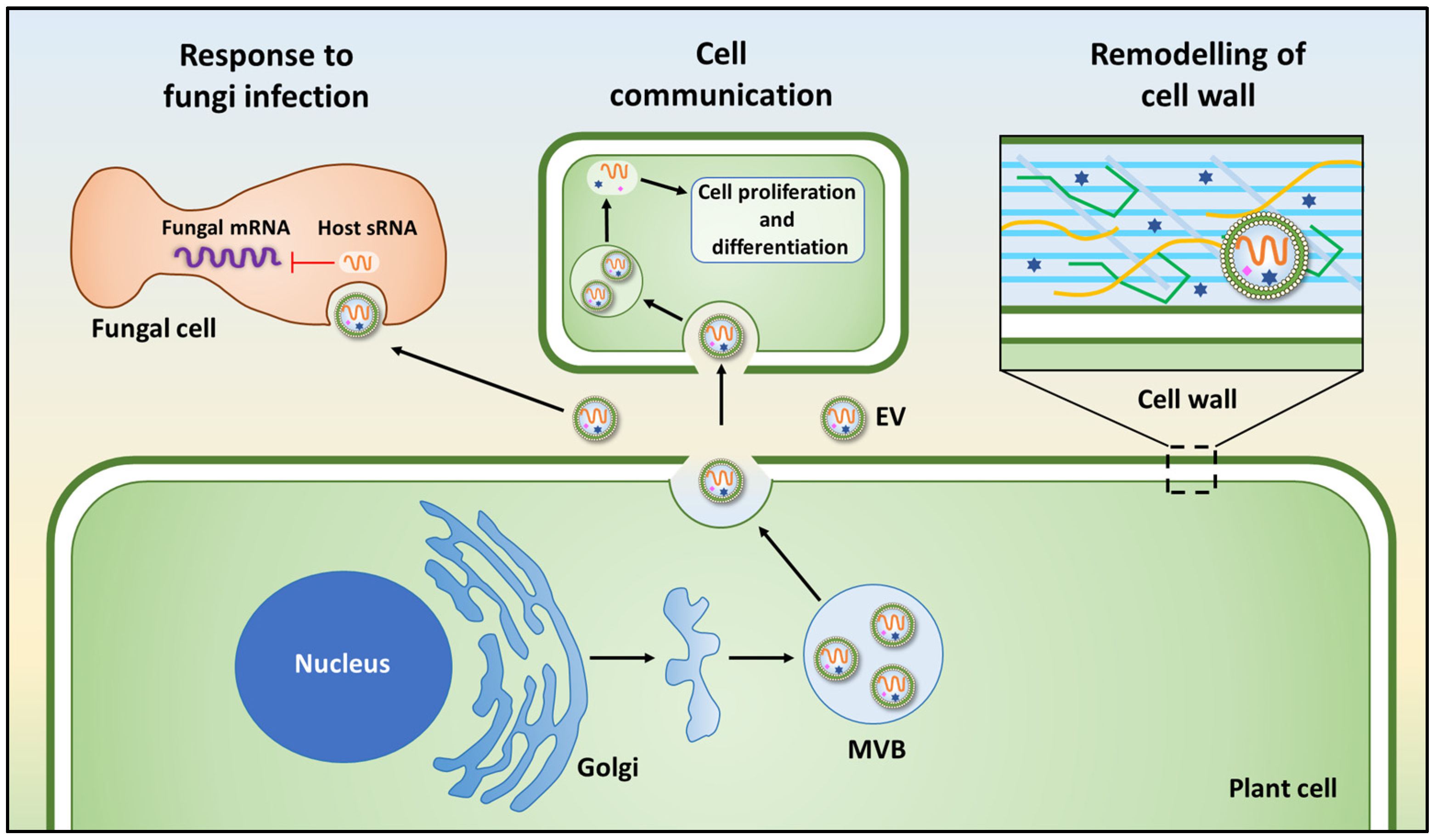
3.2. Plant Extracellular Vesicles in Plant-Microbe Interaction

Plants and animals can be under constant pathogen attack. Some pathogens and pests deliver small RNAs (sRNAs) into host cells to suppress host immunity. Conversely, hosts also transfer sRNAs into pathogens and pests to inhibit their virulence. Emerging findings have revealed that some sRNAs can, indeed, travel between hosts and interact with microbes and fungi to silence target genes. In particular, RNA interference (RNAi) is one of the primary adaptive defense mechanisms that can regulate plant immune responses against several kinds of pathogens. For example, plants such as cotton can export plant-specific miRNAs into their fungal pathogens to induce cross-kingdom gene silencing and confer disease resistance. Cotton plants produce miR-166 and miR-159, which are exported to the hyphae of pathogenic fungus Verticillium dahlia. These miRNAs target a Ca2+—dependent cysteine protease and an isotrichodermin C-15 hydroxylase that are crucial for virulence [91].
Intriguingly, PDEV secretion is increased by pathogen infection, suggesting that EVs play important roles in plant immunity [26]. In addition to mammalians, plant-derived EVs can, indeed, mediate the communication with pathogens through RNAi. In particular, emerging studies have shown that sRNA derived from plants can silence microorganism and fungi target genes [92]. As a consequence, the discovery of EV-mediated trafficking of sRNAs from plant hosts to fungal pathogens has given rise to many exciting questions. For instance, Cai et al. discovered that small interfering RNAs are delivered by Arabidopsis into B. cinerea cells, promoting the silencing of fungal genes [43]. Further studies have revealed that cross-kingdom RNA trafficking, from the host into the pathogen, to induce the silencing of pathogenic genes, depends on EVs [93,94]. Arabidopsis sRNAs transported into B. cinerea cells are also packed into plant-derived EVs, indicating that EV-mediated transport is one of the most relevant pathways for the cross-kingdom trafficking of sRNA. Cai et al. also demonstrated that sRNA-containing vesicles accumulate at the infection sites and are taken up by the fungal cells. Moreover, transferred host sRNAs induce silencing of fungal genes critical for pathogenicity. Consequently, it was reported that plant extracellular vesicles play an essential role in cross-kingdom sRNA trafficking between Arabidopsis and the fungal pathogen B. cinerea, because Arabidopsis secretes EVs to deliver host sRNAs into fungal cells to silence virulence-related genes [43].
In addition, it is known that plant cells can release EVs containing transport RNAs, defense compounds, and signaling lipids, suggesting that plant EVs can also function as important mediators in plant–microbe interactions [26,95]. Kalarikkal et al., using an in silico approach, showed that a high abundance of miRNAs in PDEVs can target SARS-CoV-2 genes [35]. In particular, target prediction analysis was carried out using RNA hybrid software with stringent prediction criteria. Critical parameters were set during target prediction to identify SARS-CoV-2 targeting miRNAs, as reported by the authors. To confirm the specificity of miRNA binding to SARS-CoV-2 genome, the authors aligned the miRNA binding site sequence within the SARS-CoV-2 genome with global isolates of SARS-CoV-2 and SARS-CoV sequence, using MEGA software. They showed that gma-miR-4995 and mdm-miR-1511, which are respectively packed into coconut- and pear-derived EVs, can target SPIKE, the gene involved in encoding the structural protein spike. Moreover, they found that Osa-miR-530-5p, a miRNA carried by PDEVs, can indirectly inhibit the synthesis of ORF1b, by not allowing ribosomal slippage, and, thus, preventing the replication of SARS-CoV-2 [35]. In the same study, the authors validated the relative expression of six miRNAs and their differential enrichment; these include (i) miRNAs that are expressed equally in both ginger and grapefruit PDEVs (gma-miR-166 m and mtr-miR-156a), (ii) miRNAs that show higher enrichment in grapefruit PDEVs (bdi-miR-5059 and osa-miR-5077), and (iii) miRNAs that show higher enrichment in ginger PDEVs (aqc-miR-159 and gma-miR-6300). In addition, using an in silico approach, the authors identified other PDEV-derived miRNAs targeting the SARS-CoV-2 genome; gma-miR-6300 target ORF3a, which codifies for an accessory protein that inhibits autophagy by blocking the fusion of autophagosomes with lysosomes [96]. In addition, aqc-miR-159 (derived from ginger PDEVs) and zma-miR-164b-3p (derived from pear PDEVs) can respectively target SARS-CoV-2′s M and N genes, codifying membrane glycoprotein and nucleocapsid protein. Finally, pvu-miR-482-5p, a miRNA packed into pea’s PDEVs, targets ORF8, another crucial gene involved in the immune evasion of SARS-CoV-2 [97]. However, as is underlined by the authors, these results are based on in silico analysis; therefore, further in vitro and in vivo investigations are necessary to evaluate the anti-viral capacity of the microRNAs carried by PDEVs. Evidence resulting from future studies may be critical for the development of an efficient therapy for COVID-19.
Interestingly, another study demonstrated that PDEV-derived miRNAs inhibit lung inflammation, induced by exosomal SARS-CoV-2 Nsp12; Nsp12 is delivered by lung epithelial cell exosomes to macrophages, leading to the activation of the macrophages via NF-κB. Furthermore, a large number of ginger-EV miRNAs can potentially bind to multiple sites of the SARS-CoV-2 viral genome. This study demonstrated that ginger-derived EVs contain aly-miR396a-5p, which can inhibit NF-κB-mediated inflammation and apoptosis in the lungs of mice. These results pave the way for the possible application of PDEV-based therapy, since the activation of NF-κB-mediated pathways plays an essential role in many inflammatory diseases, including COVID-19 [14].
In the following table (Table 1), we have summarized the studies discussed above, focusing on the PDEV sources and miRNA functional roles.

4. Conclusions

In conclusion, the role that extracellular vesicles of plant origin may play in cross-kingdom communication has attracted the attention of the scientific community, due to their ability to regulate human targets. Above all, miRNAs packaged in PDEVs may be key modulators in the regulation of human genes, representing a possible strategy in human therapies. In addition, their higher bioavailability may encourage the application of PDEVs in some pathological conditions, such as cancer, inflammation, and response to oxidative stress. Despite this, more studies need to be carried out to validate the beneficial effects of PDEVs and, most of all, to understand the mechanism behind human target regulation mediated by EV-miRNAs.

Author Contributions

Conceptualization, S.R.; writing—original draft preparation, O.U., R.G., N.R.G. and S.R.; writing—review and editing S.R and R.A.; supervision S.R. and R.A. All authors have read and agreed to the published version of the manuscript.

Funding

This research received no external funding.

Institutional Review Board Statement

Not applicable.

Informed Consent Statement

Not applicable.

Data Availability Statement

Not applicable.

Acknowledgments

Stefania Raimondo was supported by PON “Ricerca e Innovazione” 2014–2020-Azione 1.2 “Mobilità dei Ricercatori”-AIM “Attraction and International Mobility”. Ornella Urzì is a student in “Biomedicina, Neuroscienze e Diagnostica Avanzata”, XXXV ciclo, University of Palermo. Roberta Gasparro is a student in “Biomedicina, Neuroscienze e Diagnostica Avanzata”, XXXVII ciclo, University of Palermo. Nima Rabienezhad Ganji is a student in “Oncology and Experimental Surgery”, XXXV ciclo, University of Palermo.

Conflicts of Interest

The authors declare no conflict of interest.

References

  1. Thery, C.; Witwer, K.W.; Aikawa, E.; Alcaraz, M.J.; Anderson, J.D.; Andriantsitohaina, R.; Antoniou, A.; Arab, T.; Archer, F.; Atkin-Smith, G.K.; et al. Minimal information for studies of extracellular vesicles 2018 (misev2018): A position statement of the international society for extracellular vesicles and update of the misev2014 guidelines. J. Extracell. Vesicles 2018, 7, 1535750. [Google Scholar] [CrossRef] [PubMed] [Green Version]
  2. Valadi, H.; Ekstrom, K.; Bossios, A.; Sjostrand, M.; Lee, J.J.; Lotvall, J.O. Exosome-mediated transfer of mrnas and micrornas is a novel mechanism of genetic exchange between cells. Nat. Cell Biol. 2007, 9, 654–659. [Google Scholar] [CrossRef] [PubMed] [Green Version]
  3. Raimondo, S.; Urzi, O.; Conigliaro, A.; Bosco, G.L.; Parisi, S.; Carlisi, M.; Siragusa, S.; Raimondi, L.; Luca, A.; Giavaresi, G.; et al. Extracellular vesicle micrornas contribute to the osteogenic inhibition of mesenchymal stem cells in multiple myeloma. Cancers 2020, 12, 449. [Google Scholar] [CrossRef] [PubMed] [Green Version]
  4. Jung, J.H.; Ikeda, G.; Tada, Y.; von Bornstadt, D.; Santoso, M.R.; Wahlquist, C.; Rhee, S.; Jeon, Y.J.; Yu, A.C.; O’Brien, C.G.; et al. Mir-106a-363 cluster in extracellular vesicles promotes endogenous myocardial repair via notch3 pathway in ischemic heart injury. Basic Res. Cardiol. 2021, 116, 19. [Google Scholar] [CrossRef]
  5. Byun, J.S.; Lee, H.Y.; Tian, J.; Moon, J.S.; Choi, J.; Lee, S.H.; Kim, Y.G.; Yi, H.S. Effect of salivary exosomal mir-25-3p on periodontitis with insulin resistance. Front. Immunol. 2021, 12, 775046. [Google Scholar] [CrossRef] [PubMed]
  6. O’Brien, K.; Breyne, K.; Ughetto, S.; Laurent, L.C.; Breakefield, X.O. RNA delivery by extracellular vesicles in mammalian cells and its applications. Nat. Rev. Mol. Cell Biol. 2020, 21, 585–606. [Google Scholar] [CrossRef] [PubMed]
  7. Conigliaro, A.; Costa, V.; Lo Dico, A.; Saieva, L.; Buccheri, S.; Dieli, F.; Manno, M.; Raccosta, S.; Mancone, C.; Tripodi, M.; et al. Cd90+ liver cancer cells modulate endothelial cell phenotype through the release of exosomes containing h19 lncrna. Mol. Cancer 2015, 14, 155. [Google Scholar] [CrossRef]
  8. Li, Z.; Song, Y.; He, T.; Wen, R.; Li, Y.; Chen, T.; Huang, S.; Wang, Y.; Tang, Y.; Shen, F.; et al. M2 microglial small extracellular vesicles reduce glial scar formation via the mir-124/stat3 pathway after ischemic stroke in mice. Theranostics 2021, 11, 1232–1248. [Google Scholar] [CrossRef]
  9. Pinedo, M.; de la Canal, L.; de Marcos Lousa, C. A call for rigor and standardization in plant extracellular vesicle research. J. Extracell. Vesicles 2021, 10, e12048. [Google Scholar] [CrossRef]
  10. Raimondo, S.; Naselli, F.; Fontana, S.; Monteleone, F.; Lo Dico, A.; Saieva, L.; Zito, G.; Flugy, A.; Manno, M.; Di Bella, M.A.; et al. Citrus limon-derived nanovesicles inhibit cancer cell proliferation and suppress cml xenograft growth by inducing trail-mediated cell death. Oncotarget 2015, 6, 19514–19527. [Google Scholar] [CrossRef] [Green Version]
  11. Zhang, M.; Viennois, E.; Xu, C.; Merlin, D. Plant derived edible nanoparticles as a new therapeutic approach against diseases. Tissue Barriers 2016, 4, e1134415. [Google Scholar] [CrossRef] [PubMed]
  12. Alfieri, M.; Leone, A.; Ambrosone, A. Plant-derived nano and microvesicles for human health and therapeutic potential in nanomedicine. Pharmaceutics 2021, 13, 498. [Google Scholar] [CrossRef] [PubMed]
  13. Xu, X.H.; Yuan, T.J.; Dad, H.A.; Shi, M.Y.; Huang, Y.Y.; Jiang, Z.H.; Peng, L.H. Plant exosomes as novel nanoplatforms for microrna transfer stimulate neural differentiation of stem cells in vitro and in vivo. Nano Lett. 2021, 21, 8151–8159. [Google Scholar] [CrossRef] [PubMed]
  14. Teng, Y.; Xu, F.; Zhang, X.; Mu, J.; Sayed, M.; Hu, X.; Lei, C.; Sriwastva, M.; Kumar, A.; Sundaram, K.; et al. Plant-derived exosomal micrornas inhibit lung inflammation induced by exosomes SARS-CoV-2 nsp12. Mol. Ther. J. Am. Soc. Gene Ther. 2021, 29, 2424–2440. [Google Scholar] [CrossRef]
  15. Jensen, W.A. The composition and ultrastructure of the nucellus in cotton. J. Ultrastruct. Res. 1965, 13, 112–128. [Google Scholar] [CrossRef]
  16. Halperin, W.; Jensen, W.A. Ultrastructural changes during growth and embryogenesis in carrot cell cultures. J. Ultrastruct. Res. 1967, 18, 428–443. [Google Scholar] [CrossRef]
  17. Politis, D.J.G.; Goodman, R.N. Localized cell wall appositions: Incompatibility response of tobacco leaf cells to pseudomonas pisi. Phytopathology 1978, 68, 309–316. [Google Scholar] [CrossRef]
  18. An, Q.; Ehlers, K.; Kogel, K.H.; van Bel, A.J.; Huckelhoven, R. Multivesicular compartments proliferate in susceptible and resistant mla12-barley leaves in response to infection by the biotrophic powdery mildew fungus. New Phytol. 2006, 172, 563–576. [Google Scholar] [CrossRef] [Green Version]
  19. Zeyen, R.J.; Bushnel, W.R. Papilla response of barley epidermal cells caused by erysiphe graminis: Rate and method of deposition determined by microcinematography and transmission electron microscopy. Can. J. Bot. 1979, 57, 898–913. [Google Scholar] [CrossRef]
  20. An, Q.; Huckelhoven, R.; Kogel, K.H.; van Bel, A.J. Multivesicular bodies participate in a cell wall-associated defence response in barley leaves attacked by the pathogenic powdery mildew fungus. Cell Microbiol. 2006, 8, 1009–1019. [Google Scholar] [CrossRef]
  21. Regente, M.; Corti-Monzon, G.; Maldonado, A.M.; Pinedo, M.; Jorrin, J.; de la Canal, L. Vesicular fractions of sunflower apoplastic fluids are associated with potential exosome marker proteins. FEBS Lett. 2009, 583, 3363–3366. [Google Scholar] [CrossRef] [PubMed] [Green Version]
  22. Mu, J.; Zhuang, X.; Wang, Q.; Jiang, H.; Deng, Z.B.; Wang, B.; Zhang, L.; Kakar, S.; Jun, Y.; Miller, D.; et al. Interspecies communication between plant and mouse gut host cells through edible plant derived exosome-like nanoparticles. Mol. Nutr. Food Res. 2014, 58, 1561–1573. [Google Scholar] [CrossRef] [PubMed]
  23. Teng, Y.; Ren, Y.; Sayed, M.; Hu, X.; Lei, C.; Kumar, A.; Hutchins, E.; Mu, J.; Deng, Z.; Luo, C.; et al. Plant-derived exosomal micrornas shape the gut microbiota. Cell Host. Microbe 2018, 24, 637–652.e8. [Google Scholar] [CrossRef] [PubMed] [Green Version]
  24. Sundaram, K.; Miller, D.P.; Kumar, A.; Teng, Y.; Sayed, M.; Mu, J.; Lei, C.; Sriwastva, M.K.; Zhang, L.; Yan, J.; et al. Plant-derived exosomal nanoparticles inhibit pathogenicity of porphyromonas gingivalis. iScience 2019, 21, 308–327. [Google Scholar] [CrossRef] [Green Version]
  25. Regente, M.; Pinedo, M.; San Clemente, H.; Balliau, T.; Jamet, E.; de la Canal, L. Plant extracellular vesicles are incorporated by a fungal pathogen and inhibit its growth. J. Exp. Bot. 2017, 68, 5485–5495. [Google Scholar] [CrossRef]
  26. Rutter, B.D.; Innes, R.W. Extracellular vesicles isolated from the leaf apoplast carry stress-response proteins. Plant. Physiol. 2017, 173, 728–741. [Google Scholar] [CrossRef] [Green Version]
  27. Xiao, J.; Feng, S.; Wang, X.; Long, K.; Luo, Y.; Wang, Y.; Ma, J.; Tang, Q.; Jin, L.; Li, X.; et al. Identification of exosome-like nanoparticle-derived micrornas from 11 edible fruits and vegetables. PeerJ 2018, 6, e5186. [Google Scholar] [CrossRef]
  28. He, B.; Cai, Q.; Qiao, L.; Huang, C.Y.; Wang, S.; Miao, W.; Ha, T.; Wang, Y.; Jin, H. Rna-binding proteins contribute to small rna loading in plant extracellular vesicles. Nat. Plants 2021, 7, 342–352. [Google Scholar] [CrossRef]
  29. Yang, M.; Liu, X.; Luo, Q.; Xu, L.; Chen, F. An efficient method to isolate lemon derived extracellular vesicles for gastric cancer therapy. J. Nanobiotechnol. 2020, 18, 100. [Google Scholar] [CrossRef]
  30. Pocsfalvi, G.; Turiak, L.; Ambrosone, A.; Del Gaudio, P.; Puska, G.; Fiume, I.; Silvestre, T.; Vekey, K. Protein biocargo of citrus fruit-derived vesicles reveals heterogeneous transport and extracellular vesicle populations. J. Plant. Physiol. 2018, 229, 111–121. [Google Scholar] [CrossRef]
  31. Pocsfalvi, G.; Turiak, L.; Ambrosone, A.; Del Gaudio, P.; Puska, G.; Fiume, I.; Silvestre, T.; Vekey, K. Physiochemical and protein datasets related to citrus juice sac cells-derived nanovesicles and microvesicles. Data Brief 2019, 22, 251–254. [Google Scholar] [CrossRef] [PubMed]
  32. Raimondo, S.; Nikolic, D.; Conigliaro, A.; Giavaresi, G.; Lo Sasso, B.; Giglio, R.V.; Chianetta, R.; Manno, M.; Raccosta, S.; Corleone, V.; et al. Preliminary results of citraves effects on low density lipoprotein cholesterol and waist circumference in healthy subjects after 12 weeks: A pilot open-label study. Metabolites 2021, 11, 276. [Google Scholar] [CrossRef] [PubMed]
  33. Raimondo, S.; Saieva, L.; Cristaldi, M.; Monteleone, F.; Fontana, S.; Alessandro, R. Label-free quantitative proteomic profiling of colon cancer cells identifies acetyl-coa carboxylase alpha as antitumor target of citrus limon-derived nanovesicles. J. Proteom. 2018, 173, 1–11. [Google Scholar] [CrossRef] [PubMed]
  34. Garaeva, L.; Kamyshinsky, R.; Kil, Y.; Varfolomeeva, E.; Verlov, N.; Komarova, E.; Garmay, Y.; Landa, S.; Burdakov, V.; Myasnikov, A.; et al. Delivery of functional exogenous proteins by plant-derived vesicles to human cells in vitro. Sci. Rep. 2021, 11, 6489. [Google Scholar] [CrossRef] [PubMed]
  35. Kalarikkal, S.P.; Sundaram, G.M. Edible plant-derived exosomal micrornas: Exploiting a cross-kingdom regulatory mechanism for targeting SARS-CoV-2. Toxicol. Appl. Pharmacol. 2021, 414, 115425. [Google Scholar] [CrossRef]
  36. Stanly, C.; Alfieri, M.; Ambrosone, A.; Leone, A.; Fiume, I.; Pocsfalvi, G. Grapefruit-derived micro and nanovesicles show distinct metabolome profiles and anticancer activities in the a375 human melanoma cell line. Cells 2020, 9, 2722. [Google Scholar] [CrossRef]
  37. Niu, W.; Xiao, Q.; Wang, X.; Zhu, J.; Li, J.; Liang, X.; Peng, Y.; Wu, C.; Lu, R.; Pan, Y.; et al. A biomimetic drug delivery system by integrating grapefruit extracellular vesicles and doxorubicin-loaded heparin-based nanoparticles for glioma therapy. Nano Lett. 2021, 21, 1484–1492. [Google Scholar] [CrossRef]
  38. Savci, Y.; Kirbas, O.K.; Bozkurt, B.T.; Abdik, E.A.; Tasli, P.N.; Sahin, F.; Abdik, H. Grapefruit-derived extracellular vesicles as a promising cell-free therapeutic tool for wound healing. Food Funct. 2021, 12, 5144–5156. [Google Scholar] [CrossRef]
  39. De Palma, M.; Ambrosone, A.; Leone, A.; Del Gaudio, P.; Ruocco, M.; Turiak, L.; Bokka, R.; Fiume, I.; Tucci, M.; Pocsfalvi, G. Plant roots release small extracellular vesicles with antifungal activity. Plants 2020, 9, 1777. [Google Scholar] [CrossRef]
  40. Mammadova, R.; Fiume, I.; Bokka, R.; Kralj-Iglic, V.; Bozic, D.; Kisovec, M.; Podobnik, M.; Zavec, A.B.; Hocevar, M.; Gellen, G.; et al. Identification of tomato infecting viruses that co-isolate with nanovesicles using a combined proteomics and electron-microscopic approach. Nanomaterials 2021, 11, 1922. [Google Scholar] [CrossRef]
  41. Wang, Q.; Zhuang, X.; Mu, J.; Deng, Z.B.; Jiang, H.; Zhang, L.; Xiang, X.; Wang, B.; Yan, J.; Miller, D.; et al. Corrigendum: Delivery of therapeutic agents by nanoparticles made of grapefruit-derived lipids. Nat. Commun. 2016, 7, 11347. [Google Scholar] [CrossRef] [PubMed]
  42. Bokka, R.; Ramos, A.P.; Fiume, I.; Manno, M.; Raccosta, S.; Turiak, L.; Sugar, S.; Adamo, G.; Csizmadia, T.; Pocsfalvi, G. Biomanufacturing of tomato-derived nanovesicles. Foods 2020, 9, 1852. [Google Scholar] [CrossRef] [PubMed]
  43. Cai, Q.; Qiao, L.; Wang, M.; He, B.; Lin, F.M.; Palmquist, J.; Huang, S.D.; Jin, H. Plants send small rnas in extracellular vesicles to fungal pathogen to silence virulence genes. Science 2018, 360, 1126–1129. [Google Scholar] [CrossRef] [Green Version]
  44. Liu, N.J.; Bao, J.J.; Wang, L.J.; Chen, X.Y. Arabidopsis leaf extracellular vesicles in wound-induced jasmonate accumulation. Plant. Signal. Behav. 2020, 15, 1833142. [Google Scholar] [CrossRef]
  45. Liu, N.J.; Wang, N.; Bao, J.J.; Zhu, H.X.; Wang, L.J.; Chen, X.Y. Lipidomic analysis reveals the importance of gipcs in arabidopsis leaf extracellular vesicles. Mol. Plant 2020, 13, 1523–1532. [Google Scholar] [CrossRef] [PubMed]
  46. Liu, Y.; Wu, S.; Koo, Y.; Yang, A.; Dai, Y.; Khant, H.; Osman, S.R.; Chowdhury, M.; Wei, H.; Li, Y.; et al. Characterization of and isolation methods for plant leaf nanovesicles and small extracellular vesicles. Nanomed. Nanotechnol. Biol. Med. 2020, 29, 102271. [Google Scholar] [CrossRef] [PubMed]
  47. Baldrich, P.; Rutter, B.D.; Karimi, H.Z.; Podicheti, R.; Meyers, B.C.; Innes, R.W. Plant extracellular vesicles contain diverse small rna species and are enriched in 10- to 17-nucleotide “tiny” RNAs. Plant. Cell 2019, 31, 315–324. [Google Scholar] [CrossRef] [Green Version]
  48. Rutter, B.D.; Rutter, K.L.; Innes, R.W. Isolation and quantification of plant extracellular vesicles. Bio Protoc. 2017, 7, e2533. [Google Scholar] [CrossRef]
  49. Kirbas, O.K.; Bozkurt, B.T.; Asutay, A.B.; Mat, B.; Ozdemir, B.; Ozturkoglu, D.; Olmez, H.; Islek, Z.; Sahin, F.; Tasli, P.N. Optimized isolation of extracellular vesicles from various organic sources using aqueous two-phase system. Sci. Rep. 2019, 9, 19159. [Google Scholar] [CrossRef]
  50. Stanly, C.; Kim, H.; Antonucci, G.; Fiume, I.; Guescini, M.; Kim, K.P.; Ciardiello, M.A.; Giangrieco, I.; Mari, A.; Pocsfalvi, G. Crosstalk between the immune system and plant-derived nanovesicles: A study of allergen transporting. Front. Bioeng. Biotechnol. 2021, 9, 760730. [Google Scholar] [CrossRef]
  51. Stanly, C.; Fiume, I.; Capasso, G.; Pocsfalvi, G. Isolation of exosome-like vesicles from plants by ultracentrifugation on sucrose/deuterium oxide (d2o) density cushions. Methods Mol. Biol. 2016, 1459, 259–269. [Google Scholar] [PubMed]
  52. Stanly, C.; Moubarak, M.; Fiume, I.; Turiak, L.; Pocsfalvi, G. Membrane transporters in citrus clementina fruit juice-derived nanovesicles. Int. J. Mol. Sci. 2019, 2, 6205. [Google Scholar] [CrossRef] [PubMed] [Green Version]
  53. Timo, S.; Lisa, W.; Patrick, B.; Timo, W.B.; Christian, P.e.; Martin, H.; Anna, M.; Dagmar, B.; Martina, C.; Lukas, J.; et al. Host-induced gene silencing involves arabidopsis escrt-iii pathway for the transfer of dsrna-derived sirna. bioRxiv 2021. [Google Scholar] [CrossRef] [Green Version]
  54. Movahed, N.; Cabanillas, D.G.; Wan, J.; Vali, H.; Laliberte, J.F.; Zheng, H. Turnip mosaic virus components are released into the extracellular space by vesicles in infected leaves. Plant. Physiol. 2019, 180, 1375–1388. [Google Scholar] [CrossRef] [PubMed]
  55. Wang, B.; Zhuang, X.; Deng, Z.B.; Jiang, H.; Mu, J.; Wang, Q.; Xiang, X.; Guo, H.; Zhang, L.; Dryden, G.; et al. Targeted drug delivery to intestinal macrophages by bioactive nanovesicles released from grapefruit. Mol. Ther. 2014, 22, 522–534. [Google Scholar] [CrossRef] [PubMed] [Green Version]
  56. Prado, N.; Alche Jde, D.; Casado-Vela, J.; Mas, S.; Villalba, M.; Rodriguez, R.; Batanero, E. Nanovesicles are secreted during pollen germination and pollen tube growth: A possible role in fertilization. Mol. Plant. 2014, 7, 573–577. [Google Scholar] [CrossRef] [Green Version]
  57. Ju, S.; Mu, J.; Dokland, T.; Zhuang, X.; Wang, Q.; Jiang, H.; Xiang, X.; Deng, Z.B.; Wang, B.; Zhang, L.; et al. Grape exosome-like nanoparticles induce intestinal stem cells and protect mice from dss-induced colitis. Mol. Ther. 2013, 21, 1345–1357. [Google Scholar] [CrossRef] [Green Version]
  58. Woith, E.; Guerriero, G.; Hausman, J.F.; Renaut, J.; Leclercq, C.C.; Weise, C.; Legay, S.; Weng, A.; Melzig, M.F. Plant extracellular vesicles and nanovesicles: Focus on secondary metabolites, proteins and lipids with perspectives on their potential and sources. Int. J. Mol. Sci. 2021, 22, 3719. [Google Scholar] [CrossRef]
  59. Deng, Z.; Rong, Y.; Teng, Y.; Mu, J.; Zhuang, X.; Tseng, M.; Samykutty, A.; Zhang, L.; Yan, J.; Miller, D.; et al. Broccoli-derived nanoparticle inhibits mouse colitis by activating dendritic cell amp-activated protein kinase. Mol. Ther. 2017, 25, 1641–1654. [Google Scholar] [CrossRef] [Green Version]
  60. Zhuang, X.; Deng, Z.B.; Mu, J.; Zhang, L.; Yan, J.; Miller, D.; Feng, W.; McClain, C.J.; Zhang, H.G. Ginger-derived nanoparticles protect against alcohol-induced liver damage. J. Extracell Vesicles 2015, 4, 28713. [Google Scholar] [CrossRef]
  61. Perut, F.; Roncuzzi, L.; Avnet, S.; Massa, A.; Zini, N.; Sabbadini, S.; Giampieri, F.; Mezzetti, B.; Baldini, N. Strawberry-derived exosome-like nanoparticles prevent oxidative stress in human mesenchymal stromal cells. Biomolecules 2021, 11, 87. [Google Scholar] [CrossRef] [PubMed]
  62. Zhang, M.; Viennois, E.; Prasad, M.; Zhang, Y.; Wang, L.; Zhang, Z.; Han, M.K.; Xiao, B.; Xu, C.; Srinivasan, S.; et al. Edible ginger-derived nanoparticles: A novel therapeutic approach for the prevention and treatment of inflammatory bowel disease and colitis-associated cancer. Biomaterials 2016, 101, 321–340. [Google Scholar] [CrossRef] [PubMed] [Green Version]
  63. Cao, M.; Yan, H.; Han, X.; Weng, L.; Wei, Q.; Sun, X.; Lu, W.; Ye, J.; Cai, X.; Hu, C.; et al. Ginseng-derived nanoparticles alter macrophage polarization to inhibit melanoma growth. J. Immunother. Cancer 2019, 7, 326. [Google Scholar] [CrossRef] [PubMed]
  64. Sriwastva, M.K.; Deng, Z.B.; Wang, B.; Teng, Y.; Kumar, A.; Sundaram, K.; Mu, J.; Lei, C.; Dryden, G.W.; Xu, F.; et al. Exosome-like nanoparticles from mulberry bark prevent dss-induced colitis via the ahr/cops8 pathway. EMBO Rep. 2022, 23, e53365. [Google Scholar] [CrossRef]
  65. Sundaram, K.; Mu, J.; Kumar, A.; Behera, J.; Lei, C.; Sriwastva, M.K.; Xu, F.; Dryden, G.W.; Zhang, L.; Chen, S.; et al. Garlic exosome-like nanoparticles reverse high-fat diet induced obesity via the gut/brain axis. Theranostics 2022, 12, 1220–1246. [Google Scholar] [CrossRef]
  66. Lee, R.; Ko, H.J.; Kim, K.; Sohn, Y.; Min, S.Y.; Kim, J.A.; Na, D.; Yeon, J.H. Anti-melanogenic effects of extracellular vesicles derived from plant leaves and stems in mouse melanoma cells and human healthy skin. J. Extracell. Vesicles 2020, 9, 1703480. [Google Scholar] [CrossRef] [Green Version]
  67. Kim, K.; Yoo, H.J.; Jung, J.H.; Lee, R.; Hyun, J.K.; Park, J.H.; Na, D.; Yeon, J.H. Cytotoxic effects of plant sap-derived extracellular vesicles on various tumor cell types. J. Funct. Biomater. 2020, 11, 22. [Google Scholar] [CrossRef] [Green Version]
  68. Trentini, M.; Zanotti, F.; Tiengo, E.; Camponogara, F.; Degasperi, M.; Licastro, D.; Lovatti, L.; Zavan, B. An apple a day keeps the doctor away: Potential role of mirna 146 on macrophages treated with exosomes derived from apples. Biomedicines 2022, 10, 415. [Google Scholar] [CrossRef]
  69. Zhang, L.; Hou, D.; Chen, X.; Li, D.; Zhu, L.; Zhang, Y.; Li, J.; Bian, Z.; Liang, X.; Cai, X.; et al. Exogenous plant mir168a specifically targets mammalian ldlrap1: Evidence of cross-kingdom regulation by microrna. Cell Res. 2012, 22, 107–126. [Google Scholar] [CrossRef]
  70. Aquilano, K.; Ceci, V.; Gismondi, A.; De Stefano, S.; Iacovelli, F.; Faraonio, R.; Di Marco, G.; Poerio, N.; Minutolo, A.; Minopoli, G.; et al. Adipocyte metabolism is improved by tnf receptor-targeting small rnas identified from dried nuts. Commun. Biol. 2019, 2, 317. [Google Scholar] [CrossRef]
  71. Zhou, Z.; Li, X.; Liu, J.; Dong, L.; Chen, Q.; Kong, H.; Zhang, Q.; Qi, X.; Hou, D.; Zhang, L.; et al. Honeysuckle-encoded atypical microrna2911 directly targets influenza a viruses. Cell Res. 2015, 25, 39–49. [Google Scholar] [CrossRef] [PubMed]
  72. Jiang, M.; Sang, X.; Hong, Z. Beyond nutrients: Food-derived micrornas provide cross-kingdom regulation. Bioessays 2012, 34, 280–284. [Google Scholar] [CrossRef] [PubMed] [Green Version]
  73. Chin, A.R.; Fong, M.Y.; Somlo, G.; Wu, J.; Swiderski, P.; Wu, X.; Wang, S.E. Cross-kingdom inhibition of breast cancer growth by plant mir159. Cell Res. 2016, 26, 217–228. [Google Scholar] [CrossRef] [PubMed] [Green Version]
  74. Qin, Y.; Zheng, B.; Yang, G.S.; Yang, H.J.; Zhou, J.; Yang, Z.; Zhang, X.H.; Zhao, H.Y.; Shi, J.H.; Wen, J.K. Salvia miltiorrhiza-derived sal-mir-58 induces autophagy and attenuates inflammation in vascular smooth muscle cells. Mol. Ther. Nucleic Acids 2020, 21, 492–511. [Google Scholar] [CrossRef] [PubMed]
  75. Marzano, F.; Caratozzolo, M.F.; Consiglio, A.; Licciulli, F.; Liuni, S.; Sbisa, E.; D’Elia, D.; Tullo, A.; Catalano, D. Plant mirnas reduce cancer cell proliferation by targeting malat1 and neat1: A beneficial cross-kingdom interaction. Front. Genet. 2020, 11, 552490. [Google Scholar] [CrossRef]
  76. Snow, J.W.; Hale, A.E.; Isaacs, S.K.; Baggish, A.L.; Chan, S.Y. Ineffective delivery of diet-derived micrornas to recipient animal organisms. RNA Biol. 2013, 10, 1107–1116. [Google Scholar] [CrossRef] [Green Version]
  77. Witwer, K.W.; McAlexander, M.A.; Queen, S.E.; Adams, R.J. Real-time quantitative pcr and droplet digital pcr for plant mirnas in mammalian blood provide little evidence for general uptake of dietary mirnas: Limited evidence for general uptake of dietary plant xenomirs. RNA Biol. 2013, 10, 1080–1086. [Google Scholar] [CrossRef] [Green Version]
  78. Dickinson, B.; Zhang, Y.; Petrick, J.S.; Heck, G.; Ivashuta, S.; Marshall, W.S. Lack of detectable oral bioavailability of plant micrornas after feeding in mice. Nat. Biotechnol. 2013, 31, 965–967. [Google Scholar] [CrossRef]
  79. Yang, J.; Farmer, L.M.; Agyekum, A.A.; Hirschi, K.D. Detection of dietary plant-based small rnas in animals. Cell Res. 2015, 25, 517–520. [Google Scholar] [CrossRef] [Green Version]
  80. Yang, J.; Farmer, L.M.; Agyekum, A.A.; Elbaz-Younes, I.; Hirschi, K.D. Detection of an abundant plant-based small rna in healthy consumers. PLoS ONE 2015, 10, e0137516. [Google Scholar] [CrossRef] [Green Version]
  81. Hirschi, K.D.; Pruss, G.J.; Vance, V. Dietary delivery: A new avenue for microrna therapeutics? Trends Biotechnol. 2015, 33, 431–432. [Google Scholar] [CrossRef] [PubMed]
  82. Potesta, M.; Roglia, V.; Fanelli, M.; Pietrobono, E.; Gismondi, A.; Vumbaca, S.; Nguedia Tsangueu, R.G.; Canini, A.; Colizzi, V.; Grelli, S.; et al. Effect of microvesicles from moringa oleifera containing mirna on proliferation and apoptosis in tumor cell lines. Cell Death Discov. 2020, 6, 43. [Google Scholar] [CrossRef] [PubMed]
  83. Wang, H.; Luo, Q.; Feng, X.; Zhang, R.; Li, J.; Chen, F. Nlrp3 promotes tumor growth and metastasis in human oral squamous cell carcinoma. BMC Cancer 2018, 18, 500. [Google Scholar] [CrossRef] [PubMed] [Green Version]
  84. Feng, X.; Luo, Q.; Wang, H.; Zhang, H.; Chen, F. Microrna-22 suppresses cell proliferation, migration and invasion in oral squamous cell carcinoma by targeting nlrp3. J. Cell Physiol. 2018, 233, 6705–6713. [Google Scholar] [CrossRef] [PubMed]
  85. Yang, M.; Luo, Q.; Chen, X.; Chen, F. Bitter melon derived extracellular vesicles enhance the therapeutic effects and reduce the drug resistance of 5-fluorouracil on oral squamous cell carcinoma. J. Nanobiotechnol. 2021, 19, 259. [Google Scholar] [CrossRef] [PubMed]
  86. Bruno, S.P.; Paolini, A.; D’Oria, V.; Sarra, A.; Sennato, S.; Bordi, F.; Masotti, A. Extracellular vesicles derived from citrus sinensis modulate inflammatory genes and tight junctions in a human model of intestinal epithelium. Front. Nutr. 2021, 8, 778998. [Google Scholar] [CrossRef] [PubMed]
  87. Kameli, N.; Dragojlovic-Kerkache, A.; Savelkoul, P.; Stassen, F.R. Plant-derived extracellular vesicles: Current findings, challenges, and future applications. Membranes 2021, 11, 411. [Google Scholar] [CrossRef]
  88. Cavalieri, D.; Rizzetto, L.; Tocci, N.; Rivero, D.; Asquini, E.; Si-Ammour, A.; Bonechi, E.; Ballerini, C.; Viola, R. Plant micrornas as novel immunomodulatory agents. Sci. Rep. 2016, 6, 25761. [Google Scholar] [CrossRef] [Green Version]
  89. Link, J.; Thon, C.; Schanze, D.; Steponaitiene, R.; Kupcinskas, J.; Zenker, M.; Canbay, A.; Malfertheiner, P.; Link, A. Food-derived xeno-micrornas: Influence of diet and detectability in gastrointestinal tract-proof-of-principle study. Mol. Nutr. Food Res. 2019, 63, e1800076. [Google Scholar] [CrossRef]
  90. Kim, D.K.; Rhee, W.J. Antioxidative effects of carrot-derived nanovesicles in cardiomyoblast and neuroblastoma cells. Pharmaceutics 2021, 13, 1203. [Google Scholar] [CrossRef]
  91. Alshehri, B. Plant-derived xenomirs and cancer: Cross-kingdom gene regulation. Saudi J. Biol. Sci. 2021, 28, 2408–2422. [Google Scholar] [CrossRef] [PubMed]
  92. Huang, C.Y.; Wang, H.; Hu, P.; Hamby, R.; Jin, H. Small rnas—Big players in plant-microbe interactions. Cell Host Microbe 2019, 26, 173–182. [Google Scholar] [CrossRef] [PubMed] [Green Version]
  93. Hou, Y.; Zhai, Y.; Feng, L.; Karimi, H.Z.; Rutter, B.D.; Zeng, L.; Choi, D.S.; Zhang, B.; Gu, W.; Chen, X.; et al. A phytophthora effector suppresses trans-kingdom rnai to promote disease susceptibility. Cell Host Microbe 2019, 25, 153–165.e5. [Google Scholar] [CrossRef] [PubMed] [Green Version]
  94. Cai, Q.; He, B.; Wang, S.; Fletcher, S.; Niu, D.; Mitter, N.; Birch, P.R.J.; Jin, H. Message in a bubble: Shuttling small rnas and proteins between cells and interacting organisms using extracellular vesicles. Annu. Rev. Plant. Biol. 2021, 72, 497–524. [Google Scholar] [CrossRef] [PubMed]
  95. Liu, G.; Kang, G.; Wang, S.; Huang, Y.; Cai, Q. Extracellular vesicles: Emerging players in plant defense against pathogens. Front. Plant. Sci. 2021, 12, 757925. [Google Scholar] [CrossRef]
  96. Zhang, Y.; Sun, H.; Pei, R.; Mao, B.; Zhao, Z.; Li, H.; Lin, Y.; Lu, K. The SARS-CoV-2 protein orf3a inhibits fusion of autophagosomes with lysosomes. Cell Discov. 2021, 7, 31. [Google Scholar] [CrossRef]
  97. Zhang, Y.; Chen, Y.; Li, Y.; Huang, F.; Luo, B.; Yuan, Y.; Xia, B.; Ma, X.; Yang, T.; Yu, F.; et al. The orf8 protein of sars-cov-2 mediates immune evasion through down-regulating mhc-iota. Proc. Natl. Acad. Sci. USA 2021, 118, e2024202118. [Google Scholar] [CrossRef]
Figure 1. Timeline of plant-derived extracellular vesicles research. The first observation of PDEVs came from electronic transmission microscopy in the 1960s. For several years they were correlated to the response against pathogens, until it their capability to mediate the cross-kingdom communication was demonstrated. MVBs, multivesicular bodies; PDEVs, plant-derived extracellular vesicles.
Membranes 12 00352 g001
Figure 2. The physiological roles of plant-derived extracellular vesicles. The main functions of PDEVs are (i) response to fungi infection; (ii) cell–cell communication; (iii) cell wall remodeling. EV, extracellular vesicle; MVB, multivesicular body; sRNA, short RNA, mRNA, messenger RNA.
Membranes 12 00352 g002
Figure 3. The sources of plant-derived extracellular vesicles. PDEVs can be isolated from various edible plants, such as fruits and vegetables. The starting matrix can be represented by tissues or organs, juice, leaves, seeds, and roots.
Membranes 12 00352 g003
Figure 4. A schematic view for the uptake of plant-derived extracellular vesicles into the human body. PDEVs containing proteins, lipids, miRNA, and metabolites enter the human body after edible plant ingestion. In the gastrointestinal tract, where food is digested, PDEVs are absorbed and enter the bloodstream; thus, reaching the final recipient organs, such as the brain, liver, and kidney. PDEVs release their content in target organs and exert their biological properties. They can cross the BBB and reach the cells of the central nervous system, or they can be found in the urine of plant-eating humans. PDEVs, plant-derived extracellular vesicles; BBB, blood-brain barrier.
Membranes 12 00352 g004
Table 1. Origin and functional role of microRNAs packed into different plant-derived extracellular vesicles.
PDEV OriginmicroRNAFunctional RoleReferences
Bitter melonmiR-156 d, miR-162, miR-166 5p, miR-167, miR-172,miR-390, miR-394, miR-396 3p, miR-399, miR-529, miR-2111 5pPotential role in the regulation of NLRP3 mRNA[84]
SoybeanmiR-5781, miR-4996, miR-5671aRegulatation of interleukin 17A, interleukin 10, interleukin 33[27]
gma-miR-6300Targeting gene ORF3a of SARS-CoV-2[35]
mtr-miR-156aTargeting gene ORF1ab of SARS-CoV-2
Hami melonmiR-164aRegulatation of interleukin 16[27]
ath-miR-164b-5p, zma-miR-398b-5p, cme-miR-530b, cme-miR-399dTargeting gene ORF1ab of SARS-CoV-2[35]
OrangemiR-398bRegulatation of interleukin 1, alpha[27]
GingermiR-1078Regulatation of interleukin 6
miR-7267-3pSuppression of Lactobacillus rhamnosus monooxygenase ycnE mRNA, in the gut microbiome[23]
aly-miR396a-5pInhibition of the expression of inflammatory cytokines induced by Nsp12 of SARS-CoV-2; suppression of the SARS-CoV-2 cytopathic effect by inhibiting the expression of the viral S and Nsp12[14]
rlcv-miR-rL1-28-3pSuppression of the SARS-CoV-2 cytopathic effect by inhibiting the expression of the viral S and Nsp12
gma-miR-6300Targeting gene ORF3a of SARS-CoV-2[35]
aqc-miR-159Targeting gene M of SARS-CoV-2
TomatomiR-4995Regulatation of interleukin 5[27]
gma-miR-6300Targeting gene ORF3a of SARS-CoV-2[35]
gma-miR-4375, zma-miR-398b-5p, bdi-miR-5059, osa-miR-5077Targeting gene ORF1ab of SARS-CoV-2
sly-miR-1919aTargeting gene ORF10 of SARS-CoV-2
FragariamiR-166gDisruption of the morphogenesis of leaves[89]
Moringa oleiferamol-miR160h, mol-mir482b, mol-mir166, mol-mir 159c, mol-mir2118a, mol-mir167f-3p, mol-mir156e, mol-mir395d, mol-mir393a, mol-mir397a, mol-mir858b, mol-mir396aPotential regulation of proapoptotic and antiapoptotic targets[81]
WalnutsmiR-156c, miR-159aRegulation of mammalian TNF-α signaling pathway in adipocytes and regulate inflammation[69]
Coconutgma-miR-4995Targeting gene SPIKE of SARS-CoV-2[35]
mtr-miR-156aTargeting gene ORF1ab of SARS-CoV-2
Pearmdm-miR-1511Targeting gene SPIKE of SARS-CoV-2
zma-miR-164b-3pTargeting gene N of SARS-CoV-2
Peapvu-miR-482-5pTargeting gene ORF8 of SARS-CoV-2
gma-miR-156fTargeting gene ORF1ab of SARS-CoV-2
Blueberryzma-miR-398b-5p
Grapefruitbdi-miR-5059, osa-miR-5077
Kiwifruitosa-miR-530-5p
Grapesvvi-miR-3630, vvi-miR-156a/n, vvi-miR-169r/u
Publisher’s Note: MDPI stays neutral with regard to jurisdictional claims in published maps and institutional affiliations.

