26 pages, 1039 KiB  
Review
Laboratory Diagnosis of Antiphospholipid Syndrome: Insights and Hindrances
by Arne Vandevelde and Katrien M. J. Devreese
J. Clin. Med. 2022, 11(8), 2164; https://doi.org/10.3390/jcm11082164 - 13 Apr 2022
Cited by 24 | Viewed by 8703
Abstract
Diagnosis of antiphospholipid syndrome (APS) requires the presence of a clinical criterion (thrombosis and/or pregnancy morbidity), combined with persistently circulating antiphospholipid antibodies (aPL). Currently, laboratory criteria aPL consist of lupus anticoagulant (LAC), anticardiolipin antibodies (aCL) IgG/IgM, and anti-β2 glycoprotein I antibodies (aβ2GPI) IgG/IgM. [...] Read more.
Diagnosis of antiphospholipid syndrome (APS) requires the presence of a clinical criterion (thrombosis and/or pregnancy morbidity), combined with persistently circulating antiphospholipid antibodies (aPL). Currently, laboratory criteria aPL consist of lupus anticoagulant (LAC), anticardiolipin antibodies (aCL) IgG/IgM, and anti-β2 glycoprotein I antibodies (aβ2GPI) IgG/IgM. Diagnosis and risk stratification of APS are complex and efforts to standardize and optimize laboratory tests have been ongoing since the initial description of the syndrome. LAC detection is based on functional coagulation assays, while aCL and aβ2GPI are measured with immunological solid-phase assays. LAC assays are especially prone to interference by anticoagulation therapy, but strategies to circumvent this interference are promising. Alternative techniques such as thrombin generation for LAC detection and to estimate LAC pathogenicity have been suggested, but are not applicable yet in routine setting. For aCL and aβ2GPI, a lot of different assays and detection techniques such as enzyme-linked immunosorbent and chemiluminescent assays are available. Furthermore, a lack of universal calibrators or standards results in high variability between the different solid-phase assays. Other non-criteria aPL such as anti-domain I β2 glycoprotein I and antiphosphatidylserine/prothrombin antibodies have been suggested for risk stratification purposes in APS, while their added value to diagnostic criteria seems limited. In this review, we will describe laboratory assays for diagnostic and risk evaluation in APS, integrating applicable guidelines and classification criteria. Current insights and hindrances are addressed with respect to both laboratory and clinical implications. Full article
Show Figures

Figure 1

8 pages, 647 KiB  
Article
One Year of COVID-19: Lessons Learned in a Hand Trauma Center
by Marcello Covino, Camillo Fulchignoni, Silvia Pietramala, Marco Barbaliscia, Francesco Franceschi, Giulio Maccauro, Gianfranco Merendi and Lorenzo Rocchi
J. Clin. Med. 2022, 11(8), 2163; https://doi.org/10.3390/jcm11082163 - 13 Apr 2022
Cited by 4 | Viewed by 1292
Abstract
2020 will be remembered worldwide as the year of COVID-19 outbreak. The onset of this pandemic abruptly changed everybody’s life and, in a particular manner, doctors’ lives. Our hand surgery department became rapidly one of the first COVID-19-specialized wards in Italy, impacting considerably [...] Read more.
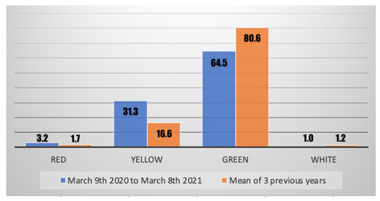
2020 will be remembered worldwide as the year of COVID-19 outbreak. The onset of this pandemic abruptly changed everybody’s life and, in a particular manner, doctors’ lives. Our hand surgery department became rapidly one of the first COVID-19-specialized wards in Italy, impacting considerably the authors’ routines and activities. In this paper, the authors focus on how the demographics of patients with hand trauma changed and how they had to modify their activity. The authors retrospectively took into consideration all patients reaching their emergency department (ED) with hand trauma between 9 March 2020 (the day of the beginning of the first lockdown in Italy) and 8 March 2021 and compared them to those who reached the ED in the three previous years. Authors have analyzed the number of patients, their gender and age, the severity of their trauma, where the trauma occurred, the type of lesion, the percentage of patients who underwent surgery, and the percentage of patients who had an emergency admission. In the last year, the number of patients reaching the ED for a hand trauma has been reduced by two thirds (975 patients during the past year), the mean age of those patients has slightly increased, the severity of cases has increased, places of trauma and type of lesions have changed, and, lastly, the percentage of patients needing surgery who were admitted immediately has increased. This paper shows how the type of patients reaching the ED changed and discusses how surgeons evolved and modified their habits in treating those patients during the first lockdown and the year that followed. Full article
Show Figures

Figure 1

9 pages, 819 KiB  
Article
Predictive Value of Elevated Neutrophil Gelatinase-Associated Lipocalin (NGAL) Levels for Assessment of Cardio–Renal Interactions among ST-Segment Elevation Myocardial Infarction Patients
by David Zahler, Ilan Merdler, Ariel Banai, Eden Shusterman, Omri Feder, Tamar Itach, Leemor Robb, Shmuel Banai and Yacov Shacham
J. Clin. Med. 2022, 11(8), 2162; https://doi.org/10.3390/jcm11082162 - 13 Apr 2022
Cited by 12 | Viewed by 1611
Abstract
Background: Elevated serum neutrophil gelatinase-associated lipocalin (NGAL) levels reflect both inflammatory reactions and renal tubular injury. Recently, associations with endothelial dysfunction and plaque instability were also proposed. We investigated the prognostic utility of elevated NGAL levels for renal and clinical outcomes among ST-segment [...] Read more.
Background: Elevated serum neutrophil gelatinase-associated lipocalin (NGAL) levels reflect both inflammatory reactions and renal tubular injury. Recently, associations with endothelial dysfunction and plaque instability were also proposed. We investigated the prognostic utility of elevated NGAL levels for renal and clinical outcomes among ST-segment elevation myocardial infarction (STEMI) patients treated with primary coronary intervention (PCI). Methods: We performed a prospective, observational, open-label trial. High NGAL was defined as values within the third tertile (>66 percentile). Results: A total of 267 patients were included (mean age 66 ± 14 years, 81% males). Short-term adverse outcomes were consistently increased in the high NGAL group with more acute kidney injury, lower mean left ventricular ejection fraction, higher 30-day mortality, and higher incidence for the composite outcome of major adverse cardiac events (MACE). In a multivariate logistic regression model, high NGAL emerged as a strong and independent predictor for MACE (OR 2.07, 95% CI 1.15–3.73, p = 0.014). Conclusions: Among STEMI patients undergoing primary PCI, elevated NGAL levels are associated with adverse renal and cardiovascular outcomes, independent of traditional inflammatory markers. Further studies are needed to assess the potentially unique role of NGAL in cardio–renal interactions. Full article
Show Figures

Figure 1

9 pages, 783 KiB  
Article
Comparison of the Effects of Propofol and Sevoflurane Anesthesia on Optic Nerve Sheath Diameter in Robot-Assisted Laparoscopic Gynecology Surgery: A Randomized Controlled Trial
by Jung Eun Kim, Seong Yoon Koh and In-Jung Jun
J. Clin. Med. 2022, 11(8), 2161; https://doi.org/10.3390/jcm11082161 - 12 Apr 2022
Cited by 5 | Viewed by 2082
Abstract
Optic nerve sheath diameter (ONSD) is used as a surrogate parameter for intracranial pressure. This study was conducted to evaluate the effect of the anesthetics (sevoflurane and propofol) on ONSD in women undergoing robotic surgery. The 42 patients who were scheduled for robot-assisted [...] Read more.
Optic nerve sheath diameter (ONSD) is used as a surrogate parameter for intracranial pressure. This study was conducted to evaluate the effect of the anesthetics (sevoflurane and propofol) on ONSD in women undergoing robotic surgery. The 42 patients who were scheduled for robot-assisted gynecology surgery were randomly allocated to the sevoflurane group or the propofol group. ONSD was recorded at 10 min after the induction of anesthesia (T0); 5 min, 20 min, and 40 min after carbon dioxide pneumoperitoneum was induced and the patients were put in a steep Trendelenburg position (T1, T2, and T3, respectively); and at skin closure after desufflation of the pneumoperitoneum (T4). Patients were observed for postoperative nausea and vomiting (PONV) during the immediate postoperative period. The propofol group had significantly lower ONSD than the sevoflurane group at T3. Mean ONSD values continuously increased from T0 to T3 in both groups. Two patients in the sevoflurane group experienced PONV. This study suggests that propofol anesthesia caused a lower increase in ONSD than sevoflurane anesthesia. Full article
(This article belongs to the Section Anesthesiology)
Show Figures

Figure 1

10 pages, 3505 KiB  
Article
The Feasibility and Safety of Temporary Transcatheter Balloon Occlusion of Bilateral Internal Iliac Arteries during Cesarean Section in a Hybrid Operating Room for Placenta Previa with a High Risk of Massive Hemorrhage
by Jin-Gon Bae, Young Hwan Kim, Jin Young Kim and Mu Sook Lee
J. Clin. Med. 2022, 11(8), 2160; https://doi.org/10.3390/jcm11082160 - 12 Apr 2022
Cited by 3 | Viewed by 2037
Abstract
This study aimed to evaluate the feasibility and safety of temporary transcatheter balloon occlusion of bilateral internal iliac arteries (TBOIIA) during cesarean section in a hybrid operating room (OR) for placenta previa (PP) with a high risk of massive hemorrhage. This retrospective study [...] Read more.
This study aimed to evaluate the feasibility and safety of temporary transcatheter balloon occlusion of bilateral internal iliac arteries (TBOIIA) during cesarean section in a hybrid operating room (OR) for placenta previa (PP) with a high risk of massive hemorrhage. This retrospective study analyzed the medical records of 62 patients experiencing PP with a high risk of massive hemorrhage (mean age, 36.2 years; age range 28–45 years) who delivered a baby via planned cesarean section with TBOIIA in a hybrid OR between May 2019 and July 2021. Operation time, estimated blood loss (EBL), amount of intra- and postoperative blood transfusion, perioperative hemoglobin level, hospital stay after operation, balloon time, fluoroscopy time, radiation dose, rate of uterine artery embolization (UAE) and hysterectomy, and complication-related TBOIIA were assessed. The mean operation time was 122 min, and EBL was 1290 mL. Nine out of sixty-two patients (14.5%) received a blood transfusion. The mean hemoglobin levels before surgery, immediately after surgery and within 1 week after surgery were 11.3 g/dL, 10.4 g/dL and 9.2 g/dL, respectively. In terms of radiation dose, the mean dose area product (DAP) and cumulative air kerma were 0.017 Gy/cm2 and 0.023 Gy, respectively. Ten out of sixty-two patients (16.1%) underwent UAE postoperatively in the hybrid OR. One out of sixty-two patients had been diagnosed with placenta percreta with bladder invasion based on preoperative ultrasound, and thus underwent cesarean hysterectomy following TBOIIA and UAE. While intra-arterial balloon catheter placement for managing PP with a high risk of hemorrhage remains controversial, a planned cesarean section with TBOIIA in a hybrid OR is effective in eliminating the potential risk of intra-arterial balloon catheter displacement, thus reducing intraoperative blood loss, ensuring safe placental removal and conserving the uterus. Full article
(This article belongs to the Section Obstetrics & Gynecology)
Show Figures

Figure 1

10 pages, 708 KiB  
Article
Is Increased Resting Heart Rate after Radiofrequency Pulmonary Vein Isolation a Predictor of Favorable Long-Term Outcome of the Procedure?
by Cezary Maciejewski, Michał Peller, Piotr Lodziński, Edward Koźluk, Agnieszka Piątkowska, Dariusz Rodkiewicz, Izabela Sierakowska, Natalia Roman, Diana Wiśniewska, Dominika Żółcińska, Dominika Rymaszewska, Grzegorz Opolski, Marcin Grabowski and Paweł Balsam
J. Clin. Med. 2022, 11(8), 2159; https://doi.org/10.3390/jcm11082159 - 12 Apr 2022
Viewed by 3053
Abstract
Background: Increased resting heart rate (RHR) after pulmonary vein isolation (PVI) for treatment of atrial fibrillation (AF) is a common observation, possibly resulting from ganglionated plexus modification during ablation. Previous trials have suggested that an increase in RHR after ablation might be related [...] Read more.
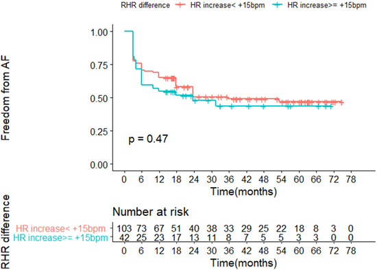
Background: Increased resting heart rate (RHR) after pulmonary vein isolation (PVI) for treatment of atrial fibrillation (AF) is a common observation, possibly resulting from ganglionated plexus modification during ablation. Previous trials have suggested that an increase in RHR after ablation might be related to higher efficacy of the procedure. The aim of this study was to determine whether or not higher increase in RHR after radiofrequency (RF) PVI might predict better long-term outcome of the procedure in a real-life cohort of patients in whom index ablation for paroxysmal AF was performed. Material and methods: The health records of patients who underwent index point-by-point or drag lesion RF PVI for paroxysmal AF in our department between January 2014 and November 2018 were analyzed. Resting heart rate (RHR) was determined from 12-lead ECG recorded prior to the ablation and before discharge to evaluate changes in RHR after PVI. Only patients in sinus rhythm before the procedure and at discharge were included in the analysis. Telephone follow-up was collected for evaluation of arrhythmia recurrence status. Results: A total of 146 patients who underwent PVI for paroxysmal AF were included. Mean follow-up time was 3.5 years. RHR increased from 64 [58.5–70], prior to procedure, to 72 [64.25–80] bpm at discharge (p < 0.001). Higher increase in RHR was not protective from arrhythmia recurrence in long-term observation in both univariable HR = 1.001 (CI 0.99–1.017, p = 0.857) and multivariable analyses HR = 1.001 (CI 0.99–1.02, p = 0.84). Conclusions: RHR after PVI increased in comparison to baseline in our cohort. However, we did not observe higher increase in RHR to be associated with more favorable long-term effectiveness of the procedure. Full article
Show Figures

Figure 1

43 pages, 2761 KiB  
Review
Development of CAR T Cell Therapy in Children—A Comprehensive Overview
by Michael Boettcher, Alexander Joechner, Ziduo Li, Sile Fiona Yang and Patrick Schlegel
J. Clin. Med. 2022, 11(8), 2158; https://doi.org/10.3390/jcm11082158 - 12 Apr 2022
Cited by 14 | Viewed by 7338
Abstract
CAR T cell therapy has revolutionized immunotherapy in the last decade with the successful establishment of chimeric antigen receptor (CAR)-expressing cellular therapies as an alternative treatment in relapsed and refractory CD19-positive leukemias and lymphomas. There are fundamental reasons why CAR T cell therapy [...] Read more.
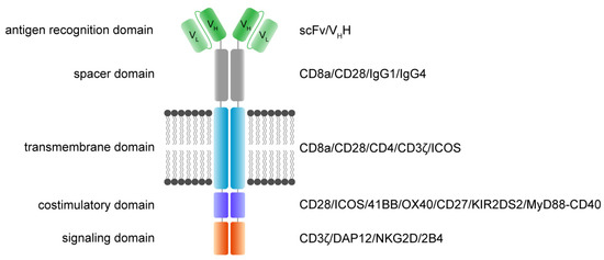
CAR T cell therapy has revolutionized immunotherapy in the last decade with the successful establishment of chimeric antigen receptor (CAR)-expressing cellular therapies as an alternative treatment in relapsed and refractory CD19-positive leukemias and lymphomas. There are fundamental reasons why CAR T cell therapy has been approved by the Food and Drug administration and the European Medicines Agency for pediatric and young adult patients first. Commonly, novel therapies are developed for adult patients and then adapted for pediatric use, due to regulatory and commercial reasons. Both strategic and biological factors have supported the success of CAR T cell therapy in children. Since there is an urgent need for more potent and specific therapies in childhood malignancies, efforts should also include the development of CAR therapeutics and expand applicability by introducing new technologies. Basic aspects, the evolution and the drawbacks of childhood CAR T cell therapy are discussed as along with the latest clinically relevant information. Full article
(This article belongs to the Special Issue Advance in the Treatment of Pediatric Leukemia)
Show Figures

Figure 1

13 pages, 3227 KiB  
Article
Role of a Chest X-ray Severity Score in a Multivariable Predictive Model for Mortality in Patients with COVID-19: A Single-Center, Retrospective Study
by Masoud Baikpour, Alex Carlos, Ryan Morasse, Hannah Gissel, Victor Perez-Gutierrez, Jessica Nino, Jose Amaya-Suarez, Fatimatu Ali, Talya Toledano, Joseph Arampulikan, Menachem Gold, Usha Venugopal, Anjana Pillai, Kennedy Omonuwa and Vidya Menon
J. Clin. Med. 2022, 11(8), 2157; https://doi.org/10.3390/jcm11082157 - 12 Apr 2022
Cited by 4 | Viewed by 2101
Abstract
Predicting the mortality risk of patients with Coronavirus Disease 2019 (COVID-19) can be valuable in allocating limited medical resources in the setting of outbreaks. This study assessed the role of a chest X-ray (CXR) scoring system in a multivariable model in predicting the [...] Read more.
Predicting the mortality risk of patients with Coronavirus Disease 2019 (COVID-19) can be valuable in allocating limited medical resources in the setting of outbreaks. This study assessed the role of a chest X-ray (CXR) scoring system in a multivariable model in predicting the mortality of COVID-19 patients by performing a single-center, retrospective, observational study including consecutive patients admitted with a confirmed diagnosis of COVID-19 and an initial CXR. The CXR severity score was calculated by three radiologists with 12 to 15 years of experience in thoracic imaging, based on the extent of lung involvement and density of lung opacities. Logistic regression analysis was used to identify independent predictive factors for mortality to create a predictive model. A validation dataset was used to calculate its predictive value as the AUROC. A total of 628 patients (58.1% male) were included in this study. Age (p < 0.001), sepsis (p < 0.001), S/F ratio (p < 0.001), need for mechanical ventilation (p < 0.001), and the CXR severity score (p = 0.005) were found to be independent predictive factors for mortality. We used these variables to develop a predictive model with an AUROC of 0.926 (0.891, 0.962), which was significantly higher than that of the WHO COVID severity classification, 0.853 (0.798, 0.909) (one-tailed p-value = 0.028), showing that our model can accurately predict mortality of hospitalized COVID-19 patients. Full article
(This article belongs to the Section Nuclear Medicine & Radiology)
Show Figures

Figure 1

13 pages, 2942 KiB  
Article
Rapid Campimetry—A Novel Screening Method for Glaucoma Diagnosis
by Fabian Müller, Khaldoon O. Al-Nosairy, Francie H. Kramer, Christian Meltendorf, Nidele Djouoma, Hagen Thieme, Michael B. Hoffmann and Friedrich Hoffmann
J. Clin. Med. 2022, 11(8), 2156; https://doi.org/10.3390/jcm11082156 - 12 Apr 2022
Cited by 1 | Viewed by 4034
Abstract
One of the most important functions of the retina—the enabling of perception of fast movements—is largely suppressed in standard automated perimetry (SAP) and kinetic perimetry (Goldmann) due to slow motion and low contrast between test points and environment. Rapid campimetry integrates fast motion [...] Read more.
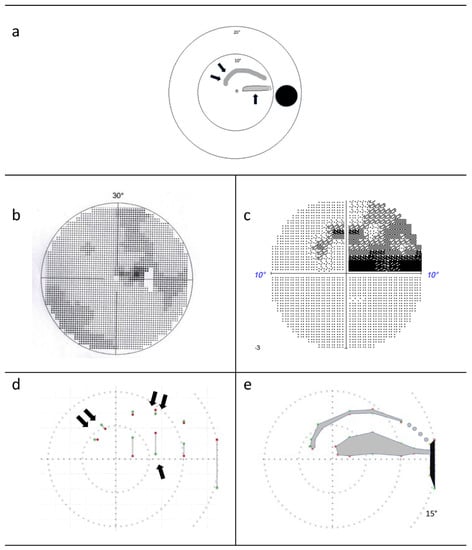
One of the most important functions of the retina—the enabling of perception of fast movements—is largely suppressed in standard automated perimetry (SAP) and kinetic perimetry (Goldmann) due to slow motion and low contrast between test points and environment. Rapid campimetry integrates fast motion (=10°/4.7 s at 40 cm patient–monitor distance) and high contrast into the visual field (VF) examination in order to facilitate the detection of absolute scotomas. A bright test point moves on a dark background through the central 10° VF. Depending on the distance to the fixation point, the test point automatically changes diameter (≈0.16° to ≈0.39°). This method was compared to SAP (10-2 program) for six subjects with glaucoma. Rapid campimetry proved to be comparable and possibly better than 10-2 SAP in identifying macular arcuate scotomas. In four subjects, rapid campimetry detected a narrow arcuate absolute scotoma corresponding to the nerve fiber course, which was not identified as such with SAP. Rapid campimetry promises a fast screening method for the detection of absolute scotomas in the central 10° visual field, with a potential for cloud technologies and telemedical applications. Our proof-of-concept study motivates systematic testing of this novel method in a larger cohort. Full article
(This article belongs to the Special Issue Going for Gaps in Glaucoma)
Show Figures

Figure 1

11 pages, 1000 KiB  
Review
Therapeutic Alternatives in Diabetic Foot Patients without an Option for Revascularization: A Narrative Review
by Gerhard Ruemenapf, Stephan Morbach and Martin Sigl
J. Clin. Med. 2022, 11(8), 2155; https://doi.org/10.3390/jcm11082155 - 12 Apr 2022
Cited by 15 | Viewed by 3336
Abstract
Background: The healing of foot wounds in patients with diabetes mellitus is frequently complicated by critical limb threatening ischemia (neuro-ischemic diabetic foot syndrome, DFS). In this situation, imminent arterial revascularization is imperative in order to avoid amputation. However, in many patients this is [...] Read more.
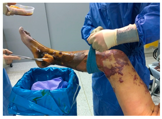
Background: The healing of foot wounds in patients with diabetes mellitus is frequently complicated by critical limb threatening ischemia (neuro-ischemic diabetic foot syndrome, DFS). In this situation, imminent arterial revascularization is imperative in order to avoid amputation. However, in many patients this is no longer possible (“too late”, “too sick”, “no technical option”). Besides conservative treatment or major amputation, many alternative methods supposed to decrease pain, promote wound healing, and avoid amputations are employed. We performed a narrative review in order to stress their efficiency and evidence. Methods: The literature research for the 2014 revision of the German evidenced-based S3-PAD-guidelines was extended to 2020. Results: If revascularization is impossible, there is not enough evidence for gene- and stem-cell therapy, hyperbaric oxygen, sympathectomy, spinal cord stimulation, prostanoids etc. to be able to recommend them. Risk factor management is recommended for all CLTI patients. With appropriate wound care and strict offloading, conservative treatment may be an effective alternative. Timely amputation can accelerate mobilization and improve the quality of life. Conclusions: Alternative treatments said to decrease the amputation rate by improving arterial perfusion and wound healing in case revascularization is impossible and lack both efficiency and evidence. Conservative therapy can yield acceptable results, but early amputation may be a beneficial alternative. Patients unfit for revascularization or major amputation should receive palliative wound care and pain therapy. New treatment strategies for no-option CLTI are urgently needed. Full article
(This article belongs to the Special Issue Peripheral Artery Disease: Epidemiology and Global Perspectives)
Show Figures

Figure 1

10 pages, 5585 KiB  
Article
Estimation of the Surface Dose in Breast Irradiation by the Beam Incident Angle and the 1 cm Depth Dose
by Tsung-Yu Yen, Kai-Cheng Chuang, Hsiao-Mei Fu, Chen-Ju Feng, Ke-Yu Lien and Shih-Ming Hsu
J. Clin. Med. 2022, 11(8), 2154; https://doi.org/10.3390/jcm11082154 - 12 Apr 2022
Cited by 3 | Viewed by 1246
Abstract
To develop a method of estimating surface dose in whole breast irradiation, we used an anthropomorphic phantom with accessories for the simulation of different breast sizes. The surface points, which are measured by TLDs, are set along with two main directions, superior-inferior and [...] Read more.
To develop a method of estimating surface dose in whole breast irradiation, we used an anthropomorphic phantom with accessories for the simulation of different breast sizes. The surface points, which are measured by TLDs, are set along with two main directions, superior-inferior and medial-lateral. The incident angle between the photon beam and the surface and the doses at 1 cm beneath the surface at every point are assessed by a computerized treatment planning system (cTPS). With the prescription dose of 200 cGy, the average surface doses under tangential irradiation are 97.73 (±14.96) cGy, 99.90 (±10.73) cGy, and 105.26 (±9.21) cGy for large, medium, and small breast volumes, respectively. The surface dose increased in the model of small breast volume without significance (p = 0.39). The linear analysis between surface dose and the incident angle is y = 0.5258x + 69.648, R2 = 0.7131 (x: incident angle and y: surface dose). We develop the percentage of skin surface dose with reference to a depth of 1 cm (PSDR1cm) to normalize the inhomogeneous dose. The relationship between incident angle and PSDR1cm is y = 0.1894x + 36.021, R2 = 0.6536 (x: incident angle and y: PSDR1cm) by linear analysis. In conclusion, the surface dose in whole breast irradiation could be estimated from this linear relationship between PSDR1cm and incident angle in daily clinical practice by cTPS. Further in vivo data should be studied to verify this formula. Full article
(This article belongs to the Special Issue Side Effects of Cancer Therapeutics in Clinical Practice)
Show Figures

Figure 1

8 pages, 237 KiB  
Article
Comparison of Cardiovascular Risk and Events among Spanish Patients with and without Ocular Pseudoexfoliation
by Nora Imaz Aristimuño, Iñaki Rodriguez Agirretxe, Ricardo San Vicente Blanco, Rafael Rotaeche Del Campo and Javier Mendicute Del Barrio
J. Clin. Med. 2022, 11(8), 2153; https://doi.org/10.3390/jcm11082153 - 12 Apr 2022
Cited by 1 | Viewed by 1215
Abstract
The purpose of this study was to calculate and compare individual cardiovascular risk (CVR) and the development of cardiovascular events and mortality in patients with and without ocular pseudoexfoliation (PEX). A cohort study was carried out to compare two groups of patients who [...] Read more.
The purpose of this study was to calculate and compare individual cardiovascular risk (CVR) and the development of cardiovascular events and mortality in patients with and without ocular pseudoexfoliation (PEX). A cohort study was carried out to compare two groups of patients who underwent cataract surgery: patients with (n = 99) and without PEX (n = 239). The CVR factors were recorded for all the subjects, and CVR was calculated for each individual using ERICE risk assessment charts. After a six-year follow-up, the cardiovascular events and mortality rates were compared between the two groups. The mean CVR was 36.41% in the PEX group and 33.72% in the non-PEX group (p = 0.13). High blood pressure was detected in significantly more PEX patients (71.4%) than non-PEX patients (58.6%, p = 0.035), yet no differences were found in the other CVR factors. The prevalence of cardiovascular events in the PEX and non-PEX patients was 17.1% and 12.5%, respectively (p = 0.26), with 5% of patients in the PEX group and 5% in the non-PEX group suffering an acute coronary event (p = 0.9). Moreover, 12% of the PEX patients and 7.5% of the non-PEX patients experienced a stroke (p = 0.17), and the six-year mortality rate was 29.3% in the PEX group and 25.9% in the non-PEX group (p = 0.52). PEX was associated with high blood pressure in our cohort of patients, although cardiovascular risk was not shown to be higher in this group. In addition, patients with PEX did not experience more cardiovascular events or have a higher mortality rate than patients without PEX during the period studied. Full article
(This article belongs to the Section Ophthalmology)
8 pages, 1173 KiB  
Article
Dynamic Ultrasound Assessment of the Anterior Tibial Translation for Anterior Cruciate Ligament Tears Diagnostic
by Anca Gabriela Stoianov, Jenel Marian Pătrașcu, Bogdan Gheorghe Hogea, Bogdan Andor, Liviu Coriolan Mișcă, Sorin Florescu, Roxana Ramona Onofrei and Jenel Marian Pătrașcu, Jr.
J. Clin. Med. 2022, 11(8), 2152; https://doi.org/10.3390/jcm11082152 - 12 Apr 2022
Cited by 4 | Viewed by 2435
Abstract
The aim of our study was to investigate the accuracy of dynamic ultrasound assessment of the anterior tibial translation, in diagnosing anterior cruciate ligament tears, and to assess its test–retest reliability. Twenty-three patients (32 ± 8.42 years; 69.56% males) with a history of [...] Read more.
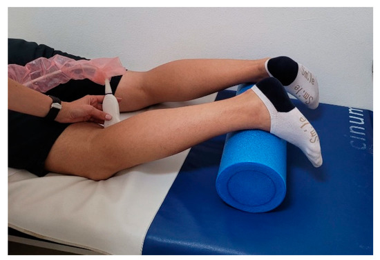
The aim of our study was to investigate the accuracy of dynamic ultrasound assessment of the anterior tibial translation, in diagnosing anterior cruciate ligament tears, and to assess its test–retest reliability. Twenty-three patients (32 ± 8.42 years; 69.56% males) with a history of knee trauma and knee instability participated in the study. Knee ultrasound was performed by an experienced orthopedic surgeon. The anterior tibial translation was measured in both knees and differences between the injured and uninjured knee were calculated. Side-to-side differences > 1 mm were considered a positive diagnosis of an ACL tear. The anterior tibial translation values were 3.34 ± 1.48 mm in injured knees and 0.86 ± 0.78 mm in uninjured knees. Side-to-side differences > 1 mm were found in 22 cases (95.65%). The diagnosis accuracy was 91.30% (95%CI: 71.96–98.92%) and sensitivity 95.45% (95%CI: 77.15–99.88%). The intraclass correlation coefficient showed an excellent test–retest reliability (ICC3,1 = 0.97 for the side-to-side difference in anterior tibial translation). The study highlights the accuracy and reliability of the dynamic ultrasound assessment of the anterior tibial translation in the diagnosis of unilateral anterior cruciate ligament tears. Ultrasound assessment is an accessible imaging tool that can provide valuable information and should be used together with physical examination in suspected cases of ACL injuries. Full article
Show Figures

Figure 1

18 pages, 1718 KiB  
Review
Epigenetic Risks of Medically Assisted Reproduction
by Romualdo Sciorio and Nady El Hajj
J. Clin. Med. 2022, 11(8), 2151; https://doi.org/10.3390/jcm11082151 - 12 Apr 2022
Cited by 17 | Viewed by 5377
Abstract
Since the birth of Louise Joy Brown, the first baby conceived via in vitro fertilization, more than 9 million children have been born worldwide using assisted reproductive technologies (ART). In vivo fertilization takes place in the maternal oviduct, where the unique physiological conditions [...] Read more.
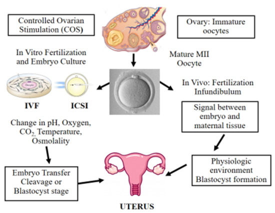
Since the birth of Louise Joy Brown, the first baby conceived via in vitro fertilization, more than 9 million children have been born worldwide using assisted reproductive technologies (ART). In vivo fertilization takes place in the maternal oviduct, where the unique physiological conditions guarantee the healthy development of the embryo. During early embryogenesis, a major wave of epigenetic reprogramming takes place that is crucial for the correct development of the embryo. Epigenetic reprogramming is susceptible to environmental changes and non-physiological conditions such as those applied during in vitro culture, including shift in pH and temperature, oxygen tension, controlled ovarian stimulation, intracytoplasmic sperm injection, as well as preimplantation embryo manipulations for genetic testing. In the last decade, concerns were raised of a possible link between ART and increased incidence of imprinting disorders, as well as epigenetic alterations in the germ cells of infertile parents that are transmitted to the offspring following ART. The aim of this review was to present evidence from the literature regarding epigenetic errors linked to assisted reproduction treatments and their consequences on the conceived children. Furthermore, we provide an overview of disease risk associated with epigenetic or imprinting alterations in children born via ART. Full article
(This article belongs to the Special Issue Hot Topics in Reproductive Medicine Research)
Show Figures

Figure 1

13 pages, 5286 KiB  
Article
Biomechanical Effects of Plastic Heel Cup on Plantar Fasciitis Patients Evaluated by Ultrasound Shear Wave Elastography
by Che-Yu Lin, Pei-Yu Chen, Shin-Han Wu, Yio-Wha Shau and Chung-Li Wang
J. Clin. Med. 2022, 11(8), 2150; https://doi.org/10.3390/jcm11082150 - 12 Apr 2022
Cited by 5 | Viewed by 2523
Abstract
The plastic heel cup has been adopted to treat plantar heel problems for years. However, its mechanisms and biomechanical effects are yet to be fully understood. The purpose of this study was to investigate the effects of the plastic heel cup on the [...] Read more.
The plastic heel cup has been adopted to treat plantar heel problems for years. However, its mechanisms and biomechanical effects are yet to be fully understood. The purpose of this study was to investigate the effects of the plastic heel cup on the microchamber and macrochamber layers of the heel pad by comparing the stiffness (in terms of the shear wave speed) and thickness of these two layers with and without a plastic heel cup during static standing. Fifteen patients with unilateral plantar fasciitis were recruited. The shear wave speed and thickness of the microchamber and microchamber layers of each symptomatic heel pad during standing measured by ultrasound shear wave elastography were compared between conditions with and without a plastic heel cup. It was found that a plastic heel cup reduced the shear wave speed of the microchamber layer to 55.5% and increased its thickness to 137.5% compared with the condition without a plastic heel cup. For the microchamber layer, the shear wave speed was reduced to 89.7%, and thickness was increased to 113.6% compared with the condition without a plastic heel cup. The findings demonstrate that a plastic heel cup can help to reduce the stiffness and increase the thickness for both layers of the heel pad during standing, suggesting that the mechanism of a plastic heel cup, and its resulting biomechanical effect, is to reduce the internal stress of the heel pad by increasing its thickness through confinement. Full article
(This article belongs to the Special Issue Updates in the Orthopedic Management of Foot Disorders)
Show Figures

Figure 1