Article

Applying Surfactin in the Removal of Blooms of Karlodinium veneficum Increases the Toxic Potential

1
College of Food and Pharmaceutical Sciences, Ningbo University, Ningbo 315211, China
2
Li Dak Sum Yip Yio Chin Kenneth Li Marine Biopharmaceutical Research Center, Ningbo University, Ningbo 315211, China
3
Key Laboratory of Applied Marine Biotechnology of Ministry of Education, Ningbo University, Ningbo 315211, China
4
School of Marine Sciences, Ningbo University, Ningbo 315211, China
*
Authors to whom correspondence should be addressed.
J. Mar. Sci. Eng. 2022, 10(2), 196; https://doi.org/10.3390/jmse10020196
Submission received: 8 January 2022 / Revised: 27 January 2022 / Accepted: 29 January 2022 / Published: 1 February 2022
(This article belongs to the Section Marine Biology)

Abstract

:
Biosurfactant has potential application value in the removal of microalgal blooms, but the ecological risks require more research. In this paper, the effects of surfactin on the toxic dinoflagellate Karlodinium veneficum were studied. The coaction of surfactin and K. veneficum was also evaluated through toxicological experiments on Artemia and juvenile clams. The results showed that: (1) in the concentration range of 0–10 mg/L, surfactin significantly killed algal cells in a dose-dependent manner within 48 h; the 24 h EC50 was 3.065 mg/L; (2) K. veneficum had the ability to restore population growth after stress reduction and the restored proliferation was positively correlated with the initial surfactin concentration; (3) the ability to restore population growth was associated with protection afforded by the promotion of antioxidant enzymes, including catalase (CAT), peroxidase (POD) and superoxide dismutase (SOD), whose increase was positively correlated with the surfactin concentration; (4) the toxicity of the coculture of surfactin and K. veneficum was significantly greater than that of the K. veneficum culture or surfactin alone and was dose and time dependent. The potential ecological risks should be considered when applying biosurfactants, such as surfactin, in the removal of harmful algal blooms.

1. Introduction

The small athecate dinoflagellate Karlodinum veneficum is a cosmopolitan toxic harmful algal bloom (HAB) species that is commonly distributed in coastal seawaters, and estuarine or mariculture ponds [1,2,3,4,5]. High-density blooms, causing much loss in fisheries or mariculture, have occurred in Asia, Australia, Europe, and North and South America [6,7,8,9]. In addition to forming dense, single-species blooms, K. veneficum is usually accompanied by other dominant HAB species and develops into dense, single-species blooms after the dominant species fade away [10]. K. veneficum produces a suite of polyketide toxin congeners (karlotoxins) [11,12,13]. A series of toxicity tests on different isolates of K. veneficum have confirmed their hemolytic, ichthyotoxic, and cytotoxic properties and their toxic effect on the development of sea urchin embryos [7,14,15,16]. K. veneficum is a constitutive mixotroph able to perform photosynthesis and to eat various prey by phagocytosis. Karlotoxins participate in predation or in grazer deterrence [3,14,17]. During predation, these toxins are produced and secreted into the water to immobilize the prey [18]. When environmental conditions do not favor the growth of K. veneficum, toxins may be released from senescent cells, and the cellular content of the toxins increases when population growth is halted [3,19]. These characteristics of K. veneficum raise questions regarding the fate of the cells when the dense blooms are terminated and regarding the changes in toxicity that occur when the cells are lysed or stressed.
Currently, the removal of HABs mainly involves physical, chemical and biological methods. Microbial lysis or feeding on blooming algal cells is an important method of biological control. The negative effects that algal growth-inhibiting microorganisms exert may occur through invasion of the interior of algal cells, secretion of certain metabolites that inhibit algal cell growth, or competition for nutrients with HAB species [20,21]. In recent years, the search for fast and efficient active substances for algal killing and growth suppression has become a new research direction for HAB control.
Surfactin is a natural microbial lipopeptide biosurfactant produced by Bacillus subtilis [22,23]. The typical structure is a cyclic lipopeptide composed of β-hydroxy fatty acids and peptides containing seven amino acids combined with lactone bonds [24]. Surfactin exhibits good stability, extremely low toxicity, high surface activity, easy biodegradation, and good biocompatibility. It is recognized as one of the most active biosurfactants identified thus far [25,26]. Surfactin can be used as an environmentally friendly alternative to chemical surfactants. The great application potential of surfactin has been demonstrated in oil exploration [27,28], high-end cosmetics [29,30], medicine [31,32], food processing [33], pesticides [34,35] and other industries. The application of surfactin as a surfactant with excellent performance in inhibiting the growth of and killing the freshwater HAB species Microcystis aeruginosa has been previously reported [36]. Applying other surfactants in the removal of HAB species has also been attempted in some studies [37,38,39,40,41]. Most of the studies have mainly focused on the efficiency of the removal of algal cells and the mechanisms underlying algal responses to surfactants. However, the ecological consequences in aquatic ecosystems during and after bloom termination have scarcely been considered.
For photosynthetic microalgae, chlorophyll fluorescence parameters can be used to characterize the photosynthetic system activity of microalgae [42,43]; changes in the parameters reflect the tolerance of algae under stresses [44,45]. Malondialdehyde (MDA) is the product of lipid peroxidation. Lipid peroxidation is triggered by ROS, which seriously affect the functioning and integrity of biofilms and cause irreversible damage to algal cells [46,47]. The antioxidant enzymes of superoxide dismutase (SOD), catalase (CAT) and peroxidase (POD) are important protective enzyme systems in algal cells and can maintain the dynamic balance of ROS in these cells [48,49]. Variations in MDA content and the activities of the antioxidant enzymes are usually measured when organisms are under various stresses.
In this paper, the alga-killing or algal growth-inhibiting effects of surfactin on K. veneficum cells and population growth were studied. The photosynthetic activity and antioxidant activity were analyzed to determine the algal physio-biochemical responses. To evaluate changes in toxicity in a system in which K. veneficum coexists with surfactin, applied to terminate the algal bloom, toxicological experiments were conducted. K. veneficum is mainly distributed and blooms in coastal mariculture waters where aquaculture organisms were the first to be affected. Two marine organisms, namely, brine shrimp nauplii and juvenile blood clams were examined in the toxicological experiments. The former is a model organism in toxicological studies and the latter is one of the important aquaculture bivalves in China. The purpose of the present study was to: (1) evaluate the alga-killing or algal growth-inhibiting effects of surfactin on K. veneficum and determine the dose dependence; (2) analyze the resistance characteristics of K. veneficum cells and populations exposed to surfactin; and (3) undertake a preliminary evaluation of the potential ecological risks of the elevated toxicity when surfactin is applied in the removal of K. veneficum blooms. This study was intended to provide evidence of the efficiency of applying surfactin in killing or inhibiting the growth of K. veneficum cells and poses a question regarding the necessity of comprehensive evaluation of ecological safety when applying surfactin or other surfactants to remove toxic HABs, such as K. veneficum blooms.

2. Materials and Methods

2.1. Algal Culture and Biosurfactant

The K. veneficum strain (NMBjah047-1, GM2), originally isolated from coastal mariculture waters in the East China Sea (ECS), was provided by the Microalgae Collection Center at Ningbo University, Ningbo, China. The cultures were grown in Erlenmeyer flasks containing sterile natural seawater (pH 8.30, salinity 25) enriched with NMB3 medium [50]. Briefly, the medium was composed of KNO3 (100 mg/L), KH2PO4 (10 mg/L), Fe-citrate·5H2O (3 mg/L), VB1 (6 μg/L) and VB12 (0.05 μg/L). The conditions in the cultivation room were as follows: 20 ± 2 °C, with 60 μmol/(m2·s) (cold white fluorescent lamp) illumination with a 12 h:12 h light-dark cycle. The culture flasks were shaken twice daily in the morning and afternoon to manually homogenize the culture.
Surfactin was purchased from the MedChemExpress LLC (Biological Company, Shanghai, China) (product number HY-129555), and the product purity was >98%. The powdered surfactin reagent was dissolved in dimethyl sulfoxide (DMSO) to make the stocking solution beforehand when applied in the experiment.

2.2. Effect of Surfactin on the Population Growth of K. veneficum

A culture of GM2 in the exponential phase was inoculated into a 250-mL Erlenmeyer flask containing 100 mL of freshly prepared culture medium. The initial cell number was 1.2 × 10⁵ cells/mL. Freshly prepared surfactin solution was added to the cultures. The experimental groups contained surfactin at different working concentrations (0.5, 1.0, 1.5, 2.0, 2.5, 5.0, and 10.0 mg/L), and a culture without added surfactin was set as the control group. Three replicates were set for each group. The flasks were placed under the same conditions as those in the abovementioned cultivation room. One milliliter of the cultures was sampled from each experimental group every day and fixed with Lugol’s iodine solution, and the number of algal cells was counted under an optical microscope using a hemocytometer. Pulse-amplitude-modulated (PAM) chlorophyll fluorescence parameters, namely, Fv/Fm, α, rETRm, and Ik, were determined daily by sampling 1 mL of the culture from each experimental group and making measurements with a Water PAM 101/103 fluorometer (Heinz Walz, Effeltrich, Germany). After 7 days of the experiment, the control and the two high-concentration groups (5.0 mg/L and 10.0 mg/L) were kept in the original culture environment, and the final cell number of the three groups were determined after 2 weeks.
The growth rate (R) was calculated as follows: R = (lnN2 − lnN1)/(t2 − t1), where R represents the growth rate (d−1), N1 represents the algal cell density at time t1, and N2 represents the algal cell density at time t2.

2.3. Acute Effects of Surfactin on K. veneficum

Two hundred milliliters of K. veneficum culture in the exponential phase was inoculated into a 250-mL sterilized Erlenmeyer flask. Freshly prepared surfactin solution was added to the microalgal culture to form surfactin solutions with final experimental concentrations of 2.5, 5.0, and 10.0 mg/L. Microalgal culture without added surfactin, but supplemented with an equal volume of DMSO solution, was used as a control. Three replicates were set for each group. The flasks were placed under the same culture conditions as those mentioned above. One milliliter of culture from each group was sampled at 0, 3, 6, 12, 24, and 48 h for cell counts to estimate the cell density, and 1 mL of each culture sample was used to measure chlorophyll fluorescence parameters. The cell number and chlorophyll fluorescence parameters were measured using the methods mentioned above.
The algacidal ratio (IR) was calculated as IR(%) = (1 − N/N0) × 100%, where N represents the algal density of the experimental group with surfactin, and N0 represents the algal density of the control.

2.4. Antioxidant Activity Variations in K. veneficum under Exposure to Surfactin

Stock cultures of K. veneficum in the exponential phase were divided into 39 200-mL aliquots in sterilized 250-mL Erlenmeyer flasks. Freshly prepared surfactin solution was added to the cultures to form two final surfactin concentration groups: 2.5 mg/L (low-concentration group, L) and 5.0 mg/L (high-concentration group, H). For both the L and H groups, 18 bottles were prepared for samples collected at different timepoints (scheduled at 3, 6, 12, 24, 36, and 48 h after the start of the experiment). Cultures with DMSO added at an equal volume as surfactin in the experimental groups were set as the control and sampled at the beginning of the experiment (0 h). Three replicates were set for each group. After all the treatments were performed, the flasks were placed under the same culture conditions as mentioned above. At each sampling timepoint, 200 mL of each culture was centrifuged at 4500 rpm for 20 min at 4 °C to collect the algal cells. The precipitated algal pellets were washed twice with phosphate buffer solution (pH 7.8). The algal cells were lysed with an ultrasonic cell disruptor. Subsequently, the disrupted-cell suspensions were centrifuged at 12,000 rpm and 4 °C for 10 min, and pellets of the precipitated algal cells were used in the analysis of the antioxidant-related activities. The malondialdehyde (MDA) content and the activities of catalase (CAT), peroxidase (POD) and superoxide dismutase (SOD) were measured using the corresponding kits (Jiancheng Biology Engineering Institute, Nanjing, China) by following the operation manuals.

2.5. Mortality of Brine Shrimp Nauplii and Clam Juveniles Affected by Surfactin and K. veneficum

The synergistic toxic effects of surfactin and K. veneficum on the organisms were the main factors assessed.

2.5.1. Toxicological Experiment on Brine Shrimp Nauplii

One gram of dormant eggs of brine shrimp (Artemia salina) was placed in filtered seawater for hatching at 27 °C for 48 h. Actively swimming nauplii at stages II~III were used in the experiments. K. veneficum cultures in the logarithmic growth phase (3.2 × 10⁵ cell/mL) were applied in the experiments. Aliquots (2 mL) of the algal culture were injected into the wells of a 24-well plate. Freshly prepared surfactin solutions were added to the algal culture in the wells to form final concentration groups of 2.5, 5.0, and 10.0 mg/L surfactin; these cultures were set as synergistic groups with low, medium, and high concentrations of surfactin, respectively. At the same time, the following control groups were set: a K. veneficum algal culture group (no surfactin), low-, medium- and high-concentration surfactin groups (no alga), a starvation group treated with only distilled filtered seawater, and a feeding group fed with a diet containing the microalga Isochrysis galbana. Nine active brine shrimp nauplii were pipetted into each well with a micropipette. The final volume was similar for each well, and this volume was calculated after considering the final surfactin concentration. Three replicates were set for all the experimental groups. The 24-well plates were placed in the cultivation room (24 ± 2 °C) with continuous illumination of 15 μmol/(m2·s). The activity and mortality of the nauplii were observed under an inverted microscope at timepoints of 0, 5, 12, 24, 36, 48, 72 and 96 h. Nauplii that sank completely to the bottom and showed no physical movement were considered dead.
The mortality of the nauplii was calculated as follows:
Pt = (Nt/9) × 100%, where Nt is the number of dead nauplii at time t.

2.5.2. Toxicological Experiment on Clam Juveniles

Bivalve juveniles of the blood clam Tegillarca granosa were applied in the experiment. Clam juveniles were collected from the Center of Breeding and Introduction of Maricultural Seed in Xiangshan County, Ningbo, Zhejiang Province, China. The shell length was 500–1200 µm. Before the experiment, the juveniles were temporarily kept in filtered seawater (salinity of 25 psu) in a polyethylene tank with continuous aeration and fed a diet of I. galbana. The seawater was changed daily. Before the experiment, the juveniles were screened through sieves, and individuals of a similar size (approximately 1000 μm in length) were selected for the experiment. One day before the experiment began, diet feeding was stopped to starve the clams.
Aliquots (200 mL) of K. veneficum algal culture in the logarithmic growth phase (3.8 × 10⁵ cell/mL) were placed into 250-mL sterilized glass beakers. Freshly prepared surfactin solutions were added to the cultures to form final concentration groups of 2.5, 5.0, and 10.0 mg/L; these groups were set as synergistic groups with low, medium and high concentrations of surfactin, respectively. The following control groups were set: a K. veneficum algal solution group, treated with only the algal culture; a surfactin control group, treated with low, medium and high concentrations of surfactin only, without algal cells; a starvation group, treated with only distilled filtered seawater; and a feeding group, fed with a diet containing I. galbana. Twenty of the starved clam juveniles were placed in each experimental group, and 3 replicates were set for each group. The experimental beakers were placed at 26–27 °C with natural light, away from direct sunlight. At 12, 24, 48, 72 and 96 h after the start of the experiment, the juveniles were removed and observed under an inverted microscope. After observation, the clams were returned to the corresponding groups. Juveniles that exhibited shell opening, abnormal foot extension and no responses to repeated stimulation were considered dead.
The mortality of the clam juveniles was calculated as Pt = (Nt/20) × 100%, where Nt is the number of dead seedlings at time t.

2.6. Data Processing and Statistical Analysis

Statistical analysis was conducted with the Statistical Package for the Social Sciences (SPSS). The half maximal effective concentration (EC50) was determined by applying a linear regression between the concentration of surfactin and the mortality of the experimental organisms (or chlorophyll fluorescence parameters). One-sample t-tests were used to determine the significant differences in the number of K. veneficum cells, chlorophyll fluorescence and antioxidant activity of surfactin. One-way ANOVA was used to analyze the significant differences in the data in the toxicological experiment. The significance level was set to p < 0.05. Origin software was used to prepare figures.

3. Results

3.1. Effect of Surfactin on the Population Growth of K. veneficum

The effect of different concentrations of surfactin on the proliferation of K. veneficum cells is shown in Figure 1. Compared with the control group, the effect of surfactin on the population growth of K. veneficum showed significant density dependence on the 1st day after inoculation (Figure 1): when the surfactin concentration was 2 mg/L or higher, the algal cell density was significantly lower than that in the control group (p < 0.05). On the 2nd day, the algal cell number continued to decrease in the groups treated with surfactin concentrations of 2.5–10 mg/L. However, algal cells in the groups in which the surfactin concentration was 2 mg/L or lower resumed proliferation. On the 3rd day, cell proliferation in the 2.5 mg/L surfactin group was restored, and the relative growth rate from day 2 to day 3 was 0.65 d−1. On the 5th day, cell proliferation in the 5 mg/L surfactin group was restored, and the relative growth rate from day 4 to day 5 was 0.94 d−1. During the 7 days of the experimental period, the algal cell density in the 10 mg/L surfactin group remained at the lowest value (0.25 × 10⁴ cell/mL) from the 1st day onward.
Figure 2 shows the changes in the chlorophyll fluorescence parameters Fv/Fm, α, rETRm and Ik of K. veneficum population after surfactin treatment. On the 1st day, compared with that in the control group, the Fv/Fm in the surfactin treatment groups showed a correlation with the concentration of surfactin (Figure 2a): there was no significant difference in the 0.5–2.5 mg/L groups (p > 0.05); however, the 5–10 mg/L groups showed significant decreases (p < 0.05). On the 2nd day, compared with the first day, the surfactin treatment groups (2–10 mg/L) began to recover. On the 3rd day, the value for the 2.5 mg/L group peaked. On the 5th day, the value for the 5 mg/L group peaked. On the 6th day, the values of Fv/Fm for the 1–5 mg/L groups were positively correlated with the concentration: compared with the control; the higher the initial surfactin concentration was, the higher the values of Fv/Fm. However, during the 6 days, compared with the values for the other groups, the value of Fv/Fm for the 10 mg/L group was significantly lower (p < 0.001) (Figure 2a).
On the 1st day, the α values (Figure 2b) in groups in which the surfactin concentration was 5 mg/L or higher were significantly lower than that in the control group (p < 0.05). On the 2nd day, the values in the 1.5–10 mg/L groups were significantly lower than that in the control group (p < 0.05). On the 3rd day, compared with the 2nd day, the surfactin-treated groups began to recover, except the 1 mg/L group. On the 4th day, the α value in the 10 mg/L group peaked. On the 6th day, the α values in all the groups decreased compared with those on the 5th day, and that in the 10 mg/L group reached its lowest value (Figure 2b).
On the 1st day (Figure 2c,d), the rETRm and Ik values when the concentration of surfactin was 2.5 mg/L or lower (for rETRm) and 5 mg/L or lower (for Ik), the values of rETRm and Ik were higher than those in the control group. The two values were lowest in the 10 mg/L group (Figure 2c,d). On the 2nd day, the values in the 0.5–2.5 mg/L groups peaked, and the values in most of the groups, including the control group, were higher than those on the 1st day. On the 3rd day, compared with the 2nd day, the values in the 0.5–2.5 mg/L group decreased significantly (p < 0.05), while those in the 5 mg/L group peaked (Figure 2c,d). During the 6 days, the changes in both rETRm and Ik among the groups showed an approximate “U”-shaped correlation with the surfactin concentrations: during the early days, lower concentrations of surfactin increased the values, while higher concentrations of surfactin decreased the values. During the later days, when cell proliferation was being restored, the “U”-shaped trend was reversed; the lower-concentration surfactin groups had lower values, while the higher-concentration groups had higher values (Figure 2c,d). For K. veneficum, the initial surfactin concentration of 5 mg/L seemed to be the most effective concentration, causing significant changes in the values of rETRm and Ik, both of which were significantly higher than those in the control group during the experimental period.
The effects on population growth were observed for the 0, 5 and 10 mg/L groups (Figure 3): after 2 weeks of maintenance, the proliferation of K. veneficum cells in the two high-concentration groups was restored. On the 21st day, the cell densities in the 5 and 10 mg/L high-concentration groups were 4.03 × 10⁵ and 3.68 × 10⁵ cell/mL, respectively, while that in the control group was 5.05 × 10⁵ cell/mL. From day 7 to day 21, the relative cell proliferation rates of the 5 and 10 mg/L groups were 0.05 d−1 and 0.54 d−1, respectively, and both were significantly higher than that of the control group (0.01 d−1).

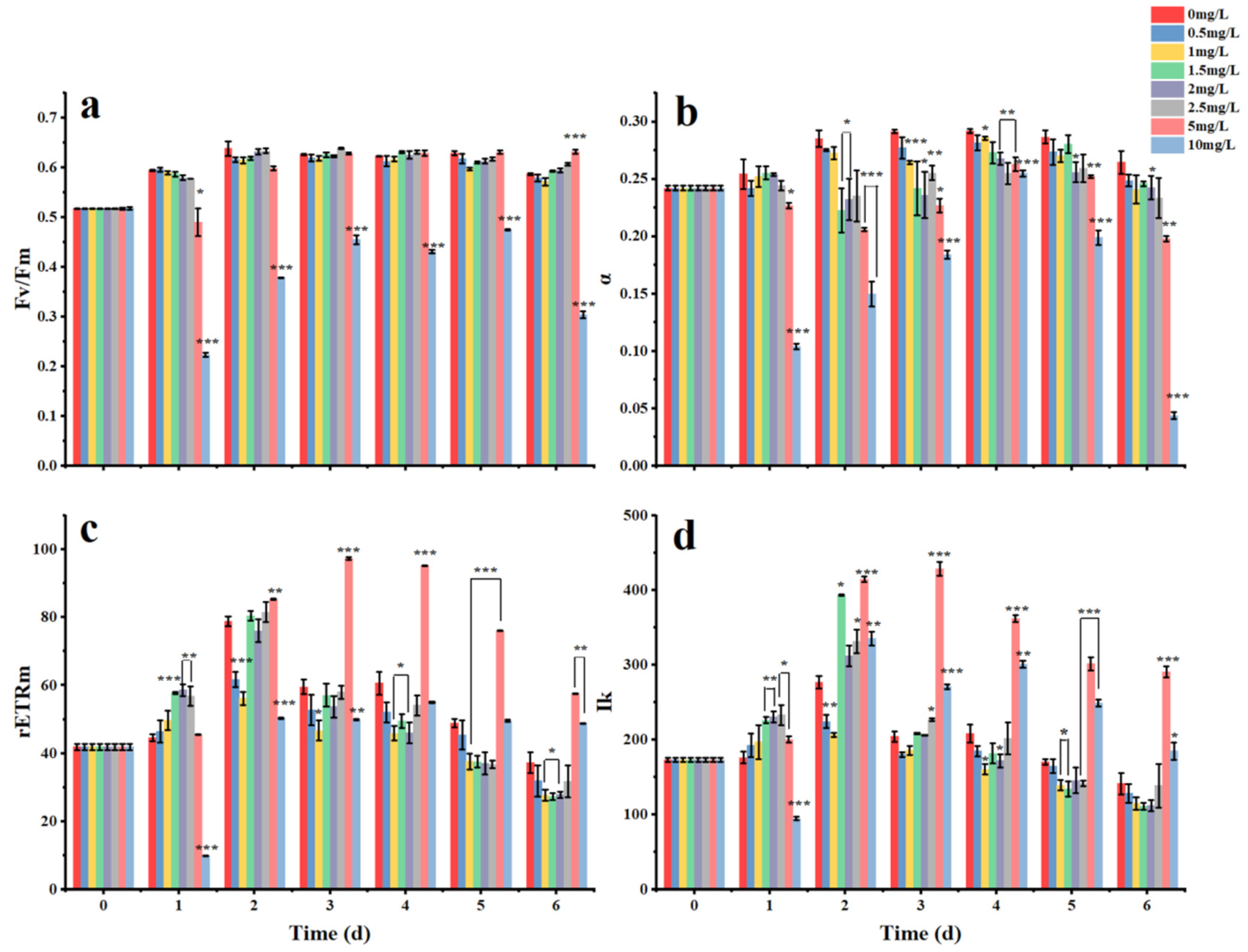
3.2. Acute Effects of Surfactin on K. veneficum

The effects of surfactin on K. veneficum were found to be time and concentration dependent during the 48 h short-term detection period (Figure 4). At 3 h, compared with the control, the inhibitory effects of surfactin on K. veneficum showed a significant density correlation (p < 0.05) (Figure 4a). At 3 h, the inhibition rates of the different surfactin concentrations (2.5, 5, and 10 mg/L) against K. veneficum were 17.8, 28.8 and 66.1%, respectively. At 6–12 h, the 5 mg/L group showed no significant difference in the inhibition rate (p > 0.05). For the 5 mg/L group, the inhibition rate at 24 h was significantly higher than that at 6 and 12 h (p < 0.05) (Figure 4b). The EC50 of surfactin against K. veneficum was 3.065 mg/L at 24 h. The cell density in the 10 mg/L group decreased to as low as 0.25 × 10⁴ cell/mL in 24 h and was maintained at a similar level until 48 h (Figure 4a). At 48 h, the inhibition rates of the different surfactin concentrations (2.5, 5, and 10 mg/L) were found to be significantly concentration dependent (p < 0.05) (Figure 4a). At 48 h, the inhibition rates of the different concentrations (2.5, 5, and 10 mg/L) increased to 58.47, 83.05 and 98.31%, respectively, showing a significant concentration dependency (p < 0.05). The EC50 was 2.884 mg/L at 48 h.
The effects of different concentrations of surfactin on the chlorophyll fluorescence parameters (Fv/Fm, α, rETRm, Ik) of K. veneficum in the short-term observation period are shown in Figure 5. The Fv/Fm value (Figure 5a) for the surfactin groups (2.5, 5, and 10 mg/L) decreased significantly and showed a positive decreasing correlation with the concentration at 3 h. The EC50 of surfactin for the Fv/Fm was 24.708 mg/L at 3 h. At 12 h, the Fv/Fm in the surfactin groups (2.5, 5, and 10 mg/L) was significantly lower than that in the control group (p < 0.05), while that in the 2.5 mg/L group reached the lowest value. The EC50 of surfactin for the Fv/Fm was 25.366 mg/L at 12 h. At 24 h, the Fv/Fm in the 2.5 mg/L group returned to the level at 6 h, while that in the 10 mg/L group reached the lowest value. At 48 h, the Fv/Fm in the 2.5 and 10 mg/L groups maintained their respective levels at 24 h, while that in the 5 mg/L group decreased significantly compared with that at 24 h (p < 0.05) (Figure 5a).
The α value (Figure 5b) in the surfactin groups at different concentrations (2.5, 5, and 10 mg/L) decreased significantly at 3 h (p < 0.05). At 12 h, the values in the surfactin groups were significantly lower than that in the control and showed a concentration dependence (p < 0.05). The EC50 of surfactin for the α value was 17.259 mg/L at 12 h. The value in the 10 mg/L group decreased to 0.127 at 3 h and was maintained at this level until 48 h (Figure 5b).
The rETRm and Ik values (Figure 5c,d) in the surfactin groups (2.5, 5, and 10 mg/L) were significantly lower than that in the control group at 3 h, and the effect was concentration dependent. The EC50 values of surfactin for the rETRm and Ik values at 3 h were 14.529 and 13.819 mg/L, respectively. At 6 h, the values in the 2.5 mg/L group were the lowest, while those in the 5 and 10 mg/L groups decreased significantly compared with those at 3 h (p < 0.05). At 24 h, the two values in the 2.5 mg/L group were restored to the level at 3 h. In the 5 and 10 mg/L groups, the rETRm value was lowest at 24 h and showed an increasing trend at 48 h. The parameter Ik in the 5 and 10 mg/L groups decreased significantly to low levels at 3 h and was maintained at these levels until 48 h without significant changes (Figure 5c,d).

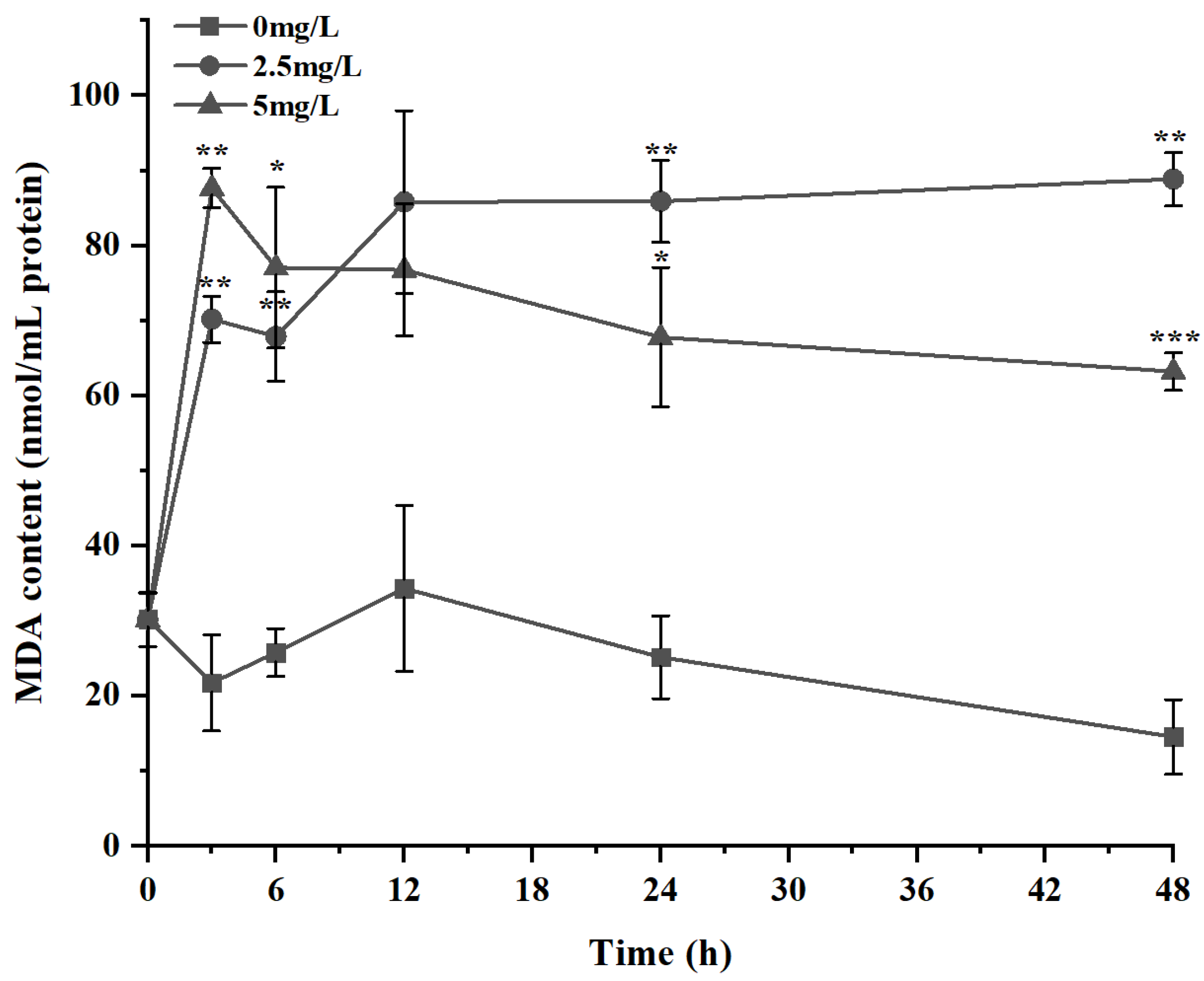
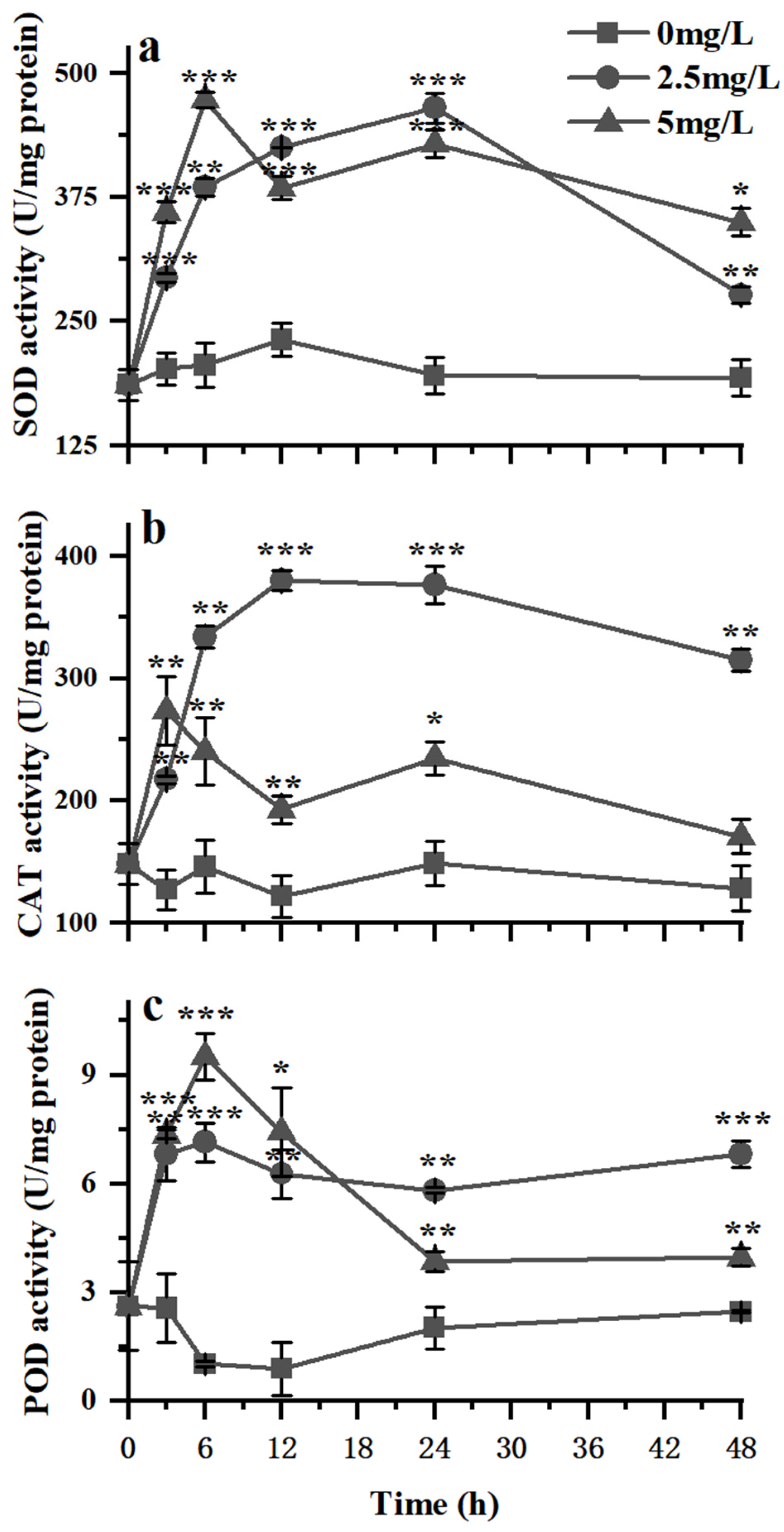
3.3. Variations in the Antioxidant Activity of K. veneficum under Exposure to Surfactin

The changes in the MDA content in K. veneficum cells under the influence of different concentrations of surfactin are shown in Figure 6. At 3 h, the MDA content of the surfactin group increased rapidly and reached 70.18 and 87.66 nmol/mg protein in the low-concentration (L) and high-concentration (H) groups, respectively. The MDA content in the H group was significantly higher than that in the L group (p < 0.05). The MDA content in the H group showed a downward trend after peaking at 3 h, while that in the L group continued to increase after 3 h. At 12 h, the MDA content in the L group reached 85.82 nmol/mg protein, while that in the H group decreased to 76.75 nmol/mg protein. At 24 h, the MDA content in the L group was 85.91 nmol/mg protein, which was significantly higher than that in the H group (67.81 nmol/mg protein) (p < 0.05). At 48 h, the MDA levels in the control and the L and H groups were 14.58, 88.87 and 63.22 nmol/mg protein, respectively.
Surfactin treatment significantly affected the activities of antioxidant enzymes (SOD, CAT, POD) (Figure 7). The SOD activity (Figure 7a) in the surfactin treatment groups was significantly higher than that in the control group within 48 h (p < 0.05). At 0–6 h, the SOD activity in the L and H groups increased linearly, and the SOD activity in the H group was significantly higher than that in the L group (p < 0.05). At 6 h, the SOD activity in the H group peaked; however, it decreased significantly at 12 h. The SOD activity continued to increase in the L group until 24 h. At 48 h, the SOD activity in the L group decreased significantly, while that in the H group was significantly higher than that in the L group (p < 0.05) (Figure 7a).
The CAT activity (Figure 7b) at 3 h, increased rapidly in both the L and H groups, but that in the H group was significantly higher than that in the L group (p < 0.05). At 6 h, the CAT activity in the L group continued to increase, while that in the H group showed a downward trend and was significantly lower than that in the L group (p < 0.05) (Figure 7b). The CAT activity in the L group peaked at 12 h and was maintained at this level until 24 h. The CAT activity in the H group was significantly lower than that in the L group from 6 h to 48 h (p < 0.05). Finally, at 48 h, the CAT activity in the L group was the highest among the 3 groups, while that in the H group was higher than that in the control group, but the difference was not statistically significant (p > 0.05) (Figure 7b).
The POD activity (Figure 7c) increased rapidly to statistically similarly high levels in both the L and H groups at 3 h (p > 0.05). From 6–48 h, the POD activity in L group was maintained at a relatively constant level as that at 3 h. However, the POD activity in the H group increased to a level significantly higher than that in the L group at 6 h. However, it decreased sharply to a level lower than that in the L group at 12 h. In 24 and 48 h, the POD activity was highest in the L group, followed by the H and control groups (p < 0.05) (Figure 7c).

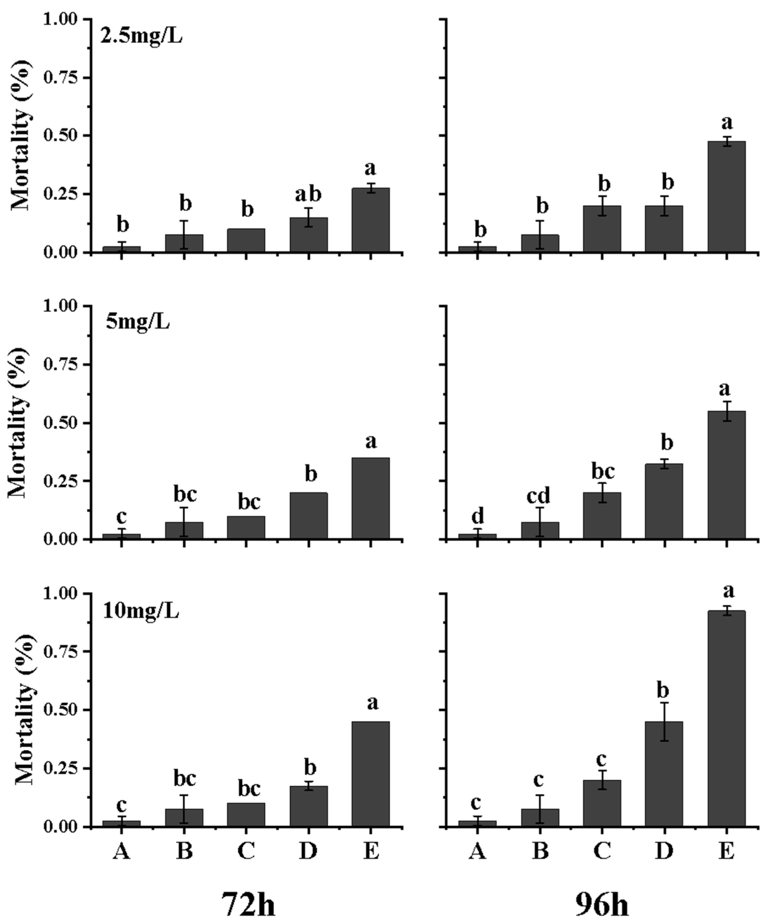
3.4. Synergistic Toxic Effect of Surfactin and K. veneficum on Brine Shrimp Nauplii

Compared with that in the control groups, the mortality of the nauplii in the groups treated with surfactin and K. veneficum increased significantly in a concentration- and time-dependent manner (Figure 8), indicating that synergistic toxic effects occurred when surfactin was applied to K. veneficum cells.
No nauplii died in the feeding group during the whole experimental period. At 5 and 12 h, no nauplii died in either of the experimental groups. At 24, 48 and 72 h, the mortality rates of nauplii in the starvation group were 0, 3.7% and 11.11%, respectively, and those in the K. veneficum group were 3.7%, 18.52% and 33.33%, respectively. Among the surfactin groups, the mortality of nauplii showed no significant difference among the low-, middle- and high-concentration groups, and the mortality of nauplii at 24 and 48 h was 3.7–11.11% (p > 0.05). At 72 h, the mortality rates of nauplii in the low-, medium- and high-concentration surfactin groups were 14.81, 29.63 and 25.93%, respectively, without a statistically significant difference (p > 0.05). At 24 h, the mortality rates of nauplii in the low-, medium- and high-concentration combined treatment groups were 14.81, 18.52 and 27.78%, respectively, which were 4, 5 and 7.5 times higher than that in the K. veneficum group. The 24 h EC50 of surfactin in the combined treatment group was 14.283 mg/L. At 48 h, the mortality rates of nauplii in the low-, medium- and high-concentration combined treatment groups were 33.33, 51.85 and 70.37%, respectively, which were 1.8, 2.8 and 3.8 times higher than that in the K. veneficum group. The 48 h EC50 of surfactin in the combined treatment group was 6.14 mg/L. At 72 h, the mortality rates of nauplii in the low-, medium- and high-concentration combined treatment groups were 66.67, 81.48 and 88.89%, respectively, which were 1.78, 2.22 and 2.56 times higher than that in the K. veneficum group. The 72 h EC50 of surfactin in the synergistic group was 3.664 mg/L. The mortality of nauplii differed significantly among the low-, medium- and high-concentration combined treatment groups (p < 0.05).

3.5. Synergistic Toxic Effect of Surfactin and K. veneficum on Clam Juveniles

The surfactin and K. veneficum combined treatment group showed a time- and concentration-dependent synergistic effect on blood clam juveniles (Figure 9).
From 0 to 48 h, there was no death in any of the experimental groups. From 72 h onward, different degrees of death were observed in the experimental groups. At 72 h, the mortality rates in the low-, medium- and high-concentrations combined treatment groups reached 27.5, 35 and 45%, respectively, but without significant differences (p > 0.05). The mortality rates in the combined treatment groups were significantly higher than that in the K. veneficum group, while those in the medium- and high-concentration groups were significantly higher than those in the respective surfactin groups (p < 0.05). The 72 h EC50 of surfactin in the combined treatment group was 9.633 mg/L. At 96 h, the mortality rates in the low-, medium- and high-concentration combined treatment groups were 47.5, 55 and 92.5%, respectively, which were 2.38, 2.75 and 4.63 times that in the K. veneficum group, respectively, and 2.38, 1.69 and 2.01 times that in the respective surfactin groups. The differences were statistically significant (p < 0.05). Among the combined treatment groups, there was a significant difference in mortality between the low- and high-concentration groups (p < 0.05). The 96 h EC50 of surfactin in the synergistic group was 4.342 mg/L.

4. Discussion

4.1. Population Growth

Surfactin had obvious concentration- and time-dependent effects in the killing or inhibition of the alga K. veneficum. While low concentrations (as low as 0.5–1.5 mg/L surfactin) had no obvious negative effects, the inhibitory effects of higher concentrations of surfactin were obvious. After being acutely inhibited by the initial application of surfactin, the population growth was able to recover afterwards. The higher the initial surfactin concentration was, the longer the time taken to recover. However, during the population recovery period, while more cells were killed by the surfactin of higher concentration (cell density was low in these groups), the surviving cells had the stronger potential to recover, showing increased photosynthetic potential and higher growth rates. K. veneficum is a typical mixotrophic species that can use a variety of trophic strategies [8,51]. It was found to swallow up its own cell debris [17]. While surfactin executes its function as a biosurfactant that causes the lysis of algal cells, the K. veneficum cells that recover from the initial inhibition might be able to take advantage of the debris to grow. In addition, those cells that survive the stronger inhibition might take advantage of the more abundant inorganic nutrients left behind after mass cell death. The superiority of the mixotrophy strategy of K. veneficum enables this universal dinoflagellate to survive and bloom constantly.
Rhamnolipids and sophorolipids are also classes of biosurfactants, and previous studies have shown that they have inhibitory effects on some harmful algae [37,38,39,40]. The previous studies showed that the inhibitory effect was related to the level of polyunsaturated fatty acids in algae. Yang et al. [41] found that, the inhibitory effects of surfactin on the blue-green algae Microcystis aeruginosa were mainly exerted on the microbial cell membrane, inhibiting its synthesis and causing apoptosis. They speculated that surfactin could synergistically act on the cell membrane of M. aeruginosa by forming holes on the cell surface, causing damage and rupture of algal cells, resulting in the loss of intracellular substances and apoptosis. These results provide a direction for further exploration of the mechanism underlying the inhibitory effect of surfactin on K. veneficum.

4.2. Chlorophyll Fluorescence Parameters

While surfactin inhibited the growth of K. veneficum or killed the cells, the photosynthetic activity of the cells was inhibited. Under surfactin stress, the Fv/Fm of algal cells decreased acutely and the decrease was positively correlated with the surfactin concentration. While the inhibition effect receded, K. veneficum cell proliferation resumed. Pokrzywinski et al. [52] found that bioactive substance (IRI–160AA), isolated from a strain of the ubiquitous bacterium Shewanella spp., had stage specific activity on K. veneficum. The highest inhibitory activity was that on K. veneficum in the logarithmic growth phase. Tilney et al. [53] further observed that IRI-160AA at a concentration of 4% (v/v) could inhibit the photosynthetic system of K. veneficum at 18 h, resulting in a decrease in Fv/Fm. The Fv/Fm returned to the control level and was stable before 42 h. The ability to restore the proliferation of K. veneficum after the inhibition effect receded was the same as the observations in our study. Liu et al. [54] found that the anionic surfactant sodium dodecyl benzene sulfonate (LAS) had a stress effect on the growth of M. aeruginosa through inhibiting the light energy capture efficiency, photochemical energy conversion efficiency and electron transfer efficiency of the algal cells. Recovery of the chlorophyll fluorescence parameters, such as qP, фPSII and rETR, etc., was observed after 2 days. In our studies for K. veneficum, not only did the recovery happen, but the phenomenon of higher growth potential in high concentration groups was also obvious during the recovery period. This phenomenon indicated the higher growth potential of the surviving cells in the higher concentration groups than that of the senescent cells in the control or of those whose growth was less strongly inhibited by lower concentrations of surfactin.

4.3. Variations in the Antioxidant Activity

The MDA content increased when K. veneficum cells were stressed by surfactin, showing that the inner membrane lipids of algal cells were oxidized. Sharply increasing cellular MDA content was also shown in Phaeocystis globosa [46] and Chlorella vulgaris and M. aeruginosa [47] when exposed to alga-lysing compounds, and the increases were concentration-dependent. All the studies showed that, with extension of exposure time after the early sharp increases, the cellular MDA content in the high-concentration group was lower than that in the low-concentration group but was significantly higher than that in the control. This may have been due to the continuous increase in ROS levels under low-concentration treatment and the constant sensitivity of algal cells, which improves the resistance of algal cells to the alga-lysing compounds to a certain extent. The MDA content in the high-concentration treatment group was lower than that in the low-concentration treatment group, which may be related to the high degree of cell membrane damage.
Changes in the activities of the antioxidant enzymes CAT, POD and SOD showed similar characteristics. The activities increased significantly in a short time when exposed to surfactin and, with the extension of time, the activities in the treatment groups remained at high levels compared to those in the control but were negatively correlated with the initial surfactin concentration. We speculate that the inhibitory effect of a low concentration of surfactin on K. veneficum was relatively weak, so the integrity of algal cells was relatively well maintained at this concentration. However, over time, the irreversible damage caused to the cells increased, resulting in the collapse of the cell antioxidant system and, therefore, in a downward trend. A similar phenomenon was observed in the stressed seaweed Hydrilla verticillate when exposed to the organic reagents toluene, ethylbenzene, xylene and fluoride [48,49]. The results from these studies likely demonstrated the common mechanisms of antioxidant enzymes in the cellular response to stress.

4.4. Synergistic Toxic Effect of Surfactin and K. veneficum

The cells and cellular extracts of K. veneficum showed different degrees of hemolysis, fish toxicity, cytotoxicity and embryotoxicity [16]. In the present study, we found that K. veneficum cells had lethal effects on brine shrimp nauplii and bivalve juveniles. The toxicities were similar to those of many toxic and harmful algae on brine shrimp nauplii and blood clam juveniles [55,56,57]. Our study demonstrated that the lethal effects of combined treatment with K. veneficum and surfactin on the nauplii and juveniles were significantly stronger than those of treatment with K. veneficum or surfactin alone, indicating that, while surfactin accelerated the killing of K. veneficum, it exhibited enhanced synergistic toxic effects on environmental organisms. This synergistic toxic effect was not only closely related to the concentration of surfactin but also related to the contact time. For toxic HAB species, when any method to eliminate the toxic blooms is applied, the release of toxins after destruction of the cell structure and the environmental effects after the disappearance of blooms are worthy of consideration. The mechanism of the coaction of K. veneficum and surfactin on other organisms requires further study.

5. Conclusions

Surfactin had a concentration-dependent algacidal effect on K. veneficum cells and an inhibitory effect on population growth. The photosynthetic activity of cells was affected as well. After the different initial inhibition effects receded, the surviving K. veneficum cells exhibited subsequent proliferation with high photosynthetic activity and growth potential. K. veneficum responded rapidly to surfactin stress by increasing antioxidant enzyme activity. K. veneficum and surfactin had synergistic effects on brine shrimp and juvenile clams, exhibiting more significant increased toxic effects than that of K. veneficum alone. A cautious approach should be adopted, given the potential risks of K. veneficum bloom termination, when using surfactants such as surfactin.

Author Contributions

X.Y. and C.Z. designed the experiments and managed the project. X.T., R.M. and Y.P. performed the experiments and the data analysis. X.T. and C.Z. mainly wrote the manuscript. All authors have read and agreed to the published version of the manuscript.

Funding

This research received no external funding.

Acknowledgments

This work was supported by the National Key Research and Development Program of China (2018YFD0900702), the National Science and Technology Basic Resources Investigation Program of China (2018FY100206), the National Key Research and Development Program of China (2018YFD0901504; 2018FY100206; 2018YFA0903003), Ningbo Science and Technology Research Projects, China (2019B10006), and Ningbo Science and Technology Research Projects, China (2019C10023).

Conflicts of Interest

The authors declare no conflict of interest.

References

  1. Larsen, J.; Moestrup, O. Guide to Toxic and Potentially Toxic Marine Algae; Institut for Sporeplanter, University of Copenhagen. Fish Inspection Service, Ministry of Fisheries: Copenhagen, Denmark, 1989; pp. 33–36. [Google Scholar]
  2. Nielsen, M.V. Chemical composition of the toxic dinoflagellate Gymnodinium galatheanum in relation to irradiance, temperature and salinity. Mar. Ecol. Prog. Ser. 1996, 136, 205–211. [Google Scholar] [CrossRef] [Green Version]
  3. Place, A.R.; Bowers, H.A.; Bachvaroff, T.R.; Adolf, J.E.; Deeds, J.R.; Sheng, J. Karlodinium veneficum-the little dinoflagellate with a big bite. Harmful Algae 2012, 14, 179–195. [Google Scholar] [CrossRef]
  4. Liu, Y.; Hu, Z.; Deng, Y.; Tang, Y.Z. Evidence for resting cyst production in the cosmopolitan toxic dinoflagellate Karlodinium veneficum and the cyst distribution in the China Seas. Harmful Algae 2020, 93, 76–83. [Google Scholar] [CrossRef]
  5. Adolf, J.E.; Parrow, M.W.; Place, A.R. Karlodinium veneficum: Still Blooming and Toxic Sixty-Two Years Later; Nova Science Publishers: New York, NY, USA, 2020; Volume 10, pp. 353–403. [Google Scholar]
  6. Bjornland, T.; Tangen, K. Pigmentation and morphology of a marine Gyrodinium (Dinophycese) with a major carotenoid different from peridinin and fucoxanthin. J. Phycol. 1979, 15, 457–463. [Google Scholar]
  7. Deeds, J.R.; Terlizzi, D.E.; Adolf, J.E.; Stoecker, D.K.; Place, A.R. Toxic activity from cultures of Karlodinium micrum (=Gyrodinium galatheanum) (Dinophyceae)—A dinoflagellate associated with fish mortalities in an estuarine aquaculture facility. Harmful Algae 2002, 1, 169–189. [Google Scholar] [CrossRef]
  8. Li, A.S.; Stoecker, D.K.; Coats, D.W. Mixotrophy in Gyrodinium galatheanum (Dinophyceae): Grazing responses to light intensity and inorganic nutrients. J. Plankton Res. 2000, 36, 33–45. [Google Scholar] [CrossRef]
  9. Hall, N.S.; Litaker, R.W.; Fensin, E.; Adolf, J.E.; Bowers, H.A.; Place, A.R.; Paerl, H.W. Environmental factors contributing to the development and demise of a toxic dinoflagellate (Karlodinium veneficum) bloom in a shallow, eutrophic, lagoonal estuary. Estuaries Coasts 2008, 31, 402–418. [Google Scholar] [CrossRef]
  10. Dai, X.; Lu, D.; Guan, W.; Wang, H.; He, P.; Xia, P.; Yang, H. Newly recorded Karlodinium veneficum dinoflagellate blooms in stratified water of the East China Sea. Deep.-Sea Res. Part II 2014, 101, 237–243. [Google Scholar] [CrossRef]
  11. Deeds, J.R.; Reimschuessel, R.; Place, A.R. Histopathological effects in fish exposed to the toxins from karlodinium micrum. J. Aquat. Anim. Health 2006, 18, 136–148. [Google Scholar] [CrossRef]
  12. Wagoner, R.; Deeds, J.R.; Satake, M.; Ribeiro, A.A.; Wright, J. Wright. Isolation and characterization of karlotoxin 1, a new amphipathic toxin from Karlodinium veneficum. Tetrahedron Lett. 2008, 49, 6457–6461. [Google Scholar] [CrossRef] [Green Version]
  13. Cai, P.; He, S.; Zhou, C.; Place, A.R.; Haq, S.; Ding, L.; Chen, H.; Jiang, Y.; Guo, C.; Xu, Y.; et al. Two new karlotoxins found in Karlodinium veneficum (strain gm2) from the East China Sea. Harmful Algae 2016, 58, 66–73. [Google Scholar] [CrossRef] [PubMed] [Green Version]
  14. Adolf, J.E.; Bachvaroff, T.; Krupatkina, D.N.; Nonogaki, H.; Brown, P.J.P.; Lewitus, A.J. Species specificity and potential roles of Karlodinium micrum toxin. Afr. J. Med. Sci. 2006, 28, 415–421. [Google Scholar]
  15. Bachvaroff, T.R.; Adolf, J.E.; Place, A.R. Strain variation in Karlodinium veneficum (Dinophyceae): Toxin profiles, pigments, and growth characteristics. J. Phycol. 2009, 45, 137–153. [Google Scholar] [CrossRef] [PubMed]
  16. Zhou, C.; Fernández, N.; Chen, H.; You, Y.; Yan, X. Toxicological Studies of Karlodinium micrum (Dinophyceae) isolated from East China sea. Toxicon 2011, 57, 9–18. [Google Scholar] [CrossRef] [PubMed]
  17. Yang, H.; Hu, Z.; Shang, L.; Deng, Y.; Tang, Y.Z. A strain of the toxic dinoflagellate karlodinium veneficum isolated from the east China sea is an omnivorous phagotroph. Harmful Algae 2020, 93, 11–27. [Google Scholar] [CrossRef]
  18. Sheng, J.; Malkiel, E.; Katz, J.; Adolf, J.; Place, A.R. A dinoflagellate exploits toxins to immobilize prey prior to ingestion. Proc. Natl. Acad. Sci. USA 2010, 107, 2082–2087. [Google Scholar] [CrossRef] [PubMed] [Green Version]
  19. Müller, M.N.; Dorantes-Aranda, J.J.; Seger, A.; Botana, M.T.; Brandini, F.P.; Hallegraeff, G.M. Ichthyotoxicity of the dinoflagellate Karlodinium veneficum in response to changes in seawater pH. Front. Mar. Sci. 2019, 6, 82. [Google Scholar] [CrossRef]
  20. Sun, P.F.; Lin, H.; Wang, G.; Lu, L.L.; Zhao, Y.H. Preparation of a new-style composite containing a key bioflocculant produced by Pseudomonas aeruginosa ZIU1 and its flocculating effect on harmful algal blooms. J. Hazard. Mater. 2015, 284, 215–221. [Google Scholar] [CrossRef]
  21. Hou, S.; Shu, W.; Tan, S.; Zhao, L.; Yin, P. Exploration of the antioxidant system and photosynthetic system of a marine algicidal bacillus and its effect on four harmful algal bloom species. Can. J. Microbiol. 2016, 62, 49–59. [Google Scholar] [CrossRef]
  22. Arima, K.; Kakinuma, A.; Tamura, G. Surfactin, a crystalline peptidelipid Surfactant produced by Bacillussubtilis: Isolation, characterization and its inhibition of fibrin clot formation. Biochemical and Biophysical Research Communications. 1968, 31, 488–494. [Google Scholar] [CrossRef]
  23. Park, G.; Nam, J.; Kim, J.; Song, J.; Kim, P.I.; Min, H.J.; Lee, C.W. Structure and Mechanism of Surfactin Peptide from Bacillus velezensis Antagonistic to Fungi Plant Pathogens. Bull. Korean Chem. Soc. 2019, 40, 704–709. [Google Scholar] [CrossRef]
  24. Abdelmawgoud, A.M.; Aboulwafa, M.; Hassouna, N.A. Characterization of surfactin produced by Bacillus subtilis isolate BS5. Appl. Biochem. Biotechnol. 2008, 150, 289–303. [Google Scholar] [CrossRef]
  25. De Oliveira, D.W.; Cara, A.B.; Lechuga-Villena, M.; García-Román, M.; Melo, V.M.M.; Gonçalves, L.R.B.; Vaz, D.A. Aquatic toxicity and biodegradability of a surfactant produced by Bacillus subtilis ICA56. Journal of environmental science and health. Part A Toxic/Hazard. Subst. Environ. Eng. 2017, 52, 174–181. [Google Scholar] [CrossRef]
  26. Mei, Y.; Yang, Z.; Kang, Z.; Yu, F.; Long, X. Enhanced surfactin fermentation via advanced repeated fed-batch fermentation with increased cell density stimulated by EDTA–Fe (II). Food Bioprod. Processing 2021, 127, 288–294. [Google Scholar] [CrossRef]
  27. Hadia, N.J.; Ottenheim, C.; Li, S.; Hua, N.Q.; Stubbs, L.P.; Lau, H.C. Experimental investigation of biosurfactant mixtures of surfactin produced by Bacillus Subtilis for EOR application. Fuel 2019, 251, 789–799. [Google Scholar] [CrossRef]
  28. Yang, Z.; Zu, Y.; Zhu, J.; Jin, M.; Cui, T.; Long, X. Application of biosurfactant surfactin as a pH-switchable biodemulsifier for efficient oil recovery from waste crude oil. Chemosphere 2020, 240, 1–8. [Google Scholar] [CrossRef] [PubMed]
  29. Kubicki, S.; Bollinger, A.; Katzke, N.; Jaeger, K.E.; Loeschcke, A.; Thies, S. Marine Biosurfactants: Biosynthesis, Structural Diversity and Biotechnological Applications. Mar. Drugs 2019, 17, 408. [Google Scholar] [CrossRef] [Green Version]
  30. Fei, D.; Zhou, G.W.; Yu, Z.Q.; Gang, H.Z.; Liu, J.F.; Yang, S.Z.; Ye, R.Q.; Mu, B.Z. Low-Toxic and Nonirritant Biosurfactant Surfactin and its Performances in Detergent Formulations. J. Surfactants Deterg. 2020, 23, 109–118. [Google Scholar] [CrossRef]
  31. Ahire, J.J.; Robertson, D.D.; Van Reenen, A.J.; Dicks, L.M.T. Surfactin-loaded polyvinyl alcohol (PVA) nanofibers alters adhesion of Listeria monocytogenes to polystyrene. Mater. Sci. Eng. C Mater. Biol. Appl. 2017, 77, 27–33. [Google Scholar] [CrossRef]
  32. Liu, J.; Li, W.; Zhu, X.; Zhao, H.; Lu, Y.; Zhang, C.; Lu, Z. Surfactin effectively inhibits Staphylococcus aureus adhesion and biofilm formation on surfaces. Appl. Microbiol. Biotechnol. 2019, 103, 4565–4574. [Google Scholar] [CrossRef]
  33. Gomes, M.Z.; Nitschke, M. Evaluation of rhamnolipid and surfactin to reduce the adhesion and remove biofilms of individual and mixed cultures of food pathogenic bacteria. Food Control 2012, 25, 441–447. [Google Scholar] [CrossRef]
  34. Desmyttere, H.; Deweer, C.; Muchembled, J.; Sahmer, K.; Jacquin, J.; Coutte, F.; Jacques, P. Antifungal Activities of Bacillus subtilis Lipopeptides to Two Venturia inaequalis Strains Possessing Different Tebuconazole Sensitivity. Front. Microbiol. 2019, 10, 2327. [Google Scholar] [CrossRef]
  35. Hazarika, D.J.; Goswami, G.; Gautom, T.; Parveen, A.; Das, P.; Barooah, M.; Boro, R.C. Lipopeptide mediated biocontrol activity of endophytic Bacillus subtilis against fungal phytopathogens. BioMed Central 2019, 19, 71. [Google Scholar] [CrossRef] [PubMed]
  36. Ahn, C.; Joung, S.; Jeon, J.; Kim, H.; Yoon, B.; Oh, H. Selective control of cyanobacteria by surfactin-containing culture broth of Bacillus subtilis C1. Biotechnol. Lett. 2003, 25, 1137–1142. [Google Scholar] [CrossRef] [PubMed]
  37. Kim, H.S.; Ahn, C.Y.; Joung, S.H.; Ahn, J.S.; Oh, H.M. Growth inhibition of Microcystis aeruginosa by a glycolipid-type compound from Bacillus subtilis C1. J. Microbiol. Biotechnol. 2010, 20, 1240–1242. [Google Scholar] [CrossRef] [Green Version]
  38. Baek, S.H.; Sun, X.X.; Lee, Y.J.; Wang, S.Y.; Han, K.N.; Choi, J.K.; Noh, J.H.; Kim, E.K. Mitigation of Harmful Algal Blooms by Sophorolipid. J. Microbiol. Biotechnol. 2003, 13, 651–659. [Google Scholar]
  39. Sun, X.X.; Choi, J.K.; Kim, E.K. A preliminary study on the mechanism of harmful algal bloom mitigation by use of sophorolipid treatment. J. Exp. Mar. Biol. Ecol. 2004, 304, 35–49. [Google Scholar] [CrossRef]
  40. Wang, X.; Gong, L.; Liang, S.; Han, X.; Zhu, C.; Li, Y. Algicidal activity of rhamnolipid biosurfactants produced by Pseudomonas aeruginosa. Harmful Algae 2004, 4, 433–443. [Google Scholar] [CrossRef]
  41. Yang, J.; Wang, Z.; Chai, J.; Chen, Y.; Lu, J.; Fang, Y.; Wang, S.; Lu, Z. Inhibitory effect of non ribosomal peptides from Bacillus subtilis fmb60 strain on the growth of Microcystis aeruginosa. Acta Bioeng. Sin. 2021, 37, 625–634. (In Chinese) [Google Scholar]
  42. Baker, N.R. A possible role for photosystem II in environmental perturbations of photosynthesis. Physiologia Plantarum 1991, 81, 563–570. [Google Scholar] [CrossRef]
  43. Ryan, K.G.; Ralph, P.J.; McMinn, A. Acclimation of Antarctic bottom-ice algal communities to lowered salinities during melting. Polar Biol. 2004, 27, 679–686. [Google Scholar] [CrossRef]
  44. Bjrkman, O.; Demmig, B. Photon yield of O2 evolution and chlorophyll fluorescence characteristics at 77 K among vascular plants of diverse origins. Planta 1987, 170, 489–504. [Google Scholar] [CrossRef] [PubMed]
  45. Henriques, F.S. Leaf Chlorophyll Fluorescence: Background and Fundamentals for Plant Biologists. Bot. Rev. 2009, 75, 249–270. [Google Scholar] [CrossRef]
  46. Tan, S.; Hu, X.; Yin, P.; Zhao, L. Photosynthetic inhibition and oxidative stress to the toxic phaeocystis globosa caused by a diketopiperazine isolated from products of algicidal bacterium metabolism. J. Microbiol. 2016, 54, 364–375. [Google Scholar] [CrossRef] [PubMed]
  47. Dong, X.; Sun, S.; Jia, R.; Xu, L.; Hou, L. Effects of sulfamethoxazole exposure on the growth, antioxidant system of Chlorella vulgaris and Microcystis aeruginosa. Bull. Environ. Contam. Toxicol. 2020, 105, 358–365. [Google Scholar] [CrossRef]
  48. Yan, S.; Zhou, Q. Toxic effects of hydrilla verticillata exposed to toluene, ethylbenzene and xylene and safety assessment for protecting aquatic macrophytes. Chemosphere 2011, 85, 1088–1094. [Google Scholar] [CrossRef]
  49. Gao, J.; Liu, C.; Zhang, J.; Zhu, S.; Shen, Y.; Zhang, R. Effect of fluoride on photosynthetic pigment content and antioxidant system of hydrilla verticillata. Int. J. Phytoremediation 2018, 20, 1257–1263. [Google Scholar] [CrossRef]
  50. Liao, K.; Chen, W.; Zhang, R.; Zhou, H.; Xu, J.; Zhou, C.; Yan, X. qPCR analysis of bivalve larvae feeding preferences when grazing on mixed microalgal diets. PLoS ONE 2017, 12, 10–22. [Google Scholar] [CrossRef] [PubMed] [Green Version]
  51. Adolf, J.E.; Stoecker, D.K.; Harding, L.W. Autotrophic growth and photoacclimation in Karlodinium micrum (dinophyceae) and Storeatula major (cryptophyceae). J. Phycol. 2010, 39, 1101–1108. [Google Scholar] [CrossRef]
  52. Pokrzywinski, K.L.; Place, A.R.; Warner, M.E.; Coyne, K.J. Investigation of the algicidal exudate produced by Shewanella sp. iri-160 and its effect on dinoflagellates. Harmful Algae 2012, 19, 23–29. [Google Scholar] [CrossRef]
  53. Tilney, C.L.; Pokrzywinski, K.L.; Coyne, K.J.; Warner, M.E. Growth, death, and photobiology of dinoflagellates (Dinophyceae) under bacterial-algicide control. J. Appl. Phycol. 2014, 26, 2117–2127. [Google Scholar] [CrossRef]
  54. Liu, L.; Chen, L.; Liu, H.; Zheng, Z. Effects of Sodium Dodecyl Benzene Sulfonate on Photosynthesis of Microcystis aeruginosa. Dly. Chem. Ind. 2008, 5, 17–21. (In Chinese) [Google Scholar]
  55. Turner, J.T.; Tester, P.A. Toxic marine phytoplankton, zooplankton grazers, and pelagic food webs. Limnol. Oceanogr. 1997, 42, 1203–1214. [Google Scholar] [CrossRef] [Green Version]
  56. Tang, Y.Z.; Gobler, C.J. Cochlodinium polykrikoides blooms and clonal isolates from the northwest Atlantic coast cause rapid mortality in larvae of multiple bivalve species. Mar. Biol. 2009, 156, 2601–2611. [Google Scholar] [CrossRef]
  57. Yang, X.; Wen, X.; Zhou, C.; Zhu, X.; Meng, R.; Luo, Q.; Yan, X. Comparative study of brine shrimp bioassay-based toxic activities of three harmful microalgal species that frequently blooming in aquaculture ponds. J. Oceanol. Limnol. 2018, 36, 1697–1706. [Google Scholar] [CrossRef]
Figure 1. Effect of different concentrations of surfactin on the population growth of K. veneficum. The error bars represent the SEs of 3 replicates.
Jmse 10 00196 g001
Figure 2. Effects of different initial concentrations of surfactin on the chlorophyll fluorescence parameters of K. veneficum during population growth in 6 days. (a): Fv/Fm; (b): α; (c): ETRm; (d): Ik. The error bars represent the SEs of 3 replicates. The asterisks indicate statistically significant differences compared with the control; *: p < 0.05, **: p < 0.01, ***: p < 0.001.
Jmse 10 00196 g002
Figure 3. Cell proliferation of K. veneficum in the control (0 mg/L) and two high-concentration surfactin treatment groups (5 and 10 mg/L) during the early growth period (0–7 days) and the later maintenance period (7–21 days). The error bars represent the SEs of 3 replicates.
Jmse 10 00196 g003
Figure 4. Acute response of cell number (a) and inhibition rate (b) when surfactin was applied to terminate K. veneficum cultures at 48 h. The error bars represent the SEs of 3 replicates. The asterisk indicates a significant difference in statistics compared with the control group; *: p < 0.05; **: p < 0.01; ***: p < 0.001.
Jmse 10 00196 g004
Figure 5. Variations of the chlorophyll fluorescence parameters of K. veneficum in 48 h when different initial concentrations of surfactin were applied to the cultures. (a): Fv/Fm; (b): α; (c): ETRm; (d): Ik. The error bars represent the SEs of 3 replicates. The asterisk indicates a significant difference in statistics compared with the control group; *: p < 0.05; **: p < 0.01; ***: p < 0.001.
Jmse 10 00196 g005
Figure 6. Changes in MDA content in K. veneficum cells when different concentrations of surfactin were applied to the algal cultures. The error bars represent the SEs of 3 replicates. The asterisk indicates a significant difference in statistics compared with the control group; *: p < 0.05; **: p < 0.01; ***: p < 0.001.
Jmse 10 00196 g006
Figure 7. Changes in antioxidant enzyme activities in K. veneficum cells when different concentrations of surfactin were applied to the algal cultures. (a): SOD; (b): CAT; (c): POD. The error bars represent the SEs of 3 replicates. The asterisk indicates a significant difference in statistics compared with the control group; *: p < 0.05; **: p < 0.01; ***: p < 0.001.
Jmse 10 00196 g007
Figure 8. Mortality of brine shrimp nauplii in different experimental groups over 24, 48 and 72 h. (A: starvation group; B: feeding group; C: K. veneficum group; D: surfactin group; E: K. veneficum and surfactin combined treatment group). The error bars represent the SEs of 3 replicates.
Jmse 10 00196 g008
Figure 9. Mortality of clam juveniles in different experimental groups over 24, 48 and 72 h (A: starvation group; B: feeding group; C: K. veneficum group; D: surfactin group; E: K. veneficum and surfactin combined treatment group). The error bars represent the SEs of 3 replicates.
Jmse 10 00196 g009
Publisher’s Note: MDPI stays neutral with regard to jurisdictional claims in published maps and institutional affiliations.

