Article

Culture of Gracilaria gracilis and Chondracanthus teedei from Vegetative Fragments in the Field and Carpospores in Laboratory

by
Malurisbel López-Campos
1,*,
José Lucas Pérez-Lloréns
1,
Felipe Barrena
1,
Claudia M. Pérez-González
2 and
Ignacio Hernández
1
1
Marine Research Institute, Department of Biology, University of Cadiz, 11510 Puerto Real, Spain
2
Department of Genetics and Molecular Biology, Faculty of Natural Sciences and Technology, Universidad de Panamá, Panamá City 3366, Panama
*
Author to whom correspondence should be addressed.
J. Mar. Sci. Eng. 2022, 10(8), 1041; https://doi.org/10.3390/jmse10081041
Submission received: 2 July 2022 / Revised: 26 July 2022 / Accepted: 27 July 2022 / Published: 28 July 2022
(This article belongs to the Section Marine Biology)

Abstract

:
Gracilarioids and Gigartinales are of great economic importance due to the phycocolloids they contain in their cell wall and are used in different industries worldwide. Field and laboratory cultures of two species of red seaweeds (Gracilaria gracilis and Chondracanthus teedei), confirmed after DNA analysis, were carried out to foster the increasing use of this species in Spain as a food source. Vegetative cultures carried out in an open-lock gate within a traditional salina in the ay of Cadiz (Southern Spain) rendered maximum growth rates in April (3.64% day−1) for G. gracilis and in November (4.68% day−1) for C. teedei, the latter showing significant differences between the months of the year. For laboratory cultures, samples of the two species used for sporulation were obtained from tidal creeks in several nearby locations of the Bay. In order to grow fertile carposporophytes from spores, Provasoli enriched seawater medium (ES medium), Miquel A + B and f/2 were used as culture medium at a temperature of 18 °C and irradiance of 30 µmol m−2 s−1 in 12:12 h photoperiod. Both species developed a basal disc after 12–15 days in ES medium and Miquel A + B, and new microscopic seedlings were observed at 20–25 days in ES medium. With f/2 medium, no growth was observed after sporulation. The life cycle of G. gracilis was completed in ES medium over a period of 11 months with a mean growth rate of 3.28% day−1. The present study is an important step towards the development of seaweed cultivation in the Bay of Cadiz, especially in integrated multi-trophic cultivation in salinas as part of the more sustainable use of the marine resources in coastal communities.

1. Introduction

Macroalgae are photosynthetic organisms of great biological and economic importance due to the ecological goods and services they provide. They are primary producers, maintain water quality, provide shelter and breeding habitat for marine flora and fauna [1,2] and have bioactive components that influence the health of marine animals [3]. Secondary metabolites synthesised by marine algae demonstrated, among others, antioxidant, anti-inflammatory or anti-cancer activity [4]. This was also observed in species of the order Gracilariales such as Gracilaria dura, Gracilaria gracilis and Gracilariopsis longissima [5]. Macroalgae were recognised by the industry as a superfood for their nutritional value and can be used as an alternative source of vegetable protein, fibre, vitamins and minerals [6]. The earliest written records of its human use originate from China, some 1700 years ago [7,8]. In addition, archaeological remains of cooked and partially eaten seaweed have been found in southern Chile as far back as 14,000 BC [9].
The genera Gracilaria and Chondracanthus produce phycocolloids (agar or carrageenans) of interest to the food and pharmaceutical industries. They also have a high content of bioactive elements and molecules [5,10,11,12,13]. In particular, the species of the genus Gracilaria are the most widely cultivated worldwide for agar extraction [8].
Major countries in the use and consumption of macroalgae, such as China, Japan or Chile, still exploit wild populations [8]. By 2019, wild harvesting stood at 1.1 million tons. Europe and America accounted for 1% of global production, but here, most of it came from the wild [14]. Industrial demand is increasing, and the ecological costs of overexploitation can have catastrophic consequences by the loss of wild populations, which directly affects the industry in the production of, for example, microbiological-grade agar obtained from Gelidium sp. [15]. For many species, actions were developed to manage and make better use of the wild resource; however, the premise remains that wild populations will not be able to sustain the demand, which is why studies of the natural stocks are carried out to regulate and control the harvest of populations, as in the case of the brown seaweed Ascophyllum nodosum, which is harvested in some northern European countries [16]. The possible solution to this potential environmental problem is macroalgae mariculture. By 2019, these crops contributed 30% of global aquaculture production, and their usefulness in different industries justifies the efforts currently being made to increase the knowledge of the biology of seaweed of high potential value [17].
The usual cultivation method is based on vegetative fragments, rather than spores, as propagation units to obtain new plants [18,19,20]. However, the thalli of many species, such as some belonging to the genus Gracilaria are small and fragile. Several studies pointed out the need for mass production and spore culture methods for Gracilaria sp. and Chondracanthus sp. since, by properly selecting strains, a better growth rate and phycocolloid production could be achieved [21,22,23,24,25,26].
Spore culture creates genetic variability. This technique is based on having fertile thalli under appropriate conditions to release the spores. Depending on the species, the culture must be maintained until the seedlings reach a minimum survival size of approximately 1.5 mm [25,27].
The Bay of Cadiz in Southern Spain hosts numerous species of macroalgae of economic interest. It is only in the last 15 years that initiatives have been developed to promote macroalgae cultivation in the region [28]. There are studies based on rope cultures and the effect of environmental variables that show that it is possible to cultivate red seaweeds in the Bay [29,30,31]. However, there is a great lack of knowledge about reproduction through spores or sporocultures, and therefore we studied the spore germination process produced by Gracilaria gracilis and Chondracanthus teedei under particular environmental conditions of temperature, salinity, irradiance and light:dark cycle in addition to several culture medium: Provasoli enriched seawater medium (ES medium), Miquel A + B, and f/2.

2. Materials and Methods

2.1. Description of Species

The species selected for this study were the carrageenophyte Chondracanthus teedei (Mertens ex Roth) Kützing and the agarophyte Gracilaria gracilis (Stackhouse) Steentoft, L. M. Irvine and Farnham. Both have a three-phase life cycle with a regularly alternating diploid tetrasporophyte with an independent and morphologically identical haploid gametophytic phase [32,33].
Chondracanthus teedei belongs to the order Gigartinales [34]. It has a cartilaginous and cylindrical base and can reach up to 30 cm in length. It is purplish red or blackish in colour and thrives in the lower intertidal zone in areas of strong hydrodynamics [35,36]. It has a high content of carrageenans, around 50–70% of dry weight [37], which makes it very interesting from an industrial and gastronomic point of view [38].
Gracilaria gracilis belongs to the order Gracilariales. It has cartilaginous thalli up to 60 cm long and usually grows in the lower intertidal and upper subtidal zone, up to 5 m deep, attached to the silty/sandy bed up to 20 cm below the sand [39].

2.2. Species Identification

In order to corroborate the specific identity of the individuals, DNA analyses were performed from 20 mg of lyophilised tissue. The DNA extraction was carried out following the instructions of the DNeasy Plant Mini Kit (Qiagen) with a final volume of 200 μL. Partial amplification of the rbcL gene was performed using the polymerase chain reaction technique in a volume of 25 μL, containing: 0.25 μL of Taq (5 u/μL) (Qiagen, Hilden, Germany), 2.5 μL of CoralLoad PCR Buffer, 2.5 μL of dNTPS (2 mM), 5 μL of Q-solution, 3.5 μL MgCl (25 mM), 1 μL of each primer (10 μM) F8 [40] and R1150 (5’-GCATTTTTGTCCGCAGCAGTGAA TACC-3’) [41], 2 μL of sample and 7.25 μL of MiliQ water. Amplification was carried out with an initial denaturation of 3 min at 94 °C, followed by 39 cycles of 1:30 min at 94 °C, 2:00 min at 47 °C (alignment temperature) and 3 min at 72 °C.
The PCR product was run on a 1.2% agarose gel stained with Red Safe. DNA sequences were edited and assembled using Geneious 10.0.9. Organism identification was performed by BLASTn, taking into account a percentage ≥ 98%.

2.3. Cultivation in the Natural Environment (In Situ)

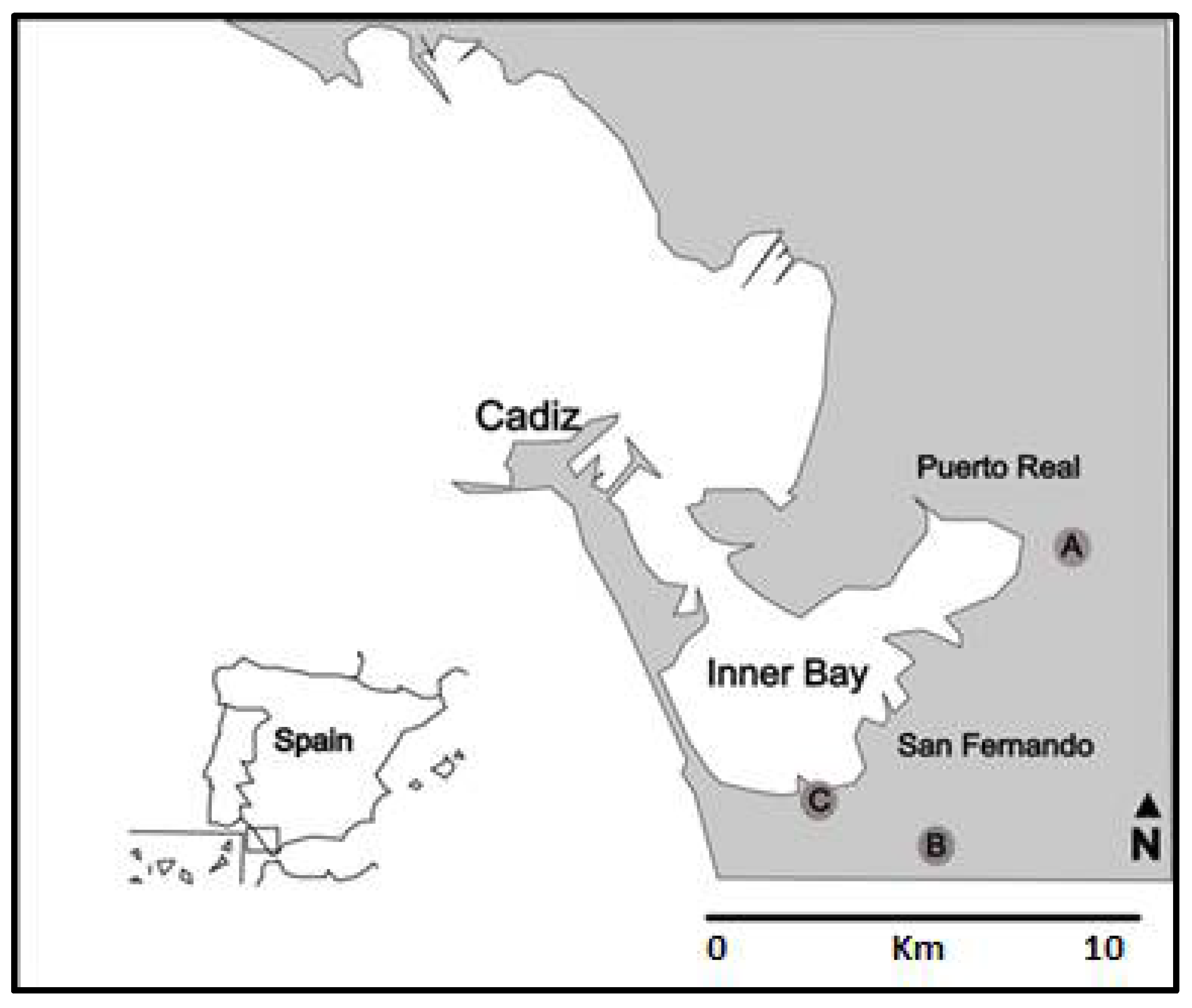
2.3.1. Characteristics of the Culture Site

The in situ culture was carried out during the months of November 2016 to October 2017 at the Salina La Esperanza in Cadiz Bay (Southern Spain; 36°30′39″ N, 6°09′35″ W). This salina is currently used for salt exploitation and extensive fish farming (e.g., seabream and European eel). The channel system included an open lock-gate to regulate the water level and to enable the free exchange of water between ponds, which allowed high hydrodynamics in the tidal channel [29]. The bottom of the culture site is rocky in nature. The mean environmental variables of the site ranged between 14.2 and 24.6 °C (water temperature) and 37.5 and 44‰ (salinity) [29,42]. The tidal range was approximately 1 m [42]. These characteristics and the high hydrodynamics made the study area very favorable for the cultivation of macroalgae.

2.3.2. Cultivation System and Growth Rate

Due to the characteristics of the study area, a rope system anchored parallel to the sluice gate was chosen. This system was based on three polypropylene ropes (three for each species) fixed to the wall with the help of spikes and carabiners so that the ropes were slightly submerged in a direction perpendicular to the flow. Between five and seven piles of the respective species were seeded in each rope.
Daily growth rate (GR), which was expressed as the percentage increase in mass per day (% day−1), was estimated as:
GR (% day−1) = ln (Wf/Wi)/t × 100
where Wf is the final weight (g) on day t, Wi is the initial weight (g) and t is the number of days of cultivation.
All ropes were weighed separately, and weekly growth was calculated, using the final biomass of the previous week as Wi.

2.4. Culture in the Laboratory

2.4.1. Collection Sites

Samples were collected in the inner Bay of Cadiz (Figure 1), in an area of tidal crevices and salinas, at ca. 1.3 m depth. Samples were collected in the proximity of sluice gates where constant water movement is created. The collection was carried out during February–August 2019 and February–March 2020, where abundant fertile thallus was found.

2.4.2. Collection and Cleaning of Carpogonial Branches

The distinction in situ of the female (haploid) gametophyte in both species is quite straightforward, as the (diploid) cystocarps can be seen with the naked eye when mature as spheres protruding from the carpogonial branches. Once the gametophytes were collected, they were placed in zip bags with seawater and transported in an isothermal container to the laboratory, where the morphological identification of the species was carried out through cross-sections of the thallus and its reproductive structures.
For the cleaning protocol, a previous test was carried out to improve the cleaning process of carpogonial branches. Therefore, different solutions of (GeO2) and antibiotics were tested in filtered and sterilised seawater. Regarding GeO2, solutions were added directly to filtered and autoclaved seawater (0.1 y 0.2 mg L−1), with final concentrations of GeO2 of 0.96 µM and 1.91 µM, respectively. Antibiotic solutions of nystatin (0.3 mg mL−1 in a concentration of 6.28 µM) and gentamicin (0.6 mg mL−1 in a concentration of 6.48 µM) were used, prepared in 100 mL of bidistilled water. Of the antibiotic solution, 1 and 2 mL of solution were added per liter of filtered and autoclaved seawater. The branches were allowed to stand for 1, 2 and 3 h in each of the following solutions: sterilised filtered seawater, sterilised filtered seawater + GeO2 (0.96 µM or 1.91 µM), filtered and sterilised seawater + antibiotics (1 mL) or (2 mL) and filtered and sterilised seawater + GeO2 (0.96 µM and/or 1.91 µM) + antibiotics (1 mL) and/or (2 mL).
According to this test, the cleaning protocol was established as follows: once the species was identified, the carpogonial branches were cleaned of impurities with filtered seawater (0.2 µm filters) and sterilised in an autoclave (121 °C for 20 min) following sterilisation recommendations [43]. Then branches were checked carefully with a stereoscopic lens to remove epiphytes. Once cleaned, they were placed back into autoclaved filtered seawater and gently shaken to remove any residue. A 0.1 g L−1 GeO2 solution (concentration of 0.96 µM) was added to the carpogonial branches for 3 h; then, thalli were cut (±3 cm in length), and fragments (n = 4) were deposited in multi-well plates.

2.4.3. Release of Carpospores

Sporulation was induced by drying and subsequent hydration of the fragments [25]. These were left to stand for 1 h, after which 3 mL of culture medium was added. It was used ES medium [44,45], Miquel A + B (or Miquel water) [46,47] to which 1 mL of vitamin solution (Thiamine . HCL 0.500 mg, Biotin 1 mL, Cyanocobalamin 1 mL) was added and f/2 [45,48,49]. Sporulation controls were performed for one week. Multi-wells with spores were kept in a culture chamber under controlled conditions (Table 1).
As the spore cultures developed into emerging thalli, it was necessary to switch them to larger volume vessels; in this case, 100 mm diameter Petri dishes (at 40 days from the beginning of the experiment), 250 mL Erlenmeyer (at 176 days), 1 L (at 200 days) and 5 L (at 228 days), when thalli were big enough to be transferred to the following culture system to continue their growth (250 days).

2.4.4. Cultivation of Thalli in the Aquarium and Growth Rate

For the following experiment, and due to space limitation, a sample of 15 individuals (developed thalli) from the spore culture was used, with 250 days of growth and an approximate weight of 0.200 g each. These were tied to a 3-tier rope system in a 10 L rectangular aquarium with artificial seawater (Pro Coral salt, containing 430–450 mg L−1 Ca, 1280–1340 mg L−1 Mg and 370–390 mg L−1 K), to which 200 mL of ES medium were added. An aquarium submersible pump was used to allow recirculation of water. The culture conditions were those described in Table 1.
The GR was estimated as above. Weightings were performed approximately every 20 days. Individuals from each rope were weighed separately. In order to calculate weekly growth, the final biomass of the previous week was used as Wi. The mean of each rope in the cultivation system was calculated, thus obtaining the mean GR of the cultivation period.

2.5. Statistical Analysis

For field cultures, the monthly means of GR of three ropes were calculated to perform an analysis of variance (ANOVA) test to determine if there were significant differences in GR in the different months of the year. For the significant ANOVAs, a Tukey test was carried out to find out the combinations where the significant difference occurred.
For laboratory cultures, an ANOVA test was also performed to assess if there were significant differences between any culture period (23, 43, 63, 84, 106 and 144 days after the start of the second phase of thalli culture). In all cases, significance was set at 5% probability.

3. Results

3.1. Species Identification

The DNA tests of the cultures performed to corroborate the species showed that these corresponded to Gracilaria gracilis and Chondracanthus teedei (Table 2). According to the percentage of similarity presented BLASTn, which in all cases was greater than 99%.

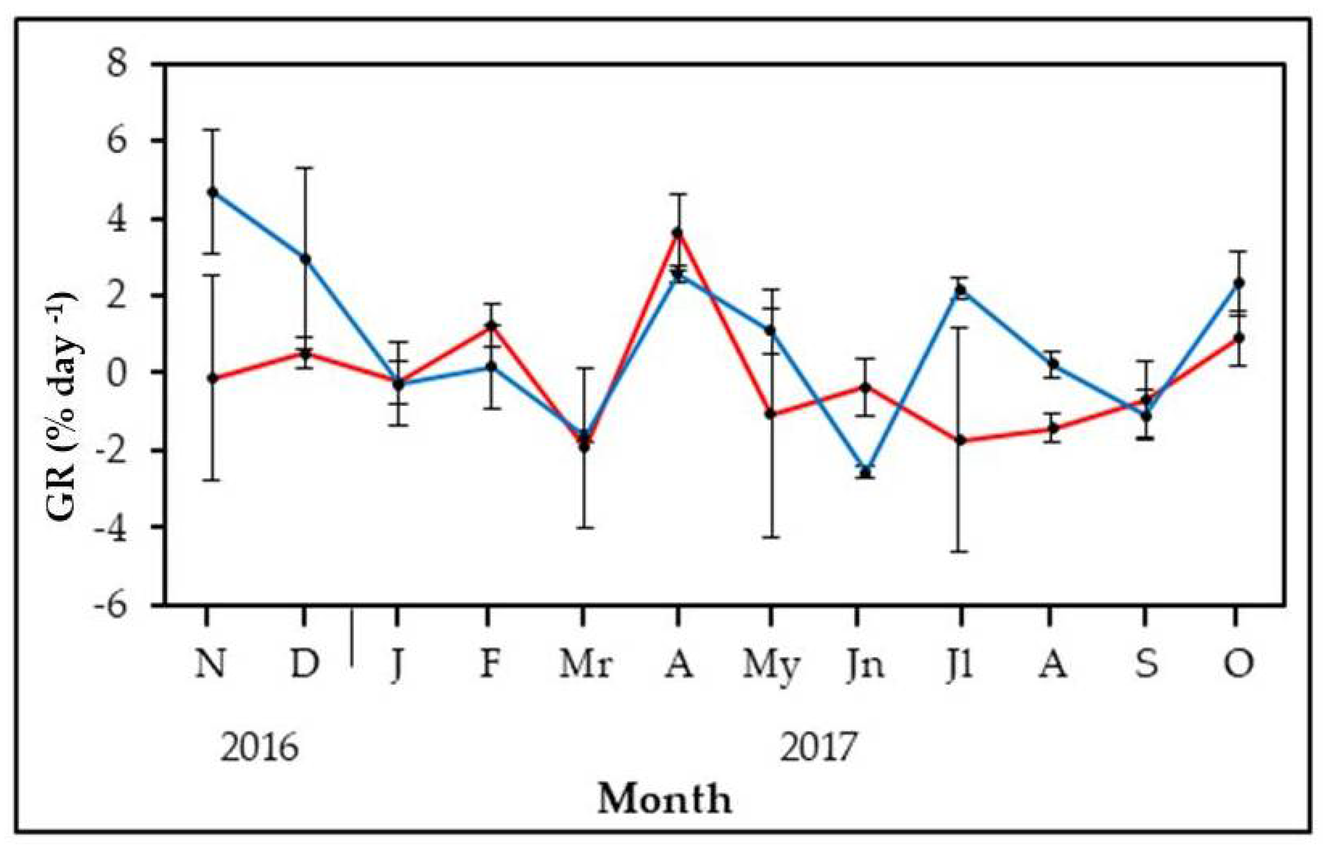
3.2. Field Growth Rates

The maximum weekly net growth rate of Gracilaria gracilis was 3.64% day−1 and was estimated in April (Figure 2). Periods of negative net growth (mainly summer) were also observed when biomass losses were found due to unfavourable environmental conditions (mostly temperature). The ANOVA showed no significant differences between the different months of the year due to the high variability of data. For C. teedei the highest weekly net growth rate was reached in November (4.68% day−1) (Figure 2). This species did not evidence a clear pattern of growth, showing alternating periods of net growth rate (November, December, April, July and October) or negative rates (particularly during January, March, June and September). The ANOVA showed significant differences in growth rates throughout the year (p < 0.001). The post hoc test evidenced significant differences between November and several months (March, June and September) and also between June, April and September.

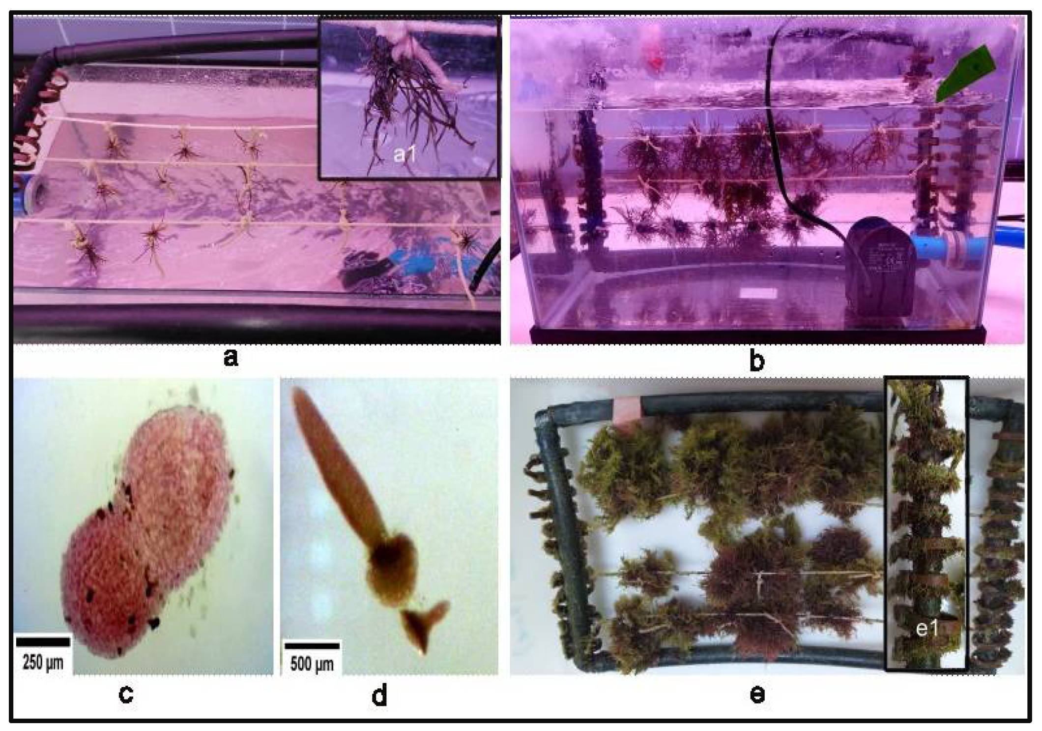
3.3. Harvesting, Cleaning and Sporulation
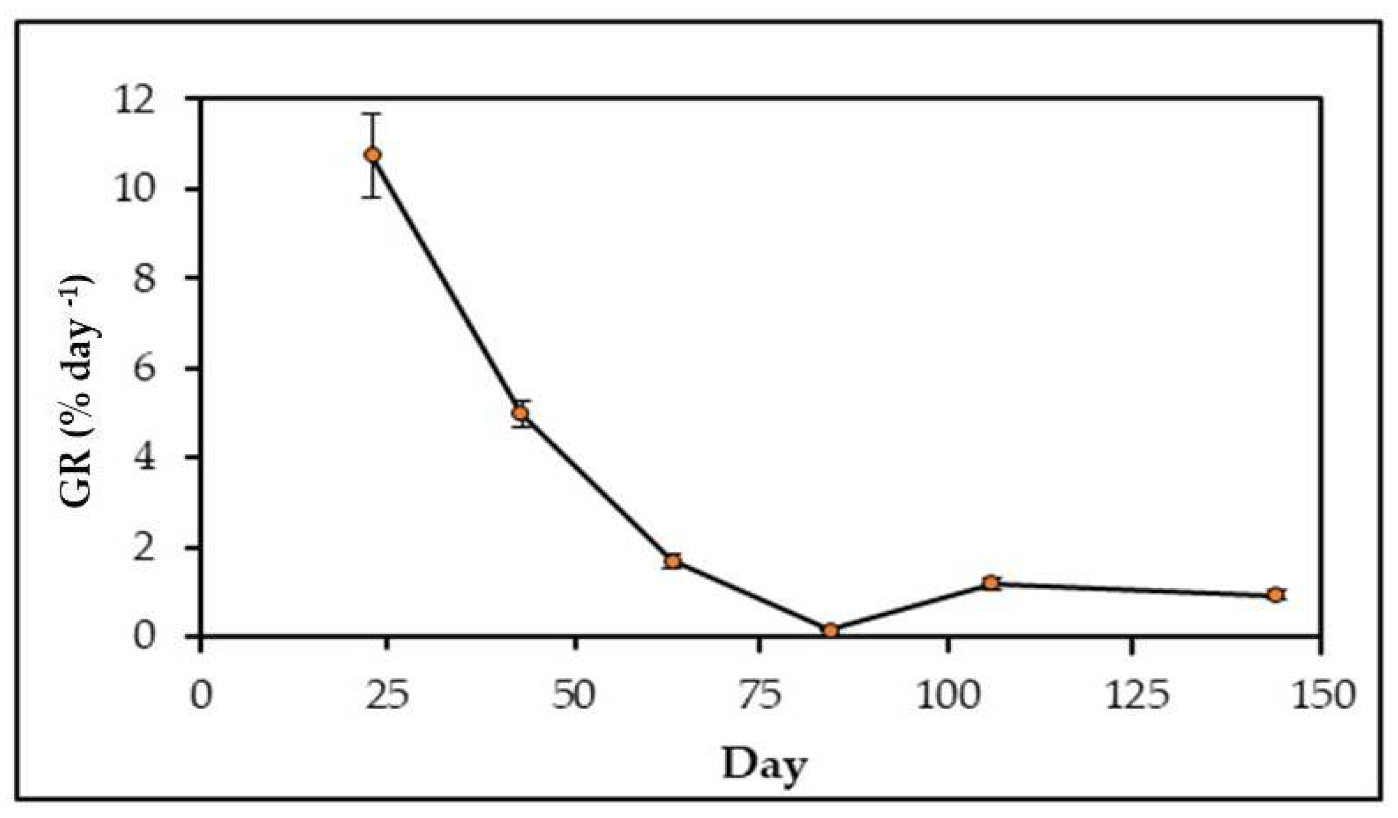
In general, specimens with carpogonial branches were observed in the natural environment in mid-February, especially in Gracilaria gracilis, and until the beginning of August. From September to January, no carpogonial branches were observed in any of the two species. (Pers. obs.).
Even though a sterilisation protocol was used, the growth of microorganisms, especially diatoms, could not be fully prevented. However, it did not affect the spores. The best concentration of GeO2 established during the cleaning process was 0.1 mg L−1 (0.96 µM). The use of antibiotics was discarded after the first trial, as there was a loss of colour of the fertile algae pieces approximately 30 min after being soaked in the solution.
Sporulation of both species was evidenced by a reddish stain on the bottom of the multi-wells. Carpospores were spherical in the two species. The carpospores released on the first day only remained viable for ca. seven days, whereas the spores released on the second day were viable for further development.
In the trials that started in March, we were able to complete all stages of growth and development in Gracilaria gracilis. In the case of Chondracanthus teedei, the trials started in May produced sporulation; basal discs were then observed, and so were the initial stages of the frond growth. The best results were observed with ES medium and Miquel A + B; however, spores on f/2 medium after 3 days were pale and lacked consistency.

3.4. Spore Culture

The mean spore size of Gracilaria gracilis was 23 ± 1.45 µm (n = 25). For the ES medium and Miquel A + B, the beginning of the formation of the basal disc was observed after 7 days; then at 12 days, a developed basal disc was visualised (Figure 3). The beginning of the growth of the frond could be seen with marked visual differences between the two culture media on day 20. In the ES medium, thalli developed with some bifurcations after approximately 27 days in culture. However, spores cultured in Miquel A + B did not continue to develop. A total of 464 thalli were grown in ES medium in the 5 L Erlenmeyer flasks for a period of 394 days and reached a final wet weight of 175 g.
The 15 individuals of Gracilaria gracilis grown and transferred to the 10 L aquarium reached a mean final fresh weight of ±15.87 g after 144 days of culture. Weakly growth rate of the carposporophytes varied significantly during the cultivation period (p < 0.001) and decreased from a maximum of 10.95 % day- 1 during the first three weeks to a 1.2% day−1 after two months of cultivation (Figure 4) when plants reached a bushy pattern and maintained a constant growth rate. The mean growth rate during the whole cultivation period was 3.28% day−1.
During this period of cultivation, the gametophytic phase was obtained from the tetrasporophyte, thus completing the three-phase life cycle of this species. The spores adhered to the aquarium walls and the culture system tubes. A basal disc was observed after 84 days of culture, and these discs developed fronds after 96 days (Figure 5).
The spores of Chondracanthus teedei had a mean size of 38 ± 4.5 µm (n = 22). Growth was only observed in the ES medium (Figure 6), where after 7 days of culture, the basal disc began to develop. After 15 days, the basal disc was observed completely developed in some germlings, and after day 25 of culture, the development of the frond was evident. After 35 days of culture, individuals could be seen in a basal disc state, but some of them evidenced poorly developed fronds. As only one germling developed an emerging frond, it was considered insufficient to further continue the germlings’ cultivation.

4. Discussion

The possibility of culturing macroalgae in different parts of the world is continuously addressed due to the economic importance of these organisms, as well as the establishment of businesses and/or economic models around this activity [50,51]. The growing need for development, improvement and diversification of marine macroalgae aquaculture in Europe is increasing [52], which requires the availability of quality seeds independently of wild populations [53]. One of these ways is the cultivation of spores from these organisms, and as far as we know, this is the first study in which the growth of the carporophyte phase of Gracilaria gracilis and Chondracanthus teedei was carried out in Spain. The study is therefore of great interest for the growing development of seaweed cultures in the region.

4.1. Species Identification

Genetic barcodes or molecular taxonomy greatly contribute to better description and recognition of species, especially in red algae [54,55,56]. This identification in algae is essential due to their phenotypic plasticity (changes in physical characteristics in response to an environmental signal) [57,58]. In particular, the taxonomy of the genus Gracilaria is difficult due to the small differences between the species and the fact that they often share morphological characteristics in the cystocarps and carposporangia [59]. The identification of Gracilaria and Gracilariopsis has proven to be extremely difficult using the available morphological typologies [60]. These DNA tests helped us to correctly identify the specimens in the current study, taking into account that both G. gracilis and Gracilariopsis longissima are found naturally in Cadiz bay.

4.2. Cleaning and Collection

When unialgal cultures are carried out, it is necessary to eliminate the presence of epiphytes since these can grow faster than the algae to be cultivated [61]. For this reason, sterilised seawater is usually used, to which GeO2 is added to reduce the presence of diatoms in different concentrations (0.1–0.2 mg L−1) depending on the species [25,43,62]. It is recommended that the maximum concentration be 1 mg L−1 in brown algae culture [61] and antibiotics for the presence of organisms such as fungi and bacteria [21,63]. In our previous experiments, the use of antibiotics weakened the spore walls, making them unviable for their development. Some authors pointed out that the use of antibiotics should be minimal because it affects growth [61]. Although it is stipulated that the elimination of epiphytes is necessary, the amount of epiphytes in our crop does not affect the development of spores.
Fertile gametophytes are required for cultivation starting with carpospores. For this reason, it is necessary to seek out the periods when the populations are fertile in the natural environment [64]. At the collection sites, a particular occurrence is that the biomass decreases from early summer to late autumn (June–December) and increases in early winter until late spring (December–June) [29]. Our observations are consistent with the studies carried out in the Bay of Cadiz, with the collection periods favourable for spore growth in the laboratory being late March for Gracilaria gracilis and May for Chondracanthus teedei, just when the biomass in the field is increasing.

4.3. Growing from Spores of Gracilaria Gracilis

In sporulation studies on Gracilaria gracilis, it was observed that the greatest spore release occurs between days 1 and 7 [25,26,65,66], although it can last up to a month [67]. This variability in sporulation periods is partly related to species distribution and adaptation to environmental conditions specific to the region of origin, such as temperature, photoperiod or irradiance [68], suggesting an ecophysiological response to sporulation processes [69].
The size of the carpospores, the times of appearance of the basal disc, and the initiation and development of the frond are similar to studies carried out with this species [25,26] and in accordance with other species within the genus such as G. dura [66]. The life cycle of G. gracilis was completed in 11 months. In general terms, the life cycle of Gracilaria cultures lasts 5–12 months [65]. Examples of this are the times reported for G. verrucosa (G. tenuistipitata) of 4.5 months [70] and five months for G. dominguensis [71].

4.4. Growing from Spores of Chondracanthus Teedei

There are studies on in situ growth and laboratory propagation of Chondracanthus teedei based on vegetative reproduction [29,36,37], with variations in environmental parameters to optimise cultures [72] and as biomitigators [30]. Spore cultures of the genus Chondracnthus have so far focused on C. chamisoi with a methodology adapted from other red algal cultures [64]. Spore size and time to basal disc and thallus attachment are similar to those reported for the species [73]. The possibility of culturing from carpospores is established according to the 35 days of growth in the laboratory. These results are consistent with those found by other authors [21,74]. The optimal growth range for C. teedei is 20–25 °C [21]. This could explain why from day 35 onwards, the cultured individuals slowed down their development, making it impossible to complete the life cycle of this species. Therefore, our methodology should be improved to complete the whole life cycle.

4.5. Growth Rates

The daily growth rates in the field allowed for progress in the knowledge of the productive behavior of the two species of macroalgae and the interactive effect of environmental variables, such as irradiance, temperature or salinity [64]. In the case of Gracilaria gracilis, cultures in the natural environment are developed under a wide range of tolerance to temperature (15–30 °C) [75,76]. The growth rate obtained in our study, both in nature and in the laboratory for both species, are similar to those reported elsewhere; for example, G. gracilis evidenced a growth rate of 0.45–2.44% day−1 and was influenced by temperature or depth [76,77]. For species of the genus Chondracanthus, rates of 5–9.3% day−1 can be estimated under controlled conditions [64]. The growth rates obtained in our study were also consistent with experiments carried out in the Bay of Cadiz when the macroalgae were cultivated in submersible rafts [29,30,31,78,79]. For several months, negative growth rates were estimated in the field, which can be explained according to the environmental factors involved and which remain controlled in the laboratory, thus allowing growth rates up to 10.74% day−1.

5. Conclusions

Both species are technically feasible for cultivation from carpospores in the laboratory, as we obtained viable spores for their development. One of the most important factors for this type of cultivation is the biomass found naturally in the Bay of Cadiz. We were able to establish with field cultures of these species the months where the biomass evidenced a high net growth rate in the natural environment.
These results are a valuable input for the establishment of pilot-scale cultures in the former salt pans, taking into account the economically important characteristics of these carrageenan and agar-producing species and previous studies on the feasibility of cultivation on ropes. Understanding the developmental pattern of the species is a crucial step in determining the success of spore cultures.
For future experiments, other culture media and growth stimulators could be considered, particularly for Chondracanthus teedei, making the most of sporulation and increasing the biomass in the laboratory for subsequent cultivation in the natural environment.

Author Contributions

Conceptualisation, M.L.-C.; Methodology, M.L.-C. and F.B.; Validation, I.H. and J.L.P.-L.; Formal analysis, M.L.-C.; Investigation, M.L.-C. and F.B.; Resources, M.L.-C., I.H., J.L.P.-L. and C.M.P.-G.; Data curation, M.L.-C.; Writing—original draft preparation, M.L.-C.; Writing—review and editing, I.H., J.L.P.-L. and C.M.P.-G.; Visualisation, I.H., J.L.P.-L. and C.M.P.-G. All authors have read and agreed to the published version of the manuscript.

Funding

Scholarship Instituto para la Formación y Aprovechamiento de Recursos Humanos and Secretaría Nacional de Ciencias, Tecnología e Innovación (IFARHU-SENACYT), contract number 270-2018-08.

Institutional Review Board Statement

Not applicable.

Informed Consent Statement

Not applicable.

Data Availability Statement

Data are contained within the article.

Acknowledgments

The research leading to these results was supported by the fellowship IFARHU-SENACYT of Panamá, Authority for the Aquatic Resources of Panamá (ARAP) and project INNOVALGA funded by the Spanish Ministry of Agriculture, Food and Fisheries.

Conflicts of Interest

The authors declare no conflict of interest.

References

  1. Pereira, L. Edible Seaweeds of the World; CRC Press: Boca Raton, FL, USA, 2016. [Google Scholar] [CrossRef]
  2. Ferreira Barbosa, D.; Lucia Pereira Dias, T.; Faria Lopes, S.; Cristina de Souza Duarte, R.; Maria Duarte do Amaral, F. Community Structure and Functional Traits of Mollusks Associated with Coastal Reef Macroalgae in Northeastern Brazil. Mar. Ecol. 2019, 40, e12563. [Google Scholar] [CrossRef]
  3. Lynch, S.A.; Breslin, R.; Bookelaar, B.; Rudtanatip, T.; Wongprasert, K.; Culloty, S.C. Immunomodulatory and Antiviral Effects of Macroalgae Sulphated Polysaccharides: Case Studies Extend Knowledge on their Importance in Enhancing Shellfish Health, and the Control of a Global Viral Pathogen Ostreid Herpesvirus-1 MicroVar. Polysaccharides 2021, 2, 202–217. [Google Scholar] [CrossRef]
  4. Ismail, M.M.; Alotaibi, B.S.; EL-Sheekh, M.M. Therapeutic Uses of Red Macroalgae. Molecules 2020, 25, 4411. [Google Scholar] [CrossRef] [PubMed]
  5. Cavallo, R.; Acquaviva, M.; Stabili, L.; Cecere, E.; Petrocelli, A.; Narracci, M. Antibacterial Activity of Marine Macroalgae against Fish Pathogenic Vibrio Species. Open Life Sci. 2013, 8, 646–653. [Google Scholar] [CrossRef]
  6. Cikoš, A.-M.; Čož-Rakovac, R.; Šubarić, D.; Jerković, I.; Ačkar, Đ.; Jokić, S. Macroalgae in the Food Industry—Opportunities and Challenges. Eng. Power 2020, 15, 14–19. [Google Scholar]
  7. Yang, L.-E.; Lu, Q.-Q.; Brodie, J. A Review of the Bladed Bangiales (Rhodophyta) in China: History, Culture and Taxonomy. Eur. J. Phycol. 2017, 52, 251–263. [Google Scholar] [CrossRef]
  8. Buschmann, A.H.; Camus, C.; Infante, J.; Neori, A.; Israel, Á.; Hernández-González, M.C.; Pereda, S.V.; Gomez-Pinchetti, J.L.; Golberg, A.; Tadmor-Shalev, N.; et al. Seaweed Production: Overview of the Global State of Exploitation, Farming and Emerging Research Activity. Eur. J. Phycol. 2017, 52, 391–406. [Google Scholar] [CrossRef]
  9. Kim, J.K.; Yarish, C.; Pereira, R. Tolerances to Hypo-Osmotic and Temperature Stresses in Native and Invasive Species of Gracilaria (Rhodophyta). Phycologia 2016, 55, 257–264. [Google Scholar] [CrossRef]
  10. Francavilla, M.; Franchi, M.; Monteleone, M.; Caroppo, C. The Red Seaweed Gracilaria gracilis as a Multi Products Source. Mar. Drugs 2013, 11, 3754–3776. [Google Scholar] [CrossRef] [Green Version]
  11. Kılınç, B.; Cirik, S.; Turan, G.; Tekogul, H.; Koru, E. Seaweeds for Food and Industrial Applications. In Food Industry, 1st ed.; Muzzalupo, I., Ed.; IntechOpen: London, UK, 2013; p. 760. [Google Scholar] [CrossRef] [Green Version]
  12. Neveux, N.; Bolton, J.J.; Bruhn, A.; Roberts, D.A.; Ras, M. The Bioremediation Potential of Seaweeds: Recycling Nitrogen, Phosphorus, and Other Waste Products. In Blue Biotechnology: Production and Use of Marine Molecules; La Barre, S., Bates, S., Eds.; Wiley-VCH Verlag GmbH & Co. KGaA: Weinheim, Germany, 2018; pp. 217–239. [Google Scholar] [CrossRef]
  13. Cherry, P.; O’Hara, C.; Magee, P.J.; McSorley, E.M.; Allsopp, P.J. Risks and Benefits of Consuming Edible Seaweeds. Nutr. Rev. 2019, 77, 307–329. [Google Scholar] [CrossRef] [Green Version]
  14. FAO. FAO Aquaculture News. No. 63 (May). Rome. 2021. Available online: http://www.fao.org/3/cb4850en/cb4850en.pdf (accessed on 10 April 2022).
  15. Callaway, E. Lab Staple Agar Runs Low: Dwindling Seaweed Harvest Imperils Reagent Essential for Culturing Microbes. Nature 2015, 7581, 171. Available online: Link.gale.com/apps/doc/A437223628/HRCA?u=anon~ac3ff0d9&sid=googleScholar&xid=b176955b (accessed on 30 January 2022). [CrossRef] [Green Version]
  16. Lauzon-Guay, J.-S.; Ugarte, R.A.; Morse, B.L.; Robertson, C.A. Biomass and Height of Ascophyllum Nodosum after Two Decades of Continuous Commercial Harvesting in Eastern Canada. J. Appl. Phycol. 2021, 33, 1695–1708. [Google Scholar] [CrossRef]
  17. Cai, J.; Lovatelli, A.; Aguilar-Manjarrez, J.; Cornish, L.; Dabbadie, L.; Desrochers, A.; Diffey, S.; Garrido Gamarro, E.; Geehan, J.; Hurtado, A.; et al. Seaweeds and Microalgae: An Overview for Unlocking Their Potential in Global Aquaculture Development; FAO: Rome, Italy, 2021. [Google Scholar] [CrossRef]
  18. Bravo, A.; Buschmann, A.H.; Valenzuela, M.E.; Uribe, M.; Vergara, P.A.; Buitano, M.S. Evaluation of Artificial Intertidal Enclosures for Gracilaria Farming in Southern Chile. Aquac. Eng. 1992, 11, 203–216. [Google Scholar] [CrossRef]
  19. Friedlander, M. Gracilaria conferta and its Epiphytes: The effect of culture conditions on growth. Bot. Mar. 1992, 35, 423–428. [Google Scholar] [CrossRef]
  20. Hurtado-Ponce, A.Q. Vertical Rope Cultivation of Gracilaria (Rhodophyta) Using Vegetative Fragments. Bot. Mar. 1990, 33, 477–482. [Google Scholar] [CrossRef]
  21. de Oliveira Bastos, E.; Horta, P.A.; Hayashi, L. Strain Selection in Chondracanthus teedei (Gigartinaceae, Rhodophyta) Using Tetraspore and Carpospore Progeny: Growth Rates, Tolerance to Temperature and Carrageenan Yield. J. Appl. Phycol. 2021, 33, 2379–2390. [Google Scholar] [CrossRef]
  22. Santelices, B.; Doty, M.S. A Review of Gracilaria Farming. Aquaculture 1989, 78, 95–133. [Google Scholar] [CrossRef]
  23. Levy, I.; Beer, S.; Friedlander, M. Strain Selection in Gracilaria spp. 2. Selection for High and Low Temperature Resistance in G. Verrucosa Sporelings. J. Appl. Phycol. 1990, 2, 163–171. [Google Scholar] [CrossRef]
  24. Glenn, E.P.; Moore, D.; Fitzsimmons, K.; Azevedo, C. Spore Culture of the Edible Red Seaweed, Gracilaria parvispora (Rhodophyta). Aquaculture 1996, 142, 59–74. [Google Scholar] [CrossRef]
  25. Michetti, K.M.; Martín, L.A.; Leonardi, P.I. Carpospore Release and Sporeling Development in Gracilaria gracilis (Gracilariales, Rhodophyta) from the Southwestern Atlantic Coast (Chubut, Argentina). J. Appl. Phycol. 2013, 25, 1917–1924. [Google Scholar] [CrossRef]
  26. Freitas, M.V.; Mouga, T.; Correia, A.P.; Afonso, C.; Baptista, T. New Insights on the Sporulation, Germination, and Nutritional Profile of Gracilaria Gracilis (Rhodophyta) Grown under Controlled Conditions. J. Mar. Sci. Eng. 2021, 9, 562. [Google Scholar] [CrossRef]
  27. Pérez Massad, I.; Ávila, M.; Contreras-Porcia, L.; Bulboa Contador, C. Spores Re-Suspending Technology, a New System Improving Spore Seeding for Culture of Commercial Red Seaweeds. Aquaculture 2020, 526, 735374. [Google Scholar] [CrossRef]
  28. Hernández, I.; Cara, C.L.; Sánchez-García, J.; Macías, M.; Love, R.; Bermejo, R. Cultivos de Macroalgas en el Litoral Gaditano: Estado Actual y Perspectivas para su Desarrollo. Algas, Bol. Inf. Soc. Esp. Ficol. 2016, 52, 5–10. [Google Scholar]
  29. Bermejo, R.; Macías, M.; Cara, C.L.; Sánchez-García, J.; Hernández, I. Culture of Chondracanthus teedei and Gracilariopsis longissima in a Traditional Salina from Southern Spain. J. App. Phycol. 2019, 31, 561–573. [Google Scholar] [CrossRef]
  30. Bermejo, R.; Cara, C.L.; Macías, M.; Sánchez-García, J.; Hernández, I. Growth Rates of Gracilariopsis longissima, Gracilaria bursa-pastoris and Chondracanthus teedei (Rhodophyta) Cultured in Ropes: Implication for N Biomitigation in Cadiz Bay (Southern Spain). J. Appl. Phycol. 2020, 32, 1879–1891. [Google Scholar] [CrossRef]
  31. Bermejo, R.; Macías, M.; Sánchez-García, F.; Love, R.; Varela-Álvarez, E.; Hernández, I. Influence of Irradiance, Dissolved Nutrients and Salinity on the Colour and Nutritional Characteristics of Gracilariopsis longissima (Rhodophyta). Algal Res. 2020, 52, 102121. [Google Scholar] [CrossRef]
  32. Pereira, L.; Silva, P. A Concise Review of the Red Macroalgae Chondracanthus teedei (Mertens Ex Roth) Kützing and Chondracanthus teedei var. lusitanicus (J.E. De Mesquita Rodrigues) Bárbara & Cremades. J. Appl. Phycol. 2021, 33, 111–131. [Google Scholar] [CrossRef]
  33. Martín, L.A.; Boraso de Zaixso, A.L.; Leonardi, P.I. Biomass Variation and Reproductive Phenology of Gracilaria gracilis in a Patagonian Natural Bed (Chubut, Argentina). J. Appl. Phycol. 2011, 23, 643–654. [Google Scholar] [CrossRef]
  34. Kützing, F.T. Phycologia Generalis: Oder, Anatomie, Physiologie und Systemkunde der Tange; Brockhaus: Leipzig, Berlin, 1843. [Google Scholar] [CrossRef] [Green Version]
  35. Cabioc’h, J.; Floc’h, J.Y.; Le Toquin, A.; Boudouresque, C.F.; Meinesz, A.; Verlaque, M. Guide to the Algae of the Seas of Europe: Atlantic and Mediterranean; Omega: Jackson, MI, USA, 1995. [Google Scholar]
  36. Schmidt, É.C.; Pereira, B.; Pontes, C.L.M.; dos Santos, R.; Scherner, F.; Horta, P.A.; de Paula Martins, R.; Latini, A.; Maraschin, M.; Bouzon, Z.L. Alterations in Architecture and Metabolism Induced by Ultraviolet Radiation-B in the Carragenophyte Chondracanthus teedei (Rhodophyta, Gigartinales). Protoplasma 2012, 249, 353–367. [Google Scholar] [CrossRef]
  37. Zinoun, M.; Cosson, J.; Deslandes, E. Influence of Culture Conditions on Growth and Physicochemical Properties of Carrageenans in Gigartina teedii (Rhodophyceae-Gigartinales). Bot. Mar. 1993, 36, 131–136. [Google Scholar] [CrossRef]
  38. Pérez-Lloréns, J.L.; Hernández, I.; Vergara, J.J.; Brun, F.G.; León, Á. Those Curious and Delicius Seaweeds a Fascinating Voyage from Biology to Gastronomy; University of Cadiz: Cadiz, Spain, 2018. [Google Scholar]
  39. Steentoft, M.; Irvine, L.M.; Farnham, W.F. Two Terete Species of Gracilaria and Gracilariopsis (Gracilariales, Rhodophyta) in Britain. Phycologia 1995, 34, 113–127. [Google Scholar] [CrossRef]
  40. Freshwater, D.W.; Fredericq, S.; Butler, B.S.; Hommersand, M.H.; Chase, M.W. A Gene Phylogeny of the Red Algae (Rhodophyta) Based on Plastid RbcL. Proc. Natl. Acad. Sci. USA 1994, 91, 7281–7285. [Google Scholar] [CrossRef] [PubMed] [Green Version]
  41. Freshwater, D.W.; Rueness, J. Phylogenetic Relationships of Some European Gelidium (Gelidiales, Rhodophyta) Species, Based on RbcL Nucleotide Sequence Analysis. Phycologia 1994, 33, 187–194. [Google Scholar] [CrossRef]
  42. Ministry of Transport, Mobility and Urban Agenda, State Ports. Available online: http://www.puertos.es/es-es/oceanografia/ (accessed on 10 March 2022).
  43. Yarish, C.; Redmond, S.; Kim, J.K. Gracilaria Culture Handbook for New England; Wrack Lines: Groton, CT, USA, 2012; Volume 72, Available online: https://opencommons.uconn.edu/wracklines/72 (accessed on 1 July 2022).
  44. Provasoli, L. Media and Prospects for the Cultivation of Marine Algae. In Proceedings of the US-Japan Conference, Hakone, Japan, 12–15 September 1966; pp. 63–75. [Google Scholar]
  45. Andersen, R.A.; Berges, J.A.; Harrison, P.J.; Watanabe, M.M. Recipes for Freshwater and Seawater Media. In Algal Culturing Techniques; Andersen, R.A., Ed.; Phycological Society; Elsevier Academic Press: Amsterdam, The Netherlands, 2005; pp. 429–532. [Google Scholar] [CrossRef]
  46. Allen, E.J.; Nelson, E.W. On the Artificial Culture of Marine Plankton Organisms. J. Mar. Biol. Assoc. 1910, 8, 421–474. [Google Scholar] [CrossRef] [Green Version]
  47. Provasoli, L.; McLaughlin, J.J.A.; Droop, M.R. The Development of Artificial Media for Marine Algae. Archiv. Mikrobiol. 1957, 25, 392–428. [Google Scholar] [CrossRef] [PubMed]
  48. Guillard, R.R.; Ryther, J.H. Studies of Marine Planktonic Diatoms. I. Cyclotella nana Hustedt and Detonula confervacea (Cleve) Gran. Can. J. Microbiol. 1962, 8, 229–239. [Google Scholar] [CrossRef]
  49. Guillard, R.R.L. Culture of Phytoplankton for Feeding Marine Invertebrates. In Culture of Marine Invertebrate Animals; Smith, W.L., Chanley, M.H., Eds.; Springer: Boston, MA, USA, 1975; pp. 29–60. [Google Scholar] [CrossRef]
  50. Angriani, E.; Made, S.; Tahang, H. Seaweed Cultivation Business Development Strategy (Gracillaria sp.) through Spores Culture in Takalar Regency. Int. J. Environ. Agric. Biotech. 2021, 6, 120–126. [Google Scholar] [CrossRef]
  51. Pérez-Vas, R.; Puime Guillén, F.; Enríquez-Díaz, J. Valuation of a Company Producing and Trading Seaweed for Human Consumption: Classical Methods vs. Real Options. Int. J. Environ. Res. Public. Health 2021, 18, 5262. [Google Scholar] [CrossRef]
  52. Barbier, M.; Charrier, B.; Araujo, R.; Holdt, S.; Jacquemin, B.; Rebours, C. Phycomorph European Guidelines for a Sustainable Aquaculture of Seaweeds. In PEGASUS; Barbier, M., Charrier, B., Eds.; COST Action FA1406. M.: Roscoff, France, 2019; p. 194. [Google Scholar] [CrossRef]
  53. Hayashi, L.; de Cantarino, J.; Critchley, A.T. Challenges to the Future Domestication of Seaweeds as Cultivated Species: Understanding Their Physiological Processes for Large-Scale Production. Adv. Bot. Res. 2020, 95, 57–83. [Google Scholar] [CrossRef]
  54. Hu, Z.; Guiry, M.D.; Duan, D. Using the Ribosomal Internal Transcribed Spacer (ITS) as a complement Marker for Species Identification of Red Macroalgae. Hydrobiologia 2009, 635, 279–287. [Google Scholar] [CrossRef]
  55. Yang, M.Y.; Geraldino, P.J.L.; Kim, M.S. DNA Barcode Assessment of Gracilaria Salicornia (Gracilariaceae, Rhodophyta) from Southeast Asia. Bot. Stud. 2013, 54, 27. [Google Scholar] [CrossRef] [Green Version]
  56. Montes, M.; Rico, J.M.; García-Vázquez, E.; Borrell, Y.J. Morphological and Molecular Methods Reveal the Asian Alga Grateloupia imbricata (Halymeniaceae) Occurs on Cantabrian Sea Shores (Bay of Biscay). Phycologia 2016, 55, 365–370. [Google Scholar] [CrossRef]
  57. Saunders, G.W. Applying DNA Barcoding to Red Macroalgae: A Preliminary Appraisal Holds Promise for Future Applications. Philos. Trans. R Soc. Lond. B Biol. Sci. 2005, 360, 1879–1888. [Google Scholar] [CrossRef] [Green Version]
  58. Hassan, R.; Othman, M.N.A.; Harith, M.N.; Md Sah, A.S.R. Morphological Diversity of Gracilaria Blodgettii Harvey 1853 (Gracilariaceae, Rhodophyta) from Sarawak, Malaysian Borneo. Scientifica 2019, 2019, 1–9. [Google Scholar] [CrossRef] [Green Version]
  59. Gurgel, C.F.D.; Fredericq, S. Systematics of the Gracilariaceae (Gracilariales, Rhodophyta): A Critical Assessment Based on Rbcl Sequence Analyses1: Systematics of the Gracilariaceae. J. Phycol. 2004, 40, 138–159. [Google Scholar] [CrossRef]
  60. Goff, L.J.; Moon, D.A.; Coleman, A.W. Molecular Delineation of Species and Species Relationships in the Red Algal Agarophytes Gracilariopsis and Gracilaria (Gracilariales). J. Phycol. 1994, 30, 521–537. [Google Scholar] [CrossRef]
  61. Kawai, H.; Motomura, T.; Okuda, K. Isolation and Purification Techniques for Macroalgae. In Algal Culturing Techniques; Andersen, R.A., Ed.; Phycological Society; Elsevier Academic Press: Amsterdam, The Netherlands, 2005; pp. 133–143. [Google Scholar] [CrossRef]
  62. Le, H.N.; Hughes, A.D.; Kerrison, P.D. Early Development and Substrate Twine Selection for the Cultivation of Sargassum muticum (Yendo) Fensholt under Laboratory Conditions. J. Appl. Phycol. 2018, 30, 2475–2483. [Google Scholar] [CrossRef] [Green Version]
  63. Garcia-Jimenez, P.; Marian, F.D.; Rodrigo, M.; Robaina, R.R. Sporulation and Sterilization Method for Axenic Culture of Gelidium canariensis. Prog. Ind. Microbiol. 1999, 35, 227–229. [Google Scholar] [CrossRef] [Green Version]
  64. Bulboa Contador, C.; Massad, I.P.; Contreras-Porcia, L.; Zapata, J.; Castañeda, F.; Ramírez, M.E.; Gil-Kodaka, P. Concise Review of Genus Chondracanthus (Rhodophyta: Gigartinales). J. Appl. Phycol. 2020, 32, 773–785. [Google Scholar] [CrossRef]
  65. Kain, J.M.; Destombe, C. A Review of the Life History, Reproduction and Phenology of Gracilaria. J. Appl. Phycol. 1995, 7, 269. [Google Scholar] [CrossRef]
  66. Mantri, V.A.; Thakur, M.C.; Kumar, M.; Reddy, C.R.K.; Jha, B. The Carpospore Culture of Industrially Important Red Alga Gracilaria dura (Gracilariales, Rhodophyta). Aquaculture 2009, 297, 85–90. [Google Scholar] [CrossRef]
  67. Lefebvre, C.A.; Destombe, C.; Godin, J. Le fonctionnement du carposporophyte de Gracilaria verrucosa et ses repercussions sur la strategie de reproduction. Cryptogam. Algol. 1987, 8, 113–126. [Google Scholar]
  68. Guzmán-Urióstegui, A.; Robledo, D. Factors Affecting Sporulation of Gracilaria cornea (Gracilariales, Rhodophyta) Carposporophytes from Yucatan, Mexico. In Sixteenth International Seaweed Symposium; Kain, J.M., Brown, M.T., Lahaye, M., Eds.; Springer: Dordrecht, The Netherlands, 1999; pp. 285–290. [Google Scholar] [CrossRef]
  69. Garza-Sánchez, F.; Zertuche-González, J.A.; Chapman, D.J. Effect of Temperature and Irradiance on the Release, Attachment and Survival of Spores of Gracilaria pacifica Abbott (Rhodophyta). Bot. Mar. 2000, 43, 205–212. [Google Scholar] [CrossRef]
  70. Bonomi, B.J.; Oliveira, E.C.; Plastino, E.M.; Oliveira, M.C. Life History, Morphological Variability and Growth Rates of the Life Phases of Gracilaria tenuistipitata (Rhodophyta: Gracilariales) in Vitro. Sci. Mar. 2010, 74, 297–303. [Google Scholar] [CrossRef] [Green Version]
  71. Guimarães, M.; Plastino, E.M.; Oliveira, E.C. Life History, Reproduction and Growth of Gracilaria domingensis (Gracilariales, Rhodophyta) from Brazil. Bot. Mar. 1999, 42, 481–486. [Google Scholar] [CrossRef]
  72. Barbosa, I.M. Efeitos da Qualidade de Radiação na Fisiologia de Chondracanthus Teedei (Gigartinales, Rhodophyta). Graduação em Oceanografia; Universidade Federal de Santa Catarina: Florianopolis, Brazil, 2019; p. 51. Available online: https://repositorio.ufsc.br/handle/123456789/197545 (accessed on 3 March 2022).
  73. Guiry, M.D. Structure, Life History and Hybridization of Atlantic Gigartina teedii (Rhodophyta) in Culture. Br. Phycol. J. 1984, 19, 37–55. [Google Scholar] [CrossRef] [Green Version]
  74. do Rosärio, M.; de Braga, A. Reproductive Characteristics of Gigartina teedii (Roth) Lamouroux (Rhodophyta, Gigartinales), a Turf-forming Species: Field and Laboratory Culture Studies. Bot. Mar. 1990, 33, 401–410. [Google Scholar] [CrossRef]
  75. Wilson, A.J.; Critchley, A.T. Studies on Gracilaria gracilis (Stackhouse) Steentoft, Farnham and Irvine and Gracilaria aculeata (Hering) Papenfuss from Southern Africa. I. The Influence of Temperature, Irradiance, Salinity and Nitrogen-Nutrition on Growth. S. Afr. J. Bot. 1997, 63, 465–473. [Google Scholar] [CrossRef] [Green Version]
  76. Capillo, G.; Savoca, S.; Costa, R.; Sanfilippo, M.; Rizzo, C.; Lo Giudice, A.; Albergamo, A.; Rando, R.; Bartolomeo, G.; Spanò, N.; et al. New Insights into the Culture Method and Antibacterial Potential of Gracilaria gracilis. Mar. Drugs 2018, 16, 492. [Google Scholar] [CrossRef] [Green Version]
  77. Mensi, F.; Nasraoui, S.; Bouguerra, S.; Ben Ghedifa, A.; Chalghaf, M. Effect of Lagoon and Sea Water Depth on Gracilaria gracilis Growth and Biochemical Composition in the Northeast of Tunisia. Sci. Rep. 2020, 10, 10014. [Google Scholar] [CrossRef]
  78. Martínez-Aragón, J.F.; Hernández, I.; Pérez-Lloréns, J.L.; Vázquez, R.; Vergara, J.J. Biofiltering Efficiency in Removal of Dissolved Nutrients by Three Species of Estuarine Macroalgae Cultivated with Sea Bass (Dicentrarchus labrax) Waste Waters 1. Phosphate. J. Appl. Phycol. 2002, 14, 365–374. [Google Scholar] [CrossRef]
  79. Hernández, I.; Pérez-Pastor, A.; Vergara, J.J.; Martínez-Aragón, J.F.; Fernández-Engo, M.Á.; Pérez-Lloréns, J.L. Studies on the Biofiltration Capacity of Gracilariopsis longissima: From Microscale to Macroscale. Aquaculture 2006, 252, 43–53. [Google Scholar] [CrossRef]
Figure 1. Collection sites in the inner Cadiz bay: (A) Salina La Esperanza (36°30′42″ N 6°09′10″ W), where in situ cultures were also carried out, (B) El Carrascon (36°27′00″ N 6°12’13″ W) and (C) Salina tres amigos (36°27′28″ N 6°13′49″ W).
Jmse 10 01041 g001
Figure 2. Weekly net growth rate of Gracilaria gracilis (red line) and Chondracanthus teedei (blue line) from November 2016 to October 2017 in salina La Esperanza (Cadiz, Spain). Error bars denote SE.
Jmse 10 01041 g002
Figure 3. Gracilaria gracilis. (a) Cystocarps (2n) on female gametophyte (n). (b) Carpospores on day 2, 24 h after sporulation, first cell division is observed in some of them. (c) Beginning of basal disc on Miquel A + B medium, after 7 days of culture. (d) Beginning of frond growth in Miquel A + B after 20 days of culture. (e) Fronds not developed at 27 days of culture in Miquel A + B medium. (f) Basal disc developed in ES medium after 12 days of culture. (g) Fusion of basal disc in some individuals in ES medium after 12 days of culture. (h) Beginning of frond growth in ES medium after 20 days of culture. (i) Prominent and bifurcate fronds in ES medium after 27 days of culture. (j) Developed thalli after 40 days of culture. (k) Developed and fused thalli in ES medium after 60 days of culture in Petris dishes. (l) Start of culture in 250 mL erlemeyer after 176 days of culture. (m) Thalli developed with a weight of 0.200 g from a 5 L Erlenmeyer flask after 228 days of culture. These thalli were transferred to a 10 L aquarium to estimate the growth rate in the laboratory. (n) Individual with 250 days.
Jmse 10 01041 g003
Figure 4. Growth rates of Gracilaria gracilis during the period of cultivation in the aquarium. Error bars denote SE.
Jmse 10 01041 g004
Figure 5. Aquarium culture of Gracilaria gracilis with both in artificial seawater and ES medium. (a) Thalli at the beginning of the culture in aquarium. (a1) Detail of the sporophyte. (b) Sporophytes (2n) after 63 days of culture. (c) Basal disc developed in a new gametophyte (n) after 84 days in culture. (d) Emerging frond of a new gametophyte (n) after 96 days in culture. (e) Culture after 144 days. (e1) Detail of developed gametophytes attached to the culture system.
Jmse 10 01041 g005
Figure 6. Chondracanthus teedei. (a) Adult specimen (female gametophyte). (b) Sporulation on the second day of cultivation in Miquel A + B medium; the red spots at the bottom of the Petri dish are enclosed in blue circles. (c) Spores in ES medium. (d) Beginning of basal disc development after ca. 7 days of cultivation. (e) Basal disc developed (15 days of cultivation). (f) Emerging frond developed after 35 days of cultivation.
Jmse 10 01041 g006
Table 1. Culture chamber conditions.
ParameterConditions
Irradiance30 µmol m−2 s−1
Temperature±18 °C
Light: dark cycle12:12
Salinity34‰
Culture mediumChanged once a week
Table 2. Identification of the worked species by sequence analysis.
Species (Initial Identification)Genbank (% Similarity)
Chondracanthus teedei (Gametophyte)Chondracanthus teedei (99.21)
Gracilariopsis longissima (Sporophyte)Gracilaria gracilis (99.72)
Gracilariopsis longissima (Gametophyte)Gracilaria gracilis (99.8)
Publisher’s Note: MDPI stays neutral with regard to jurisdictional claims in published maps and institutional affiliations.

