Review

Technological Advancements in Field Investigations of Marine Microorganisms: From Sampling Strategies to Molecular Analyses

1
Shenzhen Graduate School, Peking University, Shenzhen 518055, China
2
College of Health Science and Environmental Engineering, Shenzhen Technology University, Shenzhen 518118, China
3
Beijing International Center for Gas Hydrate, School of Earth and Space Sciences, Peking University, Beijing 100871, China
*
Authors to whom correspondence should be addressed.
J. Mar. Sci. Eng. 2023, 11(10), 1981; https://doi.org/10.3390/jmse11101981
Submission received: 28 August 2023 / Revised: 27 September 2023 / Accepted: 29 September 2023 / Published: 13 October 2023
(This article belongs to the Section Marine Biology)

Abstract

:
The special characteristics of ocean ecosystems, such as the high salinity and pressure, low temperature, and nutrition, makes marine microorganisms diverse in species, gene composition, and ecological functions. Recent advances in molecular biology techniques, together with the ongoing developments in bioinformatic and automatic technologies, have highlighted the scientific studies on marine microbial ecology, eliminating the total reliance on classical cultivation-based techniques. This review systematically summarizes the innovative aspects of a field investigation of marine microorganisms. It covers various cutting-edge sampling approaches employed in the field, highlighting the in situ high-fidelity sampling strategies with practical applications. Particular emphasis is placed on the molecular analyses for marine ecology based on recently developed omics technologies. The key technical issues and substantial contribution of the contemporary instrumental analyses are discussed accordingly. It provides references for the in situ surveys of complex biogeochemical processes from marine microorganisms to ecosystems, relying on the development of new technical concepts and scientific methodologies in field investigations.

1. Introduction

As one of the largest ecosystems, the ocean harbors vast amounts of microorganisms that account for most of the biodiversity on Earth. These marine microorganisms have attracted intensive research investigation for a long time and have been the research scope of the international ocean discovery program and many other comprehensive marine exploration programs more recently [1]. With the implementation of advanced inter-disciplinary technologies aided by modern analytical equipment and tools, considerable achievements have been made in observing, mapping, and sampling marine microorganisms, as well as in molecular analyses used to explore their biological and ecological functions [2,3]. The great complexity of marine microbial communities and their diverse suite of interactions, however, pose a considerable challenge to contemporary biological oceanographers.
As the basic premise of marine resource exploration and development, the field investigation of marine microorganisms is of great significance (Figure 1). The first prerequisite for conducting marine microbial surveys is to adequately obtain microbial samples. The physiological status and sample quality of marine microorganisms is significantly affected by surrounding matrices and the in situ environmental variables of pressure, salinity, and temperature, etc., as well as the implementation of suitable collection and preservation apparatuses, which ultimately determine the accuracy and reliability of the analysis results [4,5]. Identifying how to provide the most primitive microbial samples for scientific research has remained the focus for a long time. Beyond scientific marine biodiversity expeditions and sampling efforts, tremendous efforts have been made to isolate and culture marine microorganisms to investigate their diversity and functions, that is, to purify marine microorganisms from the natural environment, and then analyze the microbial communities in terms of their general biochemical properties or specific phenotypes [6,7,8]. In recent years, alternative methods have been applied to avoid relying on pure culture approaches [9]. The rapid development of microbial systematics and integrated meta-omics technologies, e.g., metagenomics, transcriptomics, proteomics, and metabolomics, has helped to conduct broad-scale diversity assessments of uncultured marine species [10]. The cutting-edge analytical equipment, e.g., a fluorometer, DNA sequencer, and mass spectrometer [11,12,13], is more intensively used in scientific studies on marine microorganisms. Each of these technologies investigates a different aspect of marine communities, which provides us with comprehensive information about diverse biogeochemical cycles; however, most of them need to be adapted to the marine environment with special designs and fabrications in the field investigations. Therefore, for different marine habitats and research purposes, an increasing number of modern techniques have emerged for microbial studies, and the adoption of effective sampling and analytical strategies has become a priority.
This study reviews the development of sample collection and preservation techniques, and the advancements in the sample analysis strategies for marine microorganisms to date. Starting from enumerating different sampling and analytical strategies, a comparison between traditional sampling and in situ high-fidelity collection approaches is performed for marine microorganisms in different sample matrices, followed by an illustration of the advantages and limitations of different molecular analysis methods in a complex ecological niche. The research gaps, significances, and opportunities for current and future studies are also discussed to inspire more contemporary biological oceanographers to further investigate the fundamental characteristics and biological complexity of marine microorganisms in field investigations.

2. Field Investigation of Marine Microorganisms

A typical workflow of the scientific studies on marine microorganisms includes sample collection, preservation, preparation, and analyses (Figure 2), which is described in more detail in the subsequent sections. The primary focus of the research is on the molecular biological techniques, particularly the in situ field investigations in the omics era.

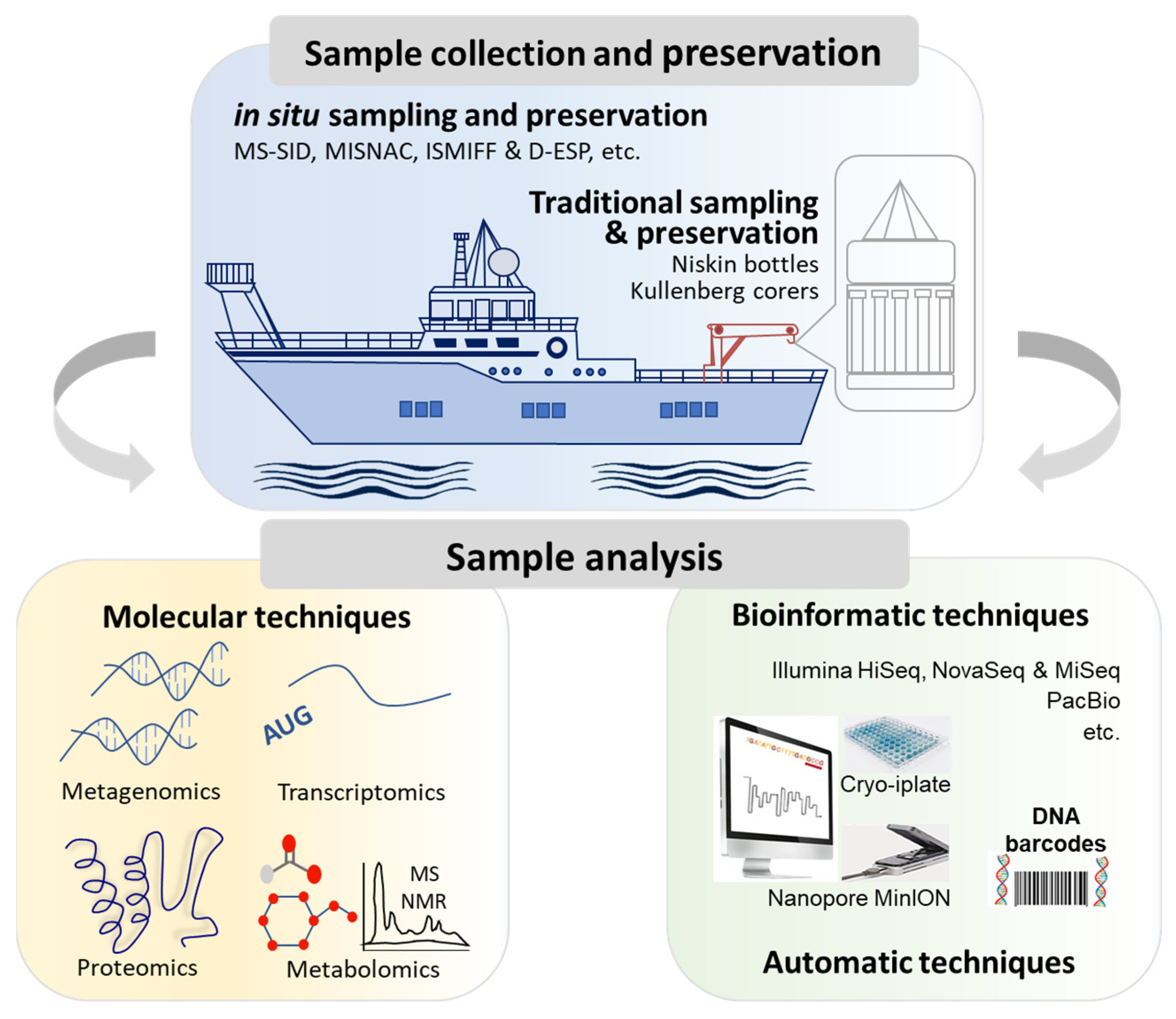
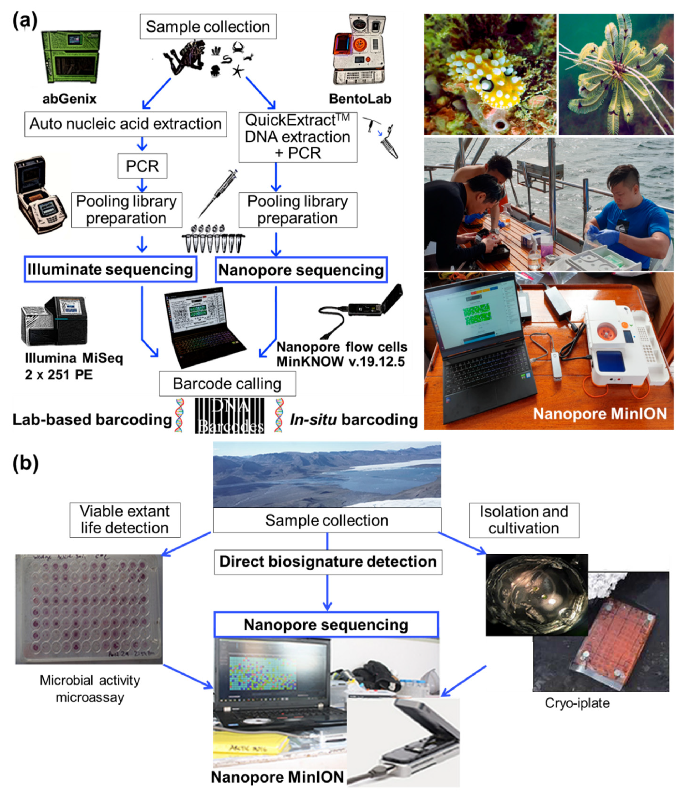
2.1. Collection of Marine Microbial Samples

The marine environments that microorganisms inhabit are usually under extreme conditions. Appropriate microbial specimens should be sampled according to the local conditions of the seawater and seabed [14]. According to the traditional sampling strategies, pre-sterilized bottles, bags, and/or corers, such as Niskin bottles and Kullenberg-type piston corers for shipboard water and sediment sample collections, should be accurately placed in predetermined column layers and seabed sites, strictly controlled at set stagnation durations, and returned in a timely manner to the deck for processing and a downstream analysis [15,16]. Considering the short lifespans of prokaryotic transcripts averages on the order of several minutes, however, traditional apparatuses are not appropriate for a nucleic acid-based analysis due to the time lapses, of up to hours, between the sample collection, preservation, and analyses [17]. In addition, the possible changes in pressure, temperature, and redox conditions from anoxic deep-sea zones to marine surfaces may alter microbial cell and community structures [4,5]. As a result, the gene expression and transcription profiles are likely to vary, caused by loss or interfered with fractions of DNA and rRNA as phylogenetic identifiers, making it difficult to obtain an accurate picture of marine microbial ecology.
In order to avoid the inaccuracy and variability of samples of a higher quality, the in situ collection, preservation, and detection of microbial samples was initiated using integrated sampling instruments in terms of deep-sea microbial sampling technology [18,19]. As shown in Table 1, the in situ sampling technologies currently used mainly focus on five technical requirements, i.e., low disturbance, pressure retaining, temperature retaining, no pollution, and no pressure drop transfer, demonstrating a better fidelity effect on marine microorganisms than the traditional methods [20]. With a pressure-retaining and thermal-insulation sampler (Figure 3a), the laboratory-measured rates of microbial methane oxidation in the Joetsu Knoll cold seep in Japan, mediated by both aerobic and anaerobic methanotrophs of gammaproteobacterial Methylococcales and ANME archaea at 10 MPa and 4 °C for 45 d, were generally consistent with the methane oxidation rates reported in other oceanographic sites [21]. By deploying a submersible-mounted sampler pressure-retaining and pressure-compensation unit, in-site sediment samples were successfully collected for the microbial communities investigation from hadal zones at a full ocean depth of 11,000 m in the West Philippine Basin, where the pressure change remained within ±6% [22]. The advent of genomic technologies has expanded the studies of gene diversity and expression in situ. The microbial sampler submersible incubation device (MS-SID) (Figure 3b), allowing for in situ tracer incubations coupled with in situ sampling and preservation, was used for profiling the gene expressions of marine microbiota in a bathypelagic water column in the Eastern Mediterranean Sea [23]. A higher percentage of MS-SID contigs were annotated (44%) than those in Niskin samples (29%), which might have contributed to the increased community complexity in the MS-SID samples with minimum environmental perturbations. More recent studies have also reported the dramatic differences in the microbial community characteristics between samples collected using multiple in situ nucleic acid collections (MISNACs), in situ microbial filtration and fixation (ISMIFF) apparatuses, and Niskin bottles [24,25], indicating the necessity for the in situ sampling and preservation of deep-sea marine samples. In the future, more high-fidelity and intelligent sampling methods must be proposed and practiced in research and development programs, and they will better meet the needs of expanding the exploration of complex marine ecosystems.

2.2. Analyses of Marine Microbial Samples

2.2.1. Molecular Analyses for Molecular Ecology

At present, marine microorganisms are phylogenetically studied according to the difference in their genetic structure and diversity, so as to explore their novel functions and, especially, uncultured microbial resources. Recent advances in studying the composition of marine microbial communities revealed several orders of magnitude of novel, uncultured species [9]. Molecular ecology, a science that studies the structure and functions of biological molecules, such as nucleic acids, proteins, and metabolites, has rapidly expanded our knowledge of marine microbial abundance, diversity, and ecological functions. Certain molecular technologies are at present commonly applied to marine microbial samples, including clone libraries, fingerprinting tools of denaturing/temperature gradient gel electrophoresis (DGGE/TGGE), catalyzed reporter deposition fluorescence in situ hybridization (CARD-FISH), real-time qPCR (qRT-PCR), and oligonucleotide microarrays (Geochip), as well as the emerging omics approaches of metagenomics, transcriptomics and proteomics, and metabolomics [27]. All of these technologies aim to understand the transmission of genetic and cellular information from marine microorganisms to marine ecosystems, each with its own advantages and limitations (Table 2). PCR-DGGE technology is one of the most commonly used methods in microbial molecular ecology, directly providing fingerprints of microbial communities in environmental samples after DNA extraction. However, there is no detailed classification and there are obvious shortcomings concerning the detection abundance and detection limit. qRT-PCR can determine the expression levels of functional genes; however, the key to its application is the design of functional gene primers [28]. Different environmental samples require redesigning primers for various functional genes, and the emergence of rapid and accurate functional Geochip technology has solved this problem. The probe labeling and hybridization of numerous genes can be completed in one experimental process with a high degree of automation [29]. CARD-FISH uses specific oligonucleotide probes to track and detect specific microorganisms; however, its sensitivity is not high, and sample processing requirements are high when dealing with complex communities [30]. Hence, the application of different molecular analysis methods needs to be evaluated based on the specific field situations.
In comparison to conventional phenotype-based markers, molecular markers can provide us with requisite landmarks for the elucidation of genetic variations in microbial ecology (Table 3). The 16S rRNA genes are the most frequently used phylogenetic markers in prokaryotic genomes. Based on 16S rRNA gene amplicon sequencing, the taxonomic identities of 26 tracer bacteria containing pathogenic and antimicrobial-resistant members were linked to coastal water pollution [34]. By sequencing the 16S rRNA genes of the V3–V4 region, 25 bacterial genera were correlated with water depth, temperature, salinity, redox, as well as Pb, Al, and aliphatic and aromatic hydrocarbon contents, deciphering the influence of specific environmental variables on the benthic microbial communities and dynamics from shallow- to deep-sea sites (44–3573 m) on spatiotemporal scales [35]. The housekeeping genes, e.g., rpoB and gyrB encoding ribosomal or DNA-linked proteins and amino-acyl synthetases, were recently highlighted as robust phylogenetic markers to precisely discriminate closely related species in microbial ecology [36,37,38]. A single copy of rpoB simplified the ecological interpretation of PCR-DGGE profiles than that of 16S rRNA genes with multiple copies and sequence heterogeneity [39]. A phylogenetic resolution of 4.5-times higher was recorded on the rpoB-based tree than on the 16S rRNA-based tree, providing more specific and sensitive DNA-sequencing subtypes for 13 Bacillus species from marine environments [40]. Additionally, the gyrB housekeeping gene was confirmed to be a valuable marker in distinguishing marine strains from the Bacillus pumilus clade [41]. As compared to 16S rRNA and housekeeping genes, functional gene amplicons are able to provide more accurate insights into the ecological potentials of highly diverse marine microorganisms in different and distant lineages. By analyzing genes for dissimilatory sulfite reductase and oxidase (dsrB and soxB) using combined high-through sequencing and qPCR, the distribution and composition of sulfur-oxidizing and sulphate-reducing bacteria in complex communities were depicted in marine sediment cores and surficial sediments along a bathymetric gradient [42,43]. In deep submarine permafrost, anaerobic methanotrophic archaea (ANME-2a/b and ANME-2d) responsible for the anaerobic oxidation of methane (AOM) were identified by functional marker genes (mcrA), CARD-FISH, and δ13C-methane signatures, suggesting the potential roles of AOM in global methane budgets [44]. With an isocitrate lyase (icl) gene maker and Geochip 2.0 targeting > 10,000 bacterial genes of ecological function with 50-mer probes, 25 bacterial isolates were determined with putative cold-adapted alleles in seacoast permafrost samples [45]. There are numerous studies on molecular markers interpreting the ecological significance of marine microorganisms, and more information will be provided by biomolecular omics technologies, advancing our knowledge of marine resource exploration, biodiversity conservation, and global climate change mitigation.

2.2.2. Molecular Analyses by Omics Technologies

Marine Metagenomics

Metagenomics is a discipline that takes the total genetic materials of all microorganisms in environmental samples as targets, sequences genomes, and screens functional genes to avoid the limitations of traditional cultivation-based microbiology [48]. It makes it possible to study the microbial samples collected directly from marine environments, showing the potential to efficiently address the taxonomic and functional compositions of heterogeneous marine microorganisms [49]. Metagenomics technologies for marine microbiota studies mainly adopt high-throughput sequencing approaches, represented by the next- or second-generation sequencing of Roche 454 and Illumina sequencing platforms [50,51]. Among them, 454 pyrosequencing can generate large-scale DNA fragments of short read lengths, which is an ideal platform for the rapid sequencing of environmental genomes. On a hybrid platform of clone-dependent long-read Sanger and high-throughput short-read 454 pyrosequencing, high-quality draft assemblies were sequenced for 6 small marine microbial genomes in the most timely and cost-effective way [50]. With complementary 18S rDNA clone libraries corresponding to 454 pyrosequencing data, near full-length sequence information on the biodiversity and community compositions was obtained for micro-, nano-, and pico-planktons in marine environments, illustrating the potential for retrieving taxa from the rare biosphere [51]. By providing millions of reads in a single run, Illumina HiSeq, MiSeq, and NovaSeq sequencers considerably contributed to the 16S rRNA gene libraries in the NCBI GenBank database with a greater sequencing depth and higher base-calling accuracy [52,53]; though, they generally suffer from limitations regarding sequence assemblies with high biodiversity and the retrieval of adaptive genes in marine prokaryotes [54]. Illumina MiniSeq, the latest benchtop Illumina sequencer, has been proved to rapidly and accurately predict antibiotic resistance by a fully automated bioinformatics system in clinical microbiology [2], which is also a potential candidate for field studies in marine microbiology.
The Pacific Biosciences (PacBio) sequencer, a third-generation platform for long-read metagenomics, allows for the direct retrieval of full-length genes to avoid the bias of sequence splicing, assembly, and annotation in bioinformatics analyses [55]. For 23 bacterial and 3 archaeal species, the PacBio complete 16S rRNA gene sequences showed less ambiguous classifications than the Illumina V4 16S rRNA gene sequences [56], demonstrating it to be a powerful tool for detecting more marine microbial species at the genus and species levels [57]. Oxford nanopore technology, another third-generation platform for single-molecule real-time sequencing, depends on a single base of nucleic acids and a protein nanopore reader. It does not employ PCR amplification or chemical labeling, leaving no theoretical limit on the reading length of DNA or RNA fragments [58]. It has been applied to decode the longer sequences of 16S rRNA, 18S rRNA, and internal transcribed spacer loci, achieving comprehensive taxonomic profiling and high-resolution mapping results for the genera and species of bacteria, protists, and fungi thriving on marine plastic debris and seawater [59,60]. Taking advantage of its scalability and portability, a mobile MinION sequencer and nanopore flow cell were incorporated into an in situ field sequencing setup (Figure 4a), where 32 samples were sequenced simultaneously for DNA barcoding and species identification onboard [61]. In a novel MICRObial life-detection platform (Figure 4b), 39 microbial isolates and an active L-serine metabolism community were identified by cryo-iPlate and a microbial activity microassay, which were further analyzed by MinION for biosignature characterizations and validated by Illumina MiSeq results. With the assistance of the base calling and uplink/downlink of nucleic acid sequences through satellite Internet, a metagenome was completed for viable microbes in the Canadian High Arctic environment [62]. With an added advantage of low capital costs, nanopore sequencers are promising shipborne solutions for the on-site analyses of complex marine microorganisms. On the whole, the development and application of long-read, real-time, and highly flexible sequencing platforms allows for the achievement of more complete metagenome-assembled genomes from marine microorganisms containing many adaptive genes with ecological functions, which significantly contribute to the exploration and conservation of marine resources.

Marine Transcriptomics and Proteomics

Transcriptomics focuses on all RNA transcripts, including those of mRNA, non-coding RNA, and small RNA, to identify their transcriptional and post-transcriptional gene expressions and regulations, aiming to interpret the underlying biological processes in response to environmental perturbations [63]. Current transcription technologies mainly include microarray or tiling arrays and transcriptome sequencing technology (RNA-Seq) [64,65,66]. Microarray or tiling arrays utilize microchips with hybridization probes of known or entire DNA sequences to specifically survey global patterns in gene expressions and regulations [67], while RNA-Seq is the high-throughput sequencing of transcribed cDNA most frequently performed on Illumina HiSeq platforms [66]. With a high-definition tiling array of marine diatom Phaeodactylum tricornutum, the first whole-genome methylome was obtained from a stramenopile to illustrate the adaptive evolutionary processes of gene-body methylation among eukaryotes, and the expression levels of methylated genes were quantified by RNA-seq data [68]. The ever-growing transcriptomic evidence can reveal niche differentiations, account for certain metagenomic limitations, or identify potential functional genes in marine microorganisms. By exhibiting various co-occurring expression patterns of carbon, nitrogen, phosphorus, and sulfur elemental cycling genes, the transcriptomic activities of three primary marine Thaumarchaeota ecotypes were found to respond differently to chlorophyll α, salinity, and nutrient concentrations that were related to seawater depth and seasonal changes [69]. With the advent of total RNA sequencing, 47 new viruses associated with vertebrate, host fish diet, or their microbiomes were identified by characterizing the viromes of 19 wild-caught fish species, exploring more of the marine virosphere [70]. Combined with Illumina HiSeq mRNA-seq, comparative transcriptomic, and qRT-PCR analyses, a series of potential genes regulating lipid fraction migration, docosahexaenoic acid biosynthesis, signal transduction, and cell transport were analyzed in a heterotrophic marine microalga, Schizochytrium sp., which could promote the industrial production of single cell oil [71].
Proteomics is an additional supplement to metagenomics and transcriptomics by comparative or quantitative, structural, and functional analyses. After protein separation by electrophoresis or liquid/gas chromatography, traditional Edman sequencing has a low throughput result and requires a large sample size, and advances in mass spectrometry (MS) have made highly sensitive proteomics more cost effective [72]. Tandem mass tag-based proteomics showed that, by screening and analyzing 437 proteins with differential expression levels, marine bacterium Halobacillus sp. P1 inhibited the nitrogen metabolism of diatom Skeletonema costatum during nutrient competition, hence revealing the algicidal mechanism in marine algae–bacteria interactions [73]. Quantitative proteomics suggested that three marine bacterial lineages, i.e., Oceanospirillaceae, Roseobacter, and Flavobacteria, profoundly altered their proteomic profiles during growth transition, with characteristic patterns in the abundance of key enzymes in energy and central carbon metabolism processes [74]. The cold and alkane-induced proteins, which were depicted by LC-MS/MS shotgun proteomics, were up to 21-fold higher in obligate hydrocarbon-degrading psychrophile Oleispira antarctica RB-8 grown on n-alkanes at 4 °C than in the non-hydrocarbon control group [75]. These studies pointed out the scientific potential of proteomics to address the divergent ecological strategies of marine microorganisms in adapting to deteriorated marine environments with algal blooms, oil spills, and other contaminants.

Marine Metabolomics

Metabolomics is an emerging omics technology following transcriptomics and proteomics, which is the closest to the biological phenotype in various omics studies. Metabolite detection commonly relies on nuclear magnetic resonance (NMR) and mass spectrometry (MS) that couples with gas chromatography (GC), liquid chromatography (LC), and capillary electrophoresis (CE) (Table 4) [76,77]. NMR can identify high concentrations of metabolites; however, it is limited to sensitivity and signal congestion in detecting metabolites at low levels [78]. The GC/LC/CE-MS detection of metabolites is superior, with a lower analysis limit and higher repeatability, and has been applied to comprehensively and intensively analyze marine metabolomics [79]. As a powerful tool to depict the internal metabolome of targeting cells, classical metabolic fingerprinting enables the identification of potential metabolite targets in marine drug discovery [80,81]. Metabolic footprinting, a more recently developed approach for detecting secreted, excreted, or consumed metabolites, is of particular interest in investigating the interactions between environment/genetic and marine ecosystems [82].
Marine metabolomics focuses on developing marine microbial resources and understanding the dynamics of marine ecosystems. In many natural product research programs, metabolomics technologies have been adopted for the separation, quantification, and elucidation of bioactive molecules. Based on LC-MS and molecular networking, five new pyrrole-derived alkaloids were isolated from marine Micromonospora sp. bacterium, where two of them exhibited antibacterial activity against methicillin-resistant strains [93]. With the advent of metabolomics, the MS/MS data of an increasing number of natural products from microorganisms inhabiting marine environments have been archived in global natural products social molecular networking (GNPS, http://gnps.ucsd.edu (accessed on 12 May 2023)). Metabolomics has also taken a central role in profiling the ecological impacts of chemical residues/pollutants on the metabolome of various marine microorganisms, so as to illustrate their biological responses to environmental changes. Under stresses of heavy metals, the NMR-based metabolomics of microalgae Chlorella sp. revealed an increased yield of extracellular polymeric substances to perform Cu uptake [94]. Felline, et al. [90] characterized the metabolome profile of the brown algae Fucus virsoides under the stress of glyphosate-based herbicides. The NMR result showed that herbicides down-regulated the metabolism of aromatic amino acids but up-regulated the shikimate content of exposed thalli. In marine microalgae Dunaliella salina exposed to polystyrene microplastics, the significant up-regulations of the amino acid biosynthesis, ATP-binding cassette transportation, and glycerophospholipid metabolism were speculated by LC-MS analysis [95]. With up-/down-regulated metabolic pathways identified under environmental stressors, it is possible to perform the biomarker identification and ecological risk assessment and achieve the toxicant action elucidation and discovery of many other marine processes.

3. Conclusions and Challenges

3.1. Conclusions

This review summarizes the cutting-edge technologies for the collection of marine microbial samples and the applications of molecular ecology on marine microorganisms.
(1) With the expansion of marine resource explorations to deeper and wider sea areas, deep-sea sampling equipment tends to be diversified in structure, highly functional, and intelligent in operation. Traditional sampling methods are usually time consuming, while in situ collection, preservation, and detection have become the mainstream methods to maintain the integrity of marine microbial samples.
(2) Metagenomics, transcriptomics, and other omics technologies help to conduct broad-scale diversity assessments of both cultured and uncultured marine species. Based on the analyses of microbial diversity, population structure, and evolutionary relationships, the functional activity, mutual collaboration, and relationship with the environment of microbial populations can be further explored.

3.2. Challenges

The global marine microbial resources investigation projects show the large groups of new species and gene resources in the ocean ecosystems. For example, the results of the Tara Oceans Expedition project indicate that the rich diversity of marine microorganisms in colony structure, metabolic pathways, physiological and biochemical reactions, metabolites, and other aspects are abundant resources with great potential [96,97]. At present, however, the research on marine microorganisms is still confronted by many challenges that need to be addressed urgently in the near future.
(1) Due to the extreme environmental conditions of ocean ecosystems, how to provide the most original marine microbial samples for scientific studies has become a worldwide research focus. The high-fidelity samples for scientific research are important indicators to measure the performance of deep-sea sampling equipment. Therefore, the development of low disturbance, pressure-retaining, and thermal-insulation deep-sea samples will better meet the needs of deep-sea resource explorations and developments, deep-sea biogeochemical cycles, and basic marine science research, and will certainly become an important development direction for marine microbial sampling strategies.
(2) How to stably preserve the collected high-fidelity samples, so as to provide reliable samples for omics research and ensure the accuracy of the research results, has become a key issue in the field investigation of marine microorganisms. The development of a stable preservation and in situ analysis platform for marine microbial samples in the future will greatly facilitate the mining of deep-sea resources.
(3) The continuous improvement of effective cultivation methods of marine microorganisms is an important aspect to promote the development of marine microbiome from descriptive to functional and applied research methods. The combination of multiple research methods further makes up for the shortcomings of single technology methods, identifies the diversity of marine microorganisms, and thoroughly explores the functions of the obtained microorganisms at the gene, protein, and metabolite levels.
To date, we achieved important information on marine biodiversity and the spatiotemporal distribution patterns of microbial communities. However, our understanding of the underlying mechanisms for maintaining the ecological functions of marine microorganisms is insufficient. With the continuous development of metagenomics, transcriptomics, proteomics, metabolomics, and other multi-omics techniques, the ecological functions of microorganisms can be revealed at molecular levels in marine ecosystems. The microbial roles can be better understood in biogeochemical processes, such as carbon cycling, nitrogen cycling, and sulfur cycling, in response to environmental changes. In addition, marine microbial research is not limited to scientific explorations and also applies to practical applications as bioenergy production, biomaterial manufacturing, and drug discovery, etc. Future research should focus more extensively on multi-scale research, from micro- to macro-scales, and from microbial single cells to complex ecosystems, in order to gain a more comprehensive understanding of the marine ecosystems driven by diverse microorganisms.

Author Contributions

Conceptualization, H.L.; writing—original draft preparation, Z.H. and F.F.; writing—review and editing, L.D., K.Y., L.Z. and H.L.; visualization, Z.H. and L.Z.; supervision, L.Z. and H.L.; project administration, H.L.; funding acquisition, H.L. and L.D. All authors have read and agreed to the published version of the manuscript.

Funding

This research was funded by the Major Project for Basic and Applied Basic Research of Guangdong Province (grant number 2020B0301030003), the China Geological Survey Project (grant numbers DD20221703 and DD20230063), and the Natural Science Foundation of Top Talent of SZTU (grant number GDRC202115).

Institutional Review Board Statement

Not applicable.

Informed Consent Statement

Not applicable.

Data Availability Statement

Not applicable.

Conflicts of Interest

The authors declare no conflict of interest.

References

  1. Bickle, M.; Arculus, R.; Barrett, P.; DeConto, R.; Camoin, G.; Edwards, K.; Fisher, A.; Inagaki, F.; Kodaira, S.; Ohkouchi, N.; et al. Illuminating Earth’s Past, Present, and Future: The Science Plan for the International Ocean Discovery Program 2013–2023; I.O.D.P.M. International: Washington, DC, USA, 2011. [Google Scholar]
  2. Kumar, N.; Raven, K.E.; Blane, B.; Leek, D.; Brown, N.M.; Bragin, E.; Rhodes, P.A.; Parkhill, J.; Peacock, S.J. Evaluation of a fully automated bioinformatics tool to predict antibiotic resistance from MRSA genomes. J. Antimicrob. Chemother. 2020, 75, 1117–1122. [Google Scholar] [CrossRef]
  3. Sun, Y.H.; Liu, Y.; Pan, J.; Wang, F.P.; Li, M. Perspectives on cultivation strategies of archaea. Microb. Ecol. 2020, 79, 770–784. [Google Scholar]
  4. Cario, A.; Oliver, G.C.; Rogers, K.L. Exploring the deep marine biosphere: Challenges, innovations, and opportunities. Front. Earth Sci. 2019, 7, 225. [Google Scholar]
  5. Garel, M.; Bonin, P.; Martini, S.; Guasco, S.; Roumagnac, M.; Bhairy, N.; Armougom, F.; Tamburini, C. Pressure-retaining sampler and high-pressure systems to study deep-sea microbes under in situ conditions. Front. Microbiol. 2019, 10, 453. [Google Scholar]
  6. Imachi, H.; Nobu, M.K.; Miyazaki, M.; Tasumi, E.; Saito, Y.; Sakai, S.; Ogawara, M.; Ohashi, A.; Takai, K. Cultivation of previously uncultured microorganisms with a continuous-flow down-flow hanging sponge (DHS) bioreactor, using a syntrophic archaeon culture obtained from deep marine sediment as a case study. Nat. Protoc. 2022, 17, 2784–2814. [Google Scholar]
  7. Liu, W.; Zheng, X.W.; Dai, X.; Zhang, Z.F.; Zhang, W.Y.; Xiao, T.; Huang, L. Isolation and characterization of the first temperate virus infecting Psychrobacillus from marine sediments. Viruses 2022, 14, 108. [Google Scholar] [CrossRef]
  8. Pope, E.; Cartmell, C.; Haltli, B.; Ahmadi, A.; Kerr, R.G. Microencapsulation and in situ incubation methodology for the cultivation of marine bacteria. Front. Microbiol. 2022, 13, 958660. [Google Scholar]
  9. Wang, F.P.; Li, M.; Huang, L.; Zhang, X.H. Cultivation of uncultured marine microorganisms. Mar. Life Sci. Technol. 2021, 3, 117–120. [Google Scholar]
  10. Caron, D.A.; Alexander, H.; Allen, A.E.; Archibald, J.M.; Armbrust, E.V.; Bachy, C.; Bell, C.J.; Bharti, A.; Dyhrman, S.T.; Guida, S.M.; et al. Probing the evolution, ecology and physiology of marine protists using transcriptomics. Nat. Rev. Microbiol. 2017, 15, 6–20. [Google Scholar]
  11. Kodama, T.; Taniuchi, Y.; Kasai, H.; Yamaguchi, T.; Nakae, M.; Okumura, Y. Empirical estimation of marine phytoplankton assemblages in coastal and offshore areas using an in situ multi-wavelength excitation fluorometer. PLoS ONE 2022, 17, e0257258. [Google Scholar]
  12. Hiraoka, S.; Sumida, T.; Hirai, M.; Toyoda, A.; Kawagucci, S.; Yokokawa, T.; Nunoura, T. Diverse DNA modification in marine prokaryotic and viral communities. Nucleic Acids Res. 2022, 50, 1531–1550. [Google Scholar] [PubMed]
  13. Bruns, S.; Wienhausen, G.; Scholz-Boettcher, B.; Wilkes, H. Simultaneous quantification of all B vitamins and selected biosynthetic precursors in seawater and bacteria by means of different mass spectrometric approaches. Anal. Bioanal. Chem. 2022, 414, 7839–7854. [Google Scholar] [PubMed]
  14. Tuit, C.B.; Wait, A.D. A review of marine sediment sampling methods. Environ. Forensics 2020, 21, 291–309. [Google Scholar] [CrossRef]
  15. Feike, J.; Juergens, K.; Hollibaugh, J.T.; Krueger, S.; Jost, G.; Labrenz, M. Measuring unbiased metatranscriptomics in suboxic waters of the central Baltic Sea using a new in situ fixation system. ISME J. 2012, 6, 461–470. [Google Scholar] [CrossRef]
  16. Lunne, T.; Long, M. Review of long seabed samplers and criteria for new sampler design. Mar. Geol. 2006, 226, 145–165. [Google Scholar]
  17. Wei, Z.-F.; Li, W.-L.; Li, J.; Chen, J.; Xin, Y.-Z.; He, L.-S.; Wang, Y. Multiple in situ nucleic acid collections (MISNAC) from deep-sea waters. Front. Mar. Sci. 2020, 7, 81. [Google Scholar] [CrossRef]
  18. Taylor, C.D.; Edgcomb, V.P.; Doherty, K.W.; Engstrom, I.; Shanahan, T.; Pachiadaki, M.G.; Molyneaux, S.J.; Honjo, S. Fixation filter, device for the rapid in situ preservation of particulate samples. Deep-Sea Res. Part I 2015, 96, 69–79. [Google Scholar] [CrossRef]
  19. Hendricks, A.; Mackie, C.M.; Luy, E.; Sonnichsen, C.; Smith, J.; Grundke, I.; Tavasoli, M.; Furlong, A.; Beiko, R.G.; LaRoche, J.; et al. Compact and automated eDNA sampler for in situ monitoring of marine environments. Sci. Rep. 2023, 13, 5210. [Google Scholar] [CrossRef]
  20. He, S.; Peng, Y.; Jin, Y.; Wan, B.; Liu, G. Review and analysis of key techniques in marine sediment sampling. Chin. J. Mech. Eng. 2020, 33, 66. [Google Scholar] [CrossRef]
  21. Case, D.H.; Ijiri, A.; Morono, Y.; Tavormina, P.; Orphan, V.J.; Inagaki, F. Aerobic and anaerobic methanotrophic communities associated with methane hydrates exposed on the seafloor: A high-pressure sampling and stable isotope-incubation experiment. Front. Microbiol. 2017, 8, 2569. [Google Scholar]
  22. He, S.; Qiu, S.; Tang, W.; Peng, Y.; Jin, Y. A novel submersible-mounted sediment pressure-retaining sampler at full ocean depth. Front. Mar. Sci. 2023, 10, 1154269. [Google Scholar] [CrossRef]
  23. Edgcomb, V.P.; Taylor, C.; Pachiadaki, M.G.; Honjo, S.; Engstrom, I.; Yakimov, M. Comparison of Niskin vs. in situ approaches for analysis of gene expression in deep Mediterranean Sea water samples. Deep-Sea Res. Part II 2016, 129, 213–222. [Google Scholar] [CrossRef]
  24. Li, J.; Xin, Y.; Cai, D.; Chen, J.; Li, W.; Wang, S.; Wei, Z.; Zhang, A. Development and scientific application of deep-sea multiple in situ nucleic acid collections (MISNAC) apparatus. In Proceedings of the Global Oceans 2020: Singapore—U.S. Gulf Coast, Biloxi, MS, USA, 5–30 October 2020; pp. 1–5. [Google Scholar]
  25. Wang, Y.; Gao, Z.-M.; Li, J.; He, L.-S.; Cui, G.-J.; Li, W.-L.; Chen, J.; Xin, Y.-Z.; Cai, D.-S.; Zhang, A.-Q. Hadal water sampling by in situ microbial filtration and fixation (ISMIFF) apparatus. Deep-Sea Res. Part I 2019, 144, 132–137. [Google Scholar] [CrossRef]
  26. Teague, J.; Scott, T.B.; Sharma, S.; Graham, G.; Allen, M.J. An Alternative Method to Niskin Sampling for Molecular Analysis of the Marine Environment. J. Mar. Sci. Eng. 2017, 5, 22. [Google Scholar] [CrossRef]
  27. He, Z.; Deng, Y.; Van Nostrand, J.D.; Tu, Q.; Xu, M.; Hemme, C.L.; Li, X.; Wu, L.; Gentry, T.J.; Yin, Y.; et al. GeoChip 3.0 as a high-throughput tool for analyzing microbial community composition, structure and functional activity. ISME J. 2010, 4, 1167–1179. [Google Scholar] [CrossRef]
  28. Zhao, X.; Yang, H.L.; Chen, M.J.; Song, X.X.; Yu, C.X.; Zhao, Y.; Wu, Y.J. Reference gene selection for quantitative real-time pcr of mycelia from lentinula edodes under high-temperature stress. Biomed Res. Int. 2018, 2018, 1670328. [Google Scholar] [CrossRef]
  29. Tu, Q.C.; Yu, H.; He, Z.L.; Deng, Y.; Wu, L.Y.; Van Nostrand, J.D.; Zhou, A.F.; Voordeckers, J.; Lee, Y.J.; Qin, Y.J.; et al. GeoChip 4: A functional gene-array-based high-throughput environmental technology for microbial community analysis. Mol. Ecol. Resour. 2014, 14, 914–928. [Google Scholar] [CrossRef]
  30. Chu, Y.H.; Heather, H.; Zhang, R.R.; Guo, Z.Y.; Ricardo, V.L. In situ hybridization: Introduction to techniques, applications and pitfalls in the performance and interpretation of assays. Semin. Diagn. Pathol. 2019, 36, 336–341. [Google Scholar] [CrossRef]
  31. Li, Z.H.; Rui, J.P.; Li, X.Z.; Li, J.B.; Dong, L.; Huang, Q.L.; Huang, C.; Wang, Z.P.; Li, L.; Xuan, P.; et al. Bacterial community succession and metabolite changes during doubanjiang-meju fermentation, a Chinese traditional fermented broad bean (Vicia faba L.) paste. Food Chem. 2017, 218, 534–542. [Google Scholar] [CrossRef]
  32. Bustin, S.A.; Benes, V.; Nolan, T.; Pfaffl, M.W. Quantitative real-time RT-PCR—A perspective. J. Mol. Endocrinol. 2005, 34, 597–601. [Google Scholar] [CrossRef]
  33. Rybalka, N.; Blanke, M.; Tzvetkova, A.; Noll, A.; Roos, C.; Boy, J.; Boy, D.; Nimptsch, D.; Godoy, R.; Friedl, T. Unrecognized diversity and distribution of soil algae from Maritime Antarctica (Fildes Peninsula, King George Island). Front. Microbiol. 2023, 14, 1118747. [Google Scholar]
  34. Pendergraft, M.A.; Belda-Ferre, P.; Petras, D.; Morris, C.K.; Mitts, B.A.; Aron, A.T.; Bryant, M.; Schwartz, T.; Ackermann, G.; Humphrey, G.; et al. Bacterial and chemical evidence of coastal water pollution from the Tijuana river in sea spray aerosol. Environ. Sci. Technol. 2023, 57, 4071–4081. [Google Scholar] [CrossRef] [PubMed]
  35. Sanchez-Soto, M.F.; Cerqueda-Garcia, D.; Aguirre-Macedo, M.L.; Garcia-Maldonado, J.Q. Spatiotemporal dynamics of benthic bacterial communities in the Perdido Fold Belt, Northwestern Gulf of Mexico. Front. Mar. Sci. 2023, 10, 1070548. [Google Scholar]
  36. Savi, D.C.; Aluizio, R.; Galli-Terasawa, L.; Kava, V.; Glienke, C. 16S-gyrB-rpoB multilocus sequence analysis for species identification in the genus Microbispora. Anton. Leeuw. 2016, 109, 801–815. [Google Scholar]
  37. Guo, S.L.; Yang, Q.H.; Feng, J.J.; Duan, L.H.; Zhao, J.P. Phylogenetic analysis of the pathogenic genus Aeromonas spp. isolated from diseased eels in China. Microb. Pathog. 2016, 101, 12–23. [Google Scholar] [PubMed]
  38. Ogier, J.C.; Pages, S.; Galan, M.; Barret, M.; Gaudriault, S. rpoB, a promising marker for analyzing the diversity of bacterial communities by amplicon sequencing. BMC Microbiol. 2019, 19, 171. [Google Scholar]
  39. Peixoto, R.S.; Coutinho, H.L.D.; Rumjanek, N.G.; Macrae, A.; Rosado, A.S. Use of rpoB and 16S rRNA genes to analyse bacterial diversity of a tropical soil using PCR and DGGE. Lett. Appl. Microbiol. 2002, 35, 316–320. [Google Scholar] [CrossRef]
  40. Ki, J.-S.; Zhang, W.; Qian, P.-Y. Discovery of marine Bacillus species by 16S rRNA and rpoB comparisons and their usefulness for species identification. J. Microbiol. Methods 2009, 77, 48–57. [Google Scholar] [CrossRef]
  41. Freitas-Silva, J.; de Oliveira, B.F.R.; Vigoder, F.d.M.; Muricy, G.; Dobson, A.D.W.; Laport, M.S. Peeling the layers away: The genomic characterization of Bacillus pumilus 64-1, an isolate with antimicrobial activity from the marine sponge Plakina cyanorosea (Porifera, homoscleromorpha). Front. Microbiol. 2021, 11, 592735. [Google Scholar]
  42. Zhang, Y.; Wang, X.; Zhen, Y.; Mi, T.; He, H.; Yu, Z. Microbial diversity and community structure of sulfate-reducing and sulfur-oxidizing bacteria in sediment cores from the East China Sea. Front. Microbiol. 2017, 8, 2133. [Google Scholar]
  43. Sanchez-Soto, M.F.; Cerqueda-Garcia, D.; Alcantara-Hernandez, R.J.; Falcon, L.I.; Pech, D.; arcega-Cabrera, F.; Aguirre-Macedo, M.L.; Garcia-Maldonado, J.Q. Assessing the diversity of benthic sulfate-reducing microorganisms in Northwestern Gulf of Mexico by Illumina sequencing of dsrB gene. Microb. Ecol. 2021, 81, 908–921. [Google Scholar] [PubMed]
  44. Winkel, M.; Mitzscherling, J.; Overduin, P.P.; Horn, F.; Winterfeld, M.; Rijkers, R.; Grigoriev, M.N.; Knoblauch, C.; Mangelsdorf, K.; Wagner, D.; et al. Anaerobic methanotrophic communities thrive in deep submarine permafrost. Sci. Rep. 2018, 8, 1291. [Google Scholar] [PubMed]
  45. Hinsa-Leasure, S.M.; Bhavaraju, L.; Rodrigues, J.L.M.; Bakermans, C.; Gilichinsky, D.A.; Tiedje, J.M. Characterization of a bacterial community from a Northeast Siberian seacoast permafrost sample. FEMS Microbiol. Ecol. 2010, 74, 103–113. [Google Scholar] [PubMed]
  46. Rani, S.; Koh, H.W.; Rhee, S.K.; Fujitani, H.; Park, S.J. Detection and diversity of the nitrite oxidoreductase alpha subunit (nxrA) gene of Nitrospina in marine sediments. Microb. Ecol. 2017, 73, 111–122. [Google Scholar]
  47. Lau, E.; Fisher, M.C.; Steudler, P.A.; Cavanaugh, C.M. The methanol dehydrogenase gene, mxaF, as a functional and phylogenetic marker for proteobacterial methanotrophs in natural environments. PLoS ONE 2013, 8, e56993. [Google Scholar]
  48. Quince, C.; Walker, A.W.; Simpson, J.T.; Loman, N.J.; Segata, N. Shotgun metagenomics, from sampling to analysis. Nat. Biotechnol. 2017, 35, 833–844. [Google Scholar]
  49. Cao, S.; Zhang, W.; Ding, W.; Wang, M.; Fan, S.; Yang, B.; McMinn, A.; Wang, M.; Xie, B.-b.; Qin, Q.-L.; et al. Structure and function of the Arctic and Antarctic marine microbiota as revealed by metagenomics. Microbiome 2020, 8, 47. [Google Scholar]
  50. Goldberg, S.M.D.; Johnson, J.; Busam, D.; Feldblyum, T.; Ferriera, S.; Friedman, R.; Halpern, A.; Khouri, H.; Kravitz, S.A.; Lauro, F.M.; et al. A Sanger/pyrosequencing hybrid approach for the generation of high-quality draft assemblies of marine microbial genomes. Proc. Natl. Acad. Sci. USA 2006, 103, 11240–11245. [Google Scholar] [CrossRef]
  51. Wolf, C.; Kilias, E.S.; Metfies, K. Evaluating the potential of 18S rDNA clone libraries to complement pyrosequencing data of marine protists with near full-length sequence information. Mar. Biol. Res. 2014, 10, 771–780. [Google Scholar] [CrossRef]
  52. Aylagas, E.; Borja, A.; Rodriguez-Ezpeleta, N. Environmental status assessment using DNA metabarcoding: Towards a genetics based marine biotic index (gAMBI). PLoS ONE 2014, 9, e90529. [Google Scholar]
  53. Singer, G.A.C.; Fahner, N.A.; Barnes, J.G.; McCarthy, A.; Hajibabaei, M. Comprehensive biodiversity analysis via ultra-deep patterned flow cell technology: A case study of eDNA metabarcoding seawater. Sci. Rep. 2019, 9, 5991. [Google Scholar] [PubMed]
  54. Haro-Moreno, J.M.; López-Pérez, M.; Rodríguez-Valera, F. Long read metagenomics, the next step? BioRxiv 2020. [Google Scholar] [CrossRef]
  55. Haro-Moreno, J.M.; Lopez-Perez, M.; Rodriguez-Valera, F. Enhanced recovery of microbial genes and genomes from a marine water column using long-read metagenomics. Front. Microbiol. 2021, 12, 708782. [Google Scholar]
  56. Singer, E.; Bushnell, B.; Coleman-Derr, D.; Bowman, B.; Bowers, R.M.; Levy, A.; Gies, E.A.; Cheng, J.-F.; Copeland, A.; Klenk, H.-P.; et al. High-resolution phylogenetic microbial community profiling. ISME J. 2016, 10, 2020–2032. [Google Scholar] [PubMed]
  57. Wang, S.; Su, X.; Cui, H.; Wang, M.; Hu, X.; Ding, W.; Zhang, W. Microbial richness of marine biofilms revealed by sequencing full-length 16s rRNA genes. Genes 2022, 13, 1050. [Google Scholar] [CrossRef] [PubMed]
  58. Xie, S.; Leung, A.W.-S.; Zheng, Z.; Zhang, D.; Xiao, C.; Luo, R.; Luo, M.; Zhang, S. Applications and potentials of nanopore sequencing in the (epi)genome and (epi)transcriptome era. Innovation 2021, 2, 100153. [Google Scholar] [PubMed]
  59. Marsay, K.S.; Koucherov, Y.; Davidov, K.; Iankelevich-Kounio, E.; Itzahri, S.; Salmon-Divon, M.; Oren, M. High-resolution screening for marine prokaryotes and eukaryotes with selective preference for polyethylene and polyethylene terephthalate surfaces. Front. Microbiol. 2022, 13, 845144. [Google Scholar]
  60. Curren, E.; Yoshida, T.; Kuwahara, V.S.; Leong, S.C.Y. Rapid profiling of tropical marine cyanobacterial communities. Reg. Stud. Mar. Sci. 2019, 25, 100485. [Google Scholar]
  61. Chang, J.J.; Ip, Y.C.; Ng, C.S.; Huang, D. Takeaways from mobile DNA barcoding with BentoLab and MinION. Genes 2020, 11, 1121. [Google Scholar] [CrossRef]
  62. Goordial, J.; Altshuler, I.; Hindson, K.; Chan-Yam, K.; Marcolefas, E.; Whyte, L.G. In situ field sequencing and life detection in remote (79 degrees 26′ N) Canadian high Arctic permafrost ice wedge microbial communities. Front. Microbiol. 2017, 8, 2594. [Google Scholar] [CrossRef]
  63. Hassan, S.; Sabreena; Khurshid, Z.; Bhat, S.A.; Kumar, V.; Ameen, F.; Ganai, B.A. Marine bacteria and omic approaches: A novel and potential repository for bioremediation assessment. J. Appl. Microbiol. 2022, 133, 2299–2313. [Google Scholar] [PubMed]
  64. Dong, X.; You, Y.; Wu, J.Q. Building an RNA sequencing transcriptome of the central nervous system. Neuroscientist 2016, 22, 579–592. [Google Scholar] [CrossRef] [PubMed]
  65. Li, W.V.; Chen, Y.; Li, J.J. TROM: A testing-based method for finding transcriptomic similarity of biological samples. Stat. Biosci. 2017, 9, 105–136. [Google Scholar] [PubMed]
  66. Page, T.M.; Lawley, J.W. The next generation is here: A review of transcriptomic approaches in marine ecology. Front. Mar. Sci. 2022, 9, 757921. [Google Scholar]
  67. Rachinger, N.; Fischer, S.; Bohme, I.; Linck-Paulus, L.; Kuphal, S.; Kappelmann-Fenzl, M.; Bosserhoff, A.K. Loss of gene information: Discrepancies between RNA sequencing, cDNA microarray, and qRT-PCR. Int. J. Mol. Sci. 2021, 22, 9349. [Google Scholar] [PubMed]
  68. Veluchamy, A.; Lin, X.; Maumus, F.; Rivarola, M.; Bhavsar, J.; Creasy, T.; O’Brien, K.; Sengamalay, N.A.; Tallon, L.J.; Smith, A.D.; et al. Insights into the role of DNA methylation in diatoms by genome-wide profiling in Phaeodactylum tricornutum. Nat. Commun. 2013, 4, 2091. [Google Scholar] [CrossRef]
  69. Lu, Y.; Lv, Y.X.; Zhang, Y.; Liu, Q.; Xu, X.W.; Xiao, X.; Xu, J. Metatranscriptomes reveal the diverse responses of Thaumarchaeota ecotypes to environmental variations in the northern slope of the South China Sea. Environ. Microbiol. 2023, 25, 410–427. [Google Scholar] [CrossRef]
  70. Geoghegan, J.L.; Giallonardo, F.D.; Wille, M.; Ortiz-Baez, A.S.; Costa, V.A.; Ghaly, T.; Mifsud, J.C.O.; Turnbull, O.M.H.; Bellwood, D.R.; Williamson, J.E.; et al. Erratum: Virome composition in marine fish revealed by meta-transcriptomics. Virus Evol. 2021, 7, veab005. [Google Scholar]
  71. Ren, L.; Hu, X.; Zhao, X.; Chen, S.; Wu, Y.; Li, D.; Yu, Y.; Geng, L.; Ji, X.; Huang, H. Transcriptomic analysis of the regulation of lipid fraction migration and fatty acid biosynthesis in Schizochytrium sp. Sci. Rep. 2017, 7, 3562. [Google Scholar]
  72. Matilda, C.S.; Madhusudan, I.; Isola, R.G.; Shanthi, C. Potential of proteomics to probe microbes. J. Basic Microbiol. 2020, 60, 471–483. [Google Scholar] [CrossRef]
  73. Zhang, S.F.; Han, B.B.; Wu, F.X.; Huang, H.H. Quantitative proteomic analysis provides insights into the algicidal mechanism of Halobacillus sp. P1 against the marine diatom Skeletonema costatum. Sci. Total Environ. 2020, 717, 137048. [Google Scholar] [PubMed]
  74. Muthusamy, S.; Lundin, D.; Branca, R.M.M.; Baltar, F.; Gonzalez, J.M.; Lehtio, J.; Pinhassi, J. Comparative proteomics reveals signature metabolisms of exponentially growing and stationary phase marine bacteria. Environ. Microbiol. 2017, 19, 2301–2319. [Google Scholar] [PubMed]
  75. Gregson, B.H.; Metodieva, G.; Metodiev, M.V.; Golyshin, P.N.; McKew, B.A. Protein expression in the obligate hydrocarbon-degrading psychrophile Oleispira antarctica RB-8 during alkane degradation and cold tolerance. Environ. Microbiol. 2020, 22, 1870–1883. [Google Scholar] [PubMed]
  76. Lin, C.Y.; Huang, F.P.; Ling, Y.S.; Liang, H.J.; Lee, S.H.; Hu, M.Y.; Tsao, P.N. Use of nuclear magnetic resonance-based metabolomics to characterize the biochemical effects of naphthalene on various organs of tolerant mice. PLoS ONE 2015, 10, e0120429. [Google Scholar]
  77. Utermann, C.; Echelmeyer, V.A.; Oppong-Danquah, E.; Bluemel, M.; Tasdemir, D. Diversity, bioactivity profiling and untargeted metabolomics of the cultivable gut microbiota of Ciona intestinalis. Mar. Drugs 2021, 19, 6. [Google Scholar] [CrossRef]
  78. Emwas, A.-H.M. The strengths and weaknesses of NMR spectroscopy and mass spectrometry with particular focus on metabolomics research. In Metabonomics: Methods in Molecular Biology; Bjerrum, J., Ed.; Humana Press: New York, NY, USA, 2015; Volume 1277, pp. 161–193. [Google Scholar]
  79. Poho, P.; Lipponen, K.; Bespalov, M.M.; Sikanen, T.; Kotiaho, T.; Kostiainen, R. Comparison of liquid chromatography-mass spectrometry and direct infusion microchip electrospray ionization mass spectrometry in global metabolomics of cell samples. Eur. J. Pharm. Sci. 2019, 138, 104991. [Google Scholar] [CrossRef]
  80. Mueller, C.; Kremb, S.; Gonsior, M.; Brack-Werner, R.; Voolstra, C.R.; Schmitt-Kopplin, P. Advanced identification of global bioactivity hotspots via screening of the metabolic fingerprint of entire ecosystems. Sci. Rep. 2020, 10, 1319. [Google Scholar]
  81. Kizhakkekalam, V.K.; Chakraborty, K. Pharmacological properties of marine macroalgae-associated heterotrophic bacteria. Arch. Microbiol. 2019, 201, 505–518. [Google Scholar]
  82. Baran, R.; Bowen, B.P.; Northen, T.R. Untargeted metabolic footprinting reveals a surprising breadth of metabolite uptake and release by Synechococcus sp. PCC 7002. Mol. Biosyst. 2011, 7, 3200–3206. [Google Scholar]
  83. Bae, M.; Chung, B.; Oh, K.B.; Shin, J.; Oh, D.C. Hormaomycins B and C: New antibiotic cyclic depsipeptides from a marine mudflat-derived Streptomyces sp. Mar. Drugs 2015, 13, 5187–5200. [Google Scholar] [CrossRef]
  84. Wang, W.J.; Li, D.Y.; Li, Y.C.; Hua, H.M.; Ma, E.L.; Li, Z.L. Caryophyllene sesquiterpenes from the marine-derived fungus Ascotricha sp. ZJ-M-5 by the one strain-many compounds strategy. J. Nat. Prod. 2014, 77, 1367–1371. [Google Scholar] [CrossRef] [PubMed]
  85. Ueoka, R.; Shinzato, N.; Kagaya, N.; Suenaga, H.; Shin-ya, K. Pseudoalteropeptide A, a novel lipopeptide from the marine bacterium Pseudoalteromonas piscicida SWA4_PA4 isolated from marine seaweed. J. Antibiot. 2021, 74, 105–110. [Google Scholar]
  86. Zhang, D.; Shu, C.Y.; Lian, X.Y.; Zhang, Z.Z. New antibacterial bagremycins F and G from the marine-derived Streptomyces sp. ZZ745. Mar. Drugs 2018, 16, 330. [Google Scholar] [CrossRef] [PubMed]
  87. Salvatore, M.M.; Nicoletti, R.; Salvatore, F.; Naviglio, D.; Andolfi, A. GC–MS approaches for the screening of metabolites produced by marine-derived Aspergillus. Mar. Chem. 2018, 206, 19–33. [Google Scholar]
  88. Al-Dhabi, N.A.; Mohammed Ghilan, A.K.; Esmail, G.A.; Valan Arasu, M.; Duraipandiyan, V.; Ponmurugan, K. Bioactivity assessment of the Saudi Arabian Marine Streptomyces sp. Al-Dhabi-90, metabolic profiling and its in vitro inhibitory property against multidrug resistant and extended-spectrum beta-lactamase clinical bacterial pathogens. J. Infect. Public Health 2019, 12, 549–556. [Google Scholar] [CrossRef]
  89. Vinale, F.; Salvatore, M.M.; Nicoletti, R.; Staropoli, A.; Manganiello, G.; Venneri, T.; Borrelli, F.; DellaGreca, M.; Salvatore, F.; Andolfi, A. Identification of the main metabolites of a marine-derived strain of Penicillium brevicompactum using LC and GC MS techniques. Metabolites 2020, 10, 55. [Google Scholar] [CrossRef]
  90. Felline, S.; Del Coco, L.; Kaleb, S.; Guarnieri, G.; Fraschetti, S.; Terlizzi, A.; Fanizzi, F.P.; Falace, A. The response of the algae Fucus virsoides (Fucales, Ochrophyta) to Roundup® solution exposure: A metabolomics approach. Environ. Pollut. 2019, 254, 112977. [Google Scholar]
  91. Bose, U.; Hewavitharana, A.K.; Ng, Y.K.; Shaw, P.N.; Fuerst, J.A.; Hodson, M.P. LC-MS-based metabolomics study of marine bacterial secondary metabolite and antibiotic production in Salinispora arenicola. Mar. Drugs 2015, 13, 249–266. [Google Scholar] [CrossRef]
  92. Zhang, X.Y.; Amin, M.; Xu, X.Y.; Qi, S.H. Antifouling potentials and metabolite profiles of two marine derived fungal isolates. Nat. Prod. Commun. 2018, 13, 423–426. [Google Scholar] [CrossRef]
  93. Teame, T.; Zhang, Z.; Ran, C.; Zhang, H.; Yang, Y.; Ding, Q.; Xie, M.; Gao, C.; Ye, Y.; Duan, M.; et al. The use of zebrafish (Danio rerio) as biomedical models. Anim. Front. 2019, 9, 68–77. [Google Scholar]
  94. Danouche, M.; El Ghachtouli, N.; El Arroussi, H. Phycoremediation mechanisms of heavy metals using living green microalgae: Physicochemical and molecular approaches for enhancing selectivity and removal capacity. Heliyon 2021, 7, e07609. [Google Scholar] [PubMed]
  95. Wang, S.C.; Liu, G.Z.; Liu, F.F. Physiological and metabolic toxicity of polystyrene microplastics to Dunaliella salina. Environ. Pollut. 2023, 316, 120544. [Google Scholar] [PubMed]
  96. Villar, E.; Farrant, G.K.; Follows, M.; Garczarek, L.; Speich, S.; Audic, S.; Bittner, L.; Blanke, B.; Brum, J.R.; Brunet, C.; et al. Environmental characteristics of Agulhas rings affect interocean plankton transport. Science 2015, 348, 1261447. [Google Scholar] [PubMed]
  97. Bork, P.; Bowler, C.; de Vargas, C.; Gorsky, G.; Karsenti, E.; Wincker, P. Tara Oceans studies plankton at planetary scale. Science 2015, 348, 873. [Google Scholar] [CrossRef]
Figure 1. An overview of the field investigation into marine microorganisms with modern techniques.
Jmse 11 01981 g001
Figure 2. A typical workflow of the scientific studies on marine microorganisms.
Jmse 11 01981 g002
Figure 3. (a) The high-pressure (HP) core. (b) The microbial sampler submersible incubation device (MS-SID) equipped with a conductivity, temperature, and depth (CTD) instrument; two turbidity sensors; an oxygen optode; a synchronous digital subscriber line (SDSL); and a photosynthetically active radiation (PAR) sensor. Adapted with permission from references [21,23]. Copyright 2017, the authors. Frontiers in Microbiology, published by Frontiers Media S.A and 2016 Elsevier Ltd.
Jmse 11 01981 g003
Figure 4. (a) A typical laboratory-based next-generation sequencing and in situ field sequencing workflow. (b) A MICRObial life-detection platform for microorganisms in remote and extreme environments. Adapted with permission from references [61,62]. Copyright 2020, the authors. Genes published by MDPI, Basel, Switzerland, 2017, the authors. Frontiers in Microbiology, published by Frontiers Media SA, Switzerland.
Jmse 11 01981 g004
Table 1. Advantages and disadvantages of different sampling strategies.
MethodsAdvantagesDisadvantagesApplication
Traditional methodsNiskin bottle [26]
  • Adjustable sampling depth
  • Quick piston closure to maintain the sample’s integrity
  • Time consuming
  • Physical and chemical changes in sample features
Water
Kullenberg pistone corer [16]Water/sediments
In situ high-fidelity collection methods [20]Pressure core barrel
  • Pressure retaining
  • Low disturbance
  • Complex structure and difficult operation
  • Low sampling speed
Water/sediments
Pressure core sampler
  • Low coring rate
  • Unable to maintain insulation
  • Pressure drops
  • Low sampling speed
Water/sediments
Advanced piston corer
  • Long core sampling
  • Quick sampling
  • Complex structure and difficult operation
  • High cost
Water/sediments
Pressure temperature core sampler
  • Pressure and temperature retaining
  • Active temperature retaining
  • Low disturbance
  • High cost
Water/sediments
Fugro pressure corer
  • Pressure and temperature retaining
  • No pressure drop transfer
  • Low disturbance
  • High cost
Water/sediments
Rotary corer
  • High cost
Water/sediments
Table 2. Advantages and limitations of different molecular analysis methods.
MethodsAdvantagesLimitations
DGGE/TGGE [31]
  • Simple operation, non radioactive, and high detection rate and specificity
  • Overlooked microorganisms with a relatively low abundance
  • Difficulties regarding high GC content and long fragment analysis
  • Overlapping spectral bands
qRT-PCR [32]
  • Sensitive, highly repetitive, and suitable for any DNA, without the need to design complex probes
  • Requirement of redesigning primers for different environmental samples and genes
  • Unable to obtain information about the entire microbial community
CARD-FISH [30]
  • Detection of abundance and special functional genes of specific microbial populations
  • Economic, safe, probe stable, short experimental cycle, and accurate positioning
  • Low detection sensitivity due to a small number of targeted molecules in cells, poor permeability of probes within cells, and low hybridization efficiency
  • False negatives or false positives
Geochip [29]
  • Up to tens of thousands of nucleotide probes with high sensitivity
  • Direct analysis of high-throughput sequences
  • Difficultiesy concerning probe design and hybridization of similar genes
Clone Library [33]
  • Short sequencing fragment with high output and resolution
  • Results cover the information of the entire microbial community
  • Expensive instruments
  • Acquisition of relative abundance
  • Detection at genus level
Table 3. The housekeeping and functional genes as molecular markers for marine microorganisms.
Gene MarkersMarine MicroorganismsMolecular TechniquesMarine Ecosystems
rpoB [40]Bacillus speciesPCRIntertidal areas, fish farms, biofilms, water, and sediments
gyrB [41]Bacillus pumilusIllumina sequencingMarine sponge
dsrB/soxB [42,43]Sulfate-reducing/sulfur-oxidizing bacteriaqPCR, Illumina sequencing, and 454 pyrosequencingMarine sediments
mcrA [44]Anaerobic methanotrophic (ANME-2a/b, ANME-2d) archaeaqPCR, CARD-FISHDeep submarine permafrost
icl [45]Firmicutes, Proteobacteria, and ActinobacteriaGeochip 2.0Seacoast permafrost
nxrA [46]Nitrospina454 pyrosequencing and qPCRMarine sediments
mxaF [47]MethylococcaceaePCRHydrothermal vent
Table 4. Identification of marine microbial metabolites based on NMR and MS techniques.
CategoriesMetabolitesMarine MicroorganismsTechniques
Depsipeptides Hormaomycins B and CStreptomyces sp.NMR [83]
Sesquiterpenes Caryophyllene derivativesAscotricha sp.NMR [84]
Ketones 1,3,6-Trihydroxy-8-methylxanthone
Lipopeptides Pseudoalteropeptide APseudoalteromonas piscicidaNMR [85]
AntibioticsBagremycins F and GStreptomyces sp.NMR [86]
Dicarboxylic acids Succinic acid, fumaric acid, malic acid, 2-methylglutaconic acid, and citric acidAspergillus sp.GC-MS [87]
Alcohols 7-Hexadecanoleicosane
Alkanes Eicosane
Ketones 7-Methyl-oxa-cyclododeca-6 and 10-dien-2-one
Pyridazines 3-Methylpyridazine and indazol-4-oneStreptomyces sp.GC-MS [88]
Alkanoic acids n-Hexadecanoic acid and octadecanoic acid
Ketones Indazol-4-one
Tetrapeptides Cis-bis(methylthio)silvatin, 6-oxo-methylthiosilvatin and deprenyl-bis(methylthio)silvatinPenicillium brevicompactumGC-MS [89]
ImmunosuppressantsMycophenolic acid
DiketopiperazinesFusaperazine A/E/F, bilain B, and saroclazine A/B
Alkaloids, antibioticsBrevianamide A/B
AlkaloidsPyrrole-derived alkaloidsMicromonospora sp.LC-MS [90]
AntibioticsRifamycins and staurosporineSalinispora arenicolaLC-MS [91]
PolyketidesSaliniketals
Alkaloidsα-Methoxyroquefortine C, roquefortine C, and isoroquefortine CAspergillus sydowii and Penicillium chrysogenumLC-MS [92]
AntibioticsMeleagrin
Disclaimer/Publisher’s Note: The statements, opinions and data contained in all publications are solely those of the individual author(s) and contributor(s) and not of MDPI and/or the editor(s). MDPI and/or the editor(s) disclaim responsibility for any injury to people or property resulting from any ideas, methods, instructions or products referred to in the content.

