Review

A Review on the Antimicrobial Activity of Chitosan Microspheres: Milestones Achieved and Miles to Go

by
Manikandan Muthu
1,
Suraj Shiv Charan Pushparaj
1,
Judy Gopal
1 and
Iyyakkannu Sivanesan
2,*
1
Department of Research and Innovation, Saveetha School of Engineering, Saveetha Institute of Medical and Technical Sciences (SIMATS), Thandalam, Chennai 602105, India
2
Department of Bioresources and Food Science, Institute of Natural Science and Agriculture, Konkuk University, 1 Hwayang-dong, Gwangjin-gu, Seoul 05029, Republic of Korea
*
Author to whom correspondence should be addressed.
J. Mar. Sci. Eng. 2023, 11(8), 1480; https://doi.org/10.3390/jmse11081480
Submission received: 4 July 2023 / Revised: 24 July 2023 / Accepted: 24 July 2023 / Published: 25 July 2023
(This article belongs to the Special Issue Sustainable Composite Materials for Marine Applications)

Abstract

:
Chitosan is a natural biodegradable polymer that is recovered from marine shell wastes. It has been widely employed in anticancer, antioxidant, and antibacterial applications due to its outstanding qualities, including biological compatibility, muco-adhesivity, hemocompatibility, and biodegradability. The contributions of this polymer have established it with respect to biomedical applications. The distinct morphologies of chitosan, such as in nanoparticulate and microparticulate for MS and as derivatives and composites have extended its visages even beyond biomedicine. This review specifically summarizes the biomedical highlights of chitosan-based MS. Special attention has been focused on the antimicrobial accomplishments of chitosan-based MS. The impact of chitosan MS against bacteria, fungi and viruses has been reviewed. The gaps in its usage for antimicrobial investigations have been addressed. The lack of significant contribution from chitosan MS towards antifungal and antiviral applications has been explicitly highlighted. Future recommendations and the scope for expansion have been suggested.

1. Introduction

The marine polymer Chitin can be partly deacetylated, to yield the multifunctional polymer known as chitosan. It is the second most common naturally occurring polysaccharide in the world after cellulose and is widely present in marine animals, particularly crustaceans, e.g., crabs, prawns and lobsters. Chitosan is a linear polymer with D- and N-acetyl-D-glucosamine units as its building blocks [1].
Chitosan has been employed in many forms that include beads, nanoparticles, nanofibers, films, microspheres, hydrogels and conjugates. Microspheres (MS) are one of the forms that are frequently employed in biomedical fields due to their superior drug loading efficiency, enormous surface area, and affinity for mucus. The natural polymers for use in biomedical and tissue engineering domains that have been the subject of the most investigation include chitosan [2], alginate [3], collagen [4], and protein [5] in the form of MS. Chitosan poorly solubilizes in water and organic solvents because it is a weak base with a pKa range of 6.2–7.0 [6]. However, it dissolves in acidic pH due to protonation of the amino group. Due to its gel-forming, self-stabilizing behavior, apart from the bioadhesive and anti-pathogenic properties, chitosan is frequently used in medical applications such as tissue repair and regeneration [6,7]. Figure 1 consolidates the chitosan properties that enable its antimicrobial activity.
The development of MS made of biopolymers, bio-glasses, and ceramics [8] among other materials [9] is currently pursued by many research groups across the globe. The properties of MS, which include their uniform size and shape, a larger surface area, lower mass density, and porosity, enable its potent use in conjunction with chitosan as a favorable functional biomaterial. These have enabled chitosan’s use for targeted drug delivery with a controlled degradation rate and ion release as well as an encapsulating agent for biomedical components [9,10,11]. In addition to the flexibility of altering porosity (external as well as internal) and structural interconnectivity, chitosan facilitates the control kinetics of drug release and biomolecule adhesion, adsorption and their proliferation [10,12]. These MS are used as is or can be fabricated as 3D scaffolds [10,11,13]. Based on these applications, the chemical composition, porosity structure and distribution of MS are varied. MS made up of ceramics [14,15,16] are predominantly investigated for dental and orthopedic tissue [8] and radionuclide therapy [17]. Some of the biomedical applications of chitosan MS covered in this article include: tissue engineering, targeted drug and gene delivery, cancer therapy, anti-microbial, enzyme immobilization and vaccination. Figure 1 consolidates the chitosan properties that enable its antibacterial activity.
This review focuses on highlighting the specific milestones achieved using chitosan MS regarding antimicrobial applications. A brief summary of the synthesis procedures and the biomedical uses of chitosan microspheres (CMs) is provided. Future directions, suggestions, and recommendations addressing the inadequacies in this field of study have been made.

2. Snapshot of Chitosan Microsphere Synthesis

CMs have been produced by various processing methods such as spray drying [18,19], internal gelation [20,21], electrospinning, emulsification [2] and freeze drying. CMs loaded with nifedipine showing high nifedipine entrapment and outstanding swelling properties obtained by emulsification technique were studied by Dhawan and Singla in 2003 [2]. Porosity in MS can be imparted by freeze drying chitosan in a solvent followed by solvent removal. Usually, chitosan solution in acetic acid is freeze dried with liquid nitrogen and vacuum dried to remove the solvents. Similarly, MS from chitosan solution with acetic acid (0.7 w/v) was obtained under pressure atomizer [18] by utilizing a pressurized atomizer at 125 °C.
Chitosan molecules cross-link when they react with a regulated amount of a multivalent anion. The fabrication of custom-made CMs has involved substantial manipulation of the cross-linking processes. Before being spray dried, the polymer must first be dissolved in the necessary solvent, mostly organic, such as acetone, dichloromethane, etc. A high-speed disperser is used to homogenize drug uniformly in the polymer solution in order to obtain it in solid form. Then, a jet of hot air is used to atomize the prepared dispersion [22]. Under atomization, tiny droplets are first produced and the solvent in it instantly evaporates to form MS with a size range of 1 to 100 µm. Consequently, MS are segregated using a cyclone separator and vacuum dried to remove the entrapped solvent. The process to obtain MS is not only rapid but also can also be performed in aseptic conditions. The coating is homogenized in a volatile solvent that is immiscible with the liquid manufacturing vehicle phase in the case of micro-encapsulation. The three phases of the liquid manufacturing vehicle, the coating material, and the core material are vigorously mixed until the coated polymer completely encloses the core polymer. Matrix-type microcapsules are created when the core material is dissolved in the coated polymer solution. With the solvent evaporation method, a variety of microcapsules with a range of core materials are obtained [22]. Chemical crosslinking can also be performed after precipitation of the polymer. Wet MS were obtained by adding an aqueous chitosan solution (3% (w/v) in 4% (v/v) glacial acetic acid) to an agitated medium under continuous stirring [23]. The wet MS were then filtered, washed, and finally allowed to air dry at room temperature. The multiple emulsion method involves creating a (o/w) primary emulsion (non-aqueous drug solution in CS solution), adding the primary emulsion to the exterior oily phase to create a (o/w/o) emulsion, and either adding glutaraldehyde or letting the organic solvent evaporate [24]. It was discovered that CMs made using this technique had good physical characteristics and a decent production yield when loaded with hydrophobic drugs such as ketoprofen. With the cross-linking technique, a balanced molar ratio between chitosan and citric acid is maintained to obtain aqueous chitosan solutions of different concentrations. Thermal crosslinking is performed at 120 °C [25] by mixing a cooled (at 0 °C) citric acid crosslinker with corn oil.
Coacervation is a process of liquid-liquid phase separation of a homogenous solution. The coacervation process is complex and CMs are formed combining polymers such as sodium alginate, Carrageenan, sodium polyacrylic acid and sodium carboxy methyl chitosan. Their formation is regulated by the interionic interaction of polymer solutions with opposing charges with KCl and CaCl2 solutions [26]. Typically, the chitosan-acetic acid solution is injected through a needle in various concentrations of tripolyphosphate/anionic mixtures. Then, the micro-beads were collected from the anionic solution and dried after repeated washing with distilled water. With the wet inversion technique, a nozzle is used to deliver the produced chitosan-acetic acid solution into the counter-ionic sodium tripolyphosphate. After being stabilized for an hour, the produced MS were washed, cross-linked with 5% ethylene glycol diglysidyl ether, and then freeze dried [27].

3. Biomedical Applications of Chitosan MS

CMs have been extensively used for biomedical applications [28]; we present a consolidated overview of the various aspects in which they are utilized. CMs are utilized to encapsulate and control the release of anticancer medicines.
For example, capecitabine has been loaded into partial interpenetrating hydrogel network made of CMs -poly(ethylene oxide-g-acrylamide) followed by emulsion crosslinking with glutaraldehyde [29]. In another study, IL-2, a substance utilized in cancer immunotherapy, has been integrated into the porous region of the MS. The prolonged release of IL-2 was found to be more effective to induce cytotoxic T lymphocytes than free IL-2 [30]. Likewise, for the treatment of human ovarian cancer cells, a novel combination of chitosan with egg-phosphatidylcholine has been employed for prolonged and localized release of paclitaxel [31]. As an alternative, biodegradable carriers for the localized delivery of paclitaxel to solid tumors have been devised using chitin and chitin-Pluronic F-108 microparticles. The malignant tissue volumes in Lewis lung carcinoma-bearing mice decreased after 6 days, according to in vivo models [32]. The oral administration of methotrexate loaded CMs have also shown promising results in mice having Ehrlich ascites tumors [33].
MS incorporated in scaffolds were found to have the inherent potency to enhance cartilage production [34,35]. A combination of three-dimensional (3-D) collagen-chitosan-glycosaminoglycan scaffold and TGF-1 loaded CMs have been fabricated for tissue engineering studies. Likewise, 3D composites of chitosan-PLGA (poly lactic-glycolic acid) porous scaffolds were developed by sintering MS for restitution of bone tissues. The alkaline phosphatase activity and expression of alkaline phosphatase genes, bone sialoprotein and osteopontin were all upregulated on cells grown on scaffolds of CMs composite [36]. The freeze-dried mixtures of PLGA chitosan were also found promising in tissue engineering for their property to tune the release kinetics of growth factors [37].
Biodegradable CMs implanted with vancomycin hydrochloride have been reported to be more effective than intramuscular injection for treating osteomyelitis in methicillin-resistant rats [38]. Similarly, the implantation of cytarabine containing CMS encased in PLGA film found these MS to be intact even after 6 months. Most importantly, the periphery of implanted matrix was found to contain conjunctive tissue, tiny blood vessels and nerve bundles [39]. The prolonged release of uracil from implanted chitosan composites, either film- or stick-type, have renewed its interest for safe and biodegradable release of other anticancer drugs [40]. The combination of chitosan-albumin MS-based delivery vehicle has demonstrated a good degree of angiogenesis around implants [41]. CMs have also been employed for targeted delivery of peptides and proteins due to their superior muco-adhesiveness [42] and permeating ability across biological surfaces. Antioxidant enzymes such as superoxide dismutase, have been encapsulated inside CMs using the coacervation process in the design of a protein delivery-based system. The encapsulation efficiency of the protein vehicle is optimized by adding polyethylene glycol to or varying pH of the protein solution [42].
Luteinizing hormone-releasing hormone (LH-RH), a decapeptide, is a naturally occurring hormone that controls sex hormone release in humans. Numerous LH-RH analogues (TX46) have been developed to regulate the menstrual cycle and treat disorders linked to steroid-dependent illnesses, sex-hormone-dependent cancers, and gynecological conditions [43]. To stop TX46 from being degraded by proteases or other enzymes, a unique type of CMs has been developed [44]. The insulin integrated CMs formed by the emulsification process with a step-by-step crosslinking procedure have been reported. The results of high insulin chemical stability (>95%), and encapsulation efficiency (>80%) with steady release behavior without any burst have been noted [45]. The chitosan gel beads obtained from chelating copper (II) ions have been studied for the delivery of peptide, protein therapeutics and insulin. Implantation into diabetic mice was used to confirm the effectiveness of the released insulin from the chitosan gel beads [46], and the effectiveness of human growth hormone encapsulated in CMs for bone osteogenesis has been demonstrated [47].
Recently, vaccines based on CMs have been formulated and evaluated for several vaccinations, including those against diphtheria, pertussis, and influenza [48]. The immune responsive agents of Bordetella bronchiseptica dermonecrotoxin, an important virulence factor causing atrophic rhinitis, has been functionalized inside CMs. By means of intranasal delivery, in vivo stimulation of immunity was studied. When RAW264.7 cells were subjected to antigen incorporated CMs, TNF- and nitric oxide were gradually released over time, indicating the CMs potency to illicit the same immunostimulatory reactions against atrophic rhinitis [49]. From days to months, a single injection of CMs matrices with tetanus toxoid has been found to keep the antibody level comparable with those obtained from booster injections of traditional vaccinations. In this way, CMs have the potential to replace the expensive PLGA polymer in the delivery of vaccines [50]. Wet phase inversion was used to create porous CMs, which consequently were altered with 3-chloro-2-hydroypropyltrimethylammonium chloride, making them suitable for antigen delivery. Antigens for Newcastle disease were sequestered within pores of CMs, and the released profile was tested [51]. Such antigen delivery strategies have been the subject of in vivo investigations on rats and hold great promise for the therapy of caries in dentistry. Moreover, chitosan was surface-coated onto PLGA MS for intranasal delivery to treat recombinant Streptococcus mutans glucan-binding protein D. Modified-cell transport of commercially available FluoSpheres® (Molecular Probes, Inc., Eugene, OR, USA) and CMs is much greater than transport by Caco-2 cell monoculture alone [52,53].
In addition to these uses, microbes have been successfully micro-encapsulated within CMs for their protectivity in refrigerated storage and safe delivery to targeted systems. For example, lactic acid bacteria have been encapsulated within chitosan and alginate to transport it to the colon region [54]. Polyphenols from olive-leaf isolate have been incorporated into CMs by spray drying, for utilizing its beneficial antioxidant property [55]. Spray drying was used to create mucoadhesive MS for the nasal delivery of drugs such as propranolol HCl. The MS were typically made up of polymers comprising chitosan, hydroxypropyl methylcellulose and carbopol 934P [56] or chitosan-poly (methyl vinyl ether-co-maleic anhydride) combinations [57]. Due to the swelling and polymeric charge nature of MS, they can affect the integrity of tight gaps without harming cells. [56].
When compared with traditional drugs, CMs loaded with the drug loratadin, to treat allergies, display better drug confinement and mild swelling [58]. Chitosan-4-thiobutylamidine (TBA) MS have the potential to be an effective formulation for the nasal administration of peptides because they have demonstrated the regulated release of fluorescein isothiocyanate-labeled insulin over 6 h. Insulin loaded with MS composites of chitosan have reported close to theoretical bioavailability in rats [59]. When compared to nasal in-take of the pure drug as a powder, the carbamazepine contained chitosan-glutamate MS has demonstrated better drug absorption (Cmax = 800 and 25 ng/mL for CMs and pure drug, respectively) [60]. For the nasal administration of insulin, CMs polymerized by ascorbyl palmitate have been synthesized by an emulsification procedure. Intravenous infusions of insulin delivered by CMs has resulted in a 67% decrease in blood sugar with a reported bioavailability value of 44% [61]. In vivo release profile of salbutamol from mucoadhesive CMs has confirmed sustained and regulated release [62]. In a sheep model, a CMs-based formulation administered nasally yielded a five- to six-fold increase in bioavailability compared to the regular mode [63].
Lately, cerebral edema has been successfully treated using CMs. In comparison to the established and topical administration of dexamethasone via intraperitoneal means, the potency of the drug when administered together with CMs for treating cold-injury-induced brain edema was much higher in Sprague-Dawley rats [64]. Indeed, the progesterone-loaded glutaraldehyde crosslinked spherical CMs (45–300 µm) that was injected intramuscularly have shown to maintain concentrations of 1–2 ng/mL in plasma even up to 5 months without any adverse burst release.
Chitosan is well known for immobilizing simple peptides to complex enzymes. It has potential applications for medical examinations and diagnostics. For instance, laccase entrapped with magnetic CMs has bettered the bio-sensing operation of fiber optic oxygen consumption with regard to analyte oxidation and provided scope for its application in medical diagnostics [65]. Another enzyme, that has been sequestered within CMs via the phase-inversion technique is catalase [66], with sulphoxine serving as a complexing resin bound to CMs and as a scaffolding matrix [67]. For liver carcinoma therapy, cisplatin-CMs were used as hepatic arterial chemo-embolization agents. The results from angiograms showed a notable reduction in arterioles in the liver with histopathological observation of nodular necrosis and liver cell degeneration in the embolized region [68]. Drugs such as mitomycin have been immobilized in alginate-coated CMs (100–400 µm) for chemoembolization [69]. The viability of these chitosan-combined alginate MS as chemoembolization agents was demonstrated by the renal angiograms obtained before/post embolization along with histological observations [70]. Suspension crosslinking has been used to create well-formed spherical CMs of 100–250 m for use as vehicles under magnetic stimuli [71]. By electrostatically adhering acrylic acid and then polymerizing it onto the chitosan-coated Fe3O4 cores, stable, chitosan-polyacrylic magnetic MS with substantial amounts of Fe3O4 were fabricated. Its potential for use in the targeted administration of pharmaceuticals was demonstrated by the sustained release of the confined ammonium glycyrrhizinate [72]. Furthermore, these superparamagnetic CMs have been tested for their use as MRI contrasting agents [73,74,75]. Instances of employing the sonochemical approach to embed these nanoparticles onto CMs (100–150 µm) were also reported [76,77].
During the 1990s, researchers investigated the possibility of chitosan as a vehicle for gene transportation via the oral route. The electrostatic nature of chitosan has enabled it to successfully bind with DNA in acidic or saline solutions without its deterioration. A complicated coacervation procedure was used in one study to encapsulate plasmid DNA (pDNA) in CMs, and subsequently gene expression was elicited after its oral intake in in vivo studies [78]. Using plasmid CMs, a high level of IL-2 expression was attained to the level comparable with lipofectin. Apparently, the combined chitosan and DNA polyplexes significantly enhance their transfection efficacy. Nevertheless, because these polyplexes have limitations as they cannot maintain the prolonged release of DNA, affecting consistent gene transfer over time, to overcome this limitation, chitosan polyplexes have been created by physically fusing PEG-grafted chitosan with PLGA using a modified version of the standard emulsion solvent evaporation process [79]. Utilizing CMs-based mucosal delivery of adenoviruses has been reported with advantageous features. Typically, the virus is encapsulated in chitosan-bile salt microparticles which preserves its infectiousness and allows for a slow release of the physiologically active particles but the timing of delivery was regulated by the host [80]. Likewise, DNA can be delivered using poly(L-lysine), however, there is a problem sustaining prolonged release. One alternative of using pDNA:poly(L-lysine) complexes enclosed in CMs has been suggested. In fact, this method proved viable for polycation-based gene carriers as its in vitro release and transfection capabilities along with pDNA integrity towards serum and DNase I were found positive [81]. In fact, CMs have been used to enclose two distinct pDNAs (pGL2 and pMK3) without compromising their functionality or structural integrity [82]. Using low density or floating hollow CMs is another intriguing method for controlled release of drugs especially one of gastroretentive. When given orally to fasting gerbils, crosslinked tetracycline CMs provide a longer residence duration than both tetracycline solution or MS made by precipitation [83]. In simulated gastric fluid, the melatonin released from floating MS were significantly delayed due to ionic interaction with matrices encompassing sodium dioctyl sulfosuccinate-CMs. Moreover, the MS kept their structure intact for greater than 3 days as opposed to non-floating MS, where the release of the drug was almost instantaneous [84]. Another novel stomach specific delivery vehicle using CMs has been created using tetracycline for its activity against Helicobacter pylori [85]. Reacetylation of CMs for regulated release of metronidazole and amoxicillin in the stomach cavity, for elimination of H. pylori associated with gastric ulcers and perhaps gastric cancer, has also been reported [86].
Using pressurized devices such as inhalers, both chemically crosslinked (glutaraldehyde) and non-crosslinked CMs have been employed as potential vehicles of delivery of proteins, peptides, and pDNA to the lungs [87]. Betamethasone-loaded CMs made of gelatin and pluronic F68 demonstrated improved drug potency (1% less hydrolysis product), a 95% entrapment rate, and a net positive surface charge of 37.5 mV. Studies conducted in vitro indicated that the drug’s release profile was excessively prolonged over 12 h, allowing for pulmonary administration [88,89,90]. In vitro studies confirmed prolonged drug release (upto 12 h) validating its utility towards pulmonary delivery. A particularly useful method for creating dry particles suitable for drug administration through the lungs is spray drying. Chitosan-tripolyphosphate nanoparticles facilitate peptide/polypeptide uptake via mucosal layer of lungs are employed to encapsulate insulin bounded CMs [91]. Elcatonin is another drug delivered to the lungs using chitosan microencapsulated PLGA nanospheres. In a study, these chitosan-based PLGA nanospheroids have been reported to become gradually expelled from the lungs following pulmonary intake. Furthermore, by maintaining its presence for 24 h, they reduced blood calcium levels to 80% of the beginning concentration [92]. In fact, a pulmonary route-based vaccine carrying pDNA encompassing restricted T cell epitopes from Mycobacterium tuberculosis has been developed. In comparison with similar delivery of plasmid solutions or intramuscular immunization, chitosan-PLGA nanospheres showed higher levels of IFN-γ secretion [93].
To transport eudragit-coated CMs (200 µm) particularly to the colon, an emulsion-solvent evaporation approach has been devised [94]. Likewise, mucoadhesive CMs-alginate containing prednisolone has been produced for colon-specific administration. The mucoadhesive nature of the MS varies depending on the method of experimental synthesis [95]. In a different study, a drug similar to albendazole was locally administered to the colon using CMs, which could bring about drug release in 24 h in colonic fluid in rat models [96].

4. Antimicrobial Activity of Chitosan Microspheres
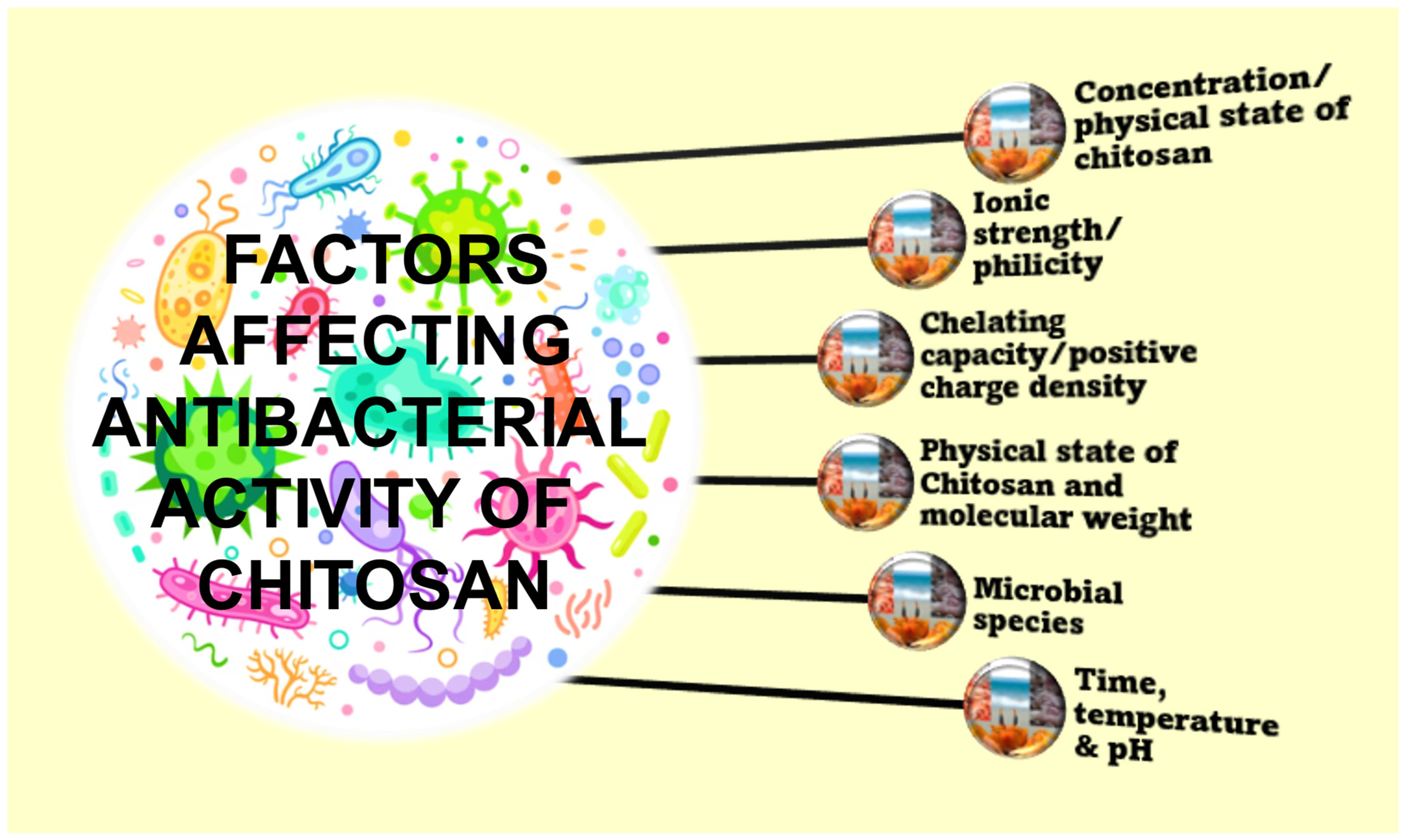
4.1. Antibacterial

Antibacterial activity is exhibited by organic as well as inorganic materials. Metals, metal oxides, and metal phosphates, belonging to the family of inorganics, have been studied for antimicrobial properties. Particularly, metal oxides, such as TiO2, MgO, ZnO and CaO are of interest as they are stable under processing conditions as well as biocompatible with humans and animals. Aromatics such as phenols and halogenated substances belong to organics group of antibacterial agents. However, nowadays, natural polymers such as chitosan gained attraction for their special antimicrobial properties [97]. The general antibacterial mechanisms of chitosan microspheres are represented in Figure 2.
The solid granular beads or MS have been of much interest for oral drug intake for numerous reasons. The uniform spread of drugs in the gastrointestinal tract, homogenous drug absorption, minimal side effects, e.g., irritation and lesser probability of their intestinal retention in comparison with singular, non-disintegrating drugs, are the major advantages [98,99]. The prolonged release of ampicillin was noted with good acid resistance and stability under simulated gastric juice for CMs crosslinked with pentasodium tripolyphosphate and methylpyrrolidinone [100]. For usage as buccal tablets to resist bacterial infections, spray-dried chlorhexidine diacetate CMs have been produced. Likewise, CMs integrated with carboxymethyl [101] and ciprofloxacin were prepared by spray drying [102] and they were found to be more efficacious than intramuscular antibiotics for treating osteomyelitis [102]. By freeze drying, MS of chitosan glutamate, aspartate and hydrochloride coated with fatty acids such as myristic, stearic, palmitic, and lauric acids were produced. The continuous release of vancomycin hydrochloride from CMs was studied to reflect peptidic model medication [103]. Due to CMs muco-adhesive property, MS loaded with clarithromycin were able to sustain for a longer time, making them suitable to treat stomach ulcers. In comparison with simple suspension-based drug ingestion, the bioavailability of clarithromycin from CMS was found to be higher as demonstrated by in vitro experiments in stomach tissues [104]. For optimal cell attachment and regulated drug release, drugs such as tetracycline have been incorporated with CMs to treat cases such as periodontitis.
It was found that drug release was slightly above the minimal dosage, sufficient to resist Staphylococcus aureus growth [105]. To increase the oral bioavailability of Polymyxin B, CMs with diameters below 3 µm were designed and reported to be absorbed by modified cells of Peyer’s patches as drug carriers to the gut-related lymphoid tissue [106]. It has also been demonstrated that the discharge of ofloxacin contained in porous N-methylated CMs (2 to 5 µm) occurs more quickly at near neutral pH of 7.4 than at acidic pH of 1.2, reaching 90 wt% in just 8 h [107]. Three anti-TB drugs—isoniazid, rifampicin and pyrazinamide—have been enclosed in alginate CMs and confirmed of their sustained release with bioavailability consistent enough for sub-therapeutic doses. Prescribed dosages of MS administered over a 10-day period to guinea pigs infected with Mycobacterium TB H37Rv resulted in the elimination of the bacteria, within 10 days, comparable to the standard 6-week treatment. Thus, the dosage of drugs is reduced by half when using MS [108]. In another trial, CMs carrying cefradine grafted ethyl cellulose retained their drug’s plasma level consistently for 24 h, helping in enhanced drug absorption in the intestine [109].
Using a gas diffusion technique, hybrid Ag-hydroxyapatite carboxylated CMs were obtained. An investigation into the antimicrobial properties of the hybrid CMs has shown better antibacterial effect against S. aureus [110]. In addition, the hybrid MS encouraged MG63 cells to proliferate and adhere. Few antimicrobial peptides (AMPs) such as MSI-78A (Pexiganan A) can eliminate Helicobacter pylori, a harmful bacterium that infects the stomach mucosa among 50% of the globe’s population [111]. About 20–40% of H. pylori-affected patients do not respond well to the conventional therapies based on antibiotics. A novel bioengineered approach of grafting AMP onto CMs has been reported. Typically, MSI-78A was grafted to CMs by thiol-maleimide using a heterobi-functional spacer (NHS-PEG113-MAL) to the C-end cysteine. The particles were pre-incubation in set up mimicking stomach conditions, the AMP borne CMs showed anti-bacterial effect against H. pylori J99 strain (a human one) at lower doses than the isolated peptide (277 μg bounded MSI-78A-SH/mL versus 512 g free MSI-78A-SH/mL). The mechanism of the H. pylori bacterial membrane damage and cytoplasmic discharge was noted in a proportion of about ten bacteria per MS after its exposure with AMP-CMs. Additionally, the electrostatic interaction of CMS facilitated H. pylori’s affinity to chitosan, which made it easier for the incorporated AMP to attack the bacterial membrane using the inverse emulsion technique; modified TiO2, with and without Ag doping, were cross linked with CMs [112]. Consequently, the antibacterial activities of composite TiO2-CMs were investigated under visible light. It was observed that TiO2 were uniformly dispersed within CMs, the particles having sizes ranging from 1 to 10 µm, showed good bactericidal effect versus E. coli, S. aureus and P. aeruginosa under the visible spectrum due to their larger surface interaction with the bacterial cell.
CMs that have been polymerized with epichlorhydrin were fabricated, and their ability to bind and form Ag+ was assessed for antibacterial potential and regulated drug release [113]. The fabricated CMs exhibited antibacterial action versus S. aureus and E. coli.
By employing Ca2+ ions as mediators for polymer densification, alginate cross-linked with CMs with uniform particle size distribution were synthesized [114]. Even at extremely low concentrations of 5–20 mg, the synthesized alginate-CMs demonstrated antibacterial activity against a wide spectrum of biofilm and pathogens of public importance. Particularly, they demonstrated effective antibiotic activity against both Gram-positive and -negative bacteria, including Enterococcus faecalis and S. aureus, as well as P. vulgaris and P. aeruginosa, respectively.
Multiphase functional MS were synthesized through emulsion-based crosslinking by mixing chitosan and gelatine solution with different concentrations of copper nanoparticles (CuNPs) [115]. The antipathogenic activity of the hybrid MS with and without CuNP was examined using agar diffusion and culture method. Typically, Gram-negative E. coli and Gram-positive E. faecalis were involved in the test. The hybrid MS with CuNP showed better antimicrobial inhibition than CuNPs and the effect was medium (solid or liquid) dependent.
The anti-bacterial and physical properties of MS can be enhanced by means of compositing microsphere with hydrogel. Typically, OAlg-CMs hydrogel is crosslinked with tetracycline hydrochloride incorporated gelatine MS [116]. In this way the antibacterial effect against E. coli ad S. aureus was improved, providing a promising future for this composite MS against bacterial infections.
There have been reports of putative antibacterial activities for chitosan/silver MS (CAgMS) [117]. The substance utilized to crosslink is glutaraldehyde. The CAgMS’s antimicrobial effectiveness was examined in experiments using fungus and bacteria. It was discovered that as Ag concentration and MS surface area increased, the inhibition potential of MS also did so.
A composite MS was made by combining green synthesized hydrogel and cryogel nanoparticles that were doped with Ag nanoparticles [118]. The antibacterial impact was then evaluated using the optical density measurement and disc diffusion methods against both Gram-positive and -negative bacteria. The hydro-cryogel combination MS has been found to have an exceptional antibacterial action when compared to commercially available conventional antibiotics.
Chitosan-quercetin (CTS-QT) was synthesized via a unique method, and their antibacterial effect was compared to that of pure CTS and QT [119]. One hundred (100) mg of CTS and equal amounts of QT and maleic anhydride were combined to produce the CTS-QT combination with carbodiiamide crosslinkers. The CTS-QT complex had a two-fold bactericidal impact on E. coli and P. aeruginosa, and a 1.5-fold effect on S. aureus, according to an antibacterial assay investigation.
The cytocompatibility and antibacterial activity of CFP-integrated polymer O-carboxymethyl chitosan (CFP-OCMC-MPs) MS towards S. aureus was assessed [120]. Additionally, the CFP-OCMC-MPs demonstrated to have a prolonged bactericidal effect.
To enhance the antimicrobial and related properties of chitosan coatings, ZnO and Ag/ZnO are incorporated with chitosan coating by casting method [121]. The modified chitosan coating with ZnO revealed better antibacterial and mechanical properties whereas Ag/ZnO exhibited optimal properties. Before modification, chitosan was able to incur minor damages on the bacterial surfaces, based on the surface charge interaction of amine group of chitosan with bacterial surfaces. Whereas the combined ZnO-chitosan coating incurred extensive damage on the bacterial cell membranes on account of charge interaction, along with hindering the synthesis of bulk protein.
CMs with oleoyl group were added to chitosan by emulsification method. The CMs had a smooth 124 μm spherical surface [122]. The antibacterial effect was found to be directly correlated with hydrophobicity and concentration of CMs.
Chitosan-polymeric and chitosan-polymeric-metal oxide of chitosan/poly vinyl alcohol (pva) and CS/PVA/ZnO) were synthesized and their antibacterial activity was tested [123]. When E. coli and S. aureus were exposed to CS/PVA/ZnO beads, it was discovered that the tri-phase beads had higher antibacterial activity than pure chitosan or bi-phasic MS. In general, a composite is usually made up of two or more materials having two or more phases with heterogeneous characters, the advantage of such composites is that compared to the single phase, the bi/or tri phases will encompass and exemplify the combined properties of all the contained phases. So, when two material components combine in a biphase, compared to those properties the triphasic beads will have an added advantage. This is what we see manifested (as higher antibacterial property) in the CS/PVA/ZnO triphasic system compared to the CS/PVA biphasic material [123].
Chitosan gel beads made of polysaccharide were created employing a twofold ionic co-crosslinking mechanism [124]. As crosslinkers, alginate and tripolyphosphate were employed. Both streptomycin and kanamycin A’s in vitro release profiles were investigated. The gels had an antibacterial effect on Gram-negative E. coli.
New environmentally friendly hydrogel made up of chitosan/genipin/cellulose seeded with dimethyldiallyl ammonium chloride (DMDAAC) was synthesized to selectively adsorb anionic dyes and these were demonstrated for their antibacterial activity against E. coli and S. aureus as well [125].
Wet milling solvent evaporation method was used to make curcumin coupled CMs (CCCMs). These CCCMs appeared to be spherical particles of size 2–5 µm [126]. The zone of inhibition was 28 mm and 23 mm against S. aureus and E. coli using CCCMs.
The core-shell MS with PLGA-glycol was prepared by emulsion-solvent evaporation method using chlorhexidine acetate and bFGF in the core of GC shell and PLGA MS respectively [127]. The release profile of CHA and bFGF have showed to retain antimicrobial and bioactive property.
To ensure prolonged resistance against bacteria, the KSL-W incorporated PLGA-CMs were developed by electrospraying and emulsion crosslinking technique [128]. Physical parameters such as surface texture, particle size and its surface, size distribution, overall drug encapsulation capacity and its in vitro release profile and antibacterial activity were all examined for various MS formulations. The study confirmed longer antibacterial resistance of formulated hybrids on oral bacteria.
A hybrid hydrogel containing graphene oxide/polyvinyl alcohol (PVA)/rose bengal (RB) were formed by freezing and de-freezing PVA and RB mixture along with CMs and Beta graphene oxide [129]. The irradiation at 808 nm and 550 nm stimulates hyperthermia condition and reactive oxygen species (ROS) production from graphene oxide and RB, respectively. These give rise to excellent antimicrobial properties within 10 min of irradiation in both in vivo and in vitro trials.

4.2. Antifungal

By spray-congealing, seven distinct formulations were created. Chitosan, sodium carboxymethylcellulose, and poloxamers (Lutrol F68 and F127) were added, along with a lipid-hydrophilic matrix (Gelucire 53/10), which served as the carrier [130]. Investigations into the MS antifungal efficacy against Candida albicans ATCC 10231 were also conducted [131]. High yields (>90%, w/w) of non-aggregated MS were obtained. The bioavailability (in vitro), solubility of the already low soluble formulation was improved significantly by poloxamer (p < 0.01). Poloxamers/Gelucire-based MP showed good inhibition against C. albicans, thereby underlining their potential for treating vaginal candidiasis with minimal administration frequency. By altering their mitochondrial structure, the monoterpene aldehyde citral has been shown to be able to suppress a wide range of pathogenic fungus [131].
By spray-congealing, seven distinct formulations were created. Chitosan, sodium carboxymethylcellulose, and poloxamers (Lutrol F68 and F127) were added, along with a lipid-hydrophilic matrix (Gelucire 53/10), which served as the carrier [129]. Investigations into the MS antifungal efficacy against Candida albicans ATCC 10231 were also conducted [130]. High yields (>90%, w/w) of non-aggregated MS were obtained. The bioavailability (in vitro), solubility of the already low soluble formulation was improved significantly by poloxamer (p < 0.01). Poloxamers/Gelucire-based MP showed good inhibition against C. albicans, thereby underlining their potential for treating vaginal candidiasis with minimal administration frequency.
By altering their mitochondrial structure, the monoterpene aldehyde citral has been shown to be able to suppress a wide range of pathogenic fungus [132]. Citral an acyclic monoterpene aldehyde has been reported to alter mitochodria cell structure of variety of fungi. Nevertheless, their chemical fragility and lability limit its use in the agricultural sector. Botrytis cinerea, an invasive plant fungus, was successfully eradicated by modified chitosan/carboxymethyl cellulose (CS/CMC) hydrogel MS incorporated with citral. In contrast to previous applications of citral as a preservative of fruits, their combination with chitosan matrixes provided scope for use in plant defense against pathogens such as Botrytis cinerea.

4.3. Antiviral

Crosslinking based on emulsion technique was used to efficiently enclose the antiviral medication acyclovir, which has low water solubility, inside chitosan and dextran that is filled with polymeric network of acrylamide [133]. The release profile showed slower release of drugs even up to 12 h for 94% acrylamide grafting efficiency. The in vivo study of acyclovir CMs into the rabbit eye showed sustained elevated concentration of acyclovir along with higher AUC values [134].
Chitosan was crosslinked with genipin, a plant-derived nontoxic reagent to form a nano/microsphere with the ability to adsorb coronaviruses [135]. Similar beads of N-(2-hydroxypropyl)-3-trimethyl chitosan (HTCC-NS/MS) were obtained from glycidyltrimethyl-ammonium chloride (GTMAC). This novel biopolymer was examined as adsorbents for Human coronavirus NL63 (HCoV-NL63), human coronavirus HCoV-OC43 and mouse hepatitis virus (MHV) in aqueous virus suspensions. An order of strong, moderate and insignificant adsorption was noted for HCoV-NL63 virus MHV and HCoV-OC43 virus, respectively. Thus, the CMs provide a means to potentially purify contaminated water by filtering or eliminating harmful viruses. The antipathogenic activities of chitosan MSs that are reported are listed in Table 1.

5. Future Recommendations and Conclusions

The antimicrobial activity of chitosan and its composites have been reviewed. The contribution of chitosan towards this cause stands unequivocally proven. With antibiotic resistance and mutants on the rise, natural alternatives for antimicrobial applications are continuously being sought after. Chitosan, known for its natural and humble origins, is an attractive option, which has been proven, as projected in this review. CMs are well established for their drug delivery applications and their antibacterial activity. Through the course of this review, we discovered that only scattered single reports are available with respect to the use of chitosan/chitosan composite MS. Moreover, these have been tested against only a handful of bacterial species. More intensive studies should be planned to expand its use against the exhaustive list of clinical pathogens.
With respect to the antibacterial, antifungal and antiviral applications of chitosan mi-crospheres, it was found that antifungal and antiviral applications were backed up by scanty research publications. Our PubMed search, using the search terms, ‘CMs antibacterial activity’ hit 216 results, implying there is a long way to go (Figure 3a). The evident gap is in the fact that antifungal and antiviral applications of CMs had only 16 (Figure 3b) and 1 (Figure 3c) supportive research publications. With the versatility of CMs antimicrobial activity well-established, the fact that almost nothing had been applied towards antifungal and antiviral applications is puzzling. With the strong potential of chitosan MS well-proven, it is necessary that it is tested for all possible applications. Given the recent COVID-19 pandemic, where a virus locked down the entire globe, extending CMs antimicrobial activity for anti-COVID solutions is a much-required endeavor. Chitosan has been prevalently used for antifungal and anti-viral applications, chitosan MS crosslinked with potential antimicrobial moieties, should far surpass the chitosan standalone effect. This review highlights the importance of focusing on this aspect. The fact that the mechanism of interaction of chitosan with almost all bacteria (with exceptions) is that the amine group acts as a positively charged moiety under right conditions, disrupting the negatively charged bacterial membrane. However, in contrast, only a fraction of viruses have such negative electron charge density on their surfaces. Hence, this might become a limiting factor against the antiviral activity of chitosan microspheres. This aspect is yet to be confirmed, affirmative research in this direction is encouraged.
CMs have been predominantly highlighted for its use in drug delivery applications owing to their unique morphologies, yet this review emphasizes that there is scope for expansion into other biomedical aspects which need to be investigated and realized.
This review surveyed the current scenario with respect to the various antimicrobial applications of CMs and their composites, the review also highlights the need to intensely look into the mechanism of antimicrobial activity of CMs as well as intricately expand its potential to fatal pathogens that are currently challenging human health and welfare.

Author Contributions

M.M., J.G. and S.S.C.P., preparation of the original draft, and revisions; I.S., participated in the review and revisions, funding. All authors have read and agreed to the published version of the manuscript.

Funding

This research received no external funding.

Institutional Review Board Statement

Not applicable.

Informed Consent Statement

Not applicable.

Data Availability Statement

Not applicable.

Acknowledgments

This article was supported by the KU Research Professor Program of Konkuk University.

Conflicts of Interest

The authors declare no conflict of interest.

References

  1. Islam, S.; Bhuiyan, M.A.R.; Islam, M.N. Chitin and Chitosan: Structure, Properties and Applications in Biomedical Engineering. J. Polym. Environ. 2017, 25, 854–866. [Google Scholar] [CrossRef]
  2. Dhawan, S.; Singla, A.K. Nifedipine Loaded Chitosan Microspheres Prepared by Emulsification Phase-Separation. Biotech. Histochem. 2003, 78, 243–254. [Google Scholar] [CrossRef] [PubMed]
  3. Lemoine, D.; Wauters, F.; Bouchend’Homme, S. Preparation and Characterization of Alginate Microspheres Containing a Model Antigen. Int. J. Pharm. 1998, 176, 9–19. [Google Scholar] [CrossRef]
  4. Hong, S.; Hsu, H.-J.; Kaunas, R.; Kameoka, J. Collagen Microsphere Production on a Chip. Lab Chip 2012, 12, 3277–3280. [Google Scholar] [CrossRef]
  5. Han, Y.; Radziuk, D.; Shchukin, D.; Moehwald, H. Stability and Size Dependence of Protein Microspheres Prepared by Ultrasonication. J. Mater. Chem. 2008, 18, 5162–5166. [Google Scholar] [CrossRef]
  6. Rodríguez-Vázquez, M.; Vega-Ruiz, B.; Ramos-Zúñiga, R.; Saldaña-Koppel, D.A.; Quiñones-Olvera, L.F. Chitosan and Its Potential Use as a Scaffold for Tissue Engineering in Regenerative Medicine. BioMed. Res. Int. 2015, 2015, 821279. [Google Scholar] [CrossRef] [PubMed] [Green Version]
  7. d’Ayala, G.G.; Malinconico, M.; Laurienzo, P. Marine Derived Polysaccharides for Biomedical Applications: Chemical Modification Approaches. Molecules 2008, 13, 2069–2106. [Google Scholar] [CrossRef] [Green Version]
  8. Bohner, M.; Tadier, S.; van Garderen, N.; de Gasparo, A.; Döbelin, N.; Baroud, G. Synthesis of Spherical Calcium Phosphate Particles for Dental and Orthopedic Applications. Biomatter 2013, 3, e25103. [Google Scholar] [CrossRef] [Green Version]
  9. Freiberg, S.; Zhu, X.X. Polymer Microspheres for Controlled Drug Release. Int. J. Pharm. 2004, 282, 1–18. [Google Scholar] [CrossRef]
  10. Cai, Y.; Chen, Y.; Hong, X.; Liu, Z.; Yuan, W. Porous Microsphere and Its Applications. Int. J. Nanomed. 2013, 8, 1111–1120. [Google Scholar]
  11. Li, S.; Nguyen, L.; Xiong, H.; Wang, M.; Hu, T.C.-C.; She, J.-X.; Serkiz, S.M.; Wicks, G.G.; Dynan, W.S. Porous-Wall Hollow Glass Microspheres as Novel Potential Nanocarriers for Biomedical Applications. Nanomedicine 2010, 6, 127–136. [Google Scholar] [CrossRef] [PubMed] [Green Version]
  12. Chen, M.; Wang, X.; Ye, Z.; Zhang, Y.; Zhou, Y.; Tan, W.-S. A Modular Approach to the Engineering of a Centimeter-Sized Bone Tissue Construct with Human Amniotic Mesenchymal Stem Cells-Laden Microcarriers. Biomaterials 2011, 32, 7532–7542. [Google Scholar] [CrossRef]
  13. Perez, R.A.; El-Fiqi, A.; Park, J.-H.; Kim, T.-H.; Kim, J.-H.; Kim, H.-W. Therapeutic Bioactive Microcarriers: Co-Delivery of Growth Factors and Stem Cells for Bone Tissue Engineering. Acta Biomater. 2014, 10, 520–530. [Google Scholar] [CrossRef]
  14. Komlev, V.S.; Barinov, S.M.; Koplik, E.V. A Method to Fabricate Porous Spherical Hydroxyapatite Granules Intended for Time-Controlled Drug Release. Biomaterials 2002, 23, 3449–3454. [Google Scholar] [CrossRef]
  15. Liu, D.M. Fabrication and Characterization of Porous Hydroxyapatite Granules. Biomaterials 1996, 17, 1955–1957. [Google Scholar] [CrossRef]
  16. Lakhkar, N.J.; Park, J.-H.; Mordan, N.J.; Salih, V.; Wall, I.B.; Kim, H.-W.; King, S.P.; Hanna, J.V.; Martin, R.A.; Addison, O.; et al. Titanium Phosphate Glass Microspheres for Bone Tissue Engineering. Acta Biomater. 2012, 8, 4181–4190. [Google Scholar] [CrossRef] [PubMed] [Green Version]
  17. Sene, F.F.; Martinelli, J.R.; Okuno, E. Synthesis and Characterization of Phosphate Glass Microspheres for Radiotherapy Applications. J. Non-Cryst. Solids 2008, 354, 4887–4893. [Google Scholar] [CrossRef]
  18. Oliveira, B.F.; Santana, M.H.A.; Ré, M.I. Spray-Dried Chitosan Microspheres Cross-Linked with d, l-Glyceraldehyde as a Potential Drug Delivery System: Preparation and Characterization. Braz. J. Chem. Eng. 2005, 22, 353–360. [Google Scholar] [CrossRef] [Green Version]
  19. Torres, M.A.; Vieira, R.S.; Beppu, M.M.; Arruda, E.J.; Santana, C.C. Production of Chemically Modified Chitosan Microspheres by a Spraying and Coagulation Method. Mater. Res. 2007, 10, 347–352. [Google Scholar] [CrossRef] [Green Version]
  20. Akamatsu, K.; Kaneko, D.; Sugawara, T. Three Preparation Methods for Monodispersed Chitosan Microspheres Using the Shirasu Porous Glass Membrane Emulsification Technique and Mechanisms of Microsphere Formation. Ind. Eng. Chem. Res. 2010, 49, 3236–3241. [Google Scholar] [CrossRef]
  21. Ribeiro, A.J.; Silva, C.; Ferreira, D.; Veiga, F. Chitosan-Reinforced Alginate Microspheres Obtained through the Emulsification/Internal Gelation Technique. Eur. J. Pharm. Sci. 2005, 25, 31–40. [Google Scholar] [CrossRef] [Green Version]
  22. Lim, S.T.; Martin, G.P.; Berry, D.J.; Brown, M.B. Preparation and Evaluation of the in Vitro Drug Release Properties and Mucoadhesion of Novel Microspheres of Hyaluronic Acid and Chitosan. J. Control. Release 2000, 66, 281–292. [Google Scholar] [CrossRef]
  23. Aggarwal, A.; Kaur, S.; Tiwary, A.K.; Gupta, S. Chitosan Microspheres Prepared by an Aqueous Process: Release of Indomethacin. J. Microencapsul. 2001, 18, 819–823. [Google Scholar] [CrossRef]
  24. Pavanetto, F.; Perugini, P.; Conti, B.; Modena, T.; Genta, I. Evaluation of Process Parameters Involved in Chitosan Microsphere Preparation by the o/w/o Multiple Emulsion Method. J. Microencapsul. 1996, 13, 679–688. [Google Scholar] [CrossRef]
  25. Orienti, I.; Aiedeh, K.; Gianasi, E.; Bertasi, V.; Zecchi, V. Indomethacin Loaded Chitosan Microspheres. Correlation between the Erosion Process and Release Kinetics. J. Microencapsul. 1996, 13, 463–472. [Google Scholar] [CrossRef]
  26. Ohya, Y.; Takei, T.; Kobayashi, H.; Ouchi, T. Release Behaviour of 5-Fluorouracil from Chitosan-Gel Microspheres Immobilizing 5-Fluorouracil Derivative Coated with Polysaccharides and Their Cell Specific Recognition. J. Microencapsul. 1993, 10, 1–9. [Google Scholar] [CrossRef]
  27. Mi, F.-L.; Shyu, S.-S.; Wong, T.-B.; Jang, S.-F.; Lee, S.-T.; Lu, K.-T. Chitosan-Polyelectrolyte Complexation for the Preparation of Gel Beads and Controlled Release of Anticancer Drug. II. Effect of PH-Dependent Ionic Crosslinking or Interpolymer Complex Using Tripolyphosphate or Polyphosphate as Reagent. J. Appl. Polym. Sci. 1999, 74, 1093–1107. [Google Scholar] [CrossRef]
  28. Varshosaz, J. The Promise of Chitosan Microspheres in Drug Delivery Systems. Expert Opin. Drug Deliv. 2007, 4, 263–273. [Google Scholar] [CrossRef]
  29. Agnihotri, S.A.; Aminabhavi, T.M. Novel Interpenetrating Network Chitosan-Poly(Ethylene Oxide-g-Acrylamide) Hydrogel Microspheres for the Controlled Release of Capecitabine. Int. J. Pharm. 2006, 324, 103–115. [Google Scholar] [CrossRef]
  30. Liu, L.-S.; Liu, S.-Q.; Ng, S.Y.; Froix, M.; Ohno, T.; Heller, J. Controlled Release of Interleukin-2 for Tumour Immunotherapy Using Alginate/Chitosan Porous Microspheres. J. Control. Release 1997, 43, 65–74. [Google Scholar] [CrossRef]
  31. Grant, J.; Blicker, M.; Piquette-Miller, M.; Allen, C. Hybrid Films from Blends of Chitosan and Egg Phosphatidylcholine for Localized Delivery of Paclitaxel. J. Pharm. Sci. 2005, 94, 1512–1527. [Google Scholar] [CrossRef] [PubMed]
  32. Nsereko, S.; Amiji, M. Localized Delivery of Paclitaxel in Solid Tumors from Biodegradable Chitin Microparticle Formulations. Biomaterials 2002, 23, 2723–2731. [Google Scholar] [CrossRef] [PubMed]
  33. Singh, U.V.; Udupa, N. Methotrexate Loaded Chitosan and Chitin Microspheres—In Vitro Characterization and Pharmacokinetics in Mice Bearing Ehrlich Ascites Carcinoma. J. Microencapsul. 1998, 15, 581–594. [Google Scholar] [CrossRef] [PubMed]
  34. Lee, J.E.; Kim, S.E.; Kwon, I.C.; Ahn, H.J.; Cho, H.; Lee, S.-H.; Kim, H.J.; Seong, S.C.; Lee, M.C. Effects of a Chitosan Scaffold Containing TGF-Beta1 Encapsulated Chitosan Microspheres on in Vitro Chondrocyte Culture. Artif. Organs 2004, 28, 829–839. [Google Scholar] [CrossRef] [PubMed]
  35. Lee, J.E.; Kim, K.E.; Kwon, I.C.; Ahn, H.J.; Lee, S.-H.; Cho, H.; Kim, H.J.; Seong, S.C.; Lee, M.C. Effects of the Controlled-Released TGF-Beta 1 from Chitosan Microspheres on Chondrocytes Cultured in a Collagen/Chitosan/Glycosaminoglycan Scaffold. Biomaterials 2004, 25, 4163–4173. [Google Scholar] [CrossRef]
  36. Jiang, T.; Abdel-Fattah, W.I.; Laurencin, C.T. In Vitro Evaluation of Chitosan/Poly(Lactic Acid-Glycolic Acid) Sintered Microsphere Scaffolds for Bone Tissue Engineering. Biomaterials 2006, 27, 4894–4903. [Google Scholar] [CrossRef]
  37. Han, G.; Chen, H.-L.; Sun, X.-Y.; Gao, J.-Q.; Liang, W.-Q. Preparation and release behavior of chitosan scaffolds encapsulating proteins loaded in PLGA microspheres. Yao Xue Xue Bao Acta Pharm. Sin. 2006, 41, 493–497. [Google Scholar]
  38. Cevher, E.; Orhan, Z.; Mülazimoğlu, L.; Sensoy, D.; Alper, M.; Yildiz, A.; Ozsoy, Y. Characterization of Biodegradable Chitosan Microspheres Containing Vancomycin and Treatment of Experimental Osteomyelitis Caused by Methicillin-Resistant Staphylococcus Aureus with Prepared Microspheres. Int. J. Pharm. 2006, 317, 127–135. [Google Scholar] [CrossRef]
  39. Blanco, M.D.; Gómez, C.; Olmo, R.; Muñiz, E.; Teijón, J.M. Chitosan Microspheres in PLG Films as Devices for Cytarabine Release. Int. J. Pharm. 2000, 202, 29–39. [Google Scholar] [CrossRef]
  40. Machida, Y.; Nagai, T.; Abe, M.; Sannan, T. Use of Chitosan and Hydroxypropylchitosan in Drug Formulations to Effect Sustained Release. Drug Des. Deliv. 1986, 1, 119–130. [Google Scholar]
  41. Elçin, Y.M.; Dixit, V.; Gitnick, G. Controlled Release of Endothelial Cell Growth Factor from Chitosan-Albumin Microspheres for Localized Angiogenesis: In Vitro and in Vivo Studies. Artif. Cells Blood Substit. Immobil. Biotechnol. 1996, 24, 257–271. [Google Scholar] [CrossRef]
  42. Çelik, Ö.; Akbuğa, J. Preparation of Superoxide Dismutase Loaded Chitosan Microspheres: Characterization and Release Studies. Eur. J. Pharm. Biopharm. 2007, 66, 42–47. [Google Scholar] [CrossRef]
  43. Schally, A.V. LH-RH Analogues: I. Their Impact on Reproductive Medicine. Gynecol. Endocrinol. 1999, 13, 401–409. [Google Scholar] [CrossRef]
  44. Xue, Z.-X.; Yang, G.-P.; Zhang, Z.-P.; He, B.-L. Application of Chitosan Microspheres as Carriers of LH-RH Analogue TX46. React. Funct. Polym. 2006, 66, 893–901. [Google Scholar] [CrossRef]
  45. Wang, L.-Y.; Gu, Y.-H.; Zhou, Q.-Z.; Ma, G.-H.; Wan, Y.-H.; Su, Z.-G. Preparation and Characterization of Uniform-Sized Chitosan Microspheres Containing Insulin by Membrane Emulsification and a Two-Step Solidification Process. Colloids Surf. B Biointerfaces 2006, 50, 126–135. [Google Scholar] [CrossRef]
  46. Kofuji, K.; Murata, Y.; Kawashima, S. Sustained Insulin Release with Biodegradation of Chitosan Gel Beads Prepared by Copper Ions. Int. J. Pharm. 2005, 303, 95–103. [Google Scholar] [CrossRef]
  47. Cho, B.C.; Kim, J.Y.; Lee, J.H.; Chung, H.Y.; Park, J.W.; Roh, K.H.; Kim, G.U.; Kwon, I.C.; Jang, K.H.; Lee, D.-S.; et al. The Bone Regenerative Effect of Chitosan Microsphere-Encapsulated Growth Hormone on Bony Consolidation in Mandibular Distraction Osteogenesis in a Dog Model. J. Craniofac. Surg. 2004, 15, 299–311; discussion 312–313. [Google Scholar] [CrossRef]
  48. Illum, L.; Jabbal-Gill, I.; Hinchcliffe, M.; Fisher, A.N.; Davis, S.S. Chitosan as a Novel Nasal Delivery System for Vaccines. Adv. Drug Deliv. Rev. 2001, 51, 81–96. [Google Scholar] [CrossRef]
  49. Jiang, H.-L.; Park, I.-K.; Shin, N.-R.; Kang, S.-G.; Yoo, H.-S.; Kim, S.-I.; Suh, S.-B.; Akaike, T.; Cho, C.-S. In Vitro Study of the Immune Stimulating Activity of an Athrophic Rhinitis Vaccine Associated to Chitosan Microspheres. Eur. J. Pharm. Biopharm. 2004, 58, 471–476. [Google Scholar] [CrossRef]
  50. Jaganathan, K.S.; Rao, Y.U.B.; Singh, P.; Prabakaran, D.; Gupta, S.; Jain, A.; Vyas, S.P. Development of a Single Dose Tetanus Toxoid Formulation Based on Polymeric Microspheres: A Comparative Study of Poly(d,l-Lactic-Co-Glycolic Acid) versus Chitosan Microspheres. Int. J. Pharm. 2005, 294, 23–32. [Google Scholar] [CrossRef]
  51. Mi, F.L.; Shyu, S.S.; Chen, C.T.; Schoung, J.Y. Porous Chitosan Microsphere for Controlling the Antigen Release of Newcastle Disease Vaccine: Preparation of Antigen-Adsorbed Microsphere and in Vitro Release. Biomaterials 1999, 20, 1603–1612. [Google Scholar] [CrossRef] [PubMed]
  52. van der Lubben, I.M.; van Opdorp, F.A.C.; Hengeveld, M.R.; Onderwater, J.J.M.; Koerten, H.K.; Verhoef, J.C.; Borchard, G.; Junginger, H.E. Transport of Chitosan Microparticles for Mucosal Vaccine Delivery in a Human Intestinal M-Cell Model. J. Drug Target. 2002, 10, 449–456. [Google Scholar] [CrossRef]
  53. Van Der Lubben, I.M.; Konings, F.A.; Borchard, G.; Verhoef, J.C.; Junginger, H.E. In Vivo Uptake of Chitosan Microparticles by Murine Peyer’s Patches: Visualization Studies Using Confocal Laser Scanning Microscopy and Immunohistochemistry. J. Drug Target. 2001, 9, 39–47. [Google Scholar] [CrossRef] [PubMed]
  54. Lee, J.S.; Cha, D.S.; Park, H.J. Survival of Freeze-Dried Lactobacillus Bulgaricus KFRI 673 in Chitosan-Coated Calcium Alginate Microparticles. J. Agric. Food Chem. 2004, 52, 7300–7305. [Google Scholar] [CrossRef] [PubMed]
  55. Kosaraju, S.L.; D’ath, L.; Lawrence, A. Preparation and Characterisation of Chitosan Microspheres for Antioxidant Delivery. Carbohydr. Polym. 2006, 64, 163–167. [Google Scholar] [CrossRef]
  56. Harikarnpakdee, S.; Lipipun, V.; Sutanthavibul, N.; Ritthidej, G.C. Spray-Dried Mucoadhesive Microspheres: Preparation and Transport through Nasal Cell Monolayer. AAPS PharmSciTech 2006, 7, E12. [Google Scholar] [CrossRef]
  57. Cerchiara, T.; Luppi, B.; Chidichimo, G.; Bigucci, F.; Zecchi, V. Chitosan and Poly(Methyl Vinyl Ether-Co-Maleic Anhydride) Microparticles as Nasal Sustained Delivery Systems. Eur. J. Pharm. Biopharm. 2005, 61, 195–200. [Google Scholar] [CrossRef]
  58. Martinac, A.; Filipović-Grcić, J.; Perissutti, B.; Voinovich, D.; Pavelić, Z. Spray-Dried Chitosan/Ethylcellulose Microspheres for Nasal Drug Delivery: Swelling Study and Evaluation of in Vitro Drug Release Properties. J. Microencapsul. 2005, 22, 549–561. [Google Scholar] [CrossRef]
  59. Krauland, A.H.; Guggi, D.; Bernkop-Schnürch, A. Thiolated Chitosan Microparticles: A Vehicle for Nasal Peptide Drug Delivery. Int. J. Pharm. 2006, 307, 270–277. [Google Scholar] [CrossRef]
  60. Gavini, E.; Hegge, A.B.; Rassu, G.; Sanna, V.; Testa, C.; Pirisino, G.; Karlsen, J.; Giunchedi, P. Nasal Administration of Carbamazepine Using Chitosan Microspheres: In Vitro/in Vivo Studies. Int. J. Pharm. 2006, 307, 9–15. [Google Scholar] [CrossRef]
  61. Varshosaz, J.; Sadrai, H.; Alinagari, R. Nasal Delivery of Insulin Using Chitosan Microspheres. J. Microencapsul. 2004, 21, 761–774. [Google Scholar] [CrossRef]
  62. Jain, S.K.; Chourasia, M.K.; Jain, A.K.; Jain, R.K.; Shrivastava, A.K. Development and Characterization of Mucoadhesive Microspheres Bearing Salbutamol for Nasal Delivery. Drug Deliv. 2004, 11, 113–122. [Google Scholar] [CrossRef]
  63. Illum, L.; Watts, P.; Fisher, A.N.; Hinchcliffe, M.; Norbury, H.; Jabbal-Gill, I.; Nankervis, R.; Davis, S.S. Intranasal Delivery of Morphine. J. Pharmacol. Exp. Ther. 2002, 301, 391–400. [Google Scholar] [CrossRef] [Green Version]
  64. Turkoglu, O.F.; Eroglu, H.; Okutan, O.; Burul, E.; Sargon, M.F.; Özer, N.; Öner, L.; Beskonaklı, E. The Efficiency of Dexamethasone Sodium Phosphate–Encapsulated Chitosan Microspheres after Cold Injury. Surg. Neurol. 2005, 64, S11–S16. [Google Scholar] [CrossRef]
  65. Jiang, D.-S.; Long, S.-Y.; Huang, J.; Xiao, H.-Y.; Zhou, J.-Y. Immobilization of Pycnoporus Sanguineus Laccase on Magnetic Chitosan Microspheres. Biochem. Eng. J. 2005, 25, 15–23. [Google Scholar] [CrossRef]
  66. Shentu, J.; Wu, J.; Song, W.; Jia, Z. Chitosan Microspheres as Immobilized Dye Affinity Support for Catalase Adsorption. Int. J. Biol. Macromol. 2005, 37, 42–46. [Google Scholar] [CrossRef]
  67. Martins, A.O.; da Silva, E.L.; Carasek, E.; Laranjeira, M.C.M.; de Fávere, V.T. Sulphoxine Immobilized onto Chitosan Microspheres by Spray Drying: Application for Metal Ions Preconcentration by Flow Injection Analysis. Talanta 2004, 63, 397–403. [Google Scholar] [CrossRef]
  68. Wang, Y.M.; Shi, T.S.; Pu, Y.L.; Zhu, J.G.; Zhao, Y.L. Studies on hepatic arterial embolization with cisplatin-chitosan-microspheres in dogs. Yao Xue Xue Bao 1995, 30, 891–895. [Google Scholar]
  69. Misirli, Y.; Oztürk, E.; Kurşaklioğlu, H.; Denkbaş, E.B. Preparation and Characterization of Mitomycin-C Loaded Chitosan-Coated Alginate Microspheres for Chemoembolization. J. Microencapsul. 2005, 22, 167–178. [Google Scholar] [CrossRef]
  70. Eroğlu, M.; Kurşaklioğlu, H.; Misirli, Y.; Iyisoy, A.; Acar, A.; Işin Doğan, A.; Denkbaş, E.B. Chitosan-Coated Alginate Microspheres for Embolization and/or Chemoembolization: In Vivo Studies. J. Microencapsul. 2006, 23, 367–376. [Google Scholar] [CrossRef]
  71. Denkbaş, E.B.; Kiliçay, E.; Birlikseven, C.; Öztürk, E. Magnetic Chitosan Microspheres: Preparation and Characterization. React. Funct. Polym. 2002, 50, 225–232. [Google Scholar] [CrossRef]
  72. Wu, Y.; Guo, J.; Yang, W.; Wang, C.; Fu, S. Preparation and Characterization of Chitosan–Poly(Acrylic Acid) Polymer Magnetic Microspheres. Polymer 2006, 47, 5287–5294. [Google Scholar] [CrossRef]
  73. Moore, A.; Basilion, J.P.; Chiocca, E.A.; Weissleder, R. Measuring Transferrin Receptor Gene Expression by NMR Imaging. Biochim. Biophys. Acta 1998, 1402, 239–249. [Google Scholar] [CrossRef] [PubMed] [Green Version]
  74. Kim, D.K.; Zhang, Y.; Kehr, J.; Klason, T.; Bjelke, B.; Muhammed, M. Characterization and MRI Study of Surfactant-Coated Superparamagnetic Nanoparticles Administered into the Rat Brain. J. Magn. Magn. Mater. 2001, 225, 256–261. [Google Scholar] [CrossRef]
  75. Morales, M.P.; Bomati-Miguel, O.; Pérez de Alejo, R.; Ruiz-Cabello, J.; Veintemillas-Verdaguer, S.; O’Grady, K. Contrast Agents for MRI Based on Iron Oxide Nanoparticles Prepared by Laser Pyrolysis. J. Magn. Magn. Mater. 2003, 266, 102–109. [Google Scholar] [CrossRef]
  76. Kim, E.H.; Ahn, Y.; Lee, H.S. Biomedical Applications of Superparamagnetic Iron Oxide Nanoparticles Encapsulated within Chitosan. J. Alloys Compd. 2007, 434–435, 633–636. [Google Scholar] [CrossRef]
  77. Lee, H.S.; Hee Kim, E.; Shao, H.; Kook Kwak, B. Synthesis of SPIO-Chitosan Microspheres for MRI-Detectable Embolotherapy. J. Magn. Magn. Mater. 2005, 293, 102–105. [Google Scholar] [CrossRef]
  78. Guliyeva, U.; Oner, F.; Ozsoy, S.; Haziroğlu, R. Chitosan Microparticles Containing Plasmid DNA as Potential Oral Gene Delivery System. Eur. J. Pharm. Biopharm. 2006, 62, 17–25. [Google Scholar] [CrossRef]
  79. Yun, Y.H.; Jiang, H.; Chan, R.; Chen, W. Sustained Release of PEG-g-Chitosan Complexed DNA from Poly(Lactide-Co-Glycolide). J. Biomater. Sci. Polym. Ed. 2005, 16, 1359–1378. [Google Scholar] [CrossRef]
  80. Lameiro, M.H.; Malpique, R.; Silva, A.C.; Alves, P.M.; Melo, E. Encapsulation of Adenoviral Vectors into Chitosan–Bile Salt Microparticles for Mucosal Vaccination. J. Biotechnol. 2006, 126, 152–162. [Google Scholar] [CrossRef]
  81. Aral, C.; Akbuga, J. Preparation and in Vitro Transfection Efficiency of Chitosan Microspheres Containing Plasmid DNA:Poly(L-Lysine) Complexes. J. Pharm. Pharm. Sci. 2003, 6, 321–326. [Google Scholar]
  82. Ozbas-Turan, S.; Aral, C.; Kabasakal, L.; Keyer-Uysal, M.; Akbuga, J. Co-Encapsulation of Two Plasmids in Chitosan Microspheres as a Non-Viral Gene Delivery Vehicle. J. Pharm. Pharm. Sci. 2003, 6, 27–32. [Google Scholar]
  83. Hejazi, R.; Amiji, M. Stomach-Specific Anti-H. Pylori Therapy; Part III: Effect of Chitosan Microspheres Crosslinking on the Gastric Residence and Local Tetracycline Concentrations in Fasted Gerbils. Int. J. Pharm. 2004, 272, 99–108. [Google Scholar] [CrossRef]
  84. El-Gibaly, I. Development and in Vitro Evaluation of Novel Floating Chitosan Microcapsules for Oral Use: Comparison with Non-Floating Chitosan Microspheres. Int. J. Pharm. 2002, 249, 7–21. [Google Scholar] [CrossRef]
  85. Hejazi, R.; Amiji, M. Stomach-Specific Anti-H. Pylori Therapy. I: Preparation and Characterization of Tetracyline-Loaded Chitosan Microspheres. Int. J. Pharm. 2002, 235, 87–94. [Google Scholar] [CrossRef]
  86. Portero, A.; Remuñán-López, C.; Criado, M.T.; Alonso, M.J. Reacetylated Chitosan Microspheres for Controlled Delivery of Anti-Microbial Agents to the Gastric Mucosa. J. Microencapsul. 2002, 19, 797–809. [Google Scholar] [CrossRef]
  87. Williams, R.O., III; Barron, M.K.; José Alonso, M.; Remuñán-López, C. Investigation of a PMDI System Containing Chitosan Microspheres and P134a. Int. J. Pharm. 1998, 174, 209–222. [Google Scholar] [CrossRef]
  88. Huang, Y.-C.; Yeh, M.-K.; Cheng, S.-N.; Chiang, C.-H. The Characteristics of Betamethasone-Loaded Chitosan Microparticles by Spray-Drying Method. J. Microencapsul. 2003, 20, 459–472. [Google Scholar] [CrossRef]
  89. Huang, Y.-C.; Chiang, C.-H.; Yeh, M.-K. Optimizing Formulation Factors in Preparing Chitosan Microparticles by Spray-Drying Method. J. Microencapsul. 2003, 20, 247–260. [Google Scholar] [CrossRef]
  90. Huang, Y.C.; Yeh, M.K.; Chiang, C.H. Formulation Factors in Preparing BTM–Chitosan Microspheres by Spray Drying Method. Int. J. Pharm. 2002, 242, 239–242. [Google Scholar] [CrossRef]
  91. Grenha, A.; Seijo, B.; Remuñán-López, C. Microencapsulated Chitosan Nanoparticles for Lung Protein Delivery. Eur. J. Pharm. Sci. 2005, 25, 427–437. [Google Scholar] [CrossRef] [PubMed]
  92. Yamamoto, H.; Kuno, Y.; Sugimoto, S.; Takeuchi, H.; Kawashima, Y. Surface-Modified PLGA Nanosphere with Chitosan Improved Pulmonary Delivery of Calcitonin by Mucoadhesion and Opening of the Intercellular Tight Junctions. J. Control. Release 2005, 102, 373–381. [Google Scholar] [CrossRef] [PubMed]
  93. Bivas-Benita, M.; van Meijgaarden, K.E.; Franken, K.L.M.C.; Junginger, H.E.; Borchard, G.; Ottenhoff, T.H.M.; Geluk, A. Pulmonary Delivery of Chitosan-DNA Nanoparticles Enhances the Immunogenicity of a DNA Vaccine Encoding HLA-A*0201-Restricted T-Cell Epitopes of Mycobacterium Tuberculosis. Vaccine 2004, 22, 1609–1615. [Google Scholar] [CrossRef]
  94. Varshosaz, J.; Jaffarian Dehkordi, A.; Golafshan, S. Colon-Specific Delivery of Mesalazine Chitosan Microspheres. J. Microencapsul. 2006, 23, 329–339. [Google Scholar] [CrossRef]
  95. Wittaya-areekul, S.; Kruenate, J.; Prahsarn, C. Preparation and in Vitro Evaluation of Mucoadhesive Properties of Alginate/Chitosan Microparticles Containing Prednisolone. Int. J. Pharm. 2006, 312, 113–118. [Google Scholar] [CrossRef] [PubMed]
  96. Rai, G.; Jain, S.K.; Agrawal, S.; Bhadra, S.; Pancholi, S.S.; Agrawal, G.P. Chitosan Hydrochloride Based Microspheres of Albendazole for Colonic Drug Delivery. Pharmazie 2005, 60, 131–134. [Google Scholar]
  97. Hosseinnejad, M.; Jafari, S.M. Evaluation of Different Factors Affecting Antimicrobial Properties of Chitosan. Int. J. Biol. Macromol. 2016, 85, 467–475. [Google Scholar] [CrossRef]
  98. Lauwo, J.A.K.; Agrawal, D.K.; Emenike, I.V. Some Pharmaceutical Studies on Sustained Release Coprbcipitates of Ampicillin Trihydrat with Acrylic Resin (Eudragit)(R)-RS. Drug Dev. Ind. Pharm. 1990, 16, 1375–1389. [Google Scholar] [CrossRef]
  99. Bodmeier, R.; Chen, H.; Tyle, P.; Jarosz, P. Pseudoephedrine HCl Microspheres Formulated into an Oral Suspension Dosage Form. J. Control. Release 1991, 15, 65–77. [Google Scholar] [CrossRef]
  100. Giunchedi, P.; Genta, I.; Conti, B.; Muzzarelli, R.A.; Conte, U. Preparation and Characterization of Ampicillin Loaded Methylpyrrolidinone Chitosan and Chitosan Microspheres. Biomaterials 1998, 19, 157–161. [Google Scholar] [CrossRef]
  101. Yin, C.; Hou, C.; Jiang, L.; Gu, Q. Research on Carboxymethyl Chitosan Acting as the Adjuvant for Implantable Degradable Microspheres. J. Biomed. Eng. Sheng Wu Yi Xue Gong Cheng Xue Za Zhi 2004, 21, 350–354. [Google Scholar]
  102. Orhan, Z.; Cevher, E.; Mülazimoglu, L.; Gürcan, D.; Alper, M.; Araman, A.; Ozsoy, Y. The Preparation of Ciprofloxacin Hydrochloride-Loaded Chitosan and Pectin Microspheres: Their Evaluation in an Animal Osteomyelitis Model. J. Bone Joint Surg. Br. 2006, 88, 270–275. [Google Scholar] [CrossRef] [Green Version]
  103. Cerchiara, T.; Luppi, B.; Bigucci, F.; Petrachi, M.; Orienti, I.; Zecchi, V. Controlled Release of Vancomycin from Freeze-Dried Chitosan Salts Coated with Different Fatty Acids by Spray-Drying. J. Microencapsul. 2003, 20, 473–478. [Google Scholar] [CrossRef]
  104. Majithiya, R.J.; Murthy, R.S.R. Chitosan-Based Mucoadhesive Microspheres of Clarithromycin as a Delivery System for Antibiotic to Stomach. Curr. Drug Deliv. 2005, 2, 235–242. [Google Scholar] [CrossRef]
  105. Govender, S.; Pillay, V.; Chetty, D.J.; Essack, S.Y.; Dangor, C.M.; Govender, T. Optimisation and Characterisation of Bioadhesive Controlled Release Tetracycline Microspheres. Int. J. Pharm. 2005, 306, 24–40. [Google Scholar] [CrossRef]
  106. Coppi, G.; Iannuccelli, V.; Sala, N.; Bondi, M. Alginate Microparticles for Polymyxin B Peyer’s Patches Uptake: Microparticles for Antibiotic Oral Administration. J. Microencapsul. 2004, 21, 829–839. [Google Scholar] [CrossRef]
  107. Peng, X.; Zhang, L. Surface Fabrication of Hollow Microspheres from N-Methylated Chitosan Cross-Linked with Gultaraldehyde. Langmuir 2005, 21, 1091–1095. [Google Scholar] [CrossRef]
  108. Pandey, R.; Khuller, G.K. Chemotherapeutic Potential of Alginate–Chitosan Microspheres as Anti-Tubercular Drug Carriers. J. Antimicrob. Chemother. 2004, 53, 635–640. [Google Scholar] [CrossRef] [Green Version]
  109. Takishima, J.; Onishi, H.; Machida, Y. Prolonged Intestinal Absorption of Cephradine with Chitosan-Coated Ethylcellulose Microparticles in Rats. Biol. Pharm. Bull. 2002, 25, 1498–1502. [Google Scholar] [CrossRef] [Green Version]
  110. Shen, J.; Jin, B.; Qi, Y.-C.; Jiang, Q.-Y.; Gao, X.-F. Carboxylated Chitosan/Silver-Hydroxyapatite Hybrid Microspheres with Improved Antibacterial Activity and Cytocompatibility. Mater. Sci. Eng. C Mater. Biol. Appl. 2017, 78, 589–597. [Google Scholar] [CrossRef]
  111. Fonseca, D.R.; Moura, A.; Leiro, V.; Silva-Carvalho, R.; Estevinho, B.N.; Seabra, C.L.; Henriques, P.C.; Lucena, M.; Teixeira, C.; Gomes, P.; et al. Grafting MSI-78A onto Chitosan Microspheres Enhances Its Antimicrobial Activity. Acta Biomater. 2022, 137, 186–198. [Google Scholar] [CrossRef]
  112. Li, J.; Xie, B.; Xia, K.; Li, Y.; Han, J.; Zhao, C. Enhanced Antibacterial Activity of Silver Doped Titanium Dioxide-Chitosan Composites under Visible Light. Materials 2018, 11, 1403. [Google Scholar] [CrossRef] [PubMed] [Green Version]
  113. Pereira, A.K.S.; Reis, D.T.; Barbosa, K.M.; Scheidt, G.N. Antibacterial Effects and Ibuprofen Release Potential Using Chitosan Microspheres Loaded with Silver Nanoparticles. Carbohydrate 2020, 488, 107891. [Google Scholar] [CrossRef]
  114. Thaya, R.; Vaseeharan, B.; Sivakamavalli, J.; Iswarya, A.; Govindarajan, M.; Alharbi, N.S.; Kadaikunnan, S.; Al-Anbr, M.N.; Khaled, J.M.; Benelli, G. Synthesis of Chitosan-Alginate Microspheres with High Antimicrobial and Antibiofilm Activity against Multi-Drug Resistant Microbial Pathogens. Microb. Pathog. 2018, 114, 17–24. [Google Scholar] [CrossRef]
  115. Fuentes, S.; Alviña, R.; Zegarra, K.; Pérez, B.; Pozo, P. Antibacterial Activities of Copper Nanoparticles in Hybrid Microspheres. J. Nanosci. Nanotechnol. 2019, 19, 4512–4519. [Google Scholar] [CrossRef] [PubMed]
  116. Chen, H.; Xing, X.; Tan, H.; Jia, Y.; Zhou, T.; Chen, Y.; Ling, Z.; Hu, X. Covalently Antibacterial Alginate-Chitosan Hydrogel Dressing Integrated Gelatin Microspheres Containing Tetracycline Hydrochloride for Wound Healing. Mater. Sci. Eng. C Mater. Biol. Appl. 2017, 70, 287–295. [Google Scholar] [CrossRef] [PubMed]
  117. An, J.; Ji, Z.; Wang, D.; Luo, Q.; Li, X. Preparation and Characterization of Uniform-Sized Chitosan/Silver Microspheres with Antibacterial Activities. Mater. Sci. Eng. C Mater. Biol. Appl. 2014, 36, 33–41. [Google Scholar] [CrossRef]
  118. Mohammed, A.M.; Hassan, K.T.; Hassan, O.M. Assessment of Antimicrobial Activity of Chitosan/Silver Nanoparticles Hydrogel and Cryogel Microspheres. Int. J. Biol. Macromol. 2023, 233, 123580. [Google Scholar] [CrossRef]
  119. Azeem, M.; Hanif, M.; Mahmood, K. Design, Synthesis, Spectroscopic Characterization, in-Vitro Antibacterial Evaluation and in-Silico Analysis of Polycaprolactone Containing Chitosan-Quercetin microspheres. J. Biomol. Struct. Dyn. 2022, 1, 1–12. [Google Scholar] [CrossRef]
  120. Liu, Z.; Wang, C.; Liu, Y.; Peng, D. Cefepime Loaded O-Carboxymethyl Chitosan Microspheres with Sustained Bactericidal Activity and Enhanced Biocompatibility. J. Biomater. Sci. Polym. Ed. 2017, 28, 79–92. [Google Scholar] [CrossRef]
  121. Wei, X.; Li, Q.; Wu, C.; Sun, T.; Li, X. Preparation, Characterization and Antibacterial Mechanism of the Chitosan Coatings Modified by Ag/ZnO Microspheres. J. Sci. Food Agric. 2020, 100, 5527–5538. [Google Scholar] [CrossRef]
  122. Kong, M.; Chen, X.G.; Liu, C.S.; Liu, C.G.; Meng, X.H.; Yu, L.J. Antibacterial Mechanism of Chitosan Microspheres in a Solid Dispersing System against E. coli. Colloids Surf. B Biointerfaces 2008, 65, 197–202. [Google Scholar] [CrossRef]
  123. Gutha, Y.; Pathak, J.L.; Zhang, W.; Zhang, Y.; Jiao, X. Antibacterial and Wound Healing Properties of Chitosan/Poly(Vinyl Alcohol)/Zinc Oxide Beads (CS/PVA/ZnO). Int. J. Biol. Macromol. 2017, 103, 234–241. [Google Scholar] [CrossRef]
  124. Tiburcio, E.; García-Junceda, E.; Garrido, L.; Fernández-Mayoralas, A.; Revuelta, J.; Bastida, A. Preparation and Characterization of Aminoglycoside-Loaded Chitosan/Tripolyphosphate/Alginate Microspheres against E. coli. Polymers 2021, 13, 3326. [Google Scholar] [CrossRef]
  125. Liu, Q.; Xia, N.; Wan, W.; Gao, Y.; Zhu, S. Selective Capture of Toxic Anionic Dyes of a Novel Prepared DMDAAC-Grafted Chitosan/Genipin/Cellulose Hydrogel Beads with Antibacterial Activity. Int. J. Biol. Macromol. 2021, 189, 722–733. [Google Scholar] [CrossRef]
  126. Saranya, T.S.; Rajan, V.K.; Biswas, R.; Jayakumar, R.; Sathianarayanan, S. Synthesis, Characterisation and Biomedical Applications of Curcumin Conjugated Chitosan Microspheres. Int. J. Biol. Macromol. 2018, 110, 227–233. [Google Scholar] [CrossRef]
  127. Chen, M.-M.; Cao, H.; Liu, Y.-Y.; Liu, Y.; Song, F.-F.; Chen, J.-D.; Zhang, Q.-Q.; Yang, W.-Z. Sequential Delivery of Chlorhexidine Acetate and BFGF from PLGA-Glycol Chitosan Core-Shell Microspheres. Colloids Surf. B Biointerfaces 2017, 151, 189–195. [Google Scholar] [CrossRef]
  128. Li, Y.; Na, R.; Wang, X.; Liu, H.; Zhao, L.; Sun, X.; Ma, G.; Cui, F. Fabrication of Antimicrobial Peptide-Loaded PLGA/Chitosan Composite Microspheres for Long-Acting Bacterial Resistance. Molecules 2017, 22, 1637. [Google Scholar] [CrossRef] [Green Version]
  129. Li, Y.; Wang, J.; Yang, Y.; Shi, J.; Zhang, H.; Yao, X.; Chen, W.; Zhang, X. A Rose Bengal/Graphene Oxide/PVA Hybrid Hydrogel with Enhanced Mechanical Properties and Light-Triggered Antibacterial Activity for Wound Treatment. Mater. Sci. Eng. C Mater. Biol. Appl. 2021, 118, 111447. [Google Scholar] [CrossRef]
  130. Jyothi, S.S.; Seethadevi, A.; Prabha, K.S.; Muthuprasanna, P.; Pavitra, P. Microencapsulation: A Review. Int. J. Pharm. Biol. Sci. 2012, 3, 509–531. [Google Scholar]
  131. Albertini, B.; Passerini, N.; Di Sabatino, M.; Vitali, B. Polymer–Lipid Based Mucoadhesive Microspheres Prepared by Spray-Congealing for the Vaginal Delivery of Econazole Nitrate. Eur. J. Pharm. Sci. 2009, 36, 591–601. [Google Scholar] [CrossRef]
  132. Ma, H.; Zhao, Y.; Lu, Z.; Xing, R.; Yao, X.; Jin, Z.; Wang, Y.; Yu, F. Citral-Loaded Chitosan/Carboxymethyl Cellulose Copolymer Hydrogel Microspheres with Improved Antimicrobial Effects for Plant Protection. Int. J. Biol. Macromol. 2020, 164, 986–993. [Google Scholar] [CrossRef] [PubMed]
  133. Rokhade, A.P.; Patil, S.A.; Aminabhavi, T.M. Synthesis and Characterization of Semi-Interpenetrating Polymer Network Microspheres of Acrylamide Grafted Dextran and Chitosan for Controlled Release of Acyclovir. Carbohydr. Polym. 2007, 67, 605–613. [Google Scholar] [CrossRef]
  134. Genta, I.; Conti, B.; Perugini, P.; Pavanetto, F.; Spadaro, A.; Puglisi, G. Bioadhesive Microspheres for Ophthalmic Administration of Acyclovir. J. Pharm. Pharmacol. 1997, 49, 737–742. [Google Scholar] [CrossRef] [PubMed]
  135. Ciejka, J.; Wolski, K.; Nowakowska, M.; Pyrc, K.; Szczubiałka, K. Biopolymeric Nano/Microspheres for Selective and Reversible Adsorption of Coronaviruses. Mater. Sci. Eng. C Mater. Biol. Appl. 2017, 76, 735–742. [Google Scholar] [CrossRef]
Figure 1. Chitosan properties that affect the antibacterial activity of chitosan. These are the key properties that enable the antibacterial activity that chitosan exhibits.
Jmse 11 01480 g001
Figure 2. Mechanism of bactericidal activity by chitosan MS. The various intracellular antibacterial effects triggered by chitosan have been indicated.
Jmse 11 01480 g002
Figure 3. Results of PubMed search using the search terms (a) chitosan MS antibacterial activity (b) chitosan MS antifungal activity (c) chitosan MS antiviral activity. These results clearly confirm the comparatively scanty research reports on chitosan microsphere’s antifungal and antiviral activity compared to its extensively researched and validated antibacterial activity.
Jmse 11 01480 g003
Table 1. Chitosan MS/MS composites and their antimicrobial activity.
Chitosan MS/
Composite Type
MicrobeActivity
Tetracycline has also been loaded into CMSStaphylococcus aureusAntibacterial activity-Growth inhibition
Carboxylated chitosan/silver-hydroxyapatite (CMCS/Ag-HA) hybrid MSStaphylococcus aureusAntibacterial activity by synergistic effect of Ag+ and CMCS
MSI-78A with a C-terminal cysteine was grafted onto chitosan MS (AMP-ChMic)Helicobacter pyloriBactericidal-membrane disruption and cytoplasmic release
Ag and TiO2 nanoparticles on the cross-linked chitosan MSE. coli, P. aeruginosa and S. aureusAntibacterial-enhancement of both electron-hole separations, oxidizing hydroxyl release,
Epichlorohydrin-crosslinked chitosan MS loaded with silver nanoparticlesE. coli and S. aureusAntibacterial
Chitosan-alginate (CS/ALG) MSGram-positive Staphylococcus aureus, Enterococcus faecalis and Gram-negative Pseudomonas aeruginosa and Proteus vulgarisAntibacterial, antibiofilm
Cephalosporins loaded in magnetic chitosan MSStaphylococcus aureus and Escherichia coliAntibacterial
CuNPs coated with chitosanGram-negative bacterium E. coli and the Gram-positive bacterium E. faecalisAntibacterial
Tetracycline hydrochloride (TH) loaded gelatin MS (GMS) integrated into the OAlg-CMCS hydrogelEscherichia coli and Staphylococcus aureusPowerful bacterial growth inhibition
Chitosan/silver MS (CAgMS)E. coli,
S. aureus, Rhizopus and Mucor
Antibacterial and antifungal
Hydrogel and cryogel MS doped with green synthesized silver nanoparticles (CS-AgNPs)Gram-positive and gram-negative bacteriaAntibacterial
Chitosan-quercetin (CTS-QT)E. coli, S. aureus and P. aeruginosaBactericidal
Cefepime loaded O-carboxymethyl chitosan MSStaphylococcus aureusLong lasting bactericidal activity
Ag/ZnO-CSShewanella putrefaciens and Pseudomonas aeruginosaBacterial cell membrane
Oleoyl-CMS (OCMS)E. coli.Antibacterial
Chitosan/poly(vinyl alcohol)/zinc oxide (CS/PVA/ZnO)Escherichia coli, and Staphylococcus aureusAntibacterial
Aminoglycoside-Loaded Chitosan/Tripolyphosphate/Alginate MSEscherichia coliGrowth inhibition
Dimethyldiallylammonium chloride (DMDAAC) grafted chitosan/genipin/cellulose hydrogel beads (CCBG-g-PDMDAAC)S. aureus and E. coliAntibacterial
Curcumin conjugated chitosan MS (CCCMS)Staphylococcus aureus and Escherichia coliAntibacterial
PLGA-glycol chitosan (GC) core-shell MSStaphylococcus aureusAntibacterial
KSL-W-loaded PLGA/chitosan composite MS (KSL/PLGA/CS MSs)Oral bacterial pathogensAntibacterial
Rose bengal/graphene oxide/PVA hybrid hydrogel immobilized with CMSHyperthermia generated ROSAntibacterial
Chitosan, sodium carboxymethylcellulose and poloxamersCandida albicansAntifungal
Chitosan/carboxymethyl cellulose (CS/CMC)Botrytis cinerea in Solanum lycopersicumAntifungal
Crosslinking of chitosan (CHIT) with genipinHuman coronavirus NL63 (HCoV-NL63), mouse hepatitis virus (MHV), and human coronavirus HCoV-OC43Antiviral
Disclaimer/Publisher’s Note: The statements, opinions and data contained in all publications are solely those of the individual author(s) and contributor(s) and not of MDPI and/or the editor(s). MDPI and/or the editor(s) disclaim responsibility for any injury to people or property resulting from any ideas, methods, instructions or products referred to in the content.

