Article

The Mediterranean Zoanthid Parazoanthus axinellae as a Novel Source of Antimicrobial Compounds

by
Loredana Stabili
1,2,3,*,
Stefano Piraino
2,3,4 and
Lucia Rizzo
4,5,*
1
Institute of Water Research, National Research Council, S.S. di Taranto, Via Roma 3, 74123 Taranto, Italy
2
National Biodiversity Future Center (NBFC), 90133 Palermo, Italy
3
Department of Biological and Environmental Sciences and Technologies, University of Salento, Via Prov.le Lecce Monteroni, 72100 Lecce, Italy
4
Consorzio Nazionale Interuniversitario per le Scienze del Mare (CoNISMa), Piazzale Flaminio 9, 00196 Rome, Italy
5
Institute of Sciences of Food Production, National Research Council (CNR-ISPA), Via Prov.le Lecce Monteroni, 73100 Lecce, Italy
*
Authors to whom correspondence should be addressed.
J. Mar. Sci. Eng. 2024, 12(2), 354; https://doi.org/10.3390/jmse12020354
Submission received: 29 December 2023 / Revised: 8 February 2024 / Accepted: 17 February 2024 / Published: 18 February 2024
(This article belongs to the Section Marine Biology)

Abstract

:
Marine bioprospecting is a dynamic research field that explores the oceans and their biodiversity as noteworthy sources of new bioactive compounds. Anthozoans are marine animals belonging to the Cnidaria phylum characterized by highly specialized mechanosensory cells used both for defence against predators and prey capture. Here, high concentration of cnidocysts have been isolated from the Mediterranean zoanthid coral Parazoanthus axinellae (Schmidt, 1862) and their antimicrobial potential has been investigated. The cnidocyst extract exerted significant antibacterial activity against some human pathogens capable of developing resistance to conventional antibiotics such as Streptococcus agalactiae and Coccus sp., and against several Vibrio species, including some microbial strains for humans and farmed fish, such as Vibrio alginolyticus, Vibrio anguillarum, Vibrio fischeri, Vibrio harveyi, and Vibrio vulnificus. Results have been discussed in light of both the ecological aspects and biotechnological value of the cnidocyst extract in the nutritional, nutraceutical, and pharmaceutical fields.

1. Introduction

Marine bioprospecting is a dynamic research field with explosive growth in recent decades, which continues to evolve. Nowadays, it is evident that the oceans and their biodiversity are a significant source of novel bioactive compounds, probably the largest resource to be discovered around the world [1]. Marine invertebrates can prove to be rich sources of natural products that display various types of biological activities employed in their defence system against microbial pathogens, parasites, and predators, or at various levels of intraspecific and interspecific communication, such as exchanging signals within marine communities [2,3]. To date, approximately 16,000 natural compounds have been discovered from marine species as described in a large number of scientific papers (e.g., [4,5,6,7,8]). The huge chemical diversity of marine bio-products with biotechnological potential and applications in the fine chemical, nutraceutical, cosmetic, pharmaceutical, and therapeutic sectors and in the agrochemical industry [9] attracts scientific and economic interest worldwide. However, currently, the number of marine bio-products on the market is small (e.g., Prialt® and Yondelis®, [10]), whereas some novel bioactive metabolites are involved in clinical steps and many others in medical trial development. Due to the complexity of problems raised during the development of these compounds, only a few authorizations for the marketing of drugs coming from the sea have been acquired, despite a consistent number in discoveries of new marine bioactive compounds [11]. Although species belonging to the phylum Porifera are the predominant source of bioactive metabolites [1,12], the biotechnological potential of other taxonomic groups of marine organisms has also attracted the interest of researchers and especially cnidarians, molluscs, sea squirts, and algae are being studied with promising results, due to the increasing efforts in bioprospects and screening unexplored marine habitats. Different from vertebrates, invertebrates cannot rely on the acquired immunity; however, their effective defensive systems include an array of cellular and humoral factors of innate immunity along with particular integuments, such as cuticles, encapsulation, mucus, or shells [13]. Cnidarians are a large taxonomic group including over 11,000 marine organisms [14]. Lacking adaptive immunity, the phylum Cnidaria is equipped with a large range of first-line defence mechanisms designed to recognize and neutralize environmental threats [15]. Among cnidarian species, some organisms exhibit peculiar features and have been recognized as venomous animals, top predators within food webs, and monopolisers of trophic inputs, and available space, particularly when wide outbreaks occur [16,17,18,19]. Over the last decades, some researchers have investigated the potential properties of several cnidarian extracts in order to isolate compounds with relevant therapeutic features. Antimicrobial peptides (AMPs) represent the innate invertebrate immunity, the evolutionarily ancient weapon against a variety of pathogen species, including viruses, bacteria, micetes, and protozoa [20,21]. Marine invertebrates live in environments generally crowded with these pathogens; however, although they are continuously exposed, they do not show particular sensitivity to pathogenic species [22]; therefore, a set of AMPs must have evolved to counteract these microbes [1,13]. As regards to Anthozoa already in the early 1990s, to identify potential new resources against marine microbes and human pathogens, screenings of soft corals extracts have been performed and allowed to identify important antimicrobial activity in Plexaura homomalla and Pseudoplexaura flagellosa extracts [23]. Recently, a purification of thermically stable proteases and AMPs from different body compartments of the sea anemones Actinia equina and Anemonia sulcata has paved the way for antimycotic treatments and applications for bio-cleaning [24]. Among the recognized antimicrobial enzymes, a lysozyme is a lytic agent capable of damaging the integrity of bacterial cells by breaking the bacterial cell walls by hydrolysing the beta-1,4- glycosidic bond between N-acetylmuramic acid (NAM) and N-acetylglucosamine (NAG) [25,26]. In cnidarians, lysozyme-like activity was assessed in the anthozoan Actinia equina [27] in the scyphozoan Rhizostoma pulmo [28] and Aurelia coerulea [2], presumably used by marine organisms as a defence against the environmental pathogens [28,29,30]. Cnidarians belonging to the class Anthozoa have highly specialized mechano-sensory cells (cnidocytes), which contain a biological structure called a cnidocyst, the “explosive” organelle that gives the poisonous protein mixtures used both for defence against predators and prey capture [15]. In this framework, we explored the antimicrobial potential of cnidocyst extracts from the Mediterranean zoanthid coral Parazoanthus axinellae (Schmidt, 1862) commonly known as the yellow cluster anemone. Within Zoantharia, this North Eastern Atlantic and Mediterranean species is among the well-known invertebrates. This zoanthid lacks a skeleton and crusting colonies made up of soft polyps. It is a common species in sublittoral rocky communities, preferring environments with low light irradiance. For this reason, it is frequently found on shaded cliffs and at cave entrances [31]. In the 1970s, the secondary metabolome of P. axinellae was first investigated through the isolation and structure revelation of zoanthoxanthins and parazoanthoxanthins, two polyaromatic alkaloids [32,33,34]. Recently, other alkaloids, named parazoanthines, were identified from this species [35]. Despite cnidocysts being morphologically complex organelles and their explosive discharge being one among the rapidest bio-mechanical processes [36,37], no data are available up to now on the presence of antimicrobial compounds in these sensory cells in the investigated species. Thus, the present paper represents a contribution to this topic and the obtained results are discussed in light of both the ecological aspects and the bioprospecting of natural products with biotechnological value in the nutritional, nutraceutical, and pharmaceutical fields.

2. Materials and Methods

2.1. Sampling

Two-hundred polyps from some P. axinellae (Cnidaria: Anthozoa) colonies were collected by SCUBA diving along the Ionian coasts of Apulia, Italy Otranto Channel (40°08′39.8″ N, 18°30′23.3″ E) at about 15 m of depth; they were transported in the laboratory under controlled temperature and immediately processed for the isolation of cnidocysts.

2.2. Isolation of Cnidocyst

In the laboratory, cnidocysts (about 5000) isolated from the colonies of P. axinellae. Polyps were washed with sterile saline, placed on sterile Petri dishes, and employed for the isolation of the cnidocysts by employing the protocol of M. Avian et al. [38]. In detail, the dissected tentacles were placed in 50–100 mL of 1 M glycerol solution in cold distilled water (GLI-DW) and stirred at 4 °C. After 3–4 h, the sample was filtered through a filter with a mesh size of at least 100 μm and the degree of cnidocyst isolation was checked by observing a drop of the liquid under the microscope. When large tissue fragments were no longer present, the sample was cold centrifuged for 10–15 min at a low speed, less than 1000 rpm, to avoid excessive compaction of the cnidocysts. The supernatant was discarded and the pellet was resuspended in the 1 M glycerol solution in cold distilled water. The centrifugation and resuspension operations were repeated 4–5 times. The last suspension was carried out in filtered sea water and then the degree of isolation of the cnidocysts in the pellet was verified by observing a drop of it under a microscope. The cnidocysts are generally devoid of staining and a stratification was initially observed in the pellet of which the upper whitish part reported the greatest concentration of isolated cnidocysts. All the operations were carried out cold and in a maximum time of 2–3 h. The pellet, containing the cnidocysts, was then sonicated at 50 duty cycles for 3 min in order to obtain the cnidocyst extract. The sonicate operation was repeated two times to ensure the rupture of as many cnidocysts as possible. The sonicate was then cold centrifuged for 10 min at low speed and the supernatant, represented by the pure cnidocyst extract, was used for antibacterial activity tests.

2.3. Lysozyme-like Activity

The presence of lysozyme activity was assessed by using the standard assay on Petri dishes, as already recently performed in other studies on cnidarians [2,27,39,40]. Dishes were prepared according to the following procedure: 700 µL of 5 mg/mL of peptidoglycan from Micrococcus luteus (Sigma, Saint Louis, MO, USA) were suspended in 7 mL of 0.05 M (Phosphate Buffer) PB-agarose (1.2%, pH 5.0), and then spread on Petri dishes. Wells with 6.3 mm diameters were sunk in the agarose gel, and each well was filled with 30 µL of cnidocyst extract. The plates were then incubated overnight at 37 °C and the enzymatic activity was evaluated by measuring the cleared diameter, due to the lysis of bacterial cell walls (at least five replicates). The diameter of the cleared zone was then compared with those of a reference sample represented by hen egg-white lysozyme (Merck, Rahway, NJ, USA).

2.4. Tested Microorganisms

The antimicrobial activity was evaluated against several microbial strains. In particular, we tested human pathogenic microbial strains capable of developing resistance to conventional antibiotics such as Streptococcus agalactiae (SA237), Salmonella spp. (SA005), Pseudomonas aeruginosa (PA016), Candida albicans (CA347), and Candida glabrata (CG975), furnished by Vito Fazzi Hospital of Lecce, along with several Vibrio strains such as Vibrio anguillarum (VA011), Vibrio alginolyticus (VA001), Vibrio harveyi (MT1), Vibrio fischeri (AT1), Vibrio vulnificus (VV937), including some pathogens for farmed fish isolated and identified from seawater and algal samples [41,42,43], and stored in the microbial collection (BioForIU) of the University of Salento.

2.5. Antimicrobial Activity

To test the antimicrobial activity, an aliquot of each bacterial or yeast suspension (108 cells/mL) was incubated with 50 µL of cnidocyst extract for 30 min at room temperature with stirring (100 rpm). Starting from this suspension, a series of dilutions were made in Marine broth (for marine bacteria), Nutrient broth (for pathogenic bacteria), and Sabouraud broth (for yeasts), and the various dilutions were plated in triplicate between two layers of nutrient agar (Marine Agar 2216E, Difco-Laboratories) for marine bacteria, PCA (Plate Count Agar) for pathogenic bacteria, and Sabouraud Agar for yeasts in order to obtain pinpoint colonies that are easily countable [44]. Colony-forming units (CFUs) were counted after 24 h of incubation at 30 °C for vibrios and at 37 °C for pathogenic bacteria and yeasts. The positive control, which allowed normal bacterial growth to be assessed, was represented by an aliquot of bacterial or yeast suspension (108 cells/mL) incubated with 50 µL of Marine broth, Nutrient broth, or Sabouraud broth. Also, in this case the various dilutions were plated in triplicate. The difference between the number of colonies observed in the control plates and in the test plates allowed to calculate the percentage of bacteria inhibited in the growth.

2.6. Scanning Electron Microscopy

The cnidocyst extract treated with Vibrio alginolyticus and the control (nutrient broth + bacterial suspension) were fixed overnight in gluteraldehyde in 0.1 M cacodylate buffer pH 7.5 (CB), then washed 3 times in CB, and post-fixed for 1 h in osmium tetroxide 2% in CB. After washing, the samples were dehydrated in a stepwise series of acetone, dried in a CPD (critical point desiccant), overlaid with gold, and then observed and recorded by using a Philips 515 scanning electron microscope at 20 KV.

2.7. Statistical Analysis

To test the effects of P. axinellae cnidocyst extract on microbial growth inhibition, permutational analyses of variance (PERMANOVA) were performed based on Euclidean distances on untransformed data (9999 random permutations) [45,46]. Pairwise tests were performed to assess the consistency of the differences among investigated levels. The p values were obtained from Monte Carlo samplings in case of a restricted number of unique permutations in the pairwise tests. The analyses were performed using the software PRIMER v. 6 [47].

3. Results

3.1. Cnidarian Sample Characterization

As shown Figure 1, high concentration of cnidocysts were isolated from P. axinellae by employing glycerol solution 1 M and the protocol of M. Avian et al. [38].

3.2. Lysozyme-like Activity

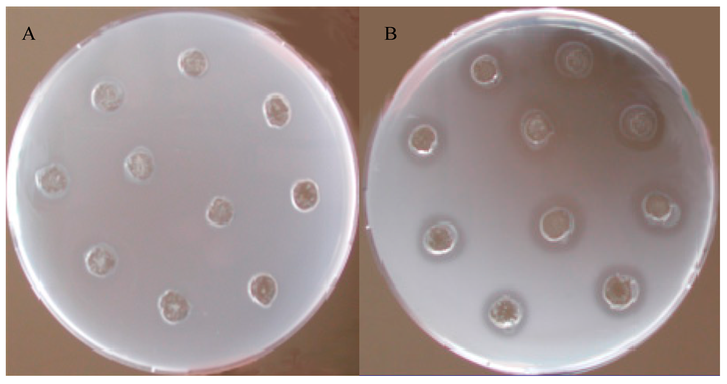
In order to evaluate the lysozyme-like activity, a standard assay on Petri dishes inoculated with Micrococcus luteus cell walls was used. The diameters of the potential cleared zone due to cnidocyst extract of P. axinellae were compared with those of a reference sample represented by hen egg-white lysozyme used at a concentration ranging from 0.2 mg/mL to 1.5 mg/mL and producing diameters of lysis comprised between 1.5 and 10.5 mm. The cnidocyst extract of P. axinellae did not record a noteworthy lysozyme-like activity and no appreciable diameter of lysis was observed. The cnidocyst extract of P. axinellae did not record a noteworthy lysozyme-like activity since no appreciable diameter of lysis was observed (Figure 2).

3.3. Antibacterial Activity

Parazoanthus axinellae cnidocyst extract exerted significant antibacterial activity against several tested microorganisms (Table 1).
In particular, the highest growth inhibition percentage (75.00 ± 0.90%) occurred against Streptococcus agalactiae, soon followed by inhibition of Vibrio alginolyticus (73.00 ± 1.30%). Although with lower percentages, a significant sensitivity to the P. axinellae cnydocyst extract was also highlighted in Vibrio fischeri (43.36 ± 5.00%), Vibrio anguillarum (40.15 ± 1.50%), Coccus sp. (37.84 ± 2.30%), Vibrio vulnificus (34.32 ± 8.00%), and Vibrio harveyi (28.00 ± 4.10%). Pseudomonas aeruginosa and Salmonella sp. were not significantly affected by treatment with P. axinellae extract. The two human pathogenic yeasts Candida albicans and C. glabrata showed no sensitivity to the tested extract. In Table 2, the Permanova analyses concerning the sensitivity of the considered microorganisms to the action of P. axinallae cnidocyst extract are reported.

3.4. Characterization of Antibacterial Activity in P. axinellae Extract

Since Vibrio alginolyticus was one of the strains most inhibited by P. axinellae cnidocyst extract, it was chosen in order to better characterize the observed activity. Antibacterial activity was tested using the bactericidal assay described above. In particular, a dose-response curve was plotted by employing increasing volumes of cnidocyst extract and maintaining the bacterial suspension at 1 × 10⁸ cells/mL (by using a spectrophotometer, optical density at 600 nm). The antibacterial activity was further characterized by increasing the bacterial concentration (from 1 × 10⁸ to 8 × 10⁸ cells/mL) of V. alginolyticus and maintaining constant the volume of P. axinellae extract (50 μL) and recording the obtained dose–response curve. Moreover, in order to determine the effect of temperature on the investigated antibacterial activity, the cnidocyst extract was held for 1 hr at 22, 37, or 56 °C. Finally, a time course of activity was also obtained by incubating 50 µL of cnidocyst extract with 10 µL of bacterial suspension (10 8 cells/mL) at 30 °C for 1, 4, 6, and 24 h.

Dose–Response Curves

Since V. alginolyticus was found to be the most sensitive microbial species, it was selected as a standard test strain for the characterization of the antibacterial activity produced by the P. axinellae cnidocyst extract.
As shown in Figure 3 a dose–response curve was obtained by increasing the volumes of the P. axinellae extract. In particular, Vibrio alginolyticus growth inhibition was 62.70 ± 0.80% when the concentration of the bacterial suspension was maintained at 1 × 10⁸ cells/mL and 20 µL of extract were employed. Increasing the extract volume at 50 and 100 µL only highlighted a small additional inhibition (73.10 ± 1.10 and 88.90 ± 0.90%, respectively). The percentage of antibacterial activity was positively correlated (R = 0.99) with the employed extract volumes.
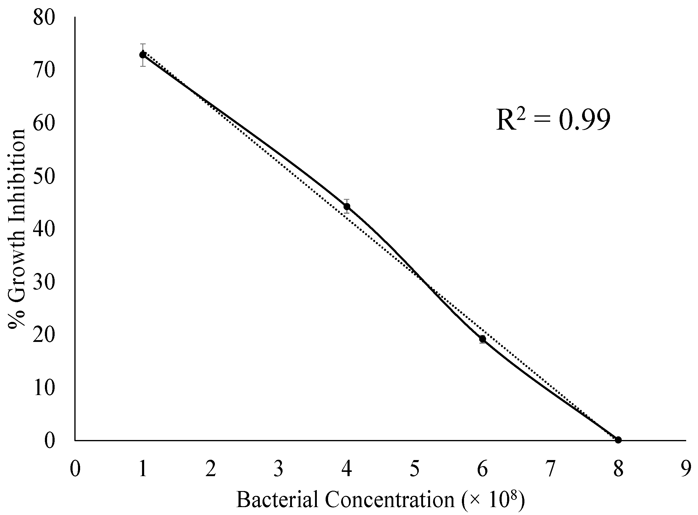
A dose–response curve was obtained by increasing the bacterial concentration (from 1 × 10⁸ to 8 × 10⁸ cells/mL) of Vibrio alginolyticus exposed to 50 μL (constant volume) of P. axinellae extract (Figure 4). Variation in bacterial concentration strongly affected the antibacterial power of the P. axinellae extract employed at a constant volume. In particular, when the concentration of the bacterial suspension was 1 × 10⁸ cells/mL, the bacterial growth inhibition percentage was 77.80 ± 2.10%. When 50 μL of P. axinellae extract were incubated with V. alginolyticus at 4 × 10⁸ and 6 × 10⁸ cells/mL, the growth inhibition decreased markedly to 44.20 ± 1.30 and 19.1 ± 0.70%, respectively. Antimicrobial activity of the P. axinellae extract was not present with a bacterial concentration of 8 × 10⁸ cells/mL. The percentage of antibacterial activity was positively correlated (R = 0.99) with the employed bacterial concentration.
The anti-V. alginolyticus activity of the tested extract significantly depended on both the employed volume of the P. axinellae cnidocyst extract (Table 3) and the concentration of the bacterial suspension (Table 4) as evidenced by the Permanova analyses.

3.5. Effect of Temperature on Antibacterial Activity

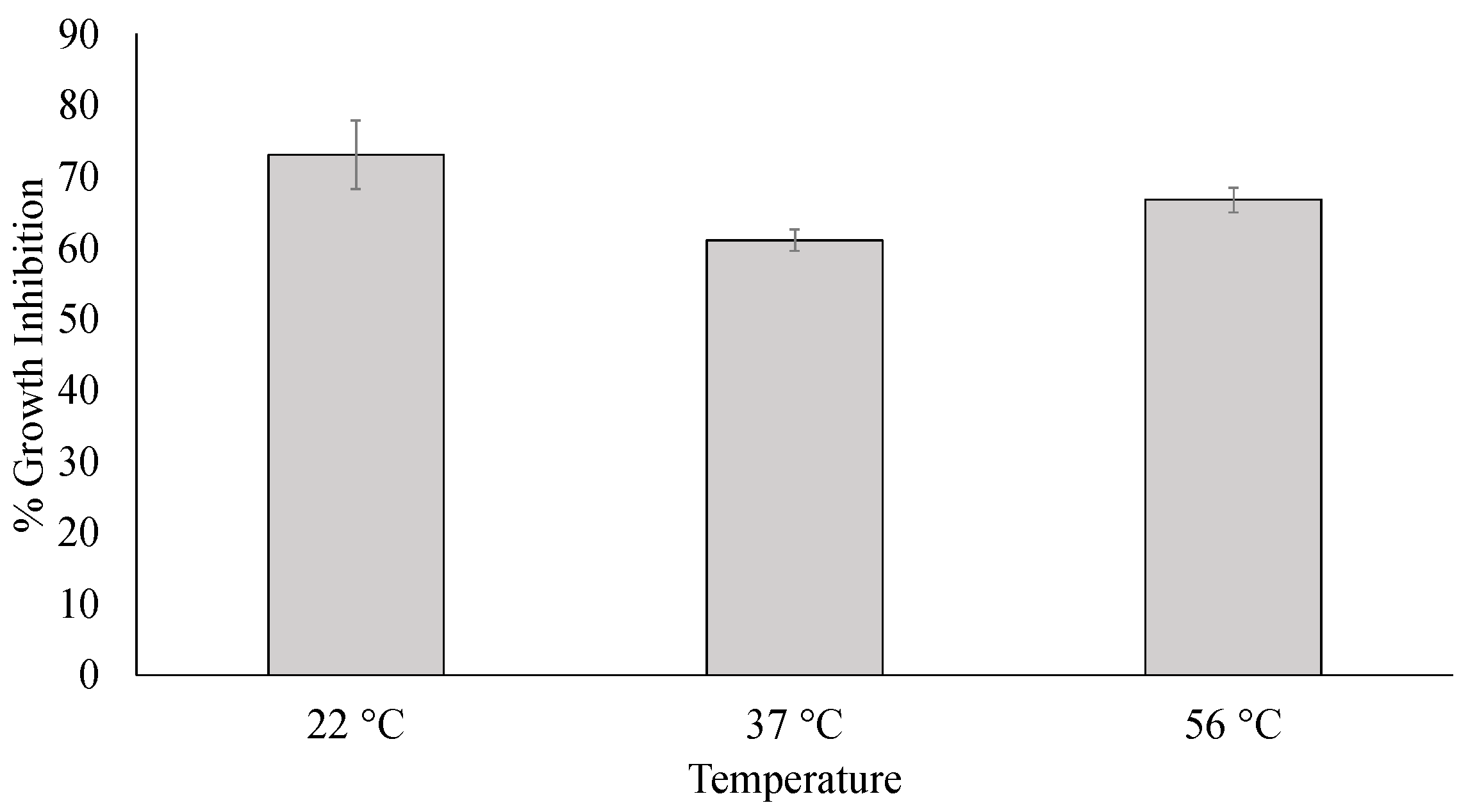
The effect of temperature on the antibacterial activity was also evaluated. When P. axinellae cnidocyst extract was maintained at 22 °C, V. alginolyticus growth inhibition percentage was 73.10 ± 3.80%, while at 37 and 56 °C, the percentages decreased accounting for 61.00 ± 1.50 and 66.70 ±1.70, respectively (Figure 5).
No significant differences in antibacterial activity were observed between the extract at 22, 37, and 56 °C (Table 5).

3.6. Time Course of Antibacterial Activity

The effect of incubation time on the antibacterial activity was also determined. After 30 min of incubation of V. alginolyticus exposed to cnidocyst extract, the maximum growth inhibition percentage was recorded, corresponding to 74.30 ± 2.70%, while, after 60 and 120 min of incubation, the percentages decreased to 43.50 ± 1.50% and 39.10 ± 1.20%, respectively (Figure 6).
Pairwise tests detected significant differences in antibacterial activity of the extract after 30 and 60, and 30 and 120 min of incubation, while no significant differences were showed in antibacterial activity of the extract after 60 and 120 min of incubation (Table 6).

3.7. Scanning Electron Microscope (SEM)

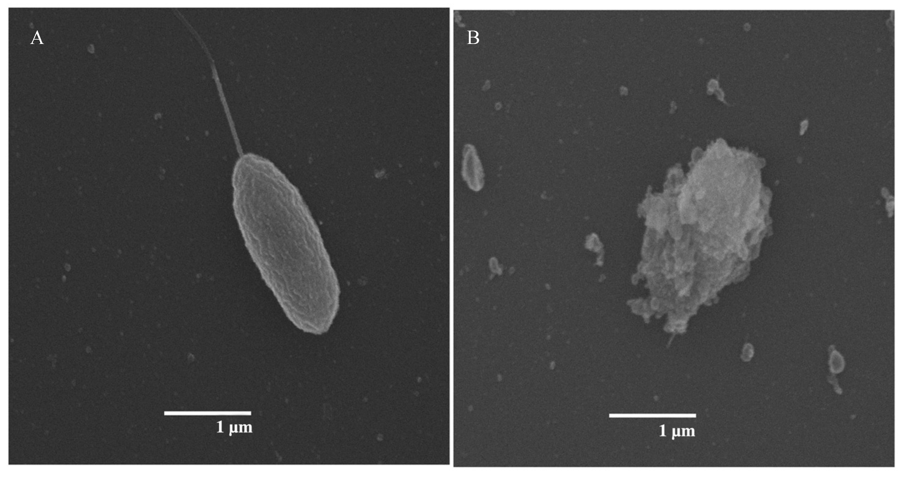
The scanning electron microscopy (SEM) revealed that the cnidocyst extract of P. axinellae strongly affected the bacterial strain Vibrio alginolyticus (Figure 7) as shown by the collapsed bacterial walls, when compared to the morphology of Vibrio alginolyticus not exposed to the tested extract.

4. Discussion

The marine environment is very complex and here animals equally fight for survival, in a complex balance between predation and defence [48,49]. Sessile organisms remain those most at risk, since they do not have the possibility to escape from the lurking dangers, whether they are other animals or changes in environmental parameters. In this framework, in the present paper, we investigated the antimicrobial activity of P. axinellae cnidocyst extract, as defence mechanism against microorganisms that normally populate the environment where this species lives. Despite the inability to move, most of the sessile organisms, including P. axinellae, indeed, have evolutionarily developed defence systems that provide for the immediate release of toxic substances. These molecules, which are generally encapsulated in specialized structures called cnidocysts, are released following an appropriate stimulus and provide a partial guarantee of survival [50]. The isolation of P. axinellae cnidocyst extract was obtained by using the method of Avian et al. [38], thus allowing us to reach a percentage of cnidocysts of about 90% used to test their antimicrobial activity. The results obtained are encouraging since the cnidocyst extract of P. axinellae shows a marked ability to inhibit the growth of some bacterial strains. The substances in the P. axinellae cnidocyst extract, responsible for the antibacterial activity observed, were thermostable and acted rapidly. In fact, only 30 min of contact with V. algynoliticus was necessary for the bacterial growth “in vitro” to be inhibited. A low percentage of bacteria (about 27%), however, survived the action of the P. axinellae cnidocyst extract and this phenomenon was most likely determined by the dose–response relationship existing between effectors and target organism. This dose–response relationship could explain why, prolonging the incubation period till 120 min, it was not possible to observe an increase in the percentage of bacterial growth inhibition as evidenced in the time course. After 1 h, indeed, the percentage of inhibition of bacterial growth was reduced to 43.50 ± 1.50%, probably because the bacteria not destroyed by the extract continued to multiply, producing a reduction in the estimated effect. The mechanism of action of P. axinellae cnidocyst extract against V. alginolyticus presumably could be reflected also against the other tested microorganisms such as Coccus sp., Streptococcus agalactiae, and vibrios. A similar trend of the antibacterial activity was already observed also in the cnidarian Anemonia sulcata [51], in the annelidan Eisenia andrei [52], in the sea urchin Paracentrotus lividus [53], and in several molluscans [54].
The bacteria treated with the P. axinellae extract showed morphological changes, when observed by scanning electron microscopy. In particular, significant lesions of the bacterial wall were highlighted suggesting a lytic action of cnidocysts. Studies conducted on other cnidarians have evidenced the ability of some molecules called “cytolysins” to lyse the plasma membrane of other cells with which they come into contact [55]. In some cases, the mechanism of action of cytolysins has been determined and appears to consist of two phases. The first one involves the formation of a bond with the plasma membrane of the target cell and the second one the oligomerization at the level of the plasma membrane with the formation of the pore [55,56,57,58,59]. Cytolysins, therefore, could be responsible for the inhibitory activity shown by P. axinellae cnidocyst extract on bacterial growth. However, the defensive system used against microorganisms living in the surrounding environment is complex. Thus, it cannot be excluded that other factors are responsible for the observed antibacterial activity or that both cytolysins and other “defensive factors” act synergistically in the antibacterial protection of P. axinellae. The presence of several defence mechanisms to preserve the integrity of P. axinellae is also demonstrated by the work of Herndl and Velimirov [60]. They indicated that the coelenteric fluid excellently controls the concentration of bacteria inside the gastric cavity of P. axinellae, degrading bacteria when their concentration rises to above a threshold value. As hypothesized by some authors, this digestion would bring advantages to the anthozoans, as they would incorporate useful carbonaceous compounds, vitamins, and essential trace elements, or antibiotic substances coming from the tissues of microorganisms [15,60,61,62,63]. It is uncertain, however, whether it is an enzymatic digestion, due to gastric processing of the same microorganisms, or whether there are specific substances at the coelenteric level that determine this phenomenon. The results obtained in the present work suggest that the survival of P. axinellae in an environment usually crowded by microorganisms is due to the relationship between the substances here investigated, encapsulated within the cnidocysts, and those freely present in an unpackaged form in the coelenteric fluid Parazoanthus axinellae, as observed in other marine organisms [64,65,66], that has probably managed over the course of evolution to develop a genetic pool capable of leading to the synthesis of specific defence molecules, acting synergistically, fighting potential pathogens. The specific genetic makeup or the mechanism of synthesis and storage of the aforementioned substances is not yet known and further studies will be conducted to clarify these aspects. The existence of a common synthesis route of molecules with antibacterial activity which are partly encapsulated in the cnidocysts and partly secreted by the external cell layers cannot be excluded.
Another particularly interesting and noteworthy aspect obtained in this work is the notable sensitivity of bacteria belonging to the genus Vibrio to the action of the cnidocyst extract of P. axinellae. These bacteria, in fact, include halophilic species and are counted among the most interesting bacterial strains present in the marine environment. Currently, numerous of these species are considered pathogenic both for humans and for the several marine organisms, including some invertebrates and fish [67,68,69,70]. The antibacterial activity of P. axinellae cnidocyst extract against vibrios, particularly V. alginolyticus, is of particular interest for the potential biotechnological applications since the extract could be used to fight vibriosis, representing one of the major problems in aquaculture with economic relapsed [71,72,73]. Some bacterial strains (especially Pseudomonas aeruginosa and Salmonella sp.) and the yeast fungi Candida albicans and Candida glabrata were not absolutely inhibited in their growth. On the contrary, we found a high antibacterial activity of P. axinellae cnidocyst extract against the bacterial strain Streptococcus agalactiae (GBS). It is a relatively frequent bacterial strain in a female gastrointestinal and genitourinary tract. However, it can be transmitted from mothers to infants at the time of birth, causing septicaemia, meningitis, sepsis, and neonatal pneumonia [74,75,76,77,78]. Among the newborns, Schindler et al. [79] showed a strong relationship between GBS infection and the risk of intrauterine foetal death. Thus, finding antibacterial capable of combating GBS represents a challenge due to its high incidence among parturients and their neonates worldwide and the development of its antibiotic resistance [80]. Moreover, previous studies showed an antimicrobial activity of P. axinellae extract incorporated nanostructures against the Gram-positive bacterial strain Staphylococcus aureus and the Gram-negative bacterial strain Aeromonas hydrophila, Aeromonas sobria, Escherichia coli, and Salmonella enterica as reported by Konuklugil et al. [81]. In this scenario, further investigations will be needed in order to isolate the potential molecules responsible for the antibacterial activities and our results pave the way for the identification of these interesting bio-compounds requiring further integrated analytical approaches, such as metabolomic, HPLC, GC-MS, and LC-MS methods. In particular metabolomic approach could be the pivotal topic of a further study in order to provide further information insight the secondary metabolites present in the cnidocyst extract elucidating also the chemical nature of the compounds involved in the here evidenced antibacterial activity.
In conclusion, on account of the antimicrobial activity of P. axinellae cnidocyst extract against vibrios and S. agalactiae, the present work encourages the potential exploitation of P. axinellae as a novel and excellent source of antibacterial compounds with several possible applications for the biotechnological and pharmacological sectors. This is crucial considering the need to discover innovative antimicrobials for the treatment of infectious diseases due to multidrug-resistant bacteria and to combine research and technological advancements.

Author Contributions

Methodology, L.S., S.P. and L.R.; analysis, L.S. and L.R.; writing—original draft preparation, L.S. and L.R.; writing—review and editing, L.S., S.P. and L.R. All authors have read and agreed to the published version of the manuscript.

Funding

This study was partially supported by the “National Biodiversity Future Center (NBFC)”, funded by European Union Next Generation EU (PNRR), Spoke 2 Activity 3 Task 3.2 CUP B83C22002930006.

Institutional Review Board Statement

Not applicable.

Informed Consent Statement

Not applicable.

Data Availability Statement

The data presented in this study are available in Table 1.

Acknowledgments

The authors thank Marcella D’Elia for the technical support in the Scanning Electron Microscope Laboratory.

Conflicts of Interest

The authors declare no conflicts of interest.

References

  1. Rocha, J.; Peixe, L.; Gomes, N.C.; Calado, R. Cnidarians as a source of new marine bioactive compounds—An overview of the last decade and future steps for bioprospecting. Mar. Drugs 2011, 9, 1860–1886. [Google Scholar] [CrossRef]
  2. Stabili, L.; Rizzo, L.; Caprioli, R.; Leone, A.; Piraino, S. Jellyfish bioprospecting in the Mediterranean Sea: Antioxidant and lysozyme-like activities from Aurelia coerulea (Cnidaria, Scyphozoa) extracts. Mar. Drugs 2021, 19, 619. [Google Scholar] [CrossRef]
  3. Jain, R.; Sonawane, S.; Mandrekar, N. Marine organisms: Potential source for drug discovery. Curr. Sci. 2008, 94, 292. Available online: https://www.currentscience.ac.in/Volumes/94/03/0292.pdf (accessed on 1 January 2023).
  4. Bhakuni, D.S.; Rawat, D.S. Bioactive Marine Natural Products; Springer Science & Business Media: Dordrecht, The Netherlands, 2006. [Google Scholar] [CrossRef]
  5. Carroll, A.R.; Copp, B.R.; Davis, R.A.; Keyzers, R.A.; Prinsep, M.R. Marine natural products. Nat. Prod. Rep. 2022, 39, 1122–1171. [Google Scholar] [CrossRef]
  6. Stabili, L.; Fraschetti, S.; Acquaviva, M.I.; Cavallo, R.A.; De Pascali, S.A.; Fanizzi, F.P.; Gerardi, C.; Narracci, M.; Rizzo, L. The potential exploitation of the Mediterranean invasive alga Caulerpa cylindracea: Can the invasion be transformed into a gain? Mar. Drugs 2016, 14, 210. [Google Scholar] [CrossRef]
  7. Stabili, L.; Acquaviva, M.I.; Cecere, E.; Gerardi, C.; Petrocelli, A.; Fanizzi, F.P.; Angilè, F.; Rizzo, L. Screening of Undaria pinnatifida (Laminariales, Phaeophyceae) lipidic extract as a new potential source of antibacterial and antioxidant compounds. J. Mar. Sci. Eng. 2023, 11, 2072. [Google Scholar] [CrossRef]
  8. Thawabteh, A.M.; Swaileh, Z.; Ammar, M.; Jaghama, W.; Yousef, M.; Karaman, R.; Bufo, S.A.; Scrano, L. Antifungal and antibacterial activities of isolated marine compounds. Toxins 2023, 15, 93. [Google Scholar] [CrossRef] [PubMed]
  9. Fusetani, N. Biotechnological potential of marine natural products. Pure Appl. Chem. 2010, 82, 17–26. [Google Scholar] [CrossRef]
  10. Shehzad, A.; Zahid, A.; Latif, A.; Amir, R.M.; Suleria, H.A.R. Marine foods: Nutritional significance and their industrial applications. In Technological Processes for Marine Foods, from Water to Fork: Bioactive Compounds, Industrial Applications, and Genomics; CRC Press: Boca Raton, FL, USA, 2019; Volume 289. [Google Scholar] [CrossRef]
  11. Glaser, K.B.; Mayer, A.M. A renaissance in marine pharmacology: From preclinical curiosity to clinical reality. Biochem. Pharmacol. 2009, 78, 440–448. [Google Scholar] [CrossRef] [PubMed]
  12. Hong, L.L.; Ding, Y.F.; Zhang, W.; Lin, H.W. Chemical and biological diversity of new natural products from marine sponges: A review (2009–2018). Mar. Life Sci. Technol. 2022, 4, 356–372. [Google Scholar] [CrossRef] [PubMed]
  13. Guryanova, S.V.; Balandin, S.V.; Belogurova-Ovchinnikova, O.Y.; Ovchinnikova, T.V. Marine invertebrate antimicrobial peptides and their potential as novel peptide antibiotics. Mar. Drugs 2023, 21, 503. [Google Scholar] [CrossRef]
  14. Daly, M.; Brugler, M.R.; Cartwright, P.; Collins, A.G.; Dawson, M.N.; Fautin, D.G.; France, S.C.; McFadden, C.S.; Opresko, D.M.; Rodriguez, E.; et al. The phylum Cnidaria: A review of phylogenetic patterns and diversity 300 years after Linnaeus. Zootaxa 2007, 1668, 127–182. [Google Scholar] [CrossRef]
  15. Parisi, M.G.; Parrinello, D.; Stabili, L.; Cammarata, M. Cnidarian immunity and the repertoire of defense mechanisms in anthozoans. Biology 2020, 9, 283. [Google Scholar] [CrossRef]
  16. Boero, F. Review of Jellyfish Blooms in the Mediterranean and Black Sea; No. 92.; Food and Agriculture Organisation: Rome, Italy, 2013. [Google Scholar] [CrossRef]
  17. Riisgård, H.U.; Madsen, C.V.; Barth-Jensen, C.; Purcell, J.E. Population dynamics and zooplankton-predation impact of the indigenous scyphozoan Aurelia aurita and the invasive ctenophore Mnemiopsis leidyi in Limfjorden (Denmark). Aquat. Invasion 2012, 7, 147–162. [Google Scholar] [CrossRef]
  18. Hashim, A.R.; Kamaruddin, S.A.; Buyong, F.; Mat Nazir, E.N.; Che Ismail, C.Z.; Tajam, J.; Abdullah, A.L.; Azis, T.M.F.; Anscelly, A. Jellyfish Blooming: Are We Responsible? In Proceedings of the ICAN International Virtual Conference 2022 (IIVC 2022) Proceedings—Navigating the VUCA World: Harnessing the Role of Industry Linkages, Community Development and Alumni Network in Academia, Arau, Malaysia, 18 August 2022; pp. 51–60. [Google Scholar]
  19. Kennerley, A.; Wood, L.E.; Luisetti, T.; Ferrini, S.; Lorenzoni, I. Economic impacts of jellyfish blooms on coastal recreation in a UK coastal town and potential management options. Ocean Coast. Manag. 2022, 227, 106284. [Google Scholar] [CrossRef]
  20. Moretta, A.; Scieuzo, C.; Petrone, A.M.; Salvia, R.; Manniello, M.D.; Franco, A.; Lucchetti, D.; Vassallo, A.; Vogel, H.; Sgambato, A.; et al. Antimicrobial peptides: A new hope in biomedical and pharmaceutical fields. Front. Cell. Infect. Microbiol. 2021, 11, 668632. [Google Scholar] [CrossRef]
  21. Hoffmann, J.A.; Kafatos, F.C.; Janeway Jr, C.A.; Ezekowitz, R.A.B. Phylogenetic perspectives in innate immunity. Science 1999, 284, 1313–1318. [Google Scholar] [CrossRef]
  22. Bosch, T.C.G. The path less explored: Innate immune reactions in cnidarians. In Innate Immunity of Plants, Animals, and Humans; Heine, H., Ed.; Nucleic Acids and Molecular Biology; Springer: Berlin/Heidelberg, Germany, 2008; pp. 27–42. [Google Scholar] [CrossRef]
  23. Kim, K. Antimicrobial activity in gorgonian corals (Coelenterata: Octocorallia). Coral Reefs 1994, 13, 75–80. [Google Scholar] [CrossRef]
  24. Barresi, G.; di Carlo, E.; Trapani, M.R.; Parisi, M.G.; Chille, C.; Mule, M.F.; Cammarata, M.; Palla, F. Marine organisms as source of bioactive molecules applied in restoration projects. Herit. Sci. 2015, 3, 17. [Google Scholar] [CrossRef]
  25. Phillips, D.C. The three-dimensional structure of an enzyme molecule. Sci. Am. 1966, 215, 78–90. [Google Scholar] [CrossRef]
  26. Sava, G. Pharmacological aspects and therapeutic applications of lysozymes. Exs 1996, 75, 433–449. [Google Scholar] [CrossRef]
  27. Stabili, L.; Schirosi, R.; Parisi, M.G.; Piraino, S.; Cammarata, M. The Mucus of Actinia equina (Anthozoa, Cnidaria): An Unexplored Resource for Potential Applicative Purposes. Mar. Drugs 2015, 13, 5276–5296. [Google Scholar] [CrossRef]
  28. Stabili, L.; Rizzo, L.; Fanizzi, F.P.; Angilè, F.; Del Coco, L.; Girelli, C.R.; Lomartire, S.; Piraino, S.; Basso, L. The jellyfish Rhizostoma pulmo (Cnidaria): Biochemical composition of ovaries and antibacterial lysozyme-like activity of the oocyte lysate. Mar. Drugs 2019, 17, 17. [Google Scholar] [CrossRef]
  29. Stabili, L.; Rizzo, L.; Basso, L.; Marzano, M.; Fosso, B.; Pesole, G.; Piraino, S. The microbial community associated with Rhizostoma pulmo: Ecological significance and potential consequences for marine organisms and human health. Mar. Drugs 2020, 18, 437. [Google Scholar] [CrossRef]
  30. Basso, L.; Rizzo, L.; Marzano, M.; Intranuovo, M.; Fosso, B.; Pesole, G.; Piraino, S.; Stabili, L. Jellyfish summer outbreaks as bacterial vectors and potential hazards for marine animals and humans health? The case of Rhizostoma pulmo (Scyphozoa, Cnidaria). Sci. Total Environ. 2019, 692, 305–318. [Google Scholar] [CrossRef]
  31. Cachet, N.; Genta-Jouve, G.; Ivanisevic, J.; Chevaldonné, P.; Sinniger, F.; Culioli, G.; Pérez, T.; Thomas, O.P. Metabolomic profiling reveals deep chemical divergence between two morphotypes of the zoanthid Parazoanthus axinellae. Sci. Rep. 2015, 5, 8282. [Google Scholar] [CrossRef]
  32. Cariello, L.; Crescenzi, S.; Prota, G.; Capasso, S.; Giordano, F.; Mazzarella, L. Zoanthoxanthin, a natural 1,3,5,7-tetraazacyclopent[f]azulene from Parazoanthus axinellae. Tetrahedron 1974, 30, 3281–3287. [Google Scholar] [CrossRef]
  33. Cariello, L.; Crescenzi, S.; Prota, G.; Zanetti, L. New zoanthoxanthins from the Mediterranean zoanthid Parazoanthus axinellae. Experientia 1974, 30, 849–850. [Google Scholar] [CrossRef]
  34. Cariello, L.; Crescenzi, S.; Prota, G.; Giordano, F.; Mazzarella, L. Zoanthoxanthin, a heteroaromatic base from Parazoanthus axinellae (Zoantharia). Structure confirmation by x-ray crystallography. J. Chem. Soc. Chem. Commun. 1973, 3, 99–100. [Google Scholar] [CrossRef]
  35. Cachet, N.; Genta-Jouve, G.; Regalado, E.L.; Mokrini, R.; Amade, P.; Culioli, G.; Thomas, O.P. Parazoanthines A-E, hydantoin alkaloids from the mediterranean sea anemone Parazoanthus axinellae. J. Nat. Prod. 2009, 72, 1612–1615. [Google Scholar] [CrossRef]
  36. David, C.N.; Ozbek, S.; Adamczyk, P.; Meier, S.; Pauly, B.; Chapman, J.; Hwang, J.S.; Gojobori, T.; Holstein, T.W. Evolution of complex structures: Minicollagens shape the cnidarian nematocyst. Trends Genet. 2008, 24, 431–438. [Google Scholar] [CrossRef]
  37. Nuchter, T.; Benoit, M.; Engel, U.; Ozbek, S.; Holstein, T.W. Nanosecond-scale kinetics of nematocyst discharge. Curr. Biol. 2006, 16, R316–R318. [Google Scholar] [CrossRef]
  38. Avian, M.; Del Negro, P.; Sandrini, L.R. A comparative analysis of nematocysts in Pelagia noctiluca and Rhizostoma pulmo from the North Adriatic Sea. Hydrobiologia 1991, 216, 615–621. [Google Scholar] [CrossRef]
  39. Manzari, C.; Fosso, B.; Marzano, M.; Annese, A.; Caprioli, R.; D’Erchia, A.M.; Gissi, C.; Intranuovo, M.; Picardi, E.; Santamaria, M.; et al. The influence of invasive jellyfish blooms on the aquatic microbiome in a coastal lagoon (Varano, SE Italy) detected by an Illumina-based deep sequencing strategy. Biol. Invasions 2015, 17, 923–940. [Google Scholar] [CrossRef]
  40. Canicatti, C.; Roch, P. Studies on Holothuria polii (Echinodermata) antibacterial proteins. I. Evidence for and activity of a coelomocyte lysozyme. Experientia 1989, 45, 756–759. [Google Scholar] [CrossRef]
  41. Stabili, L.; Gravili, C.; Tredici, S.M.; Piraino, S.; Talà, A.; Boero, F.; Alifano, P. Epibiotic Vibrio luminous bacteria isolated from some hydrozoa and bryozoa species. Microb. Ecol. 2008, 56, 625–636. [Google Scholar] [CrossRef]
  42. Rizzo, L.; Fraschetti, S.; Alifano, P.; Pizzolante, G.; Stabili, L. The alien species Caulerpa cylindracea and its associated bacteria in the Mediterranean Sea. Mar. Biol. 2016, 163, 4. [Google Scholar] [CrossRef]
  43. Rizzo, L.; Fraschetti, S.; Alifano, P.; Tredici, M.S.; Stabili, L. Association of Vibrio community with the Atlantic Mediterranean invasive alga Caulerpa cylindracea. J. Exp. Mar. Biol. Ecol. 2016, 475, 129–136. [Google Scholar] [CrossRef]
  44. Boman, H.G.; Nilson-Faye, I.; Paul, K.; Raswson, T., Jr. Insect immunity. 1. Characteristics of an inolcible cell-free antibacterial reaction in hemolymph of Semie Cynthie pupal. Immunity 1974, 10, 136–145. [Google Scholar] [CrossRef]
  45. Anderson, M.; Braak, C.T. Permutation tests for multi-factorial analysis of variance. J. Stat. Comput. Simul. 2003, 73, 85–113. [Google Scholar] [CrossRef]
  46. Anderson, M.J. A new method for non-parametric multivariate analysis of variance. Austral Ecol. 2001, 26, 32–46. [Google Scholar] [CrossRef]
  47. Anderson, M.J.; Gorley, R.N.; Clarke, K.R. PERMANOVA+ for PRIMER: Guide to Software and Statistical Methods; PRIMER-E: Plymouth, UK, 2015. [Google Scholar]
  48. Bashevkin, S.M.; Morgan, S.G. Predation and competition. Nat. Hist. Crustac. 2020, 7, 360–382. [Google Scholar] [CrossRef]
  49. Abrams, P.A. Adaptive responses of predators to prey and prey to predators; the failure of the arms race analogy. Evolution 1986, 40, 1229–1247. [Google Scholar] [CrossRef]
  50. Oppegard, S.C.; Anderson, P.A.; Eddington, D.T. Puncture mechanics of cnidarian cnidocysts: A natural actuator. J. Biol. Eng. 2009, 3, 17. [Google Scholar] [CrossRef]
  51. Trapani, M.R.; Parisi, M.G.; Toubiana, M.; Coquet, L.; Jouenne, T.; Roch, P.; Cammarata, M. First evidence of antimicrobial activity of neurotoxin 2 from Anemonia sulcata (Cnidaria). Invertebr. Surviv. J. 2014, 11, 182–191. [Google Scholar] [CrossRef]
  52. Hirigoyenberry, F.; Lassegues, M.; Roch, P. Antibacterial activity of Eisenia fetida andrei coelomic fluid: Immunological study of the two major antibacterial proteins. J. Invertebr. Pathol. 1992, 59, 69–74. [Google Scholar] [CrossRef]
  53. Stabili, L.; Pagliara, P.; Roch, P. Antibacterial activity in the coelomocytes of the sea urchin Paracentrotus lividus. Comp. Biochem. Physiol. B Biochem. Mol. Biol. 1996, 113, 639–644. [Google Scholar] [CrossRef]
  54. Li, H.; Parisi, M.G.; Parrinello, N.; Cammarata, M.; Roch, P. Molluscan antimicrobial peptides, a review from activity-based evidences to computer-assisted sequences. Invertebr. Surviv. J. 2011, 8, 85–97. Available online: https://www.isj.unimore.it/index.php/ISJ/article/view/239 (accessed on 1 January 2023).
  55. Anderluh, G.; Maček, P. Cytolytic peptide and protein toxins from sea anemones (Anthozoa: Actiniaria). Toxicon 2002, 40, 111–124. [Google Scholar] [CrossRef]
  56. Kulma, M.; Anderluh, G. Beyond pore formation: Reorganization of the plasma membrane induced by pore-forming proteins. Cell. Mol. Life Sci. 2021, 78, 6229–6249. [Google Scholar] [CrossRef]
  57. Yap, W.Y.; Hwang, J.S. Response of cellular innate immunity to cnidarian pore-forming toxins. Molecules 2018, 23, 2537. [Google Scholar] [CrossRef]
  58. Rojko, N.; Dalla Serra, M.; Maček, P.; Anderluh, G. Pore formation by actinoporins, cytolysins from sea anemones. Biochim. Biophys. Acta (BBA)-Biomembr. 2016, 1858, 446–456. [Google Scholar] [CrossRef]
  59. Kristan, K.Č.; Viero, G.; Dalla Serra, M.; Maček, P.; Anderluh, G. Molecular mechanism of pore formation by actinoporins. Toxicon 2009, 54, 1125–1134. [Google Scholar] [CrossRef] [PubMed]
  60. Herndl, G.J.; Velimirov, B. Bacteria in the coelenteron of Anthozoa: Control of coelenteric bacterial density by the coelenteric fluid. J. Exp. Mar. Biol. Ecol. 1985, 93, 115–130. [Google Scholar] [CrossRef]
  61. Stabili, L.; Parisi, M.G.; Parrinello, D.; Cammarata, M. Cnidarian interaction with microbial communities: From aid to animal’s health to rejection responses. Mar. Drugs 2018, 16, 296. [Google Scholar] [CrossRef] [PubMed]
  62. Sorokin, Y.I. On the feeding of some scleractinian corals with bacteria and dissolved organic matter. Limnol. Oceanogr. 1973, 18, 380–386. [Google Scholar] [CrossRef]
  63. Burkholder, P.R. The ecology of marine antibiotics and coral reefs. Biol. Geol. Coral Reefs 1973, 2, 117–182. [Google Scholar] [CrossRef]
  64. Lauritano, C.; Ianora, A. Chemical defense in marine organisms. Mar. Drugs 2020, 18, 518. [Google Scholar] [CrossRef]
  65. Bachère, E.; Rosa, R.D.; Schmitt, P.; Poirier, A.C.; Merou, N.; Charrière, G.M.; Destoumieux-Garzón, D. The new insights into the oyster antimicrobial defense: Cellular, molecular and genetic view. Fish Shellfish Immunol. 2015, 46, 50–64. [Google Scholar] [CrossRef]
  66. Patrzykat, A.; Douglas, S.E. Gone gene fishing: How to catch novel marine antimicrobials. Trends Biotechnol. 2003, 21, 362–369. [Google Scholar] [CrossRef]
  67. Zhang, X.H.; He, X.; Austin, B. Vibrio harveyi: A serious pathogen of fish and invertebrates in mariculture. Mar. Life Sci. Technol. 2020, 2, 231–245. [Google Scholar] [CrossRef]
  68. Austin, B.; Zhang, X.H. Vibrio harveyi: A significant pathogen of marine vertebrates and invertebrates. Lett. Appl. Microbiol. 2006, 43, 119–124. [Google Scholar] [CrossRef]
  69. Roux, F.L.; Wegner, K.M.; Baker-Austin, C.; Vezzulli, L.; Osorio, C.R.; Amaro, C.; Ritchie, J.M.; Defoirdt, T.; Destoumieux-Garzón, D.; Blokesch, M.; et al. The emergence of Vibrio pathogens in Europe: Ecology, evolution, and pathogenesis (Paris, 11–12th March 2015). Front. Microbiol. 2015, 6, 830. [Google Scholar] [CrossRef]
  70. Baker-Austin, C.; Oliver, J.D. Vibrio vulnificus: New insights into a deadly opportunistic pathogen. Environ. Microbiol. 2018, 20, 423–430. [Google Scholar] [CrossRef]
  71. Trinanes, J.; Martinez-Urtaza, J. Future scenarios of risk of Vibrio infections in a warming planet: A global mapping study. Lancet Planet. Health 2021, 5, e426–e435. [Google Scholar] [CrossRef]
  72. Ina-Salwany, M.Y.; Al-saari, N.; Mohamad, A.; Mursidi, F.A.; Mohd-Aris, A.; Amal, M.N.A.; Kasai, H.; Mino, S.; Sawabe, T.; Zamri-Saad, M. Vibriosis in fish: A review on disease development and prevention. J. Aquat. Anim. Health 2019, 31, 3–22. [Google Scholar] [CrossRef]
  73. Igbinosa, E.O. Detection and antimicrobial resistance of Vibrio isolates in aquaculture environments: Implications for public health. Microb. Drug Resist. 2016, 22, 238–245. [Google Scholar] [CrossRef]
  74. Miselli, F.; Frabboni, I.; Di Martino, M.; Zinani, I.; Buttera, M.; Insalaco, A.; Stefanelli, F.; Lugli, L.; Berardi, A. Transmission of Group B Streptococcus in late-onset neonatal disease: A narrative review of current evidence. Ther. Adv. Infect. Dis. 2022, 9, 20499361221142732. [Google Scholar] [CrossRef]
  75. Rao, G.G.; Khanna, P. To screen or not to screen women for Group B Streptococcus (Streptococcus agalactiae) to prevent early onset sepsis in newborns: Recent advances in the unresolved debate. Ther. Adv. Infect. Dis. 2020, 7, 2049936120942424. [Google Scholar] [CrossRef]
  76. George, C.R.R.; Jeffery, H.E.; Lahra, M.M. Infection of mother and baby. In Keeling’s Fetal and Neonatal Pathology; Springer: Cham, Switzerland, 2022; pp. 207–245. [Google Scholar] [CrossRef]
  77. Baker, C.J. Interview with Carol J. Baker, M.D. Prevention of neonatal Group B streptococcal disease. Pediatr. Infect. Dis. 1983, 2, 1–5. [Google Scholar] [CrossRef]
  78. Dillon, H.C., Jr.; Khare, S.; Gray, B.M. Group B streptococcal carriage and disease: A 6-year prospective study. J. Pediatr. 1987, 110, 31–36. [Google Scholar] [CrossRef] [PubMed]
  79. Schindler, Y.; Rahav, G.; Nissan, I.; Madar-Shapiro, L.; Abtibol, J.; Ravid, M.; Maor, Y. Group B Streptococcus serotypes associated with different clinical syndromes: Asymptomatic carriage in pregnant women, intrauterine fetal death, and early onset disease in the newborn. PLoS ONE 2020, 15, e0244450. [Google Scholar] [CrossRef] [PubMed]
  80. Adu-Afari, G. Streptococcus agalactiae Infection among Parturients and Their Neonates at the Cape Coast Teaching Hospital: An Evaluation of Different Diagnostic Methods, Prevalence and Risk Factors. Ph.D. Dissertation, University of Cape Coast, Cape Coast, Ghana, 2021. [Google Scholar]
  81. Konuklugil, B.; Uras, I.S.; Karsli, B.; Demirbas, A. Parazoanthus axinellae extract incorporated hybrid nanostructure and its potential antimicrobial activity. Chem. Biodivers. 2023, 20, e202300744. [Google Scholar] [CrossRef] [PubMed]
Figure 1. Cnidocysts discharged from the P. axinellae tentacles immersed in glycerol solution.
Jmse 12 00354 g001
Figure 2. Standard assay on Petri dish inoculated with Micrococcus luteus cell walls to detect the lysozyme-like activity of cnidocyst extract from P. axinellae (A). Standard hen egg-white lysozyme (HEWL) was used as positive control (B).
Jmse 12 00354 g002
Figure 3. Dose–response curve of antibacterial activity against Vibrio alginolyticus recorded at different volumes of cnidocyst extract (from 20 to 100 μL). Values are given as means ± standard error.
Jmse 12 00354 g003
Figure 4. Dose–response curve of antibacterial activity against different bacterial concentration of Vibrio alginolyticus exposed to a constant volumes of P. axinellae cnidocyst extract (50 μL). Values are given as means ± standard error.
Jmse 12 00354 g004
Figure 5. Power of the P. axinellae extract to inhibit the growth of Vibrio alginolyticus under different experimental temperatures, n = 3. Values are given as means ± standard error.
Jmse 12 00354 g005
Figure 6. Power of the P. axinellae extract to inhibit the growth of Vibrio alginolyticus under different experimental incubation time. Values are given as means ± standard error.
Jmse 12 00354 g006
Figure 7. Power of the P. axinellae extract to inhibit the growth of Vibrio alginolyticus (A) = control: Vibrio alginolyticus with only nutrient broth, (B) = treatment: Vibrio alginolyticus with cnidocyst extract.
Jmse 12 00354 g007
Table 1. Results of an in-vitro test showing the antibacterial activity of P. axinellae cnidocyst extract. Each value represents the average of three replications ± SE. Concentration of microbial strains = 1 × 10⁸ cells/mL.
Bacterial Strain% Growth Inhibition
Candida albicans0.00 ± 0.00
Candida glabrata0.00 ± 0.00
Coccus sp.37.84 ± 2.30
Pseudomonas aeruginosa0.00 ± 0.00
Salmonella sp.0.00 ± 0.00
Streptococcus agalactiae75.00 ± 0.90
Vibrio alginolyticus73.00 ± 1.30
Vibrio anguillarum40.15 ± 1.50
Vibrio fischeri43.36 ± 5.00
Vibrio harveyi28.00 ± 4.10
Vibrio vulnificus34.32 ± 8.00
Table 2. Results of the PERMANOVA tests on percentages of microbial growth inhibition produced by P. axinellae cnidocyst extract. Abbreviations used: df—degrees of freedom; MS—mean squares; Pseudo-F—Pseudo-F statistic; P(MC)—probability level after Monte Carlo simulations; ** p ≤ 0.01; *** p ≤ 0.001; ns—not significant.
SourcedfMSPseudo-FP(MC)MSPseudo-FP(MC)
Candida albicansCandida glabrata
Factor11.67 × 10−51ns1.67 × 10−51ns
Residual41.67 × 10−5 1.67 × 10⁻⁵
Total5
Coccus sp.Pseudomonas aeruginosa
An12147.80271.18***1.67 × 10−51ns
Res47.92 1.67 × 10⁻⁵
Total5
Salmonella sp.Streptococcus agalactiae
Factor11.67 × 10−51.00ns8437.506934.20***
Residual41.67 × 10−5 1.22
Total5
Vibrio alginolyticusVibrio anguillarum
Factor17993.503157.90***2418715.39***
Residual42.53 3.38
Total5
Vibrio fischeriVibrio harveyi
Factor12820.1075.21**1176.0046.657**
Residual437.50 25.20
Total5
Vibrio vulnificus
Factor11766.8018.39**
Residual496.05
Total5
Table 3. Results of the PERMANOVA tests on percentages of bacterial growth inhibition exhibited by several cnidocyst extract volumes of P. axinellae against Vibrio alginolyticus. Abbreviations used: df—degrees of freedom; MS—mean squares; Pseudo-F—Pseudo-F statistic; P(MC)—probability level after Monte Carlo simulations; t—pairwise tests; *** p ≤ 0.001; V20 = 20 μL of cnidocyst extract of P. axinellae; V50 = 50 μL of cnidocyst extract of P. axinellae; V100 = 100 μL of cnidocyst extract of P. axinellae; vs. = versus.
SourcedfMSPseudo-FP(MC)PairwisetP(MC)
Volume2522.12195.46***V20 vs. V507.63***
Residual62.67 V20 vs. V10021.72***
Total8 V50 vs. V10011.10***
Table 4. Results of the PERMANOVA tests on percentages of bacterial growth inhibition exhibited by the cnidocyst extract of P. axinellae against several bacterial concentrations of Vibrio alginolyticus. Abbreviations used: df—degrees of freedom; MS—mean squares; Pseudo-F—Pseudo-F statistic; P(MC)—probability level after Monte Carlo simulations; t—pairwise tests; *** p ≤ 0.001; C1 = 1 × 10⁸ cells/mL; C4 = 4 × 10⁸ cells/mL; C6 = 6 × 10⁸ cells/mL; C8 = 8 × 10⁸ cells/mL; vs. = versus.
SourcedfMSPseudo-FP(MC)PairwisetP(MC)PairwisetP(MC)
Concentration32980.70602.58***C1 vs. C411.58***C4 vs. C617.02***
Residual84.95 C1 vs. C624.25***C4 vs. C833.91***
Total11 C1vs. C834.58***C6 vs. C827.10***
Table 5. Results of the PERMANOVA tests on percentages of bacterial growth inhibition exhibited by the cnidocyst extract of P. axinellae against Vibrio alginolyticus under several experimental temperatures. Abbreviations used: df—degrees of freedom; MS—mean squares; Pseudo-F—Pseudo-F statistic; P(MC)—probability level after Monte Carlo simulations; ns—not significant.
SourcedfMSPseudo-FP(MC)
Temperature2109.933.90ns
Residual628.15
Total8
Table 6. Results of the PERMANOVA tests on percentages of bacterial growth inhibition exhibited by the cnidocyst extract of P. axinellae against Vibrio alginolyticus under several experimental temperatures. Abbreviations used: df—degrees of freedom; MS—mean squares; Pseudo-F—Pseudo-F statistic; P(MC)—probability level after Monte Carlo simulations; *** p ≤ 0.001; ns—not significant.
SourcedfMSPseudo-FP(MC)PairwisetP(MC)
Incubation21103.50100.35***I30 vs. I609.96***
Residual611.00 I30 vs. I12011.90***
Total8 I60 vs. I1202.29ns
Disclaimer/Publisher’s Note: The statements, opinions and data contained in all publications are solely those of the individual author(s) and contributor(s) and not of MDPI and/or the editor(s). MDPI and/or the editor(s) disclaim responsibility for any injury to people or property resulting from any ideas, methods, instructions or products referred to in the content.