Article

Genetic Differentiation in Red and Green Noctiluca scintillans in Jakarta Bay, Indonesia

1
Graduate School of Agricultural Sciences & WPI-Advanced Institute for Marine Ecosystem Change, Tohoku University, Sendai 980-8572, Japan
2
Graduate School of Bioresources, Mie University, 1577 Kurima-machiya, Tsu, Mie 514-8507, Japan
3
Department of Marine Science and Technology, Faculty of Fisheries and Marine Sciences, IPB University Dramaga-Bogor, West Java 16680, Indonesia
*
Author to whom correspondence should be addressed.
J. Mar. Sci. Eng. 2025, 13(5), 866; https://doi.org/10.3390/jmse13050866 (registering DOI)
Submission received: 7 April 2025 / Revised: 24 April 2025 / Accepted: 25 April 2025 / Published: 26 April 2025

Abstract

:
Marine dinoflagellate Noctiluca scintillans occurs in two forms, red and green, that overlap in distribution across the Arabian Sea and Southeast Asian coastal waters, particularly in Indonesia. However, limited genetic data on Indonesian N. scintillans cast uncertainty on their taxonomic status. In this study, we introduce the first 28S PCR primer set specifically designed for Noctiluca to enable sequence analysis. The Indonesian red-type N. scintillans (RNS) sequences show considerable divergence from other documented populations, suggesting a potentially adapted population in Jakarta Bay. Similarly, the RNS in Indonesia differ genetically from co-occurring green-type N. scintillans (GNS). Morphological differences were also observed between Indonesian and Japanese RNS, with Indonesian cells exhibiting a smaller size and rougher surface, in contrast to the larger size and smoother surface observed in Japanese specimens.

1. Introduction

Harmful algal blooms (HABs) are a global natural phenomenon characterized by the rapid proliferation of algae and microalgae. These organisms show high growth and reproduction rates during these outbreaks, leading to substantial ecological impacts. HABs pose serious threats to ecosystems, including massive fish mortality and toxin accumulation in seafood [1,2]. HAB events have been increasing globally, with a particularly pronounced trend in Asia that is associated with increased fisheries activities. HAB causes are varied and complex; however, they are often linked to phytoplankton, particularly those with flagellates, such as dinoflagellates. These organisms can perform vertical migration and accumulate at the sea surface, resulting in water discoloration known as a red tide, often accompanied by a decline in water quality, including oxygen depletion [3]. This study focused on one of the most widespread HAB species, Noctiluca scintillans.
Noctiluca scintillans was first described as Medusa scintillans by Macartney in 1810 from specimens collected off the coast of Kent, England, and was later assigned to the genus Noctiluca by Suriray in 1836. Kofoid and Swezy (1921) [4] unified the nomenclature under the currently accepted binomial Noctiluca scintillans (Macartney) Kofoid & Swezy. Phylogenomic analyses now place Noctiluca within the early-branching dinoflagellate order Noctilucales, where it forms a sister group to Amphidinium [5,6].
Noctiluca scintillans, a large marine heterotrophic dinoflagellate, is known for causing red tide events in coastal areas worldwide, primarily in temperate regions from spring to early summer [7,8,9]. This species exhibits voracious feeding behavior, consuming various sources such as zoo- and phytoplankton, fish and copepod eggs, detritus, and bacteria [10,11]. Additionally, its vacuoles contain ammonia, nitrogen, and inorganic phosphorus [12]. Besides its role in red tide events, N. scintillans plays a crucial role in marine ecosystems. Studies have demonstrated that it substantially contributes to phytoplankton production by excreting relatively large amounts of ammonia and inorganic phosphorus into the surrounding environment [13], particularly during peak blooms in spring and summer. Furthermore, a study reports that N. scintillans aids in cleaning up the sea surface from crude oil pollution by ingesting and incorporating crude oil droplets into the marine food web [14]. Although it has beneficial roles, N. scintillans is considered a HAB species. It can cause substantial losses in fish and invertebrate yields in aquaculture areas due to massive blooms, despite being reportedly less harmful than other HAB species [1]. Fish mortality during N. scintillans blooms is typically caused by oxygen depletion, gill clogging, and high ammonia levels [12]. A recent regional review documented multiple N. scintillans-related fish-kill events in Southeast Asia, including Jakarta Bay, highlighting the socio-economic significance of these blooms [15]. Furthermore, N. scintillans can act as a vector for toxic microalgae by grazing on them and transferring phycotoxins to higher trophic levels, such as fish [2]. A recent study also found that a bloom of N. scintillans in the Gulf of Mannar (between India and Sri Lanka) resulted in the death of over 70% of the hard coral colonies in the affected area [16].
The genus Noctiluca currently comprises only one species. However, two distinct types of N. scintillans have been reported based on their bloom color and other characteristics: red-type N. scintillans (RNS) and green-type N. scintillans (GNS) [17,18]. RNS are heterotrophic and exclusively phagotrophic, while GNS are mixotrophic and harbors Protoeuglena noctilucae (Chlorophyta, Pedinophyceae) as endosymbiont [19,20]. This symbiotic relationship allows GNS to thrive in low-food-resource environments by utilizing the photosynthetic products of its symbiont as an energy source [17]. Despite the distinct ecological niches occupied by RNS and GNS, whether they represent different taxa [21] remains unsubstantiated. However, suggestions have been made due to the habitat preference of GNS for warmer waters. Noctiluca scintillans is generally found in various coastal areas globally, exhibiting a wide optimal tolerance to seawater temperatures ranging from 15 °C to 25 °C. The lowest observed temperature for N. scintillans was recorded at a coastal station in Alaska (3 °C) [22], while the highest temperature was documented in the Arabian Sea (33 °C) [23]. Regarding distribution, RNS is present globally and is commonly encountered in temperate to subtropical waters. In contrast, GNS exhibit a more confined habitat, typically restricted to tropical waters, including the Arabian Sea, India, and Southeast Asia [18].
Currently, GenBank features 28S rDNA sequences of N. scintillans from China (RNS), the United States (RNS), Mexico (RNS), and the Arabian Sea (GNS). Even within RNS, geographic sequence variations have been reported among these countries [24]. Consistent with that pattern, using three loci of the rRNA gene cistron, Pan et al. (2016) [25] showed that Chinese coastal populations have low within-region diversity but display divergence from American populations. However, no genetic information is available on N. scintillans from Indonesia, despite it displaying the coexistence of red and green N. scintillans. The semi-enclosed nature of Jakarta Bay indicates a potential locally adapted population. Therefore, this study aimed to explore the genetic information of red and green N. scintillans collected from Jakarta Bay, Indonesia, to ascertain whether they constitute the same genotype. A Noctiluca-specific PCR primer set was designed and developed to facilitate N. scintillans sequence analysis. This primer set can selectively amplify the 28S rDNA sequence of red and green N. scintillans, even in the presence of multiple prey, enabling direct sequence analysis without gene cloning. Furthermore, RNS cells were collected from the Japanese coast and compared with those from Indonesia for morphological difference examination.

2. Materials and Methods

2.1. Sample Collection and Morphological Study

Samples for morphology and genetic comparisons were collected from Jakarta Bay, Indonesia, and two sites in Japan, Miyagi and Mie (Figure 1). Specifically, 183 cells of N. scintillans, comprising 94 from Indonesia and 89 from Japan, were successfully analyzed (Table 1). Seawater samples were collected from the surface layer using an 80 µm plankton net and promptly fixed in 1% final concentration of neutral buffered formalin in 50 mL centrifuge tubes (Model 91051, TPP, Trasadingen, Switzerland). The sample tubes were transported back to the laboratory and stored at 4 °C until further examination.
Noctiluca scintillans cells obtained from the sampling point were isolated using a pipette under a binocular microscope (CX23LED Model, Olympus, Tokyo, Japan). Only cells with optimal morphology that remained intact were selected. Cell morphology and surface characteristics were observed in photomicrographs of N. scintillans obtained using NIH ImageJ version 1.54g software [26]. The vertical and horizontal diameters of the specimens from Indonesia, Miyagi, and Mie were also measured. This study obtained measurements by drawing two perpendicular lines intersecting in the cell’s center. Since most cells exhibited a spherical to slightly oval shape, measurements were conducted along the vertical and horizontal diameters. Specifically, the vertical diameter was aligned with the cytostome opening and flagellum growth point, while the horizontal diameter was oriented perpendicular to the cytostome opening (Supplementary Material Figure S1). Results are represented as the length of the vertical diameter (µm) and the length of the horizontal diameter (µm).

2.2. Genetic Analysis

During the isolation process, each cell was washed three times with filter-sterilized seawater to prevent contamination. Subsequently, each cell was placed in a 0.2 mL PCR tube (TreffLab, Nolato Treff AG, Dagersheim, Switzerland) containing 15 µL of 10% Chelex-100 Resin (Bio-Rad Labs., CA, USA) as the extraction medium. The sample tubes were incubated in a thermocycler at 96 °C for 20 min for DNA extraction. Single-cell PCR targeting the nuclear 28S rDNA was conducted using an Applied Biosystems MiniAmp Thermal Cycler (ThermoFisher Scientific, MA, USA). The reaction mixture (20 µL) comprised 1.0 µL of template DNA, 0.2 mM of each dNTP, 1X PCR buffer, 1.2 mM Mg2+, 0.75 U KOD-Plus-ver.2 (TOYOBO, Osaka, Japan) with intensive 3′→5′ exonuclease activity, and 0.2 µM of each primer. The newly developed PCR primers (n28S-Noct-2319F/n28S-Noct-3020R) and sequencing primers (n28S-Noct-2596FS/n28S-Noct-2754RS) used in this study are listed in Supplementary Material Table S1. The PCR cycling conditions were as follows: initial activation at 94 °C for 2 min; 36 cycles of duration at 98 °C for 10 s; annealing at 64 °C for 30 s; extension at 68 °C for 30 s; and a final hold step at 4 °C. The products obtained after PCR completion were subjected to 1.5% TAE agarose gel electrophoresis to confirm the presence and quality of the PCR products. Subsequently, the PCR products were treated with ExoSAP-IT (78201, ThermoFisher Scientific, MA, USA) to remove excess primers and dNTPs and obtain clean and readable DNA. The DNA sequences of N. scintillans were directly determined, without gene cloning, using a DYEnamic ET Terminator Cycle Sequencing Kit (GE Healthcare, Little Chalfont, UK). They were analyzed using a 3730xl DNA Analyzer (ThermoFisher Scientific, MA, USA).
The forward and reverse sequences were aligned and analyzed utilizing the MUSCLE method in GENETYX v.12 software (Genetyx Corporation, Tokyo, Japan). The resulting alignment files were imported into MEGA X version 10.2.6 software [27] for phylogenetic analyses. A phylogenetic tree was constructed using sequences from this study (Indonesia and Japan) along with reference sequences from previous studies (China, Arabian Sea, Mexico, and the USA) obtained from NCBI GenBank. For clarity and chronological context, clade numbering was assigned based on the year each clade-forming sequence was registered in GenBank. The tree was generated using the maximum-likelihood method and analyzed under the Tamura–Nei model [28]; identical sequences were pooled together with one representative registered sequence. The phylogenetic tree was generated with 1000 bootstrap replicates. Pairwise sequence comparisons were carried out among 26 selected N. scintillans specimens using MEGA X version 10.2.6 software to determine genetic distance scores. Furthermore, a haplotype network was established using the median-joining method in PopART version 1.7 software [29].

3. Results

3.1. Morphological Observation and Cell Size Measurement

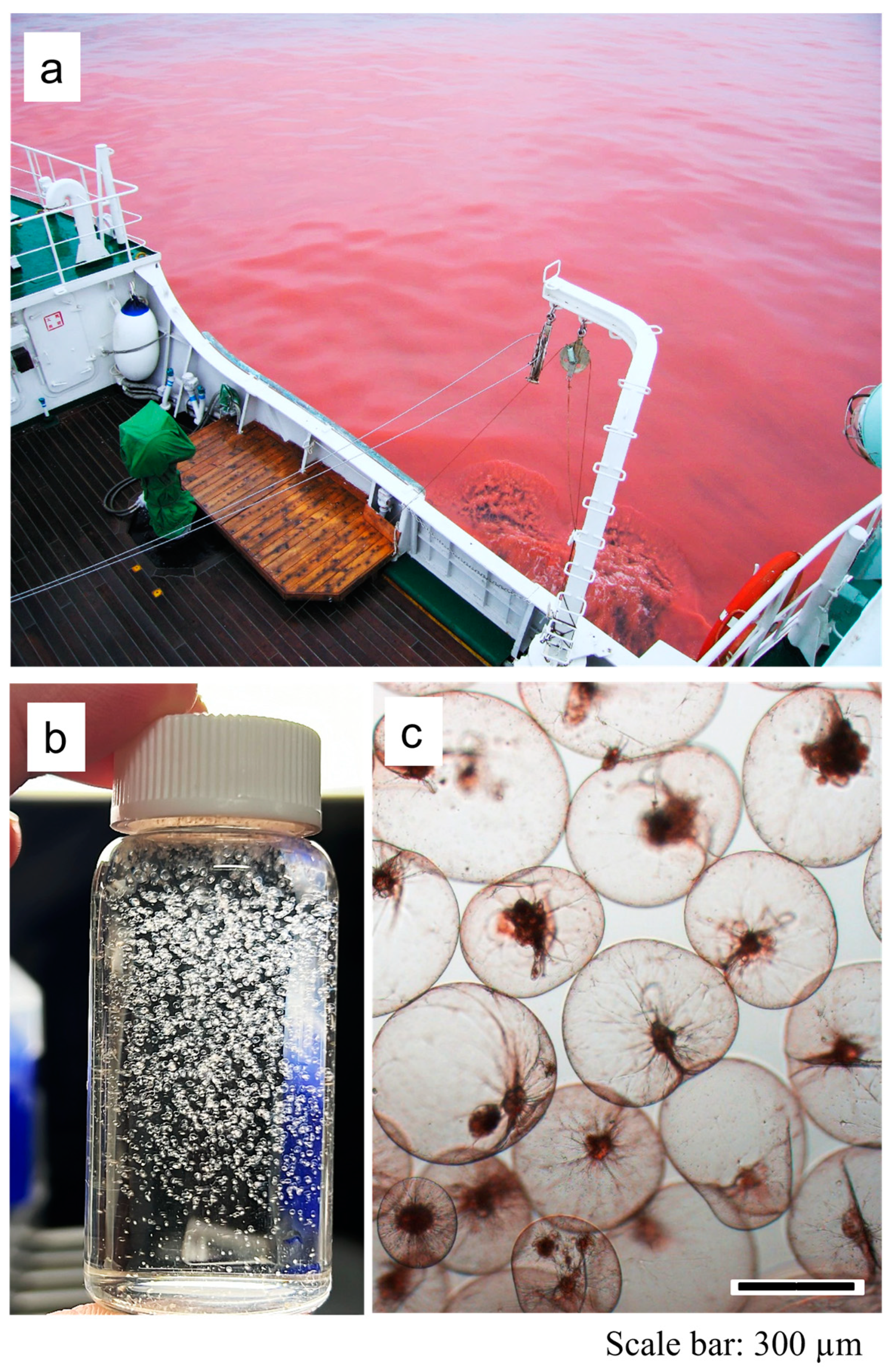
In this study, samples were collected from three distinct locations: Jakarta Bay (Indonesia) and Mie and Miyagi (Japan) (Figure 1). Specifically, the Mie sample was obtained during a blooming event of red N. scintillans in Ise Bay (Figure 2).
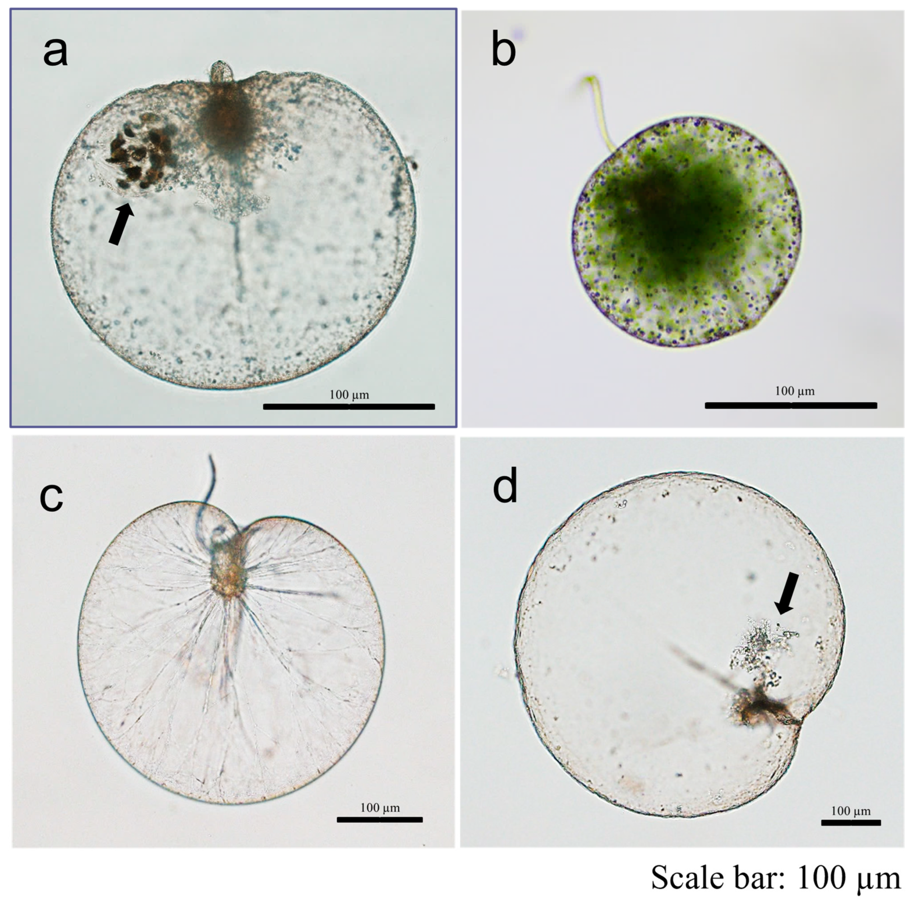
Morphological observations of the RNS specimens from Indonesia and Japan showed that, while their general appearance was comparable, there were notable differences in cell size and cell surface appearance. Noctiluca scintillans specimens from Indonesia (Jakarta Bay) exhibited a rough, bumpy cell surface, whereas those from Japanese waters (Mie and Miyagi) showed a smooth and highly transparent surface (Figure 3). The Indonesian N. scintillans population includes both red- and green-type individuals. Compared with the red-type, green-type cells contain free-swimming green symbionts (Figure 3).
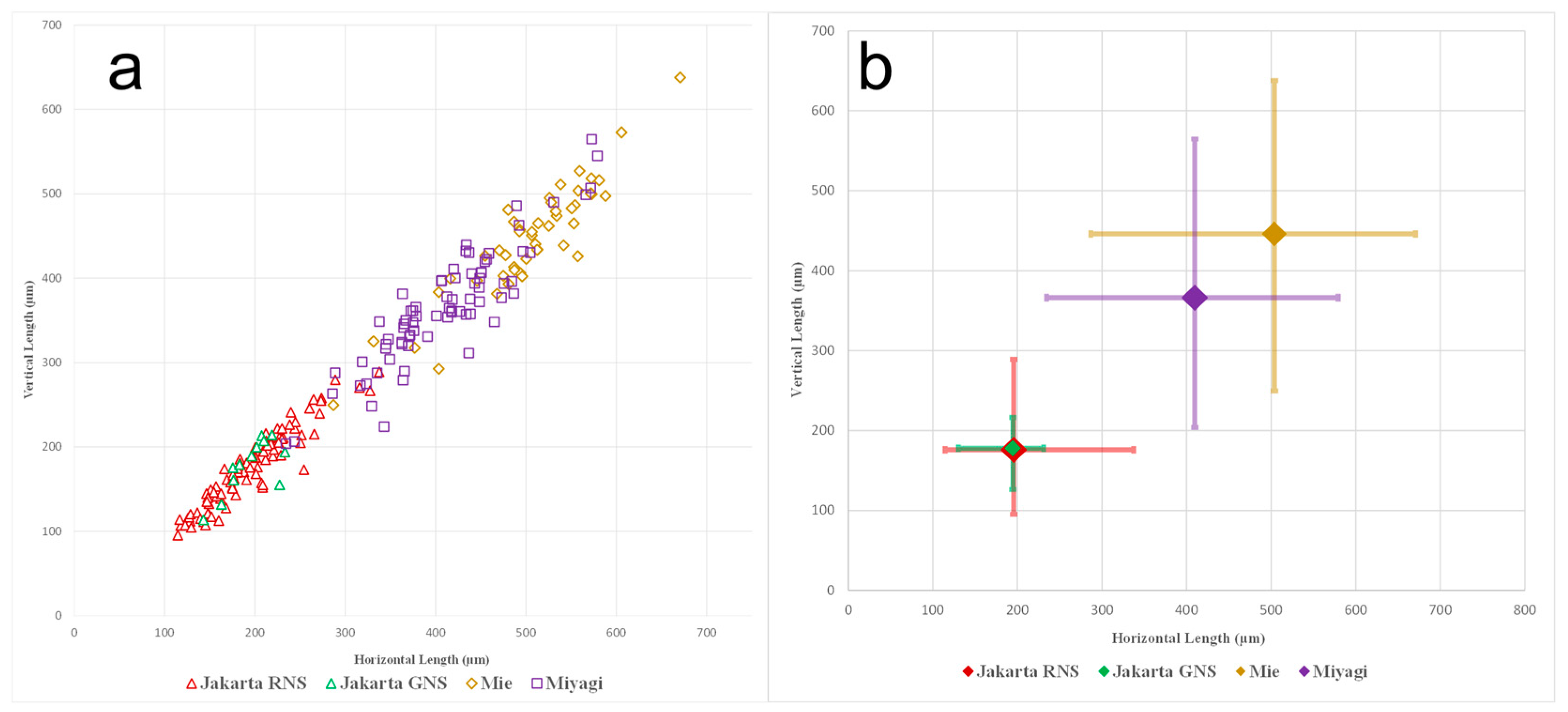
The RNS Jakarta population exhibited an average horizontal diameter of 195.7 µm and an average vertical diameter of 175.9 µm. In comparison, the GNS population from the same location displayed an average horizontal diameter of 194.7 µm and an average vertical diameter of 177.8 µm. Conversely, the average cell size of N. scintillans from Miyagi was larger, with an average horizontal diameter of 409.8 µm and an average vertical diameter of 366.2 µm. The largest cell size was observed in Mie, with an average horizontal diameter of 504.0 µm and an average vertical diameter of 446.0 µm (Table 2, Figure 4).
The t-test was employed to identify statistically significant differences in cell sizes among these populations (Supplementary Material Tables S2 and S3). The results indicate that the cell sizes of Indonesian RNS and GNS populations differed significantly from those of Japanese RNS populations. The calculated p-values for all paired locations were significant (p < 0.05), except the JakartaRNS–JakartaGNS pair. However, the t-Stat values for the Miyagi–Mie and JakartaRNS–JakartaGNS pairs were lower than the t-critical value (t-Stat < 1.96).

3.2. Phylogenetic Relationship and Genetic Divergence

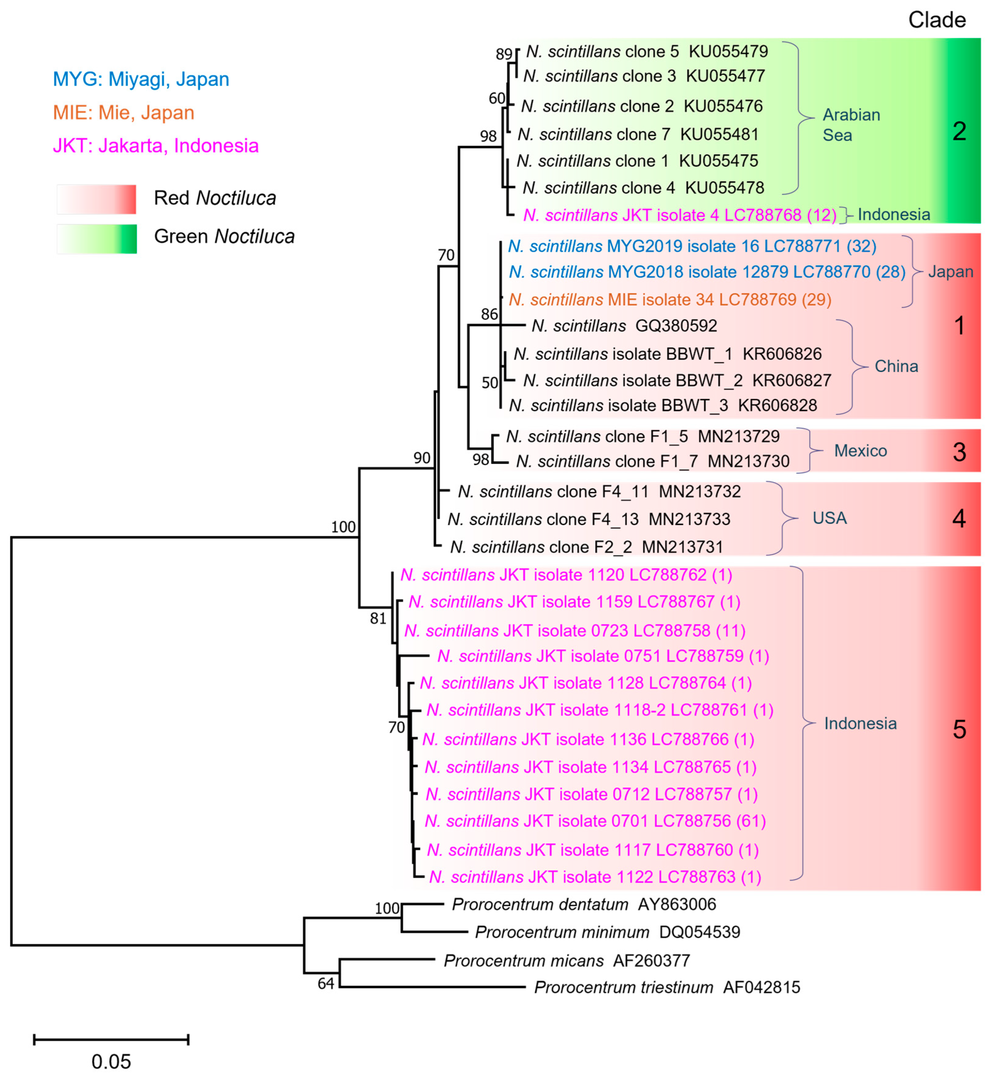
We utilized newly developed primer sets to generate PCR amplicons of the 28S rDNA, yielding fragments approximately 650 base pairs in length. Then, a phylogenetic tree was constructed utilizing 172 sequences obtained from this study (Indonesia and Japan) in conjunction with several GenBank sequences, including those from China, the Arabian Sea, Mexico, and the United States. Molecular analysis identified five distinct N. scintillans clades in the phylogenetic tree (Figure 5), with clustering patterns largely corresponding to geographic origin. Clade 1 included RNS specimens from Japan and China, while Clade 2 grouped GNS specimens from the Arabian Sea and Indonesia. Clade 3 consisted of RNS sequences from Mexico, and Clade 4 comprised RNS specimens from the United States. Notably, Clade 5 contained only Indonesian RNS specimens, demonstrating a clear genetic divergence from the other clades.
A haplotype network was constructed to support the phylogenetic tree analysis, revealing that each geographical area forms a distinct haplotype node. As an exception, GNS specimens from Indonesia and the Arabian Sea are included in the same haplotype group (refer to Supplementary Material Figure S2). Despite significant morphological differences between GNS and RNS due to the endosymbiosis of green algae, the haplotype network indicates that Indonesian RNS is more distantly related to other clades than GNS. No genetic diversity was detected among the 89 Japanese RNS cells, whereas ten haplotypes were identified in the 82 Indonesian RNS cells and one haplotype in the 12 Indonesian GNS cells. This study demonstrates that the N. scintillans population in Jakarta comprises genetically distinct red and green types.
Genetic distances (p-distance, pairwise) among randomly selected N. scintillans cells, based on large subunit (LSU) rDNA sequences, are provided in Supplementary Material Table S4 to enhance the comprehension of genetic differences between Indonesian RNS and other reported populations. Genetic variability among all N. scintillans cells obtained in this study ranged from 0 to 0.114, with an overall average of 0.032. Supplementary Table S4 illustrates genetic distances within Indonesian RNS cells (ranging from 0 to 0.015) and between Indonesian RNS and GNS (ranging from 0.057 to 0.067) despite being sampled from the same location. Indonesian RNS displayed high genetic distances when compared with specimens from other locations, ranging from 0.046 to 0.114. Furthermore, Indonesian GNS exhibited a notable similarity to GNS from the Arabian Sea, with p-distances ranging from 0 to 0.006.

4. Discussion

4.1. Morphology and Cell Size

This study reveals distinct morphological characteristics among RNS populations in Indonesia and Japan. Specifically, specimens from Indonesia exhibited bumpy cellular surfaces that appear rough, while those from Japan displayed smooth and highly transparent cellular surfaces (Figure 3). The presence of rough structures may represent a morphological trait of N. scintillans populations in Indonesia. It is important to note that although rough cell surfaces in N. scintillans have sometimes been associated with stages of the life cycle such as gametogenesis, our observations suggest that this is not the case for the Jakarta RNS specimens. We examined hundreds of individual cells one by one under light microscopy and identified that these cells were not undergoing gametogenesis. According to our observation and a report by Fukuda et al. (2006) [37], cells in the gametogenesis phase typically lose prominent structures such as the tentacle and cytostome, and the gametes are observed to accumulate on one side of the cell. In contrast, the rough-surfaced specimens from Jakarta retained visible tentacles and other characteristic structures, suggesting they were in a normal vegetative state. We also considered the possibility that surface roughness could result from deformation due to the ingestion of large prey items. However, based on our observations, none of the rough-surfaced cells had engulfed prey larger than their own diameter. In fact, the rough surface was consistently present across all observed Jakarta specimens, most of which did not contain large food vacuoles with visible prey item. For comparison, Japanese N. scintillans specimens maintained a smooth and transparent surface, even when they had ingested large prey items, as also reported by Nishitani et al. (2020) [11]. Another possibility is that these structures could be remnants of digested food items; however, such material is typically observed in clumped formations, as shown in Figure 3a,d. However, the Indonesian RNS (Figure 3a) displays a bumpy appearance across the entire cell surface, differing from the smooth and transparent surface seen in the Japanese RNS (Figure 3c,d). These findings suggest that the rough cell surface observed in Jakarta RNS is likely a morphological characteristic, rather than a temporary state due to prey ingestion or life cycle stage. To our knowledge, no previous studies have reported such morphological differences in N. scintillans. This disparity in cell-surface features may stem from variations in N. scintillans genotype in Indonesia and Japan. As our preliminary SEM trials on Indonesian RNS specimens have so far been unsuccessful, future research should prioritize the development of optimized scanning electron microscopy (SEM) or transmission electron microscopy (TEM) techniques to better examine this phenomenon without compromising cell integrity.
Cell size measurements of N. scintillans in this study indicate distinct characteristics in Indonesian and Japanese RNS populations based on geographical location. Noctiluca scintillans exhibited substantial cell size variability, as indicated in Table 2. Examples of large cell sizes have been reported, such as in certain blooming events in India, where N. scintillans cell diameters ranged from 400 to 1200 µm [33,36]. Similar size variations have been observed in Rio Grande do Sul, Brazil, ranging from 600 to 1000 µm [34]. Conversely, locations like the Belgian part of the North Sea reported smaller cell size variations, ranging from 350 to 570 µm [31]. Comparable size ranges, specifically 220 to 500 µm and 150–500 µm, were reported in Mazatlan Bay, Mexico [30], and Chabahar Bay, Iran [35], respectively. Additionally, samples obtained in this study from Miyagi and Mie in Japan exhibited sizes ranging from 300 to 650 µm, closely resembling those previously reported in Sagami Bay, Japan (approximately 300–800 µm) [32]. Among the diverse locations where N. scintillans has been reported, Indonesian specimens display the smallest size population, ranging from 95 to 338 µm, and an average size of approximately 195 µm. This size range is notably smaller than in specimens from other countries, including Japan, supporting the notion of morphological differentiation within Indonesian populations, especially those from Jakarta Bay. It is therefore interesting to further investigate N. scintillans populations across Indonesia’s diverse and environmentally complex coastal waters.

4.2. Insights into Phylogenetic Patterns and Genetic Divergence

In this study, we developed a Noctiluca scintillans-specific primer targeting the 28S rRNA gene. This was necessary because universal primers failed to amplify N. scintillans specimens from Indonesia. The newly designed 28S primer successfully amplified N. scintillans from all examined localities, providing a tool for genotype identification and comparative genetic analysis. The 18S rRNA region was not targeted in this study because it is highly conserved among marine planktonic protists, making it difficult to design Noctiluca scintillans-specific primers. Additionally, N. scintillans is a heterotrophic species capable of ingesting a wide variety of prey, which increases the likelihood of co-amplifying non-target DNA from ingested organisms when using universal 18S primers. Moreover, attempts to amplify the ITS region with newly designed primers were unsuccessful for our Indonesian isolates, despite extensive optimization of annealing temperature, MgCl2 concentration, and cycle number. Given these constraints, the LSU-D1/D2 fragment used in this study remains the most practical locus for reliable, species-specific amplification in N. scintillans. Future surveys that apply this primer set to additional oceanic regions may facilitate the global tracking of its movement or transportation, enabling predictions of future N. scintillans blooming events.
Regarding phylogenetic analysis, Valiadi et al. (2019) [24] demonstrated variations in the 28S rDNA sequences of red and green N. scintillans across different countries and locations (USA, Mexico, China, and the Arabian Sea). Our findings further support and reinforce these results. The phylogenetic tree (Figure 5) illustrates that N. scintillans sequences segregate into four distinct red-type clades and one green-type clade, with noticeable region-specific disparities. The Japanese population (Clade 1) comprises sequences from Miyagi and Mie in Japan, exhibiting similar haplotypes to those from China, consistent with their geographical proximity. Conversely, the Washington, USA, population (Clade 4) displays genetic divergence from the Gulf of Mexico (Clade 3), likely attributable to geographical isolation. Notably, the USA cells lack bioluminescence, whereas populations in Mexico, the Arabian Sea, and China exhibit bioluminescence [24]. This may account for the genetic similarity of the bioluminescent Japanese population with Mexican populations, despite their considerable geographical distance. However, it is important to consider the potential influence of interpopulation transportation of plankton via ocean current and ballast water on large ships or tankers as a contributing factor to genetic similarity.
Although 1000-replicate bootstrapping indicates that the LSU-D1/D2 region captures genuine phylogenetic structure, future long-read sequencing may still uncover low-frequency LSU paralogues that bridge the apparent RNS and GNS split. Such hidden diversity has already been demonstrated for the 18S rDNA V4 of N. scintillans [38], and for other bloom-forming dinoflagellates [39].
From a broader phylogenetic perspective, Clades 1, 3, and 4 (RNS), along with Clade 2 (GNS), may collectively represent a larger overarching clade group. Within this context, the RNS population from Indonesia stands out as genetically different, as evidenced by the separation of Clade 5 from the other related clades. This finding warrants further investigation, as it highlights that the Indonesian population may be a locally adapted genotype of Noctiluca. Further analyses of populations from other regions such as Europe, Africa, South America, Australia, and other Southeast Asian areas is necessary to comprehensively understand the diversity of this genus. This notion is supported by our observations of morphological differences between the Indonesian and Japanese populations (Figure 3), as well as the smaller size of Indonesian N. scintillans compared with Japanese populations (Figure 4). In addition, the RNS population in Jakarta Bay, Indonesia, may exhibit a distinct genetic identity compared with other populations worldwide, owing to this region’s unique oceanographic conditions. The semi-enclosed nature of the Jakarta Bay waters, situated between Java, Sumatra, and Borneo islands, may limit the impact of strong ocean currents from the Indian and Pacific Oceans, resulting in a degree of genetic isolation. This isolation may have led to the adaptation of the Jakarta Bay population. However, the presence of several straits surrounding the area may enable limited cell transportation from other populations, leading to a certain genetic variability within the N. scintillans population in Jakarta Bay, which is represented by the diverse haplotypes of the Indonesian RNS analyzed in this study (Figure 5 and Figure S2).
This discussion brings up the Indonesian archipelago’s oceanographic history within the realm of biogeography. It is widely acknowledged in the field that cladogenetic events, signifying the divergence of a single species into multiple distinct species, are prevalent in the Pacific Ocean, particularly within the Indo-Australian archipelago [40]. This phenomenon is attributed to recurrent glacial cycles and rapid sea-level fluctuations, which facilitate the isolation of various populations. Such occurrences are exemplified in species such as labrid fishes of the genus Cirrhilabrus [40] and are likely applicable to other organisms, including plankton species such as N. scintillans. The findings of this study indicate that red N. scintillans from Jakarta Bay exhibits genetic distinctiveness from both its green counterpart and other populations worldwide. This result supports the potential of a locally adapted genotype of N. scintillans in Indonesian waters, driven by geological and oceanographic factors such as ocean currents and the geography of the Indonesian archipelago. However, this concept does not preclude the potential transportation of certain plankton via ocean currents traversing specific straits or through ballast water in vessels. Considering these possibilities, we infer that GNS in Jakarta Bay, identical to GNS in the Arabian Sea, result from plankton transportation through naturally occurring pathways such as ocean currents or artificial ones such as ballast water discharge.
For a more detailed discussion, we selected representatives of each detected haplotype and compared their genetic distances. There was substantial variability in the 28S rDNA sequences of the sampled N. scintillans specimens, with an overall average genetic distance of 0.032. Notably, the Indonesian RNS population displayed a high genetic distance with other RNS and GNS N. scintillans populations, ranging from 0.046 to 0.114, with an average of 0.063 (Supplementary Material Table S4). This value is comparable with those observed in multispecies dinoflagellates of other genera. For comparison (Table 3), 11 species from the genus Dinophysis exhibited p-values ranging from 0.005 to 0.117, averaging at 0.026. Consequently, genetic distances greater than 0.026 within this genus typically indicate separate species. The genus Gambierdicus displayed an average genetic distance of 0.033, smaller than that of Noctiluca. Conversely, the average distances were higher in the genera Fukuyoa, Paragymnodinium, and Karlodinium, at 0.042, 0.067, and 0.071, respectively. These findings indicate that average genetic distances among separate species within dinoflagellates vary across the genera.
Although the genus Noctiluca currently comprises only one species, Indonesian RNS may open up the possibility of a locally adapted genotype. This is because, when comparing Indonesian RNS specimens, they exhibit very high similarity to each other, with an average genetic distance of 0.005. In contrast, compared with RNS populations from different locations, they display substantially higher genetic distances, averaging at 0.063 (Supplementary Material Table S4). This study presents the first reported genetic data of N. scintillans from Indonesia. In addition, RNS from Japan collected in this study exhibit low genetic distances ranging from 0 to 0.027, with an average of 0.009, when compared with RNS from the USA and Mexico. Identifying a separate species within the genus Noctiluca is difficult. However, we aim to note the possibility of genetic diversity among populations in different regions. The 28S rDNA analysis offers a way to explore each population’s local genetic identity, which could be valuable for mitigating or tracking blooming events.

4.3. Red and Green Noctiluca scintillans

Our study also elucidated the differentiation between red and green N. scintillans in Jakarta Bay. Geographically, RNS exhibit a wide distribution in temperate to subtropical waters, whereas GNS inhabit tropical waters, such as the Arabian Sea, India, and Southeast Asia, notably Indonesia. While numerous reports exist of red tides caused by N. scintillans in the Gulf of Oman (near the Arabian Sea), including the most recent record of RNS blooms in the southeastern Arabian Sea [45], none provide gene sequence data. The only GNS population previously examined is from the Arabian Sea, with no registered RNS sequences. Previous studies have solely analyzed the DNA sequences of RNS from Japan, South Korea, China, the US, and Mexico. Indonesia exhibits an overlap in the geographic distribution of both N. scintillans types. The present study is the first to obtain and analyze RNS and GNS specimens from the same location and time.
This study sheds light on the relationship between red and green N. scintillans and provides evidence of their coexistence in Jakarta Bay, Indonesia. The results revealed genetic differences between the two types (Figure 5 and Figure S2 and Table 3), indicating they are not likely to be interchangeable. Moreover, in the laboratory, we attempted to isolate living green N. scintillans and observe them under a microscope. The symbiont cells were seen swimming freely within the host cell. We then attempted to burst the host cell to expose the symbionts with the surrounding water. Initially, the exposed symbiont cells were moving, but they lost their motility within one hour. Based on this observation, we assume that the symbiont cells are either unable to survive or have extremely limited viability outside the Noctiluca host cell. Although the evidence is not entirely conclusive, these findings suggest that the presence of green symbiotic microalgae in GNS cells is a stable characteristic, unlikely to be altered by the absence or presence of symbionts in open waters. Consequently, it is less likely that heterotrophic RNS could convert into mixotrophic GNS by acquiring these microalgae. Thus, in addition to the possibility of a locally adapted RNS genotype from Jakarta Bay, this study’s findings also sustain the hypothesis that red and green N. scintillans are potentially distinct varieties. Further research is needed to ascertain the taxonomic status of these two types of N. scintillans.

5. Conclusions

The current study utilized newly developed primers to specifically sequence any N. scintillans specimens, resulting in sequences from Indonesian specimens that had not been previously registered in GenBank. Morphological observations, phylogenetic tree analysis, and haplotype network analysis yielded mutually supportive findings, indicating that red-type N. scintillans in Jakarta Bay (Indonesia) possess a genetic differentiation from previously reported populations. This discovery raises the possibility of an adapted genotype within the genus Noctiluca. Furthermore, the analysis of 94 cells from the Jakarta Bay sample revealed the presence of 11 different haplotypes (28S rDNA), highlighting the high genetic variability of the N. scintillans population in Indonesia. Additionally, the coexistence of red and green N. scintillans with notably distant genetic differences at the same time and location reduces the likelihood that one form could transform into the other, supporting the uniqueness of RNS from Jakarta Bay. Although defining separate Noctiluca species remains challenging, our 28S rDNA results expose a hidden regional diversity which could aid in bloom monitoring.

Supplementary Materials

The following supporting information can be downloaded at: https://www.mdpi.com/article/10.3390/jmse13050866/s1, Figure S1: Illustration of the cell size measurement methodology used in this study; Figure S2: Haplotype network of Noctiluca scintillans based on LSU rDNA; Table S1: List of newly developed primers used in this study; Table S2: Comparative statistical calculation of cell size measurements among Jakarta, Miyagi, and Mie populations; Table S3: Comparative statistical calculation of cell measurement across populations; Table S4: Estimated genetic distance (p-distance, pairwise) values among selected Noctiluca scintillans specimens collected from different geographical areas based on large subunit rDNA sequences.

Author Contributions

Conceptualization, M.I.N., G.N., and B.S.; sample collection, M.I.N., A.I., and S.H.; formal analysis, M.I.N.; data interpretation and visualization, M.I.N. and G.N.; writing and draft preparation, M.I.N.; review and editing, M.I.N., G.N., A.I., S.H., W.S.-O., and B.S. All authors have read and agreed to the published version of the manuscript.

Funding

This study was supported by a Grant-in-Aid for Scientific Research (B) from the Japan Society for the Promotion of Science (JSPS) (KAKENHI; No. 23H02283), a grant from The Japan Science and Technology Agency (JST) (JST SPRING; No. JPMJSP2114), Hashimoto-road Co., Ltd., Miyagi Pref., Japan, and WPI-AIMEC, World Premier International Research Center Initiative (WPI), MEXT, Japan.

Data Availability Statement

Representative sequences data that support the findings of this study have been deposited in in the DDBJ/EMBL/GenBank databases, each allocated a unique accession number (LC788756-LC788771). (https://www.ddbj.nig.ac.jp/).

Conflicts of Interest

The authors declare that this study received funding from Hashimoto-road Co., Ltd. The funder was not involved in the study design, collection, analysis, interpretation of data, the writing of this article or the decision to submit it for publication.

Abbreviations

The following abbreviations are used in this manuscript:
HABHarmful algal bloom
RNSRed Noctiluca scintillans
GNSGreen Noctiluca scintillans
PCRPolymerase chain reaction

References

  1. Huang, C.; Qi, Y. The abundance cycle and influence factors on red tide phenomena of Noctiluca scintillans (Dinophyceae) in Dapeng Bay, the South China Sea. J. Plankton Res. 1997, 19, 303–318. [Google Scholar] [CrossRef]
  2. Escalera, L.; Pazos, Y.; Moroño, Á.; Reguera, B. Noctiluca scintillans may act as a vector of toxigenic microalgae. Harmful Algae 2007, 6, 317–320. [Google Scholar] [CrossRef]
  3. Sellner, K.G.; Doucette, G.J.; Kirkpatrick, G.J. Harmful algal blooms: Causes, impacts and detection. J. Ind. Microbiol. Biotechnol. 2003, 30, 383–406. [Google Scholar] [CrossRef]
  4. Kofoid, C.A.; Swezy, O. The Free-Living Unarmored Dinoflagellata; Memoirs of the University of California: Berkeley, CA, USA, 1921. [Google Scholar]
  5. Saldarriaga, J.F.; Taylor, F.J.R.; Cavalier-Smith, T.; Menden-Deuer, S.; Keeling, P.J. Molecular data and the evolutionary history of dinoflagellates. Eur. J. Protistol. 2004, 40, 85–111. [Google Scholar] [CrossRef]
  6. Cooney, E.C.; Leander, B.S.; Keeling, P.J. Phylogenomics shows unique traits in Noctilucales are derived rather than ancestral. PNAS Nexus 2022, 1, pgac202. [Google Scholar] [CrossRef] [PubMed]
  7. Baek, S.H.; Shimode, S.; Kim, H.C.; Han, M.S.; Kikuchi, T. Strong bottom-up effects on phytoplankton community caused by a rainfall during spring and summer in Sagami Bay, Japan. J. Mar. Syst. 2009, 75, 253–264. [Google Scholar] [CrossRef]
  8. do Rosário Gomes, H.; Goes, J.I.; Matondkar, S.P.; Buskey, E.J.; Basu, S.; Parab, S.; Thoppil, P. Massive outbreaks of Noctiluca scintillans blooms in the Arabian Sea due to spread of hypoxia. Nat. Commun. 2014, 5, 4862. [Google Scholar] [CrossRef]
  9. Mikaelyan, A.S.; Malej, A.; Shiganova, T.A.; Turk, V.; Sivkovitch, A.E.; Musaeva, E.I.; Kogovšek, T.; Lukasheva, T.A. Populations of the red tide forming dinoflagellate Noctiluca scintillans (Macartney): A comparison between the Black Sea and the northern Adriatic Sea. Harmful Algae 2014, 33, 29–40. [Google Scholar]
  10. Quevedo, M.; Gonzalez-Quiros, R.; Anadon, R. Evidence of heavy predation by Noctiluca scintillans on Acartia clausi (Copepoda) eggs off the central Cantabrian coast (NW Spain). Oceanol. Acta 1999, 22, 127–131. [Google Scholar] [CrossRef]
  11. Nishitani, G.; Shiromoto, M.; Sato-Okoshi, W.; Ishikawa, A. Molecular approach for analysis of in situ feeding by the dinoflagellate Noctiluca scintillans. Harmful Algae 2020, 99, 101928. [Google Scholar] [CrossRef]
  12. Okaichi, T. Identification of ammonia as the toxic principle of red tide of Noctiluca miliaris. Bull Plank Soc Jpn. 1976, 23, 75–80. [Google Scholar]
  13. Drits, A.V.; Nikishina, A.B.; Sergeeva, V.M.; Solov’ev, K.A. Feeding, respiration, and excretion of the Black Sea Noctiluca scintillans MacCartney in summer. Oceanology 2013, 53, 442–450. [Google Scholar] [CrossRef]
  14. Almeda, R.; Connelly, T.L.; Buskey, E.J. Novel insight into the role of heterotrophic dinoflagellates in the fate of crude oil in the sea. Sci. Rep. 2014, 4, 7560. [Google Scholar] [CrossRef]
  15. San Diego-McGlone, M.L.; Yñiguez, A.T.; Benico, G.; Lum, W.M.; Hii, K.S.; Leong, S.C.Y.; Leaw, C.P.; Iwataki, M.; Lim, P.T. Fish Kills Related to Harmful Algal Bloom Events in Southeast Asia. Sustainability 2024, 16, 10521. [Google Scholar] [CrossRef]
  16. Raj, K.D.; Mathews, G.; Obura, D.O.; Laju, R.L.; Bharath, M.S.; Kumar, P.D.; Arasamuthu, A.; Kumar, T.A.; Edward, J.P. Low oxygen levels caused by Noctiluca scintillans bloom kills corals in Gulf of Mannar, India. Sci. Rep. 2020, 10, 22133. [Google Scholar] [CrossRef] [PubMed]
  17. Hansen, P.J.; Miranda, L.; Azanza, R. Green Noctiluca scintillans: A dinoflagellate with its own greenhouse. Mar. Ecol. Prog. Ser. 2004, 275, 78–87. [Google Scholar] [CrossRef]
  18. Harrison, P.J.; Furuya, K.; Glibert, P.M.; Xu, J.; Liu, H.B.; Yin, K.; Lee, J.H.; Anderson, D.M.; Gowen, R.; Al-Azri, A.R.; et al. Geographical distribution of red and green Noctiluca scintillans. Chin. J. Oceanol. Limnol. 2011, 29, 807–831. [Google Scholar]
  19. Wang, L.; Lin, X.; Goes, J.I.; Lin, S. Phylogenetic Analyses of Three Genes of Pedinomonas noctilucae, the Green Endosymbiont of the Marine Dinoflagellate Noctiluca scintillans, Reveal its Affiliation to the Order Marsupiomonadales (Chlorophyta, Pedinophyceae) under the Reinstated Name Protoeuglena noctilucae. Protist 2016, 167, 205–216. [Google Scholar]
  20. Manigandan, V.; Muthukumar, C.; Shah, C.; Logesh, N.; Sivadas, S.K.; Ramu, K.; Murthy, M.R. Phylogenetic affiliation of Pedinomonas noctilucae and green Noctiluca scintillans nutritional dynamics in the Gulf of Mannar, Southeastern Arabian Sea. Protist 2024, 175, 126019. [Google Scholar] [CrossRef]
  21. Elbrachter, M.; Qi, Y.Z. Aspects of Noctiluca (Dinophyceae) population dynamics. Physiol. Ecol. Harmful Algal Blooms 1998, 41, 315–335. [Google Scholar]
  22. Tibbs, J.F. On Some Planktonic Protozoa Taken from the Track of Drift Station ARLIS I, 1960–1961. Arct. Inst. N. Am. 1967, 20, 247–254. [Google Scholar] [CrossRef]
  23. Sulochanan, B.; Dineshbabu, A.P.; Saravanan, R.; Subramanya Bhat, G.; Lavanya, S. Occurrence of Noctiluca scintillans bloom off Mangalore in the Arabian Sea. Indian J. Fish. Sci. 2014, 61, 42–48. [Google Scholar]
  24. Valiadi, M.; de Rond, T.; Amorim, A.; Gittins, J.R.; Gubili, C.; Moore, B.S.; Iglesias-Rodriguez, M.D.; Latz, M.I. Molecular and biochemical basis for the loss of bioluminescence in the dinoflagellate Noctiluca scintillans along the west coast of the U.S.A. Limnol. Oceanogr. 2019, 64, 2709–2724. [Google Scholar] [CrossRef]
  25. Pan, Y.; Wang, L.; Zhang, W.; Liu, G.; Lin, S. Genetic analysis of Noctiluca scintillans populations indicates low latitudinal differentiation in China but high China-America differences. J. Exp. Mar. Biol. Ecol. 2016, 477, 31–39. [Google Scholar] [CrossRef]
  26. Schneider, C.A.; Rasband, W.S.; Eliceiri, K.W. NIH Image to ImageJ: 25 years of image analysis. Nat. Methods 2012, 9, 671–675. [Google Scholar] [CrossRef]
  27. Kumar, S.; Stecher, G.; Li, M.; Knyaz, C.; Tamura, K. MEGA X: Molecular evolutionary genetics analysis across computing platforms. Mol. Biol. Evol. 2018, 35, 1547–1549. [Google Scholar] [CrossRef]
  28. Tamura, K.; Nei, M. Estimation of the Number of Nucleotide Substitutions in the Control Region of Mitochondrial DNA in Humans and Chimpanzees. Mol. Biol. Evol. 1993, 10, 512–526. [Google Scholar] [PubMed]
  29. Leigh, J.W.; Bryant, D. POPART: Full-feature software for haplotype network construction. Methods Ecol Evol. 2015, 6, 1110–1116. [Google Scholar] [CrossRef]
  30. Rodríguez, R.A.; Ochoa, J.L.; Uribe Alcocer, M. Grazing of heterotrophic dinoflagellate Noctiluca scintillans (Mcartney) Kofoid on Gymnodinium catenatum Graham. Rev. Latinoam. Microbiol. 2005, 47, 6–10. [Google Scholar]
  31. Ollevier, A.; Mortelmans, J.; Aubert, A.; Deneudt, K.; Vandegehuchte, M.B. Noctiluca scintillans: Dynamics, Size Measurements and Relationships With Small Soft-Bodied Plankton in the Belgian Part of the North Sea. Front. Mar. Sci. 2021, 8, 777999. [Google Scholar] [CrossRef]
  32. Ara, K.; Nakamura, S.; Takahashi, R.; Shiomoto, A.; Hiromi, J. Seasonal variability of the red tide-forming heterotrophic dinoflagellate Noctiluca scintillans in the neritic area of Sagami Bay, Japan: Its role in the nutrient-environment and aquatic ecosystem. Plankton Benthos Res. 2013, 8, 9–30. [Google Scholar] [CrossRef]
  33. Padmakumar, K.B.; SreeRenjima, G.; Fanimol, C.L.; Menon, N.R.; Sanjeevan, V.N. Preponderance of heterotrophic Noctiluca scintillans during a multi-species diatom bloom along the southwest coast of India. Int. J. Ocean. Oceanogr. 2010, 4, 55–63. [Google Scholar]
  34. Cardoso, L.d.S. Bloom of Noctiluca scintillans (Macartney) Kofoid & Swezy (Dinophyceae) in Southern Brazil. Braz. J. Oceanogr. 2012, 60, 265–268. [Google Scholar]
  35. Asefi, M.A.; Attaran-Fariman, G. Harmful blooming of Noctiluca scintillans in the southeast coastal waters of Iran, Oman Sea. Iran. J. Fish. Sci. 2023, 22, 261–277. [Google Scholar]
  36. Gopakumar, G.; Sulochanan, B.; Venkatesan, V. Bloom of Noctiluca scintillans (Macartney) in Gulf of Mannar, southeast coast of India. J. Mar. Biol. Assoc. India 2009, 51, 75–80. [Google Scholar]
  37. Fukuda, Y.; Endoh, H. New details from the complete life cycle of the red-tide dinoflagellate Noctiluca scintillans (Ehrenberg) McCartney. Eur. J. Protistol. 2006, 42, 209–219. [Google Scholar] [CrossRef]
  38. Liu, K.; Huang, X.; Ding, X.; Chen, N. The high molecular diversity in Noctiluca scintillans is dominated by intra-genomic variations revealed by single cell high-throughput sequencing of 18S rDNA V4. Harmful Algae 2024, 132, 102568. [Google Scholar] [CrossRef]
  39. Miranda, L.N.; Zhuang, Y.; Zhang, H.; Lin, S. Phylogenetic analysis guided by intragenomic SSU rDNA polymorphism refines classification of “Alexandrium tamarense” species complex. Harmful Algae 2012, 16, 35–48. [Google Scholar] [CrossRef]
  40. Tea, Y.K.; Xu, X.; Dibattista, J.D.; Lo, N.; Cowman, P.F.; Ho, S.Y.W. Phylogenomic Analysis of Concatenated Ultraconserved Elements Reveals the Recent Evolutionary Radiation of the Fairy Wrasses (Teleostei: Labridae: Cirrhilabrus). Syst. Biol. 2022, 71, 1–12. [Google Scholar] [CrossRef]
  41. Funaki, H.; Nishimura, T.; Yoshioka, T.; Ataka, T.; Tanii, Y.; Hashimoto, K.; Yamaguchi, H.; Adachi, M. Toxicity and growth characteristics of epiphytic dinoflagellate Gambierdiscus silvae in Japan. Harmful Algae 2022, 115, 102230. [Google Scholar]
  42. Leung, P.T.; Yan, M.; Lam, V.T.; Yiu, S.K.; Chen, C.Y.; Murray, J.S.; Harwood, D.T.; Rhodes, L.L.; Lam, P.K.; Wai, T.C. Phylogeny, morphology and toxicity of benthic dinoflagellates of the genus Fukuyoa (Goniodomataceae, Dinophyceae) from a subtropical reef ecosystem in the South China Sea. Harmful Algae 2018, 74, 78–97. [Google Scholar] [CrossRef] [PubMed]
  43. Yokouchi, K.; Horiguchi, T. Paragymnodinium verecundum sp. nov. (Gymnodiniales, Dinophyceae), a new species of mixotrophic dinoflagellate from Japan. Phycol. Res. 2021, 69, 124–136. [Google Scholar] [CrossRef]
  44. Cen, J.; Wang, J.; Huang, L.; Lin, Y.; Ding, G.; Qi, Y.; Lü, S. Karlodinium elegans sp. nov. (Gymnodiniales, Dinophyceae), a novel species isolated from the East China Sea in a dinoflagellate bloom. J. Oceanol. Limnol. 2021, 39, 242–258. [Google Scholar] [CrossRef]
  45. Shaju, S.S.; Akula, R.R.; Jabir, T. Characterization of light absorption coefficient of red Noctiluca scintillans bloom in the South Eastern Arabian Sea. Oceanologia 2018, 60, 419–425. [Google Scholar] [CrossRef]
Figure 1. Noctiluca scintillans sampling points in East and Southeast Asia. The sampling points of this study are indicated by the red circles: Miyagi, Mie, and Jakarta.
Jmse 13 00866 g001
Figure 2. Red-type N. scintillans during a bloom event in Ise Bay, Mie Prefecture, Japan. (a) Surface seawater discolored by a red tide of N. scintillans; (b) Whitish–transparent particles represent N. scintillans cells, visible to the naked eye; (c) Close-up of red-type N. scintillans showing some ingested food items (Dr. Kazunori Taguchi, Mie University, provided Figure 2a).
Jmse 13 00866 g002
Figure 3. Photomicrographs of N. scintillans cells observed in this study. (a) Red-type N. scintillans from Indonesia (Jakarta Bay); (b) Green-type N. scintillans from Indonesia (Jakarta Bay) with visible green endosymbiont within the cell; (c) Red-type N. scintillans from Japan (Miyagi); (d), Red-type N. scintillans from Japan (Mie). The black arrow indicates possible remnants of digested prey or organic matter. Specimens from Jakarta Bay (line bordered picture) exhibit rough cell surface, while specimens from Japan display highly transparent and smooth cell surface.
Jmse 13 00866 g003
Figure 4. Cell size comparison of N. scintillans from each sampling point. (a) Scatter plot of all isolated specimens. (b) Averaged cell size, with error bars indicating the samples’ maximum and minimum cell sizes.
Jmse 13 00866 g004
Figure 5. Phylogenetic tree of Noctiluca scintillans based on 28S rDNA sequences known to date, including sequences from this study. Using the maximum-likelihood algorithm, the phylogenetic relationships between N. scintillans specimens from different geographical areas were inferred from large subunit rDNA sequences. Sequences obtained in this study are denoted by colored letters, and numbers in parentheses after specimen names represent the number of cells analyzed. The color of the shades indicates the cell type, i.e., red and green N. scintillans. Numbers beside each cluster indicate the clade number.
Jmse 13 00866 g005
Table 1. The number of cells isolated from each sampling location.
Sampling Location Collection TimeNumber of Isolated Cells
JapanIse Bay, Mie Pref.June 201829 (RNS)
Matsushima Bay, Miyagi Pref.May 201828 (RNS)
August 201932 (RNS)
IndonesiaJakarta BaySeptember 201951 (RNS)
September 202031 (RNS)
September 202012 (GNS)
Total 183
Table 2. Noctiluca scintillans sizes in different localities.
LocationSize (µm)RNS/GNSReference
Jakarta, Indonesia95–338RNSThis study
Miyagi, Japan203–580RNSThis study
Mie, Japan250–671RNSThis study
Mazatlan Bay, Mexico220–500RNSRodriguez et al. (2005) [30]
North Sea, Belgium350–570RNSOllevier et al. (2021) [31]
Sagami Bay, Japan300–800RNSAra et al. (2013) [32]
Southwest coast of India500–1000RNSPadmakumar et al. (2010) [33]
Rio Grande do Sul, Brazil600–1000RNSCardoso (2012) [34]
Jakarta, Indonesia114–233GNSThis study
Chabahar Bay, Iran150–500GNSAsefi & Attaran-Fariman (2023) [35]
Gulf of Mannar, India400–1200GNSGopakumar et al. (2009) [36]
Table 3. Pairwise distances of large subunit rDNA sequences of several dinoflagellate genera.
GenusNo. of Speciesp-Distance *Reference
Dinophysis110.026GenBank, calculated in this study
Gambierdiscus40.033Funaki et al. (2022) [41]
Noctiluca10.039This study
Fukuyoa60.042Leung et al. (2018) [42]
Paragymnodinium40.067Yokouchi et al. (2020) [43]
Karlodinium160.071Cen et al. (2021) [44]
* Average value.
Disclaimer/Publisher’s Note: The statements, opinions and data contained in all publications are solely those of the individual author(s) and contributor(s) and not of MDPI and/or the editor(s). MDPI and/or the editor(s) disclaim responsibility for any injury to people or property resulting from any ideas, methods, instructions or products referred to in the content.