Article

Laboratory Culture-Based Characterization of the Resting Stage Cells of the Brown-Tide-Causing Pelagophyte, Aureococcus anophagefferens

1
CAS Key Laboratory of Marine Ecology and Environmental Sciences, Institute of Oceanology, Chinese Academy of Sciences, Qingdao 266071, China
2
University of Chinese Academy of Sciences, Beijing 100049, China
3
Laboratory for Marine Ecology and Environmental Science, Qingdao National Laboratory for Marine Science and Technology, Qingdao 266237, China
4
Center for Ocean Mega-Science, Chinese Academy of Sciences, Qingdao 266071, China
5
School of Marine and Atmospheric Sciences, Stony Brook University, Stony Brook, NY 11790, USA
*
Authors to whom correspondence should be addressed.
Current address: School of Life Science and Engineering, Handan University, Handan 056005, China.
J. Mar. Sci. Eng. 2020, 8(12), 1027; https://doi.org/10.3390/jmse8121027
Submission received: 17 November 2020 / Revised: 10 December 2020 / Accepted: 14 December 2020 / Published: 16 December 2020
(This article belongs to the Special Issue Taxonomy and Ecology of Marine Algae)

Abstract

:
Life history (life cycle) plays a vital role in the ecology of some microalgae; however, the well-known brown-tide-causing pelagophyte Aureococcus anophagefferens has been barely investigated in this regard. Recently, based mainly on detections in marine sediments from China, we proved that this organism has a resting stage. We, therefore, conducted a follow-up study to characterize the resting stage cells (RSCs) of A. anophagefferens using the culture CCMP1984. The RSCs were spherical, larger than the vegetative cells, and smooth in cell surface and contained more aggregated plastid but more vacuolar space than vegetative cells. RSCs contained a conspicuous lipid-enriched red droplet. We found a 9.9-fold decrease in adenosine triphosphate (ATP) content from vegetative cells to RSCs, indicative of a "resting" or dormant physiological state. The RSCs stored for 3 months (at 4 °C in darkness) readily reverted back to vegetative growth within 20 days after being transferred to the conditions for routine culture maintenance. Our results indicate that the RSCs of A. anophagefferens are a dormant state that differs from vegetative cells morphologically and physiologically, and that RSCs likely enable the species to survive unfavorable conditions, seed annual blooms, and facilitate its cosmopolitan distribution that we recently documented.

1. Introduction

The non-motile, picoplanktonic (2–3 μm) pelagophyte Aureococcus anophagefferens Hargraves et Sieburth has caused numerous ecosystem disruptive algal blooms (EDABs), commonly known as "brown tides", in U.S. estuaries since 1985 [1,2]. This species lacked morphological features easily distinguishing it from other similar sized forms under light microscopy, but ultrastructural observations exhibited that each cell has a single chloroplast, nucleus, and mitochondrion and an unusual exocellular polysaccharide-like layer [2]. Although nontoxic to humans, A. anophagefferens blooms have significantly negative effects on the seagrass beds, shellfish industry, algal grazers, and zooplankton in the affected area [1]. In the summer of 1985, the first A. anophagefferens blooms have been reported in several estuaries of the northeastern U.S. [2,3,4], and there has been an extra-large extension of the known range of A. anophagefferens along the U.S. East Coast since then, from Florida north to New Hampshire [5]. Aureococcus anophagefferens blooms have been reported for the first time in Saldanha Bay, South Africa in 1997 and several more during 1998–2003 [6,7]. Surprisingly, A. anophagefferens suddenly bloomed in the coastal waters of Qinhuangdao, China in early summer from 2009 to 2011, which caused significant negative impacts on the shellfish mariculture industry and large economic losses [8,9]. Its discontinuous global distribution and seemingly rapid geographic expansion, however, has been a vital but highly controversial issue [10]. It was hypothesized that A. anophagefferens was possibly introduced both within and outside (South Africa) the U.S. via ships’ ballast water [11]. Previous studies have indeed shown that A. anophagefferens could endure the prolonged darkness for 30 days [12,13], a characteristic facilitating its anthropogenic transport by ships’ ballast water. As a novel brown-tide-forming species in China, whether A. anophagefferens was an alien species recently introduced to China via anthropogenic transport processes or has been an indigenous species existing with a background abundance prior to the first reported bloom has thus become a question of ecological significance. A comparison of nearly the entire length of the 18S ribosomal RNA (rRNA) gene sequences of A. anophagefferens from Qinhuangdao, China with that from the USA has shown that there was relatively little genetic variability (0–6 bp differences) [9], which suggested that A. anophagefferens was possibly an alien species in China [10,14].
In nature, microalgae have evolved many survival strategies to withstand adverse environments such as forming resting cysts or spores [15,16,17], reduction in their metabolic rates [18,19], or reliance on the alternation of nutrition modes [20,21]. Inactive or resting stages are common in the life history of many microalgae (diatoms, dinoflagellates, haptophytes, green algae, cyanobacteria, raphidophytes, chrysophytes, euglenophytes, and cryptophytes) [22,23] and may provide tolerance to unfavorable conditions [17,22,23]. Resting stages refer to all types of cells that greatly reduce metabolic rate, cease cell division, but remain viable [24], and are often characterized by distinct morphological and compositional changes in cells such as thickened membranes and the formation of starch granules [25], lipid droplets [26], or red accumulation bodies [16]. Resting cells that undergo changes in morphology and physiology leading to a resting stage do not undergo major changes in cell surface or enclosing cell structures, which distinguishes them from resting cysts or spores [27]. The life history (life cycle) of A. anophagefferens had not been uncovered [2] until recently when a resting stage in another brown-tide-causing species, Aureoumbra lagunensis, was firstly described in pelagophytes from laboratory cultures [28], which provided insights into the life cycle of A. anophagefferens. One of the key questions regarding ballast water hypothesis of A. anophagefferens introduction is whether or not the species has a dormant stage in its life history, because it has been well established that all microalgal species that have been proved to be transported and introduced via ships’ ballast tanks are cyst-forming species [10]. Therefore, answering whether or not A. anophagefferens has a resting stage will greatly help to elucidate the origin and seeding of A. anophagefferens found in China and its apparent geographic expansion around the world.
In our recently published work [29], we proved that A. anophagefferens has a resting stage in its life history via germination experiments of a sediment sample collected from the coast of Qinhuangdao, China, where blooms of this species occurred, and also found that this species has an extremely wide geographic distribution (a range of ~30° in latitude, ~15.7° in longitude) and a more than 1500-year presence in China and thus is not an alien species. With the support of mining the supplementary dataset in a recent work [30], we also found that A. anophagefferens in fact distributes globally [29]. As A. anophagefferens is a small-sized (2–3 μm) and morphologically simple alga, we were not able to recognize and isolate single resting stage cells (RSCs) from the marine sediment sample for morphological observations and also failed in establishing cultures from the germlings in germination experiments. A follow-up work using laboratory cultures is, therefore, highly desirable to characterize the morphology and physiological status of the RSCs and to observe the transformation process of resting stage cells into vegetative cells. The present study describes the basic morphological characteristics and physiological status of RSCs as well as their ability to revert from resting stage back to vegetative growth based on the long laboratory-raised culture of A. anophagefferens strain CCMP1984 (Provasoli-Guillard National Center for Culture of MarinePhytoplankton (West Boothbay Harbor, ME, USA), CCMP). We believe the knowledge of these aspects will be significantly important in understanding the basic biology and ecology of this notorious EDABs-causing pelagophyte.

2. Materials and Methods

2.1. Culture Information and RSC Formation

The culture of A. anophagefferens strain CCMP1984 was obtained from Stony Brook University. The culture was routinely maintained in natural seawater-based f/2-Si medium [31] supplied with 10−8 M (final concentration) selenium (salinity 32) at 21°C in an incubator with a 12:12 h light: dark cycle and an irradiance of 100 µmol photons m−2·s−1. An antibiotic solution (10,000 IU·mL−1 penicillin and 10,000 μg·mL−1 streptomycin, Solarbio, Beijing, China) was added into the medium immediately (final concentration 2%) before inoculation to discourage growth of bacteria. The RSCs were produced in the routinely maintained batch cultures of CCMP1984 at the late stationary growth stages (>20 days) and particularly in the cultures that were cultured to the late exponential phase and then placed in the dark for a few weeks or longer. The morphological observations on RSCs below were conducted with RSCs produced this way, except for that stated otherwise.

2.2. Basic Morphological Observation

To observe and contrast the general morphology of vegetative cells (VCs) and RSCs of A. anophagefferens, 5 mL VCs and RSCs were added into a 10 mL conical centrifuge tube, respectively. After being rinsed with filtered, sterilized seawater three times and diluted to appropriate concentration, the samples were transferred to clean slides and observed under an optical microscope (BX53, Olympus, Japan).

2.3. BODIPY 505/515 Fluorescence Staining

To detect lipid-rich structures in the RSCs of A. anophagefferens, an optimized BODIPY 505/515 fluorescence staining method was used by following the protocol described previously [32] with minor modifications, as described below: BODIPY 505/515 (4, 4-difluro-1, 3, 5, 7-tetramethyl-4-bora-3a, 4adiaza-s-indacene; Invitrogen Molecular Probes, USA) was dissolved in DMSO as a stock solution (0.5 mg·mL−1) and stored in a brown sample bottle away from light. RSCs of the A. anophagefferens culture were collected onto a polycarbonate filter (pore size 0.2 μm) and rinsed with 500 μL 0.22 μm-filtered, sterilized seawater three times. The cells were then stained with BODIPY 505/515 (final concentration 0.1 μg·mL−1) and incubated at 21 °C for 10 min in the dark. After incubation, the samples were centrifuged at 2000× g for 3 min and rinsed three times using filtered, sterilized seawater. The cells were then resuspended in 100 μL filtered, sterilized seawater and were transferred to clean slides for observation under an inverted microscope (IX73, Olympus, Japan) equipped with dichroic filters BP450–480 nm and BA >515 nm (Olympus, Japan).

2.4. Adenosine Triphosphate (ATP) Assays

To characterize intracellular metabolic activity of RSCs and normal vegetative cells, the cellular content of ATP was quantified by using an Enhanced ATP assay Kit (S0027; Beyotime Biotechnology, Shanghai, China) following the manufacturer’s protocol. Three 50 mL samples of RSCs and VCs of A. anophagefferens were collected onto a polycarbonate filter (pore size 0.2 μm) and rinsed with 500 μL filtered, sterilized seawater three times and then to be determined for the ATP content. The cellular ATP content was estimated according to an ATP standard curve and expressed as nmol/107 cells.

2.5. Germination Experiments

To evaluate the ability of RSCs to revert back to vegetative growth after being stored in a prolonged (3 months) darkness at 4 °C, which resembles winter environmental conditions of marine sediments, germination experiments were conducted using both culture plates and Erlenmeyer flasks as described below. RSCs in culture plates were first stored in darkness at 4 °C for 1, 2, and 3 months, with addition of antibiotics mixture to a final 2% concentration (10,000 IU·mL−1 penicillin and 10,000 μg·mL−1 streptomycin) in darkness every 10 d to discourage growth of bacteria [33]. Once the RSCs were stored for 1, 2, and 3 months, a subset of RSCs in plates was transferred to three Erlenmeyer flasks containing fresh medium, placed in the incubator for RSC germination with normal conditions used for culture maintenance, and monitored for cell density on day 0, 20, and 30, respectively. Moreover, daily observations on the germination of RSCs and subsequent vegetative growth were performed using culture plates. The RSCs were inoculated into wells of culture plates containing fresh culture medium, placed at normal culturing conditions, and examined daily with an Olympus microscope (IX73, Tokyo, Japan).

2.6. Statistical Analyses

For the germination experiments and intracellular metabolic activity of RSCs and normal vegetative cells, one-way ANOVA was used to assess the differences among cell densities of RSCs and vegetative cells at different incubation time, and G-test for ATP contents in RSCs and vegetative cells. In all cases, significance levels were set at p < 0.05.

3. Results

3.1. Morphological Characteristics of Resting Cells

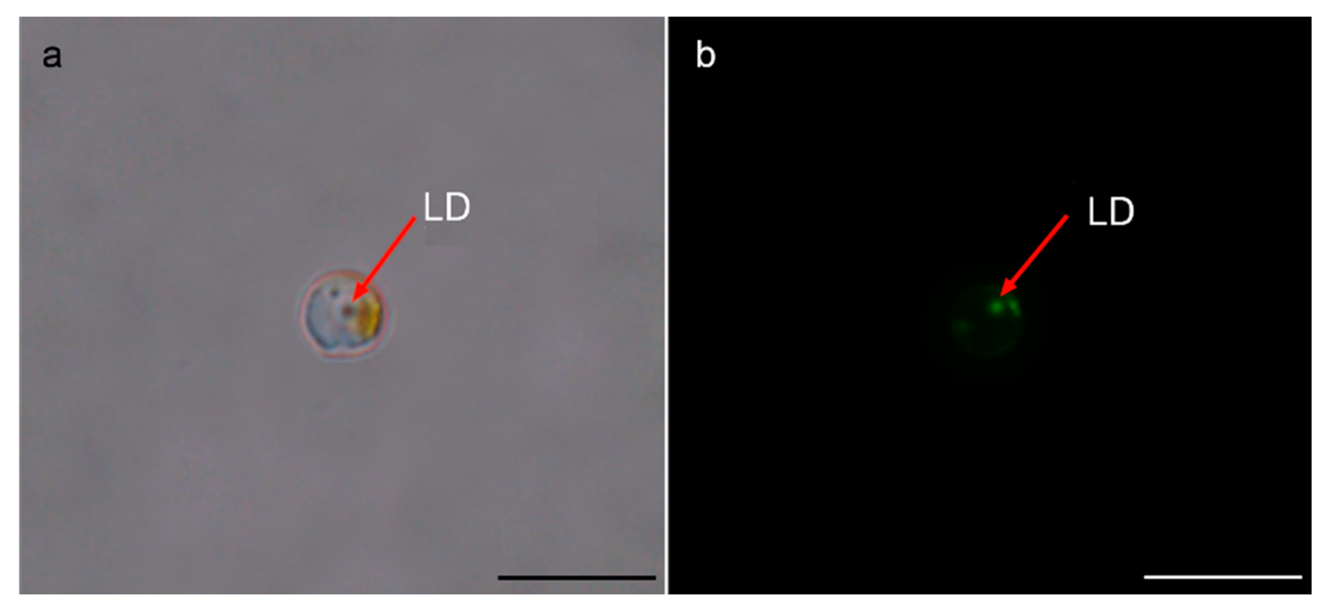
In the batch cultures of CCMP1984 that was routinely maintained as described above (salinity 32, 21 °C, 12:12 h photoperiod, an irradiance of 100 µmol photons m−2·s−1, with 2% antibiotics mixture), RSCs were generally formed at the late stationary growth stages (>20 days). Similar to normal vegetative cells, RSCs were spherical and of a relatively smooth surface (Figure 1b,d). Contrast to a size of 2–3 μm for vegetative cells, RSCs were characterized by an approximately cell diameter two-fold larger than vegetative cells (4–6 μm; Figure 1d). In RSCs, the granular cytoplasm became denser, the plastid became pale, fewer, and densely aggregated, and the vacuole space expanded (Figure 1b). All RSCs contained a large and conspicuous red droplet-like organelle (Figure 1d), which was proven to be lipid-enriched “droplet” by BODIPY 505/515 fluorescence staining and appeared green under epi-fluorescence microscopy (Figure 2b). These morphological features are generally similar to RSCs of another pelagophyte, A. lagunensis [28].

3.2. Metabolic Activity of RSCs as Indicated in Cellular ATP Content

Resting stage cells of A. anophagefferens were characterized by a significantly lowered cellular ATP content relative to that of normal vegetative cells, while the content of ATP in normal vegetative cells was 31.6 ± 0.51 nmol/107 cells, which was significantly higher than that in RSCs (2.9 ± 0.05 nmol/107 cells; Table 1; p < 0.05). This 9.9-fold drop in cellular ATP content from normal vegetative cells to RSCs indicated a significantly reduced metabolic activity in RSCs.

3.3. Resumption of Vegetative Growth from Resting State

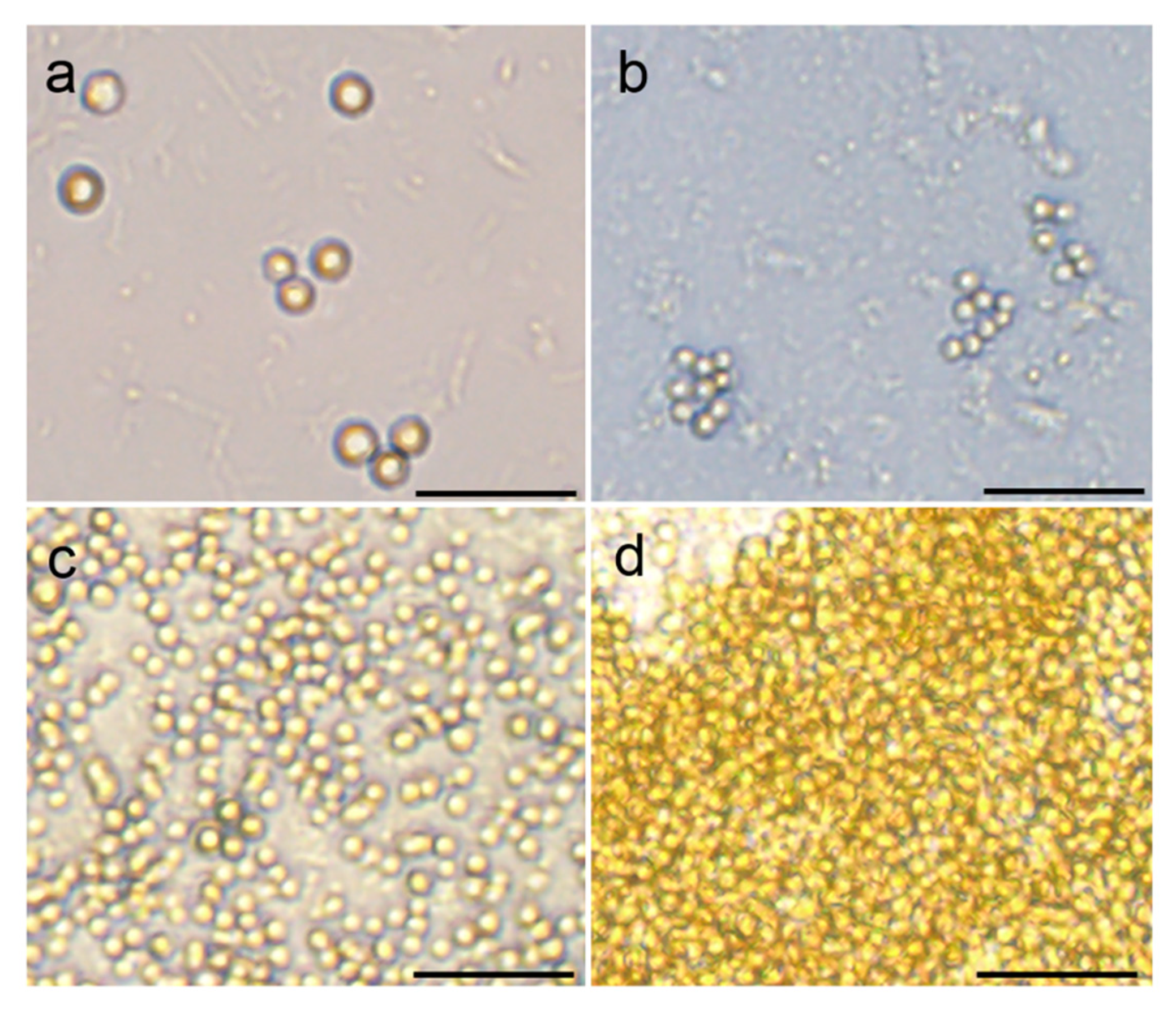
To investigate the germination or transformation processes of the RSCs into vegetative cells, a time series observation was performed via culturing RSCs in fresh medium under 21 °C at 100 µmol photons m−2·s−1. While we did not observe the details of germination processes of RSCs into vegetative cells due to the small sizes of RSCs and a quick process, we observed that almost all RSCs transformed into vegetative cells within 10 days, resumed rapid vegetative growth, and reached a high cell density within 20 days (Figure 3). It was noteworthy that the germination of RSCs was a direct morphological transformation and quick resumption of cell division, similar to that in another pelagophyte A. lagunensis [28], but different from other species of Ochrophyta, e.g., three types of statospores (uninucleate, asexual; binucleate, asexual (potentially autogamic); binucleate, sexual (zygotic)) in the chrysophyte Dinobryon cylindricum [34,35], new germling escaped from the structure underneath the lid of germination pore in Heterosigma akashiwo (Raphidophyceae) [36], and dinoflagellates, in which a germination process within the cyst wall and a germling release through an archeopyle that has been numerously observed in the germination of resting cysts [33,37,38].

3.4. The Ability of RSCs to Survive Prolonged Darkness and Coldness

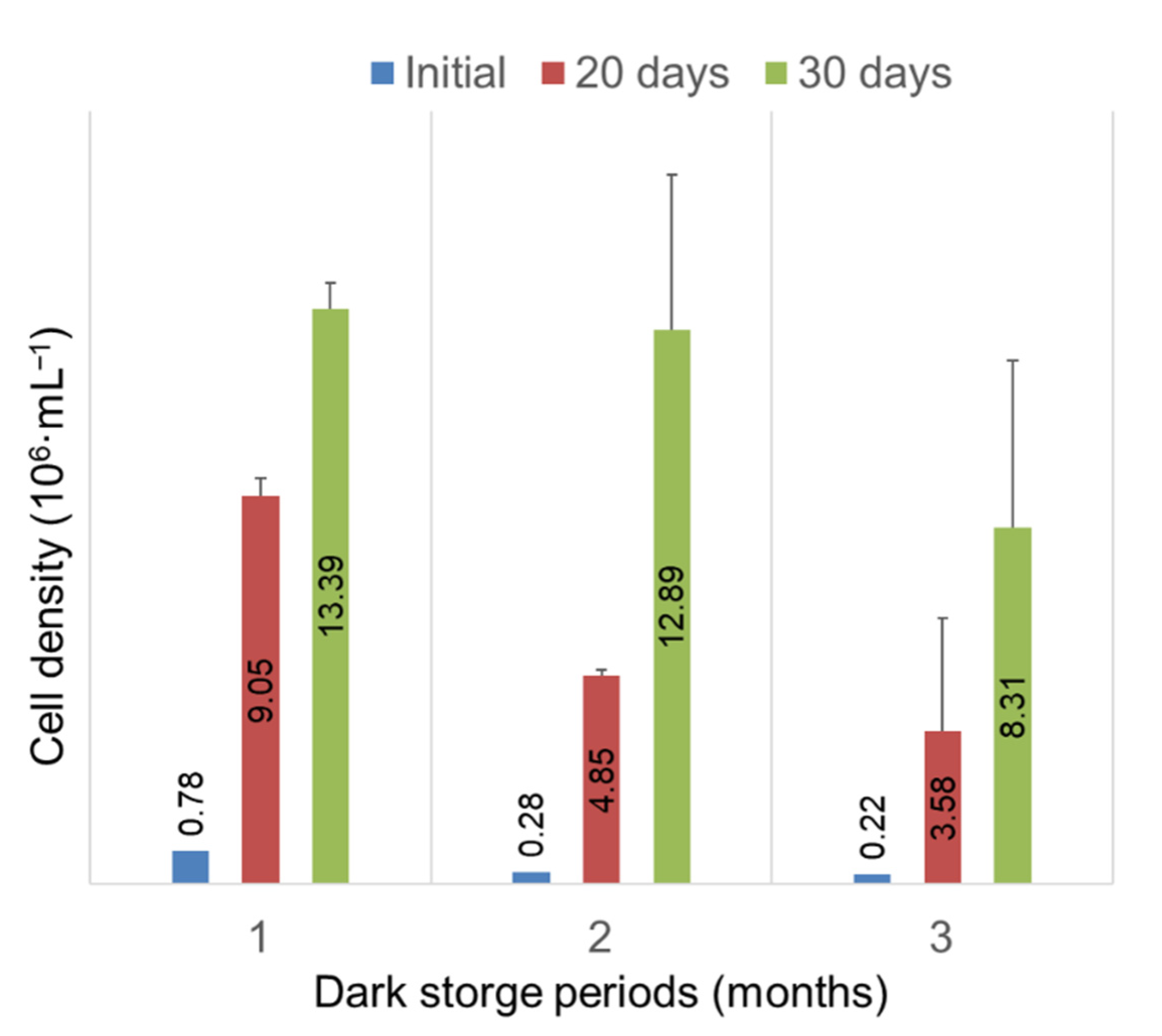
The ability of RSCs of A. anophagefferens to survive prolonged darkness and coldness and to resume growth was assessed via storing RSCs in darkness at 4 °C for one to three months and incubating RSCs in fresh medium under routine culture maintenance conditions. Although the number of RSCs decreased significantly over time during the storage, which died during the storage because of light or nutrient shortage, or decomposed by bacteria, only about 28% of RSCs survived 3 months of darkness coldness (Figure 4 and Figure 5). Under normal culture conditions, RSCs that survived 1, 2, and 3 months of storage could germinate and grow to a high cell density within 20 days (Figure 5). While RSCs declined from 7.8 × 10⁵ cells·mL−1 to 2.2 × 10⁵ cells·mL−1 after 3 months of storage (p < 0.05), the density of vegetative cells increased to 3.58 × 10⁶ (~16 folds) and 8.31 × 10⁶ (~38 folds) cells·mL−1 on day 20 and 30, respectively (p < 0.05), when RSCs were incubated in fresh medium under routine culturing conditions (Figure 5).

4. Discussion

Aureococcus anophagefferens blooms (also known as brown tides) have caused destructive environmental impacts and massive losses in aquaculture in the past three decades [39,40,41,42]. For a long period, however, brown tides were only reported in the U.S. East Coast until the first brown tides occurred at Saldanha Bay, South Africa in 1997 [6] and suddenly occurred in the coastal waters of Qinhuangdao, China in 2009 [9]. A hypothesis was put forward that ballast water vectoring could provide an explanation for the geographical spreading behavior of A. anophagefferens [10,11]. However, there have been many uncertainties regarding the hypothesis. Focal, unresolved questions include whether or not the species can produce a resting stage and/or has been existing at lower abundance in regions where brown tides seemed to occur suddenly [10]. It has been found that A. anophagefferens could survive prolonged darkness for more than 30 days in darkness, a characteristic facilitating its anthropogenic transport by ships [12]. A physiological change and adjustment of cells has been suggested to contribute to the capability [13]. It had been generally believed that pelagophytes may not form dormant cysts or resting stage cells [1] until the recent description of a resting stage in the pelagophyte A. lagunensis [28]. We recently proved the existence of a resting stage in A. anophagefferens via sediment germination experiments and also found that the alga in fact distributes globally [29]. Although sediment had been stored in darkness at 4 °C for more than one year prior to the germination experiments, the RSCs of A. anophagefferens in the sediment sample could recover (or “germinate”) to normal vegetative growth within a reasonable time (a few weeks) when returned to favorable conditions [29]. However, it is noteworthy that our discovery of RSCs in A. anophagefferens did not support the “recent invasion of alien species” hypothesis for the sudden appearance of brown tides in Bohai Sea, because the sedimental record of A. anophagefferens demonstrated that the species has been existing in Bohai Sea for at least 1500 years [29]. On the other hand, this finding also does not disprove the possible roles of RSCs in the initiation and geographic expansion of brown tides and in the general ecology of this species. It is still possible that the RSCs provided the facility for A. anophagefferens to seed annual brown tides and to expand its geographical distribution via either artificial (e.g., ships’ ballast water) or natural pathways up to a globally cosmopolitan distribution [29]. Therefore, following the abovementioned discovery of RSCs in A. anophagefferens, we characterized the general morphological features, physiological status, and the ability to resume vegetative growth of RSCs on the basis of observations on the laboratory-raised culture CCMP1984 in the present study.
Once the culture of A. anophagefferens that had been cultured to the late exponential phase was placed in the dark for a few weeks, almost all vegetative cells could transform into RSCs. Consumption of nutrients in the culture medium [34,43], growth of bacteria, and sustained darkness [26,44,45] may all be the cues driving the transformation of vegetative cells into RSCs. Whether or not a temperature stress (low or high and/or a rapid change) could drive the formation of RSCs also needs to be considered in future investigations [15,46]. In the investigation on A. lagunensis, vegetative cells were observed to quickly transform into resting cells upon exposure to higher temperatures [28].
RSCs of A. anophagefferens have a relatively smooth cell surface, which is different from the resting cysts or spores of many species that undergo more substantial changes in cell surface and/or surface structures including thickened walls or modified enclosing inorganic or organic thecae, as seen in dinoflagellates and some diatoms [27,47,48,49,50,51]. Similar to vegetative cells, RSCs of A. anophagefferens are spherical in shape, but are so different from vegetative cells in cell size: RSCs are significantly larger (doubled in cell diameter). RSCs of A. anophagefferens were also characterized by aggregated plastid and a large, red, and lipid-enriched droplet, both similar to the RSCs of A. lagunensis [28]. These red, lipid-enriched droplets may be biochemically similar to the red accumulation bodies found in the resting cysts of many dinoflagellates, although the latter has been hypothesized to be enriched with pigments [17,52,53]. It is likely that the red droplet in RSCs of A. anophagefferens is a lipid reserve used as energy storage for cellular metabolism during prolonged resting periods and germination [26]. Furthermore, lipid is an essential component of the membranes of microorganisms to control membrane fluidity and permeability [54,55,56,57,58]. Given that cell transition from resting stage to vegetative growth involves going from larger cells with a low ratio of surface to volume (cell membrane) to smaller cells with a high ratio, lipid may be utilized for membrane formation when vegetative growth resumes. Microalgae may increase lipid content in response to stress at the expense of protein and polysaccharides, which are considered as short-term energy storage [59]. Considering the findings mentioned above, we speculate that A. anophagefferens cells may accumulate photosynthetic products during normal growth stage and produce lipid-enriched droplets from the photosynthetic products under adverse conditions such as nutrient shortage and prolonged darkness as an adaptive mechanism for persistence as a resting cell. This process seems to take one or two weeks as indicated in the formation of RSCs. In this study, RSCs of A. anophagefferens readily reverted back to vegetative cells when optimal conditions provided after storage for 3 months (at 4°C in darkness). Similarly, the RSCs of A. anophagefferens collected from marine sediment could also germinate and resume vegetative growth swiftly under favorable conditions even after having been stored in darkness and coldness for longer than one year [29]. These results indicated that the metabolic activity of RSCs was at a low level during the resting period, which made an extended resting stage possible in the field. The low metabolic activity of RSCs was also evidenced by an about 10-fold difference in the cellular content of ATP between the vegetative cells and RSC.
Almost all RSCs that were stored in darkness and coldness for 1–3 months could “germinate” (transform) into vegetative cells within a few to 10 days and then begin to divide and grow rapidly to a high cell density in ~20 days when they were placed at normal culturing conditions. This indicated that RSCs of A. anophagefferens are not in a deep dormancy and thus sensitive to alterations of environmental conditions. Since we did not capture the details of the cell transformation processes of RSCs when they were germinating into vegetative cells due to the resolution limitation of light microscopy for the small-sized RSCs and the swiftness of process, it is important that future studies investigate further the details of the cellular transformation during germination and whether the resting cells are formed via sexual or asexual processes, as has been conducted in dinoflagellates and many other microalgal groups [33,37,38,48,51].

5. Conclusions

Since Aureococcus anophagefferens is a globally cosmopolitan species [29], it is, therefore, changes in the environmental setting where the species forms blooms that account for the occurrences of A. anophagefferens brown tides. Our results indicate that the RSCs of A. anophagefferens are in a dormant state that differ both morphologically and physiologically from the vegetative cells. The ability of A. anophagefferens to form RSCs likely enables the species to survive unfavorable conditions and inoculate annual blooms and may have facilitated its geographic expansion and its globally cosmopolitan distribution that we recently documented. In conclusion, the novel knowledge of RSCs in the life history will be significantly important not only in obtaining insights into the basic biology of A. anophagefferens, but also the ecology of this brown-tide-causing pelagophyte.

Author Contributions

Conceptualization, Y.Z.T.; methodology, Z.M.; validation, Y.Z.T., Z.M., Z.H., Y.D., L.S., and C.J.G.; formal analysis, Z.M., Y.Z.T., Z.H., Y.D. and L.S.; investigation, Z.M.; resources, Y.Z.T.; data curation, Z.M.; writing—original draft preparation, Y.Z.T. and Z.M.; writing—review and editing, Y.Z.T., Z.H., Z.M. and C.J.G.; supervision, Y.Z.T. and Z.H.; funding acquisition, Y.Z.T. All authors have read and agreed to the published version of the manuscript.

Funding

This research was funded by Science & Technology Basic Resources Investigation Program of China, grant number 2018FY100200, the Marine S&T Fund of Shandong Province for Pilot National Laboratory for Marine Science and Technology (Qingdao), grant number 2018SDKJ0504-2, and National Natural Science Foundation of China, grant numbers 61533011, 41776125, 41976134.

Conflicts of Interest

The authors declare no conflict of interest.

References

  1. Gobler, C.J.; Sunda, W.G. Ecosystem disruptive algal blooms of the brown tide species, Aureococcus anophagefferens and Aureoumbra lagunensis. Harmful Algae 2012, 14, 36–45. [Google Scholar] [CrossRef]
  2. Sieburth, J.M.; Johnson, P.W.; Hargraves, P.E. Ultrastructure and ecology of Aureococcus anophageferens gen. et sp. nov. (Chrysophyceae): The dominant picoplankter during a bloom in Narragansett Bay, Rhode Island, summer 1985. J. Phycol. 1988, 24, 416–425. [Google Scholar] [CrossRef]
  3. Cosper, E.M.; Dennison, W.; Milligan, A.; Carpenter, E.J.; Lee, C.; Holzapfel, J.; Milanese, L. An examination of environmental factors important to initiating and sustaining “Brown Tide” blooms. In Novel Phytoplankton Blooms: Causes and Impacts of Recurrent Brown Tides and Other Unusual Blooms; Cosper, E.M., Bricelj, V.M., Carpenter, E.J., Eds.; Springer: Berlin/Heidelberg, Germany, 1989; pp. 317–340. [Google Scholar]
  4. Olsen, P.S. Development and distribution of a brown-water algal bloom in Barnegat Bay, New Jersey with perspective on resources and other red tides in the region. In Novel Phytoplankton Blooms: Causes and Impacts of Recurrent Brown Tides and Other Unusual Blooms; Cosper, E.M., Bricelj, V.M., Carpenter, E.J., Eds.; Springer: Berlin/Heidelberg, Germany, 1989; pp. 189–212. [Google Scholar]
  5. Popels, L.C.; Cary, S.C.; Hutchins, D.A.; Forbes, R.; Pustizzi, F.; Gobler, C.J.; Coyne, K.J. The use of quantitative polymerase chain reaction for the detection and enumeration of the harmful alga Aureococcus anophagefferens in environmental samples along the United States East Coast. Limnol. Oceanogr. Methods 2003, 1, 92–102. [Google Scholar] [CrossRef]
  6. Probyn, T.; Pitcher, G.; Pienaar, R.; Nuzzi, R. Brown tides and mariculture in Saldanha Bay, South Africa. Mar. Pollut. Bull. 2001, 42, 405–408. [Google Scholar] [CrossRef]
  7. Probyn, T.A.; Bernard, S.; Pitcher, G.C.; Pienaar, R.N. Ecophysiological studies on Aureococcus anophagefferens blooms in Saldanha Bay, South Africa. Harmful Algae 2010, 9, 123–133. [Google Scholar] [CrossRef]
  8. Kong, F.; Yu, R.; Zhang, Q.; Yan, T.; Zhou, M. Pigment characterization for the 2011 bloom in Qinhuangdao implicated “brown tide” events in China. Chin. J. Oceanol. Limnol. 2012, 30, 361–370. [Google Scholar] [CrossRef]
  9. Zhang, Q.-C.; Qiu, L.-M.; Yu, R.-C.; Kong, F.-Z.; Wang, Y.-F.; Yan, T.; Gobler, C.J.; Zhou, M.-J. Emergence of brown tides caused by Aureococcus anophagefferens Hargraves et Sieburth in China. Harmful Algae 2012, 19, 117–124. [Google Scholar] [CrossRef]
  10. Smayda, T.J. Reflections on the ballast water dispersal—Harmful algal bloom paradigm. Harmful Algae 2007, 6, 601–622. [Google Scholar] [CrossRef]
  11. Doblin, M.A.; Popels, L.C.; Coyne, K.J.; Hutchins, D.A.; Cary, S.C.; Dobbs, F.C. Transport of the harmful bloom alga Aureococcus anophagefferens by oceangoing ships and coastal boats. Appl. Environ. Microbiol. 2004, 70, 6495–6500. [Google Scholar] [CrossRef] [Green Version]
  12. Popels, L.C.; Hutchins, D.A. Factors affecting dark survival of the brown tide alga Aureococcus anophagefferens (Pelagophyceae). J. Phycol. 2002, 38, 738–744. [Google Scholar] [CrossRef]
  13. Popels, L.C.; MacIntyre, H.L.; Warner, M.E.; Zhang, Y.; Hutchins, D.A. Physiological responses during dark survival and recovery in Aureococcus anophagefferens (Pelagophyceae). J. Phycol. 2007, 43, 32–42. [Google Scholar] [CrossRef]
  14. Bolch, C.J.S.; de Salas, M.F. A review of the molecular evidence for ballast water introduction of the toxic dinoflagellates Gymnodinium catenatum and the Alexandriumtamarensis complex” to Australasia. Harmful Algae 2007, 6, 465–485. [Google Scholar] [CrossRef]
  15. Anderson, D.M.; Coats, D.W.; Tyler, M.A. Encystment of the dinoflagellate Gyrodinium uncatenum: Temperature and nutrient effects. J. Phycol. 1985, 21, 200–206. [Google Scholar] [CrossRef]
  16. Matsuoka, K.; Fukuyo, Y. Technical Guide for Modern Dinoflagellate Cyst Study; WESTPAC-HAB/WEATPAC/IOC; Japanese Society for the Promotion of Science: Tokyo, Japan, 2000; pp. 1–97. [Google Scholar]
  17. Bravo, I.; Figueroa, R.I. Towards an ecological understanding of dinoflagellate cyst functions. Microorganisms 2014, 2, 11–32. [Google Scholar] [CrossRef] [Green Version]
  18. Smayda, T.J.; Mitchell-Innes, B. Dark survival of autotrophic, planktonic marine diatoms. Mar. Biol. 1974, 25, 195–202. [Google Scholar] [CrossRef]
  19. Dehning, I.; Tilzer, M.M. Survival of Scenedesmus acuminatus (Chlorophyceae) in darkness. J. Phycol. 1989, 25, 509–514. [Google Scholar] [CrossRef]
  20. Deventer, B.; Heckman, C.W. Effects of prolonged darkness on the relative pigment content of cultured diatoms and green algae. Aquat. Sci. 1996, 58, 241–252. [Google Scholar] [CrossRef]
  21. Wilken, S.; Huisman, J.; Naus-Wiezer, S.; Van Donk, E. Mixotrophic organisms become more heterotrophic with rising temperature. Ecol. Lett. 2013, 16, 225–233. [Google Scholar] [CrossRef] [PubMed]
  22. Ellegaard, M.; Ribeiro, S. The long-term persistence of phytoplankton resting stages in aquatic ‘seed banks’. Biol. Rev. 2018, 93, 166–183. [Google Scholar] [CrossRef] [Green Version]
  23. Figueroa, R.I.; Estrada, M.; Garcés, E. Life histories of microalgal species causing harmful blooms: Haploids, diploids and the relevance of benthic stages. Harmful Algae 2018, 73, 44–57. [Google Scholar] [CrossRef] [PubMed]
  24. von Dassow, P.; Montresor, M. Unveiling the mysteries of phytoplankton life cycles: Patterns and opportunities behind complexity. J. Plankton Res. 2011, 33, 3–12. [Google Scholar] [CrossRef] [Green Version]
  25. Chapman, D.V.; Dodge, J.D.; Heaney, S.I. Cyst formation in the freshwater dinoflagellate Ceratium hirundinella (Dinophyceae). J. Phycol. 1982, 18, 121–129. [Google Scholar] [CrossRef]
  26. Anderson, O.R. The ultrastructure and cytochemistry of resting cell formation in Amphora coffaeformis (Bacillariophyceae). J. Phycol. 1975, 11, 272–281. [Google Scholar] [CrossRef]
  27. Sicko-Goad, L.; Stoermer, E.F.; Kociolek, J.P. Diatom resting cell rejuvenation and formation: Time course, species records and distribution. J. Plankton Res. 1989, 11, 375–389. [Google Scholar] [CrossRef]
  28. Kang, Y.; Tang, Y.-Z.; Taylor, G.T.; Gobler, C.J. Discovery of a resting stage in the harmful, brown-tide-causing pelagophyte, Aureoumbra lagunensis: A mechanism potentially facilitating recurrent blooms and geographic expansion. J. Phycol. 2017, 53, 118–130. [Google Scholar] [CrossRef]
  29. Tang, Y.Z.; Ma, Z.; Hu, Z.; Deng, Y.; Yang, A.; Lin, S.; Yi, L.; Chai, Z.; Gobler, C.J. 3000 km and 1500-year presence of Aureococcus anophagefferens reveals indigenous origin of brown tides in China. Mol. Ecol. 2019, 28, 4065–4076. [Google Scholar] [CrossRef]
  30. de Vargas, C.; Audic, S.; Henry, N.; Decelle, J.; Mahé, F.; Logares, R.; Lara, E.; Berney, C.; Le Bescot, N.; Probert, I.; et al. Eukaryotic plankton diversity in the sunlit ocean. Science 2015, 348, 1261605. [Google Scholar] [CrossRef] [Green Version]
  31. Guillard, R.R.L. Culture of Phytoplankton for Feeding Marine Invertebrates. In Culture of Marine Invertebrate Animals, Proceedings of the 1st Conference on Culture of Marine Invertebrate Animals Greenport, New York, October, 1972; Smith, W.L., Chanley, M.H., Eds.; Springer: Boston, MA, USA, 1975; pp. 29–60. [Google Scholar]
  32. Wu, S.; Zhang, B.; Huang, A.; Huan, L.; He, L.; Lin, A.; Niu, J.; Wang, G. Detection of intracellular neutral lipid content in the marine microalgae Prorocentrum micans and Phaeodactylum tricornutum using Nile red and BODIPY 505/515. J. Appl. Phycol. 2014, 26, 1659–1668. [Google Scholar] [CrossRef]
  33. Tang, Y.Z.; Gobler, C.J. The toxic dinoflagellate Cochlodinium polykrikoides (Dinophyceae) produces resting cysts. Harmful Algae 2012, 20, 71–80. [Google Scholar] [CrossRef]
  34. Sandgren, C.D. Characteristics of sexual and asexual resting cyst (statospore) formation in Dinobryon cylindricum imhof (Chrysophyta). J. Phycol. 1981, 17, 199–210. [Google Scholar] [CrossRef]
  35. Sandgren, C.D. Chrysophyte reproduction and resting cysts: A paleolimnologist’s primer. J. Paleolimnol. 1991, 5, 1–9. [Google Scholar] [CrossRef]
  36. Kim, J.-H.; Park, B.S.; Wang, P.; Kim, J.H.; Youn, S.H.; Han, M.-S. Cyst morphology and germination in Heterosigma akashiwo (Raphidophyceae). Phycologia 2015, 54, 435–439. [Google Scholar] [CrossRef]
  37. Tang, Y.Z.; Gobler, C.J. Sexual resting cyst production by the dinoflagellate Akashiwo sanguinea: A potential mechanism contributing to the ubiquitous distribution of a harmful alga. J. Phycol. 2015, 51, 298–309. [Google Scholar] [CrossRef] [PubMed]
  38. Chai, Z.Y.; Hu, Z.; Liu, Y.Y.; Tang, Y.Z. Proof of homothally of Pheopolykrikos hartmannii and details of cyst germination process. J. Oceanol. Limnol. 2020, 38, 114–123. [Google Scholar] [CrossRef]
  39. Lomas, M.W.; Gobler, C.J. Aureococcus anophagefferens research: 20 years and counting. Harmful Algae 2004, 3, 273–277. [Google Scholar] [CrossRef]
  40. Wazniak, C.E.; Glibert, P.M. Potential impacts of brown tide, Aureococcus anophagefferens, on juvenile hard clams, Mercenaria mercenaria, in the Coastal Bays of Maryland, USA. Harmful Algae 2004, 3, 321–329. [Google Scholar] [CrossRef]
  41. Gobler, C.J. Harmful Algal Species Fact Sheet: Aureococcus anophagefferens Hargraves et Sieburth & Aureoumbra lagunensis De Yoe et Stockwell—Brown Tides. In Harmful Algal Blooms: A Compendium Desk Reference; Shumway, S.E., Burkholder, J.M., Morton, S.L., Eds.; John Wiley & Sons, Ltd.: Hoboken, NJ, USA, 2018; pp. 583–584. [Google Scholar]
  42. Huang, B.; Liang, Y.; Pan, H.; Xie, L.; Jiang, T.; Jiang, T. Hemolytic and cytotoxic activity from cultures of Aureococcus anophagefferens—A causative species of brown tides in the north-western Bohai Sea, China. Chemosphere 2020, 247, 125819. [Google Scholar] [CrossRef]
  43. Figueroa, R.I.; Bravo, I.; Garcés, E. Effects of nutritional factors and different parental crosses on the encystment and excystment of Alexandrium catenella (Dinophyceae) in culture. Phycologia 2005, 44, 658–670. [Google Scholar] [CrossRef]
  44. Anderson, D.M.; Taylor, C.D.; Armbrust, E.V. The effects of darkness and anaerobiosis on dinoflagellate cyst germination. Limnol. Oceanogr. 1987, 32, 340–351. [Google Scholar] [CrossRef]
  45. Itakura, S.; Nagasaki, K.; Yamaguchi, M.; Imai, I. Cyst formation in the red tide flagellate Heterosigma akashiwo (Raphidophyceae). J. Plankton Res. 1996, 18, 1975–1979. [Google Scholar] [CrossRef]
  46. Anderson, D.M. Effects of temperature conditioning on development and germination of Gonyaulax tamarensis (Dinophyceae). J. Phycol. 1980, 16, 166–172. [Google Scholar] [CrossRef]
  47. Mertens, K.N.; Gu, H.; Gurdebeke, P.R.; Takano, Y.; Clarke, D.; Aydin, H.; Li, Z.; Pospelova, V.; Shin, H.H.; Li, Z.; et al. A review of rare, poorly known, and morphologically problematic extant marine organic-walled dinoflagellate cyst taxa of the orders Gymnodiniales and Peridiniales from the Northern Hemisphere. Mar. Micropaleontol. 2020, 159, 101773. [Google Scholar] [CrossRef]
  48. Liu, Y.; Hu, Z.; Deng, Y.; Tang, Y.Z. Evidence for resting cyst production in the cosmopolitan toxic dinoflagellate Karlodinium veneficum and the cyst distribution in the China seas. Harmful Algae 2020, 93, 101788. [Google Scholar] [CrossRef] [PubMed]
  49. Van Nieuwenhove, N.; Head, M.J.; Limoges, A.; Pospelova, V.; Mertens, K.N.; Matthiessen, J.; De Schepper, S.; de Vernal, A.; Eynaud, F.; Londeix, L.; et al. An overview and brief description of common marine organic-walled dinoflagellate cyst taxa occurring in surface sediments of the Northern Hemisphere. Mar. Micropaleontol. 2020, 159, 101814. [Google Scholar] [CrossRef]
  50. Limoges, A.; Van Nieuwenhove, N.; Head, M.J.; Mertens, K.N.; Pospelova, V.; Rochon, A. A review of rare and less well known extant marine organic-walled dinoflagellate cyst taxa of the orders Gonyaulacales and Suessiales from the Northern Hemisphere. Mar. Micropaleontol. 2020, 159, 101801. [Google Scholar] [CrossRef]
  51. Liu, Y.; Hu, Z.; Deng, Y.; Tang, Y.Z. Evidence for production of sexual resting cysts by the toxic dinoflagellate Karenia mikimotoi in clonal cultures and marine sediments. J. Phycol. 2020, 56, 121–134. [Google Scholar] [CrossRef]
  52. Bibby, B.T.; Dodge, J.D. The encystment of a freshwater dinoflagellate: A light and electron-microscopical study. Br. Phycol. Bull. 1972, 7, 85–100. [Google Scholar] [CrossRef]
  53. Loeblich, A.R.; Loeblich, L.A. 13—Dinoflagellate Cysts. In Dinoflagellates; Spector, D.L., Ed.; Academic Press: San Diego, CA, USA, 1984; pp. 443–480. [Google Scholar]
  54. Bloch, K. Sterol molecule: Structure, biosynthesis, and function. Steroids 1992, 57, 378–383. [Google Scholar] [CrossRef]
  55. Volkman, J. Sterols in microorganisms. Appl. Microbiol. Biotechnol. 2003, 60, 495–506. [Google Scholar] [CrossRef]
  56. Pichrtová, M.; Arc, E.; Stöggl, W.; Kranner, I.; Hájek, T.; Hackl, H.; Holzinger, A. Formation of lipid bodies and changes in fatty acid composition upon pre-akinete formation in Arctic and Antarctic Zygnema (Zygnematophyceae, Streptophyta) strains. FEMS Microbiol. Ecol. 2016, 92. [Google Scholar] [CrossRef] [Green Version]
  57. Barati, B.; Gan, S.Y.; Lim, P.E.; Beardall, J.; Phang, S.M. Green algal molecular responses to temperature stress. Acta Physiol. Plant. 2019, 41, 26. [Google Scholar] [CrossRef]
  58. Da Costa, E.; Silva, J.; Mendonça, S.H.; Abreu, M.H.; Domingues, M.R. Lipidomic approaches towards deciphering glycolipids from microalgae as a reservoir of bioactive lipids. Mar. Drugs 2016, 14, 101. [Google Scholar] [CrossRef] [PubMed] [Green Version]
  59. Smith, A.E.; Morris, I. Synthesis of lipid during photosynthesis by phytoplankton of the Southern Ocean. Science 1980, 207, 197–199. [Google Scholar] [CrossRef]
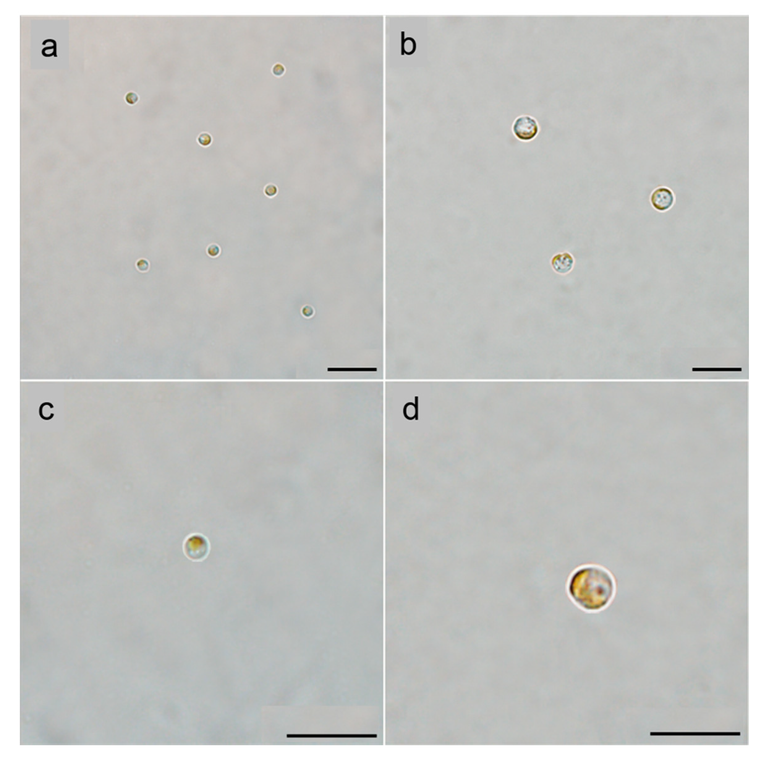
Figure 1. Light microscopic observations of the resting stage cells (RSCs) and normal vegetative cells (VCs) of A. anophagefferens strain CCMP1984. (a,c) VCs at magnifications of 400 (a) and 1000 (c); (b,d) RSCs at magnifications of 400 (b) and 1000 (d). Scale bars = 10 µm.
Jmse 08 01027 g001
Figure 2. Lipid-enriched droplets (Ld) in the RSCs of A. anophagefferens strain CCMP1984 as indicated by the arrows. The RSCs of A. anophagefferens shown in (a) was observed under bright field microscopy, while (b) shows the same cell, but it was observed under epi-fluorescence microscopy (filters settings BP450–480 nm for excitation and BA > 515 nm for emission), as it was stained by BODIPY 505/515. Scale bars = 10 µm.
Jmse 08 01027 g002
Figure 3. Germination of resting stage cells of A. anophagefferens strain CCMP1984 and subsequent rapid growth. The observations were conducted under the light microscope with the same magnification and sample volume (10 mL). Resting stage cells completely reverted to vegetative cells in about 10 days after being transferred to fresh f/2-Si medium and incubated under favorable condition (21 °C) and grew to a very high cell density in ~20 days. (a) 0 day, (b) 10th day, (c) 20th day, (d) 30th day. Scale bars = 10 µm.
Jmse 08 01027 g003
Figure 4. Appearance of resting stage cells of A. anophagefferens strain CCMP1984 after being stored in prolonged periods (a) 1 month, (b) 2 months, and (c) 3 months in darkness at 4 °C. Note that most of the cells died during the storage. Scale bars = 10 µm.
Jmse 08 01027 g004
Figure 5. Cell density recovery of A. anophagefferens strain CCMP1984 after the cultures were stored in the dark at 4 °C for 1 to 3 months. Cell densities were quantified microscopically at the time points of 0 (immediately after inoculation), 20, and 30 d after the dark-stored resting cells were inoculated to fresh f/2-Si medium and incubated at the conditions (12:12 h photoperiod, 21 °C, 100 µmol photons m−2·s−1) that were used for the routine maintenance of cultures. Each data point was shown as mean ± SD (standard deviation) from triplicate samples.
Jmse 08 01027 g005
Table 1. Measurements of intracellular content of ATP in the vegetative cells and resting stage cells of A. anophagefferens strain CCMP1984. Standard deviation (± SD) of the data from three independent experiments. For each experiment, three technical replicates were performed.
Cell TypeATP Content (nmol/107 Cells)
Resting stage cells2.9 ± 0.5
Vegetative cells31.6 ± 5.1
Publisher’s Note: MDPI stays neutral with regard to jurisdictional claims in published maps and institutional affiliations.